Abstract Degenerative disc disease (DDD) is a common finding in MRI scans and X-rays. However, their correlation to morphological and bio-chemical changes is not well estab-lished. In this study, radiological and MRI parameters of DDD were as-sessed and compared with morpho-logical and biochemical findings of disc degeneration. Thirty-nine hu-man lumbar discs (L1–S1), age 19–86 years, were harvested from eight cadavers. Within 48 h post-mortem, MRIs in various spin-echo sequences and biplanar radiographs of intact spines were obtained. Indi-vidual discs with endplates were then sectioned in the mid-sagittal plane and graded according to the morpho-logical appearance. Samples from the nucleus of each disc were har-vested for biochemical analysis in-cluding water and proteoglycan con-tents. On MRIs, T2-signal intensity, Modic changes, disc extension be-yond the interspace (DEBIT), nucleus pulposus shape, annular tears, osteo-phytes and endplate integrity were graded. On radiographs, an indepen-dent observer classified the parame-ters disc height, endplate sclerosis, osteophytes, Schmorl’s nodes, in-tradiscal calcifications and endplate shape. General linear-regression mod-els were used for statistical analysis. Backward elimination with a 10% significance cut-off level was used to identify the most significant parame-ters, which then were summed to
create composite scores for radiogra-phy, MRI and the combination of both methods. The grading was per-formed by three observers, and a re-liability analysis using Cronbach’s alpha model was used to control in-terobserver agreement. The three ra-diographic parameters height-loss, osteophytes and intradiscal calcifica-tions correlated significantly with the morphological degree of degenera-tion (p<0.001, R2=642). Significant differences of even one morphologi-cal grade could also be differentiated in the composite radiological score (p<0.05), except at the extremes be-tween grades 1 and 2 and grades 4 and 5. All MRI parameters corre-lated significantly with the morpho-logical grade (p<0.05); however Modic changes, T2-intensity and os-teophytes accounted for 83% of the variation in the data. T2-signal inten-sity correlated significantly with H2O and proteoglycan content (p<0.001), and was best for detecting highly de-generated discs. Regression showed that the combined score was better correlated with the morphological grade (p<0.001, R2=775) than either the composite radiographic (p<0.001, R2=642) or composite MRI (p<0.001, R2=696) alone. Based on the com-bined score, a backwards elimination of the regression was performed, in which the parameters Modic changes, and T2-intensity loss (MRI) as well as calcifications (X-ray) accounted for 87% of the variability. The inter-Lorin M. Benneker Paul F. Heini Suzanne E. Anderson Mauro Alini Keita Ito
Correlation of radiographic
and MRI parameters to morphological
and biochemical assessment
of intervertebral disc degeneration
Received: 10 January 2003 Revised: 16 October 2003 Accepted: 11 May 2004 Published online: 26 June 2004 © Springer-Verlag 2004
L. M. Benneker · M. Alini · K. Ito AO Research Institute, Davos, Switzerland P. F. Heini (✉)
Department of Orthopaedic Surgery, Inselspital, University of Berne, 3010 Berne, Switzerland Tel.: +41-31-6322111, e-mail: [email protected] S. E. Anderson
Department of Radiology, Inselspital, University of Berne, Berne, Switzerland
Introduction
Low back pain is one of the most common causes of dis-ability for individuals of working age in developed coun-tries [1, 32]. There are many causes of low back pain, and it is generally believed that degenerative disc disease (DDD) is one of the most prevalent [7, 27, 38]. Although the mechanisms by which DDD may cause low back pain are not clear, the severity of DDD is associated with onset of symptoms [29]. Degeneration of the intervertebral disc (IVD) is associated with progressive changes in material properties, matrix composition, and morphology. One ac-cepted method for grading IVD degeneration severity is pathomorphological [42]. This method has been shown to be both reliable and repeatable. Biochemical methods have also been established [2]. These two methods have been shown to correlate well with each other and are often used in research where cadaveric tissue is analyzed. However, because they have limited clinical utility, a non-invasive system for grading severity of DDD would be useful.
Obviously changes in IVD morphology and matrix composition are observable with various imaging modali-ties. Even prior to disc degeneration, it has been suggested that vertebral endplate changes may occur [4, 35, 36], and endplate changes such as sclerosis, Schmorl’s Nodes, end-plate shape alterations and calcifications can be observed on plain radiographs and on MR [13, 17, 37].
During the first phase of IVD degeneration, loss of proteoglycans and collagen type II has been observed [2]. Because the technique of MRI is based on proton density, water content and chemical environment of protons [28], with proteoglycan denaturation MR can detect the associ-ated dehydration as a loss of the signal intensity on T2-weighted images. This method is commonly accepted and mostly visually classified [41, 43]. In addition to direct changes in the disc, Modic noticed that signal intensity changes in the vertebral-body marrow adjacent to the end-plate are also strongly associated with clinical symptoms related to DDD of the same level [23].
With progressive matrix alterations, changes in disc morphology also become visible in radiographs. One such change is a reduction in disc height, which can be quanti-fied by comparison with a database of age-, body height-and gender-corrected normal values [11]. Another change is osteophyte formation, which is understood to be a
com-pensation mechanism to distribute the increasing axial and shear forces (due to instability) on a larger articulating sur-face. Macnab et al. differentiated osteophytes of the spine into two types: traction and claw spurs. The first is thought to be the result of abnormal shear loads and is proposed as a sign of instability, while the second is proposed to arise from compressive loads and is a benign, age-related find-ing. The displacement of the intervertebral disc may lead to traction at the site of osseous attachment (Sharpey fibers) of the annulus fibrosus or short perivertebral liga-ments to the vertebral surface. Osteophytes develop at this location, several millimeters from the disco-vertebral junction and are well depicted in radiographs [20, 33].
Nevertheless, even with such changes, MRI is believed to provide a more detailed picture of disc morphology in-cluding the endplates. The axial deformation of the disc – disc extension beyond the interspace (DEBIT) – has been classified by Jensen et al. into five categories (1) intact, (2) bulge, (3) protrusion, (4) extrusion and (5) sequestration) [16]. Viikkari graded the shape of the nucleus into four degrees (1) round/oval, (2) extension into inner annulus, (3) extension into outer annulus and (4) extension beyond outer annulus, using sagittal MR slides [43]. Yu divided annular tears into three degrees: (I) concentric tears: fluid-filled space between the annular lamellae; (II) radial tears: rupture of all annular layers; and (III) transversal tears, plus rupture of Sharpey fibers. Types II and III are visible on MRI as high-intensity zones on T2-weighted images [44]. Because many of these individual parameters have been associated with severity of IVD degeneration, they are cur-rently being examined and combined to develop a non-in-vasive grading scheme for imaging. Yet most of these stud-ies use clinical data and cannot be compared to established grades of IVD degeneration [31]. Hence, the aim of this study was to compare various imaging parameters used to describe IVD degeneration with gross-morphology-IVD-degeneration grades and biochemical IVD-matrix compo-sition, and to create grading methods based only on sig-nificant parameters.
Methods Specimen
Thirty-nine human lumbar discs (L1–S1) were collected from eight cadavers (age range: 19–86 years, average 54 years) and stored at observer validation showed a high
correlation for all three scores (Cronbach’s alpha values ranging from 0.95 to 0.97). Conclusion: selective imaging parameters and a newly created scoring scheme were found to correlate with disc degener-ation as determined in a
morphologi-cal manner. Surprisingly, radio-graphic parameters were able to dis-tinguish different stages of degenera-tion, whereas MRI could only detect advanced stages of disc degenera-tion. We conclude that X-rays may remain a cost-effective, non-invasive in vivo-grading method to detect
early disc degeneration, and, com-bined with MRI, correlate best with morphological and biochemical as-sessment of disc degeneration.
Keywords Intervertebral disc · Degeneration · Radiographs · MRI
4°C, wrapped in saline-soaked gauze to prevent dehydration. In-tersegmental ligaments and the paralumbar musculature were left intact for better contrast during MRI examination. The spines were prepared within 48 h postmortem to minimize water-content changes and enzymatic degeneration of the disc matrix. Donors with any pathology affecting the spine were excluded from this study. (Table 1)
Imaging
Prior to freezing, MRI scans were obtained with a GE 1.5-T unit (General Electric Medical Systems, Milwaukee, WI) and using a spine array coil (5×11 in.). The following spin-echo sequences
were used:
– Axial localizer (spoiled gradient) – Sagittal T1 (TE minimum full/TR 400)
– Sagittal T2 (TE 100/TR 4000), sagittal proton density (TE 10–20/ TR 2000)
– Axial T1 (TE minimum full/TR 400) (thickness 4 mm/spacing 0.4 mm, matrix 512×512, FOV 26 cm)
Three observers (one radiologist, S.E.A., and two orthopedic sur-geons, P.F.H. and L.M.B.) examined all images and assessed the following eight parameters:
1. T2-intensity loss 2. Modic changes 3. Endplate cartilage loss 4. DEBIT score
5. Annular tears 6. Osteophytes
7. Nucleus pulposus (NP) shape 8. Endplate integrity
Assessment was performed according to recommendations of other authors [16, 23, 26, 41, 43, 44]. (Table 2, Fig. 1)
While frozen, anteroposterior (A-P) and lateral radiographs were taken (Faxitron 804, Field Emission, McMinnville, OR, USA) The following settings were used: 60 kV for lateral view and 65 kV for A-P projection, exposure time 5 min, film-focus distance 53 cm with a 0.5 cm Al filter. The three observers classified the following parameters into three or four categories: (1) disc height, (2) end-plate sclerosis, (3) osteophytes, (4) Schmorl’s nodes, (5) intradiscal calcifications and (6) endplate shape [11, 14, 17]. (Table 3, Fig. 1)
Morphometry
After thawing, functional spine units (FSU) consisting of both end-plates and intervertebral disc were isolated and the posterior ele-ments removed. A double-bladed cutting device was used to sagit-tally section the disc into three parts: (1) half, (2) 1 cm thick para-midsagittal slab and (3) the remainder. Digital photos were taken of the mid-sagittal section surface (Minolta RD175 digital camera, Minolta, Japan) and graded using a five-category grading scheme for assessing the gross morphology of the IVD [42]. (Table 4, Fig. 1)
Biochemistry
Samples for biochemical analysis (water and proteoglycan con-tent) were harvested from the nucleus pulposus of the mid-sagittal slab. Water content was calculated by weighing the samples before and after drying (110°C, 5 days) [3]. For proteoglycan (PG) con-tent, samples were digested in proteinase K before measuring the glycosaminoglycan content with a dimethylmethylene blue dye binding colometric assay at 530 nm (Perkin Elmer HTS 7000, Shelton, CT, USA) [10].
Table 1 Spine donor data
Spine Segments Gender Age Morphological degeneration grade Cause of death
(total 39) [42]
1 L2–S1 Female 19 - 1 1 1 2 Motor vehicle accident
2 L1–S1 Female 32 3 2 2 2 2 Lupus e.
3 L1–S1 Male 51 2 3 3 3 3 Myocard infarct
4 L1–S1 Female 53 2 2 3 4 3 Myocard infarct
5 L1–S1 Male 53 5 5 4 3 3 Heart failure
6 L1–S1 Male 64 3 4 5 5 5 Myocard infarct
7 L1–S1 Female 65 3 3 4 3 3 Myocard infarct
8 L1–S1 Female 86 4 4 4 4 4 Pneumonia
Score T2-signal intensity
DEBIT [16] Nucleus shape [43]
Annular tears [44]
Modic changes [22]
Endplate integrity Osteo-phytes
0 Normal Intact Round/oval Intact Normal Intact Absent
1 Intermediate loss
Bulge Extension into inner annulus
Concentric tears
Type I Isolated defects Marginal 2 Marked loss Protrusion Extension into
outer annulus
Radial tears Type II Schmorl’s node<5 mm
Dis-continuous 3 Absent signal Extrusion/
sequestration
Extension beyond outer annulus
Transversal tears
Type III Schmorl’s node>5 mm
Continuous, table osteophyte Table 2 Magnetic resonance imaging parameters and scores. For
axial and sagittal images: T2-intensity loss, Modic changes, osteo-phytes and nuclear pulposus (NP) shape correlated most signifi-cantly with degeneration. Modic type 1: decreased signal intensity on T1-weighted spin-echo images and increased signal intensity
on T2-weighted images; Modic type 2: increased signal intensity on T1-weighted images and isointense or slightly increased signal intensity on T2-weighted images; DEBIT disc extension beyond the interspace
Statistical analysis
General linear regression models were used to assess the correlation between radiographic or MRI grades, i.e., the sum of scores for se-lected parameters, and morphological grade. To determine the most significant parameters to be selected for each composite imaging
score, a backward elimination was applied. Parameters below 10% significance level were omitted until parameters could no longer be rejected. This resulted in a composite score for X-ray (CRS) and MRI (CMS) using only the most significant parameters of the MRI and radiography results. Furthermore, a combined imaging score (CIS) using both CRS and CMS was determined, using the Fig. 1 a Grade 1: segment L4–L5 of a 19-year-old female. The
nu-cleus is gelatinous and round, or oval-shaped, on MR with a high T2-signal intensity. The annulus show discrete fibrous lamellas. The endplate is intact, with a uniformly thick hyaline cartilage layer. The vertebral body still has rounded margins, with no Schmorl’s nodes or sclerosis; b grade 2: segment L2–L3 of a 53-year-old fe-male. In the nucleus, peripherally, white fibrous tissue becomes visible. The annulus presents mucinous material between lamellas. The cartilage layer of the endplate becomes irregular; c grade 3: segment L3–L4 of a 53-year-old female. Consolidated fibrous tis-sue in the nucleus area and loss of the annular nuclear demarca-tion. On radiographs, reduction of the disc height and early
osteo-phytes formation is visible. Frequently, rim calcification and scle-rosis are also visible. On MRI, T2-signal intensity is reduced; the nucleus flattens and can extend into the inner annulus; isolated de-fects of the endplate and concentric tears of the annulus appear; d grade 4: segment L3–L4 of an 86-year-old female. Horizontal clefts parallel to the endplate are visible macroscopically and on MRI. Osteophytes, small Schmorl’s nodes and intranuclear calcifi-cation are common findings. The nucleus can extend into the outer annulus; e grade 5: segment L4–L5 of a 64-year-old male. Disc height is markedly reduced. The nucleus has disappeared totally (absent T2 signal), and big, sometimes-bridging, osteophytes ap-pear. Diffuse and severe sclerosis and calcifications dominate
same backward-elimination technique. In order to assess the dis-criminating potential between distinct grades of the three scoring systems, both parametric (one-way ANOVA with Tukey’s post hoc testing) and non-parametric (Kruskal–Wallis with Mann–Whitney post hoc testing) statistical analyses were performed.
Simple linear analysis of variance regression was used to ex-amine the correlation of nuclear proteoglycan and H2O content with
the MRI imaging parameters. One-way ANOVA with Tukey’s post hoc testing was used to assess differences between grades. For all statistical analyses a significant level of p<0.05 was used.
Cohen’s kappa statistic is often used to test for interobserver reliability between a pair of observers. When there are more than two observers, this statistic is calculated for all possible pairs. However, in this study, due to the high cell-level variation, i.e., the number of grades and number of cases, it was not possible to cal-culate the kappa statistics. As an alternative, a reliability analysis using Cronbach’s alpha model was used to analyze interobserver agreement between all three observers simultaneously.
Results X-Ray
Regression of the radiological grade with the morpholog-ical grade showed that disc height loss, osteophytes and intradiscal calcifications were most significantly correlated with the morphological grade, accounting for 80% of the total variation, or 96% of the variation within our speci-men population. Out of these three parameters, disc height loss correlated with the morphological grade much more significantly (p<0.001) than the other two parameters (os-teophytes p<0.014 and calcifications p<0.010). The corre-lation of this composite radiographic score (CRS) with the morphological grade of degeneration was, again, highly
significant (p<0.001), with a Pearson correlation coeffi-cient of 0.77 (Fig. 2). Furthermore, discs of different mor-phological grades had significantly different composite ra-diographic scores (p<0.001 for both parametric and non-parametric tests). However, both non-parametric and non-para-metric post hoc testing showed that this discrimination was only significant between discs of grades 1 or 2 vs grade 3 or higher, and grade 3 vs grades 4 or 5.
Table 3 Parameters (and scores) on plain anteroposterior and lateral radiographs; disc height, osteophytes and calcifi-cations correlated significantly with degeneration
Score Height Osteo-phytes Schmorl’s Intradiscal Sclerosis Endplate
loss [11] nodes calcification shape
0 0–10% Margins rounded Not present No calcifications None Continuous 1 10–20% Margins pointed Present Rim calcification Moderate Irregular
2 20–30% <2 mm – Intranuclear Severe Disrupted
calcification
3 >30% >2 mm – – – –
Table 4 The Thompson grading scheme [42] for gross morphology of the human lumbar intervertebral disc
Grade Nucleus Annulus Endplate Vertebral body
I Bulging gel Discrete fibrous lamellas Hyaline, uniformly thick Margins rounded II White fibrous tissue
peripherally
Mucinous material between lamellas
Thickness irregular Margins pointed III Consolidated fibrous
tissue
Extensive mucinous infiltration; loss of annular-nuclear demarcation
Focal defects in cartilage Early chondrophytes or osteophytes at margins IV Horizontal clefts
parallel to endplate
Focal disruptions Fibro cartilage extending from subchondral bone, irregularity and focal sclerosis in subchondral bone
Osteophytes less than 2 mm V Clefts extend through
nucleus and annulus
– Diffuse sclerosis Osteophytes greater
than 2 mm
Fig. 2 Composite radiographic score (CRS) vs morphological de-generation. Data points represent the mean of the sum of parame-ters: disc height loss, osteophytes and intradiscal calcifications, with error bars representing one standard error of the mean
MRI
Of all MRI score parameters, only T2-intensity loss (p<0.001), Modic changes (p=0.019) and osteophytes (p=0.016) were significantly correlated with morphologi-cal grade, accounting for 83% of the total variation in the data, or 98% of the variation within our specimen popula-tion. Parametric and non-parametric analyses revealed that an MRI score with only these three parameters did not discriminate well between different morphological grades of degeneration, with significantly different MRI scores only between grades 1, 2 or 3 vs grades 4 or 5 (p<0.001). However, a composite MRI score (CMS) also including NP shape was still significantly correlated with morphological grades of degeneration (p<0.001), and this score exhibited significant differences between the higher grades of degeneration, i.e., grades 1, 2 and 3 were not different from each other but were significantly different from grades 4 (p<0.001) and 5 (p<0.001). Grades 4 and 5 were also significantly different from each other (p=0.01) (Fig. 3).
MR findings and biochemical assessments
T2-intensity loss correlated significantly (p<0.001) with both water and PG content (Fig. 4). However, the water and PG content of all three T2-intensity loss levels did not significantly differ from each other. There was only a sig-nificant difference in the water (p<0.011) and PG (p<0.003) content of normal T2-intensity discs as com-pared with those with either moderate or marked T2-in-tensity loss, but not between the two severities. It should
also be noted that care must be taken in regard to these results, as the number of discs with marked T2-intensity loss was small, n≤4, especially for water content, for
which a number of outliers had to be rejected due to des-iccation during preparation, caused by the extremely dry ambient conditions in Davos (1,560 m altitude).
Scores
The composite radiographic (CRS) and composite MRI (CMS) scores were merged to see if this combined imag-ing score (CIS) would enhance the correlation with the morphological grade. Regression showed that the com-bined imaging score was better correlated to the morpho-logical grade (p<0.001, R2=775) than either the composite radiographic score (p<0.001, R2=642) or the composite MRI score (p<0.001, R2=696) alone. Parametric and non-Fig. 3 Composite MRI score (CMS) vs morphological
degenera-tion. Data points represent the mean of the sum of parameters: Modic changes, osteophytes, nucleus shape and T2-intensity loss, with error bars representing one standard error of the mean
Fig. 4 Mean proteoglycan content vs the nucleus pulposus to T2-signal intensity loss, with error bars representing one standard error of the mean
Fig. 5 Composite imaging score (CIS) vs morphological degener-ation. Data points represent the mean of the sum of parameters: disc height loss (X-ray), osteophytes (X-ray), calcifications (X-ray), T2-intensity loss (MRI), Modic changes (MRI) and nucleus shape (MRI), with error bars representing one standard error of the mean
parametric analyses showed that the combined imaging score is not much better than the composite radiographic score at distinguishing individual morphological grades; i.e., only grades 1 vs 2 and grades 4 vs 5 were not signif-icantly different (Fig. 5). Again, this result may simply be due to the limited numbers of disc samples available in grades 1 and 5. In a backward elimination of the regres-sion for the combined imaging score, the parameters Modic changes (MRI), calcifications (X-ray) and T2-in-tensity loss (MRI) accounted for 87% of the variability; but this reduced score did not enhance discrimination be-tween grades. Finally, the interobserver-agreement analy-sis using Cronbach’s alpha model of reliability showed a high correlation for all three observers, i.e., low interob-server error. The intra-class correlation coefficients (alpha values) ranged from 0.95 for CRS and CMS to 0.97 for CIS and are supposed to be highly significant. Interest-ingly, the interobserver error was markedly smaller be-tween the two surgeons than it was for a comparison with the radiologist (Table 5, Table 6).
Discussion
In this study, we investigated eight parameters of degen-eration for MR and six for conventional radiographs, and
correlated these findings with the biochemical and mor-phological degree of IVD degeneration. The frequency and distribution of the pathological findings in our data population was comparable to the results of earlier studies used as references [15, 22, 26, 41, 43, 44]. By elimination of less important/significant parameters, we created an easy-to-use grading scheme for radiograph, MR and com-bined imaging that allows accurate and non-invasive clas-sification of IVD degeneration.
There have been other proposed classifications for IVD degeneration in plain radiography as well as in MRI. Pfirrmann et al. has proposed an MRI score based on ho-mogeneity of the NP, distinction of NP as compared with annulus fibrosus, T2-signal intensity and disc height on fast spin-echo MRI [31]. They have shown that this clas-sification system tested on actual patient images has good reliability. However, accuracy, e.g., morphological grade or symptoms, was not evaluated. In comparison, our re-sults also show that T2-signal intensity, nuclear calcifica-tions, NP shape (extensions into annulus) and disc height are important. However, by contrast, the other parameters that most significantly correlated to morphological grades of degeneration were specific to the vertebral body, i.e., Modic changes, osteophytes and calcifications of the rim. Hence, neglecting the adjacent osseous structure, may limit our interpretation of images when assessing IVD de-generation severity.
Surprisingly, some pathological MR findings such as annular tears, DEBIT, Schmorl’s nodes, sclerosis and end-plate integrity in our data did not correlate as well with the morphological appearance, used as the gold standard. The reasons may be manifold, but one reason is that, in our sample population of donors, without history of spinal disorders, these findings were rare or difficult to detect. Annular tears were detected in six out of 39 examined discs (five concentric, one radial), which is the expected frequency for asymptomatic individuals [15]. Osti showed that normal MRI does not exclude significant changes in the peripheral structure of the intervertebral disc and that discography is more accurate [25]. With nine bulges, three protrusions and one extrusion out of 39 discs, DEBIT was noted in a slightly lower prevalence than expected for our donor population compared with literature [15]. Sclerosis was observed only in six out of 78 vertebral endplates (four moderate, two severe). Katz described five different types of endplate sclerosis, which did not always show the classic radiographic band-like pattern along the vertebral endplate. The atypical patterns may be difficult to recog-nize as degenerative in origin, since infection, Paget’s dis-ease, metabolic disorders and metastasis can manifest as radiographical sclerosis [17]. Additional T2-weighted MRI allows differentiation between discogenic (degener-ative) and infection-induced sclerosis [40]. Endplate carti-lage loss, which is associated with aging and degenera-tion, was only seen in five of 78 samples examined by MR [34]. Finally, Schmorl’s nodes are also difficult to de-Table 5 Disc degeneration in relation to radiography/MRI scores
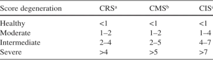
Score degeneration CRSa CMSb CISc
Healthy <1 <1 <1
Moderate 1–2 1–2 1–4
Intermediate 2–4 2–5 4–7
Severe >4 >5 >7
aComposite X-ray score CRS: sum of disc height loss, osteophytes
and intradiscal calcifications
bComposite MRI score CMS: sum of Modic changes, osteophytes,
nucleus shape and T2-intensity loss
cCombined imaging score CIS: sum of disc height loss (X-ray),
os-teophytes (X-ray), calcifications (X-ray), T2-intensity loss (MRI), Modic changes (MRI) and nucleus shape (MRI)
Table 6 Analysis of interobserver agreement on intervertebral disc degeneration grading, using Cronbach’s alpha model of relia-bility (CRS composite X-ray score, CMS composite MRI score,
CIS combined imaging score)
CRS CMS CIS
Correlation coefficient observer 1–2 0.86 0.89 0.91 Correlation coefficient observer 2–3 0.95 0.92 0.96 Correlation coefficient observer 1–3 0.78 0.82 0.89
Alpha values 0.95 0.95 0.97
Ave. measure intraclass correlation 0.91 0.92 0.95 (alpha): lower 95%
Ave. measure intraclass correlation 0.97 0.97 0.98 (alpha): upper 95%
1. Andersson GB (1998) Epidemiology of low back pain. Acta Orthop Scand [Suppl] 281:28–31
2. Antoniou J, Steffen T, Nelson F, Winterbottom N, Hollander AP, Poole RA, Aebi M, Alini M (1996) The hu-man lumbar intervertebral disc: evi-dence for changes in the biosynthesis and denaturation of the extracellular matrix with growth, maturation, age-ing, and degeneration. J Clin Invest 98:996–1003
3. Antoniou J, Pike GB, Steffen T, Baramki H, Poole AR, Aebi M, Alini M (1998) Quantitative magnetic reso-nance imaging in the assessment of de-generative disc disease. Magn Reson Med 40:900–907
4. Benneker LM, Anderson S, Heini P, Alini M, Ito K (2002) Vertebral end-plate marrow contact channel occlu-sions: a mechanism for intervertebral disc degeneration? ISSLS (unpublished data)
5. Boden SD, Davis DO, Dina TS, Patronas NJ, Wiesel SW (1990) Ab-normal magnetic-resonance scans of the lumbar spine in asymptomatic subjects. A prospective investigation. J Bone Joint Surg Am 72:403–408 6. Braithwaite I, White J, Saifuddin A,
Renton P, Taylor BA (1998) Vertebral end-plate (Modic) changes on lumbar spine MRI: correlation with pain repro-duction at lumbar discography. Eur Spine J 7:363–368
7. Buckwalter J, Martin J (1996) Interver-tebral disk degeneration and back pain. In: Weinstein JN (ed) Low back pain: A scientific and clinical overview. American Academy of Orthopedic Surgeons, Rosemont, IL, pp 607–623 8. Crock HV (1982) Traitement
chirurgi-cal de la chirurgi-calcification du nucleus pul-posus du disque intervertebral dorsal et lombaire, chez l’adulte. Rev Chir Or-thop Reparatrice Appar Mot 68:171– 177
9. Fahey V, Opeskin K, Silberstein M, Anderson R, Briggs C (1998) The pathogenesis of Schmorl’s nodes in relation to acute trauma. An autopsy study. Spine 23:2272–2275 References
pict on plain lateral radiographs as corroborated by our re-sults (one of 78 endplates on radiographs vs 13 of 78 on MRs, 7.7%) and others (0–47%)[9, 13, 21]. However, its value for disc degeneration remains unclear and contro-versial. Whereas in some older studies a relation of the appearance of Schmorl’s nodes to degeneration was ob-served [14], more recent work with additional MRI as well as our results cannot confirm this correlation [9, 13, 30]. The radiological pattern of Schmorl’s nodes seems multifactorial in origin, and degeneration, among other factors such as predisposition and axial traumas, is only one possible pathomechanism. So, before these parame-ters such as annular tears, Schmorl’s nodes, sclerosis and endplate integrity can be used as indicators for IVD, fur-ther studies are needed to evaluate and differentiate their importance for degeneration.
The difficulties we faced at distinguishing extreme grades (Thompson grades 1/2 and 4/5) can be due to the low number of samples available at these grades. On the other hand, the clinically relevant bandwidth, e.g., the on-set of discogenic symptoms, is between grades 2 to 4 (mild-to-severe degeneration) [27, 38]. Surprisingly, the composite radiographic score (and the combined imaging score) were better at differentiating degeneration grades than the composite MRI score alone (compare Figs. 2, 3 and 5). This may indicate that changes of the vertebral structure occur in an early phase of degeneration. Still, these radiographically defined parameters can also be quantified by MRI, and MR data should be used when-ever available. Probably, the use of quantitative MR for T2-signal intensity analysis would improve the discrimi-nation potential of the composite MRI score; but, on the other hand, we tried to use clinical conditions [3].
The results of the biochemical evaluation were within the range of former studies, and proteoglycan and water
content correlated as expected to the T2-signal intensity loss [2]. Again, the poor visual three-grade classification for T2-signal intensity limited the detection of early signal loss. The use of a computed signal-intensity measurement with a region-of-interest capability would be an alterna-tive for a more accurate classification [26].
The clinical relevance of our scores is open. Many studies reveal a high ratio of abnormal findings in MR and radiographs in asymptomatic people [5, 15, 26, 39]. The parameters that showed the strongest correlation with morphological degeneration – and, therefore, were in-cluded in the scores – are associated with low back pain [6, 8, 12, 18, 19, 24, 26]. Thus, the correlation of the scores with clinical symptoms is likely but needs to be ex-amined in further studies.
Conclusions
Selective imaging parameters and a newly created, repeat-able scoring scheme was found to correlate with disc de-generation as determined in a morphological manner. Sur-prisingly, in our sample population, radiographic parame-ters were better able to distinguish different stages of de-generation, whereas MRI could only detect advanced stages of disc degeneration. We conclude that conven-tional radiography remains a cost-effective, non-invasive in vivo grading method to detect early disc degeneration and, combined with MRI, correlates best with morphome-trical and biochemical assessment of disc degeneration. Acknowledgements This project was supported by a grant from the AO Foundation, Switzerland. The authors wish to thank the University of Basel and McGill University, Montreal, for provid-ing spine specimens, K. Zwygart for conductprovid-ing the MRI scans; and D. Pfluger for statistical assistance
10. Farndale RW, Buttle DJ, Barrett AJ (1986) Improved quantitation and dis-crimination of sulphated glycosamino-glycans by use of dimethylmethylene blue. Biochim Biophys Acta 883:173– 177
11. Frobin W, Brinckmann P, Biggemann M (1997) Objektive Messung der Höhe lumbaler Bandscheiben aus seitlichen Röntgen-Übersichtsaufnahmen. Z Or-thop Ihre Grenzgeb 135:395–402 12. Frymoyer JW, Newberg A, Pope MH,
Wilder DG, Clements J, MacPherson B (1984) Spine radiographs in patients with low-back pain. An epidemiologi-cal study in men. J Bone Joint Surg Am 66:1048–1055
13. Hamanishi C, Kawabata T, Yosii T, Tanaka S (1994) Schmorl’s nodes on magnetic resonance imaging. Their in-cidence and clinical relevance. Spine 19:450–453
14. Hilton RC, Ball J, Benn RT (1976) Vertebral end-plate lesions (Schmorl’s nodes) in the dorsolumbar spine. Ann Rheum Dis 35:127–132
15. Jensen MC, Brant-Zawadzki MN, Obuchowski N, Modic MT, Malkasian D, Ross JS (1994) Magnetic resonance imaging of the lumbar spine in people without back pain [see comments]. N Engl J Med 331:69–73
16. Jensen MC, Kelly AP, Brant-Zawadzki MN (1994) MRI of degenerative dis-ease of the lumbar spine. Magn Reson Q 10:173–190
17. Katz ME, Teitelbaum SL, Gilula LA, Resnick D, Katz SJ (1988) Radiologic and pathologic patterns of end-plate-based vertebral sclerosis. Invest Radiol 23:447–454
18. Lamer TJ (1999) Lumbar spine pain originating from vertebral osteophytes. Reg Anesth Pain Med 24:347–351 19. Luoma K, Riihimaki H, Raininko R,
Luukkonen R, Lamminen A, Viikari-Juntura E (1999) Low back pain in relation to lumbar disc degeneration. Spine 25:487–492
20. MacNab I (1971) The traction spur. An indicator of segmental instability. J Bone Joint Surg Am 53:663–670 21. Malmivaara A, Videman T, Kuosma E,
Troup JD (1987) Plain radiographic, discographic, and direct observations of Schmorl’s nodes in the thoracolum-bar junctional region of the cadaveric spine. Spine 12:453–457
22. Modic MT, Ross JS (1991) Magnetic resonance imaging in the evaluation of low back pain. Orthop Clin North Am 22:283–301
23. Modic MT, Steinberg PM, Ross JS, Masaryk TJ, Carter JR (1988) Degen-erative disk disease: assessment of changes in vertebral body marrow with MR imaging. Radiology 166:193–199 24. O’Neill TW, McCloskey EV, Kanis
JA, Bhalla AK, Reeve J, Reid DM, Todd C, Woolf AD, Silman AJ (1999) The distribution, determinants, and clinical correlates of vertebral osteo-phytosis: a population based survey. J Rheumatol 26:842–848
25. Osti OL, Fraser RD (1992) MRI and discography of annular tears and inter-vertebral disc degeneration. A prospec-tive clinical comparison [see comments]. J Bone Joint Surg Br 74:431–435 [pub-lished erratum appears in J Bone Joint Surg Br 1992 Sep; 74(5):793] 26. Paajanen H, Erkintalo M, Kuusela T,
Dahlstrom S, Kormano M (1989) Mag-netic resonance study of disc degenera-tion in young low-back pain patients. Spine 14:982–985
27. Paajanen H, Erkintalo M, Parkkola R (1997) Age-dependent correlation of low-back pain and lumbar disc degen-eration. Arch Orthop Trauma Surg 116:106–107
28. Panagiotacopulos ND, Pope MH, Krag MH, Block R (1987) Water content in human intervertebral discs. Part I. Measurement by magnetic resonance imaging. Spine 12:912–917
29. Peterson CK, Bolton JE, Wood AR (2000) A cross-sectional study corre-lating lumbar spine degeneration with disability and pain. Spine 25:218–223 30. Pfirrmann CW, Resnick D (2001)
Schmorl nodes of the thoracic and lumbar spine: radiographic-pathologic study of prevalence, characterization, and correlation with degenerative changes of 1,650 spinal levels in 100 cadavers. Radiology 219:368–374 31. Pfirrmann CW, Metzdorf A, Zanetti M,
Hodler J, Boos N (2001) Magnetic res-onance classification of lumbar inter-vertebral disc degeneration. Spine 26: 1873–1878
32. Praemer A, Furner S, Rice D (1992) Musculoskeletal conditions in the United States. American Academy of Orthopaedic Surgeons, Park Ridge, IL, USA
33. Resnick D (1985) Degenerative dis-eases of the vertebral column. Radiol-ogy 156:3-14
34. Roberts S, Menage J, Urban JP (1989) Biochemical and structural properties of the cartilage end-plate and its rela-tion to the intervertebral disc. Spine 14:166–174
35. Roberts S, Menage J, Eisenstein SM (1993) The cartilage end-plate and in-tervertebral disc in scoliosis: calcifica-tion and other sequelae. J Orthop Res 11:747–757
36. Roberts S, Urban JP, Evans H, Eisenstein SM (1996) Transport prop-erties of the human cartilage endplate in relation to its composition and cal-cification. Spine 21:415–420 37. Roberts S, McCall IW, Menage J,
Haddaway MJ, Eisenstein SM (1997) Does the thickness of the vertebral sub-chondral bone reflect the composition of the intervertebral disc? European Spine Journal 6:385–389
38. Salminen JJ, Erkintalo MO, Pentti J (1999) Recurrent low back pain and early disc degeneration in the young. Spine 24:1316–1321
39. Savage RA, Whitehouse GH, Roberts N (1997) The relationship between the magnetic resonance imaging appear-ance of the lumbar spine and low back pain, age and occupation in males. Eur Spine J 6:106–114
40. Sobel DF, Zyroff J, Thorne RP (1987) Diskogenic vertebral sclerosis: MR imaging. J Comput Assist Tomogr 11: 855–858
41. Tertti M, Paajanen H, Laato M, Aho H, Komu M, Kormanc M (1991) Disc degeneration in magnetic resonance imaging. A comparative biochemical, histologic, and radiologic study in ca-daver spines. Spine 16:629–634 42. Thompson JP, Pearce RH, Schechter
MT, Adams ME, Tsang IK, Bishop PB (1990) Preliminary evaluation of a scheme for grading the gross morphol-ogy of the human intervertebral disc. Spine 15:411–415
43. Viikari-Juntura E, Raininko R, Videman T, Porkka L (1989) Evalua-tion of cervical disc degeneraEvalua-tion with ultralow field MRI and discography. An experimental study on cadavers. Spine 14:616–619
44. Yu SW, Sether LA, Ho PS, Wagner M, Haughton VM (1988) Tears of the an-nulus fibrosus: correlation between MR and pathologic findings in cadav-ers. AJNR Am J Neuroradiol 9:367– 370
![Table 4 The Thompson grading scheme [42] for gross morphology of the human lumbar intervertebral disc](https://thumb-eu.123doks.com/thumbv2/123doknet/14864548.636774/5.892.71.814.278.475/table-thompson-grading-scheme-gross-morphology-lumbar-intervertebral.webp)