Received: 28 March 2003 Published online: 29 May 2003 © Springer-Verlag 2003
Abstract Introduction: Desmoplas-tic infantile gangliogliomas (DIG) are rare cerebral glioneural tumors usually occurring in early childhood. DIGs are generally benign although rare cases with poor outcome are known. Total resection, if possible, is the treatment of choice, without further adjuvant therapy. After in-complete resection, adjuvant chemo-and/or radiotherapy is generally ap-plied, despite the potential negative side effects in such young patients. Case reports: We describe two girls with DIG, one who twice underwent subtotal resection at 3 and 5 months, the other who underwent total resec-tion at 2 years. Neither had adjuvant
therapy and there was no tumor re-currence. Conclusions: Our own experience and a review of the liter-ature suggest that in most DIGs adjuvant therapy is not justified even after incomplete resection. After tumor recurrence a second surgical intervention should be con-sidered instead of adjuvant therapy. An exception may be made for rare, deep-seated DIGs, which are more aggressive and have a poorer out-come.
Keywords Desmoplastic infantile ganglioglioma · Surgery · Adjuvant therapy · Outcome
Childs Nerv Syst (2003) 19:359–366
DOI 10.1007/s00381-003-0754-9 C A S E R E P O R T
Heidi Bächli Pierino Avoledo Otmar Gratzl Marcus Tolnay
Therapeutic strategies and management
of desmoplastic infantile ganglioglioma:
two case reports and literature overview
Introduction
Desmoplastic infantile gangliogliomas (DIGs) are rare neoplasms that were first described by VandenBerg et al. in 1987 [19]. They typically occur in infants under 2 years. However, three cases of young adults (14–19 years) have also been recently reported, so that DIG does not seem to be a specific entity of infancy [5, 12, 20]. The huge size and relative lack of obtundation with such large masses suggest very slow growth, and the early age of onset has led to the speculation that this tu-mor could be of congenital origin [14]. At presentation the tumor is usually very large (up to 13 cm diameter), with associated cysts and attachment to the dura. The su-pratentorial region is preferentially involved, especially the frontal and parietal lobes, followed by the temporal [17], and rarely the occipital region. The symptoms, which usually appear soon after birth, include increasing head circumference, bulging fontanel, sunset sign,
hemi-paresis, and frequent seizures (irritation of surrounding brain or neuronal components may also be intrinsically epileptogenic). On MRI these tumors are radiologically characterized by a hypointense cystic mass with an isoin-tense peripheral solid component on the T1-weighted scan that enhances with gadolinium; on T2-weighted im-ages the cystic component is hyperintense and the solid portion is heterogeneous. The cysts are generally large, uni- or multiloculated, with clear or xanthochronic fluid. The solid superficial extracerebral part of the tumor is firm or rubbery in consistency, gray or white in color, and typically with dural attachment. Histologically, the tu-mors are characterized by a prominent desmoplastic stro-ma and mixture of neoplastic astrocytes and neuronal cells at varying stages of differentiation [14]. Mitotic ac-tivity is very low, and there is usually no necrosis. How-ever, when present, mitoses and foci of necrosis are most-ly restricted to areas with poormost-ly differentiated neuroepi-thelial cells and they are not considered to be anaplastic H. Bächli (
✉
) · O. GratzlDepartment of Neurosurgery, University Hospital Basel,
Spitalstrasse 21, 4031 Basel, Switzerland e-mail: hbaechli@uhbs.ch
Fax: +41-61-2657138 P. Avoledo
Division of Oncology/Hematology, University Children’s Hospital, Basel, Switzerland
M. Tolnay
Department of Neuropathology, University Basel,
or malignant features. Recently, the formerly termed “desmoplastic infantile neuroepithelial tumors”, either with or without ganglion cells, have been grouped collec-tively as “desmoplastic infantile astrocytoma (DIA) and ganglioglioma (DIG)”, and included in common classifi-cations of cerebral tumors [7, 11, 17]. Up to now about 60 cases of DIA/DIG have been described [14]. Whether DIA indeed represents a separate entity, or is just a form of DIG in which neurons have not been detected in the biopsied tissue, is a matter of debate [10, 14].
If possible, the preferred treatment of DIGs is total surgical resection. There is no unique practice in cases in which only incomplete resection has been carried out.
Adjuvant chemotherapy and/or radiotherapy have been administered in most of these cases, with a maximal fol-low-up of 14 years. In this paper we describe two DIG patients, one with subtotal and one with total surgical re-section, both of whom had a favorable outcome without adjuvant therapy.
Case reports
Case 1Pregnancy was uneventful. At birth, this girl had a wide non-reac-tive pupil on the left side and paresis of the superior musculus rec-tus. These symptoms were interpreted as a birth trauma. Later hemiparesis developed on the right side. Neuropediatric consulta-tion led to MRI, which revealed a large cystic tumor (7×5 cm) with inhomogeneous gadolinium enhancement, which completely filled the left hemisphere and caused a marked midline shift (Fig. 1A). The child was admitted to our Neurosurgical Clinic at the age of 3 months. The fontanel was plane, there was no patho-logical growth of the head, but clinically there was left-sided amaurosis, VII paresis on the left and hemiparesis on the right. Preoperative angiography showed a marked tumor blush, the Fig. 1A–D Desmoplastic infantile ganglioglioma (DIG) in the
left hemisphere of a 3-month-old girl. A Coronal post-contrast T1-weighted MRI scan showing a huge cystic tumor with typical dural attachment and midline shift. B Digital subtraction cerebral angiogram. C Coronal MRI scan after first operation with incom-pletely resected tumor. D Coronal post-contrast MRI scan 4 years after surgery without tumor recurrence
361
Fig. 2A–C Right occipital DIG in a 2-year-old girl. A Coronal post-contrast T1-weighted MRI scan with huge cystic tumor right parieto-occipital with extensive dural attachment on the tentorium. B Digital substraction cerebral angiogram. C Coronal MRI 3 months post-operatively without tumor recurrence
branches of the middle cerebral artery were displaced towards the front and there was a large vein draining into the left transverse sinus (Fig. 1B). The tumor, which was attached to the dura, was composed of multiple cysts and soft fleshy tumor tissue with cal-cification and signs of previous hemorrhages. Because of the at-tachment to the middle cerebral artery and branches of the posteri-or cerebral artery, the tumposteri-or could not be completely resected. The child recovered well, and postoperatively the hemiparesis was reduced, but the left-sided amaurosis remained. Epileptic seizures were successfully treated with valium infusions and a diabetes in-sipidus was substituted. A control MRI showed left fronto-parietal remains of the tumor , with a midline shift (Fig. 1C). In a second operation 2 months later some of the tumor and cyst were re-moved, but the rest attached to the arteries had to be left. Because of new cyst formation, a cysto-peritoneal drainage was inserted. Recovery was good, without any further neurological deficits. MRI control studies have been carried out at 3-month intervals. MRI 4 years after surgery showed no pathological contrast en-hancement and no tumor recurrence (Fig. 1D).
Case 2
A 2-year-old girl, whose birth and family history were unremark-able, developed partial seizures on the left hand lasting for 30 min. She was admitted to our Children’s Hospital, where again a partial seizure on the left side occurred (arm and leg). The neurological
examination showed a homonymous hemianopsia on the left side, with no further neurological deficits. MRI showed a huge cystic, right parietal-occipital tumor with typical dural attachment and in-homogeneous contrast enhancement. There was little edema and a slight midline shift (Fig. 2A). Preoperative angiography showed a huge avascular, right parieto-occipital tumor with displacement of the right posterior cerebral artery towards the cranial. There was no tumor blush and no patency of the transverse sinus (Fig. 2B). The tumor could be completely resected. Because of the extensive dural attachment to the tentorium, resection was only possible step by step because of repeated bleeding. Postoperatively, the child recovered well, although the left homonymous hemianopsia per-sisted. Under phenobarbital medication no seizures occurred. In the 3-month follow-up there was no tumor recurrence (Fig. 2C).
Material and methods
Sections (4µm thick) were prepared from formalin-fixed/paraffin-embedded specimens and were stained with hematoxylin and eosin, Masson’s trichom and reticulin stains. Immunohistochemistry was performed using primary antibodies (monoclonal, unless otherwise specified): neuron-specific enolase (NSE; 1:8000; Dako, Copen-hagen, Denmark), glial fibrillary acidic protein (GFAP, polyclonal; 1:200; Dako); synaptophysin (1:200; Dako), neurofilament (1:500; Bio-Genex, San Ramon, CA), epithelial membrane antigen (EMA; 1:50; Dako) and MIB-1 (1:800; Dianova, Hamburg, Germany).
Fig. 3A–F Desmoplastic infantile ganglioglioma. Histology and immunohistochemistry. A Astrocytic tumor cells within marked desmoplastic areas. B Collagen and reticulum fibers surrounding tumor cells. C Tumor regions with many ganglion and
ganglion-like cells. D Foci with aggregates of poorly differentiated neuro-epithelial cells. E Glial fibrillary acidic protein immunoreactive neoplastic astrocytes. F Synaptophysin staining of ganglion cells and their process. A–F×220; A–D H&E stains
Results
Histology
The multiple tissue fragments from both children showed largely similar histological features, consisting mainly of spindle, elongated tumor cells arranged in fas-cicles or in a storiform pattern (Fig. 3A), intermixed with collagen and reticulum fibers surrounding tumor cells (Fig. 3B). Some cells also resembled gemistocytes. In addition, larger ganglion-like cells were found in areas with scanty extracellular matrix (Fig. 3C). To variable extents, aggregates of poorly differentiated cells with small basophilic nuclei and minimal surrounding peri-karya were present in both cases (Fig. 3D). Overall, however, cellularity and pleomorphism were moderate, mitoses were very rare and necrosis was absent.
Immunohistochemistry
Most of the spindle shaped cells in the desmoplastic stro-ma and the larger gemistocytes stained strongly for GFAP (Fig. 3E) while such an immunoreactivity was on-ly found in a small subset of the pooron-ly differentiated cells. The ganglion cells stained intensely for NSE and synaptophysin (Fig. 3F). Only a few small ganglion cell processes stained for neurofilament. All tumor cells were negative for EMA. In both tumors the mean MIB-1 pro-liferation rate was 1.5%.
The diagnosis of desmoplastic infantile ganglioglio-ma was confirmed by the Armed Forces Institute of Pathology (AFIP) in case 1 (AFIP Accession number 268098) and by the German Reference Center for Brain Tumors in case 2 (Accession number R-21517).
Discussion
Desmoplastic infantile gangliogliomas are rare cerebral tumors classified as WHO grade I with a generally ex-cellent prognosis [1, 18, 19]. Many reports deal with his-tological, immunohistochemical, ultrastructural and/or molecular aspects and there is not much information about the management and therapeutic strategies of these tumors.
Although DIGs are considered to be benign tumors, some cases may exhibit at least focal histological fea-tures (e.g. aggregates of poorly differentiated cells with low to moderate mitotic activity, foci of necrosis) that may lead to the erroneous diagnosis of malignancy [11, 16, 19]. Since there is usually long-term survival after gross total resection, these changes are not considered to be anaplastic or malignant. Moreover, proliferation rates in DIGs are usually low with reported Ki-67 labeling in-dices ranging between 0.5 and 5% [1, 6, 20]. However,
DIGs with overt anaplastic and malignant histological features, including necrosis with pseudopalisading of tumor cells, vascular proliferation, high mitotic rate and high Ki-67 labeling indices have also been described [1, 8, 9]. It is of interest that the outcome of these cases seems to be dependent on the location of the tumor. Thus the two anaplastic DIG cases with superficially located tumors reported by Kuchelmeister et al. [9] did not show recurrence after gross complete removal without further adjuvant therapy. In contrast, the unusual deeply seated anaplastic DIG case reported by De Munnynck et al. [1], presenting with intracerebral and pial metastases, showed an aggressive behavior after incomplete resec-tion and adjuvant chemotherapy. A DIG case with deep tumor location but no anaplastic features was also re-ported, which finally led to death after incomplete resec-tion and adjuvant chemotherapy [6, 13]. Similarly, there is a report of a deeply located desmoplastic infantile as-trocytoma lacking anaplastic features, which presented with CSF dissemination and resulted in a poor outcome [15]. An additional deeply seated DIG involving the brainstem has recently been reported by Fan et al. [4]. Although there were no anaplastic features in this case, there was rapid tumor progression even after near-total resection and aggressive chemotherapy. A more favor-able outcome for two deep-seated DIGs, one of them with anaplastic histological features, was reported by Duffner et al. [2]. In both cases more than 75% of the tu-mor could be resected and chemotherapy was applied. An overview of the literature about deep-seated DIGs/ DIAs is given in Table 1.
Based on these findings we can conclude—most like-ly irrespective of the presence of histological anaplastic features—that the outcome of DIGs is at least partially determined by the tumor location. DIGs with a superfici-al location amenable to totsuperfici-al gross surgicsuperfici-al resection have a favorable prognosis, while DIGs with a deep lo-cation (possibly accompanied by CSF dissemination) show more aggressive behavior with a probably fatal outcome [4, 15].
At the moment there are no molecular genetic data that would enable us to predict tumor progression in DIGs [7].
The treatment of choice is complete surgical resec-tion, if possible. Because most of the patients are very young, careful surgical planning and management are necessary. Two cases of death during surgery have been described [11, 16]. The extent of the surgical challenge and blood loss should not be underestimated in this pop-ulation of patients. It is essential to know the relationship of the tumor to the sinuses and deeper vascular struc-tures, and if this cannot be adequately seen on MRI it would be wise to perform preoperative angiography.
With reference to the Low Grade Glioma Study, we decided in our cases to carry out a follow-up with only 3-monthly MRIs without any additional therapy. No tu-363
T
able
1
Overview of cases of deep-seated DIG/DIA described in the literature
Case Refer -Age, Sex Presentation Imaging T reatment Survival and Histological ence month follow-up anaplastic features 1 [1] 24 Girl Macrocephaly , vomiting MRI, lar ge cystic tumor Partial resection, Recurrence 1 month after Y es
right hemisphere, pial
chemotherapy , tamoxifen sur gery , died 1 1 months
enhancement along brainstem
later
and optochiasmatic cistern, ependyma enhancement along IV ventricle, IV ventricle, nodular lesion hypothalamic region, spinal dif
fuse pial +dural
enhancement, horacic +lumbar region
2 [6, 13] 3.5 Boy Macrocephaly , vomiting,
CCT cystic + solid tumor
Biopsy , chemotherapy T umor progression, No emesis (2 months), ventral diencephalon, (vincristine, actinomycin D) died after 6 years
tens fontanel, poor head
4th vessel angiogram: control, lethar gy avascular tumor , size 7 cm (3.5 months) 3 [4] 6 Boy
Progressive left head
CCT + MRI solid + cystic
Partial resection, 1 week 5 months postoperative No tilt (10 days), lethar gy ,
mass right hemisphere with
later near total resection,
tumor
↑
, 2
months
visual deterioration,
portion left
ponto-chemotherapy postoperative tumor ↑ motor skills mesencephalic chemotherapy , MIB-1 rare 4 [2] 8 Boy Seizures, vomiting CCT lar ge cystic tumor , left, Debulk (>75%), 42
months, child alive
No
temporal, deepest portion
chemotherapy (12
months)
and well, tumor stable
peduncle
5
[2]
4
Boy
Right partial seizures
CCT cystic (3 × 2 cm) and Debulk (>75%), Postoperative right Y es (for 1 week) solid tumor (5 × 7 × 4 cm) left chemotherapy (24 months)
hemiparesis, III nerve palsy
,
temporal, deep peduncle +
2
months later tumor
↑→
↓
,
adherent to inferior tentorium,
5
years free of disease
myelogram, CSF test 6 [15] 4 Boy Macrocephaly , Lar
ge deep tumor suprasellar
No
nystagmus
and hypothalamic region,
Biopsy
, partial resection
mor recurrence was seen in either case 1 with subtotal resection and a 4-year follow-up or in case 2 with total resection and a 3-month follow-up.
The use of adjuvant therapy is still controversial, especially in incompletely resected tumors. The value of radiotherapy has not been established [2, 4, 11, 18], al-though chemotherapy may be required in cases of pro-gressive disease with anaplastic features. There are not many reports dealing with the side effects of these types of treatment in children under 2 years. Four of out of five second malignancies occurred in children younger than 2 years of age at diagnosis with a cumulative risk at 8 years of 18.9% (CI 0–70%) [3].
Considering the young age of most patients and the danger of side-effects, we conclude from the available
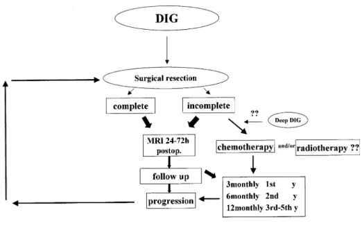
lit-erature and our own experience that only follow-up by im-aging is justified in these partially resected DIGs [3, 11, 14]. In cases where the tumor cannot be completely re-moved in one step the child should be allowed to recover and a second operation should be considered, as in our first case. In Fig. 4 we show a therapeutic plan for the management of a desmoplastic infantile ganglioglioma.
There may be a need for adjuvant chemotherapy in cases of deep seated tumors with aggressive behavior. However, because of the small number of patients, our conclusions should be interpreted with caution and fur-ther investigations are needed. We should think about a multicentric interdisciplinary study group that could col-lect data and give advice about the therapy for these rare tumors.
365
Fig. 4 Therapeutic plan for desmoplastic infantile ganglio-glioma
References
1. De Munnynck K, Van Gool S, Van Calenbergh F, Demaerel P, Uyttebroeck A, Buyse G, Sciot R (2002) Desmo-plastic infantile ganglioglioma: a potentially malignant tumor? Am J Surg Pathol 26:1515–1522 2. Duffner PK, Burger PC, Cohen ME,
Sanford RA, Krischer JP, Elterman R, Aronin PA, Pullen J, Horowitz ME, Parent A, Martin P (1994) Desmo-plastic infantile ganglioglioma: an approach to therapy. Neurosurgery 34:583–589
3. Duffner PK, Krischer JP, Horowitz ME, Cohen ME, Burger PC, Friedman HS, Kun LE (1998) Second malignan-cies in young children with primary brain tumors following treatment with prolonged postoperative chemotherapy and delayed irradiation: a Pediatric Oncology Group Study. Ann Neurol 44:313–316
4. Fan X, Larson TC, Jennings MT, Tulipan NB, Ton SA, Johnson MD (2001) December 2000: 6 month old boy with 2 week history of pro-gressive lethargy. Brain Pathol 11:265–266
5. Galatioto S, Gullotta F (1996) Desmo-plastic non-infantile ganglioglioma. J Neurosurg Sci 40:235–238
6. Komori T, Scheithauer BW, Parisi JE, Watterson J, Priest JR (2001) Mixed conventional and desmoplastic infan-tile ganglioglioma: an autopsied case with 6-year follow up. Mod Pathol 14:720–726
7. Kros JM, Delwel EJ, de Jong TH, Tanghe HL, van Run PR, Vissers K, Alers JC (2002) Desmoplastic infantile astrocytoma and ganglioglioma: a search for genomic characteristics. Acta Neuropathol (Berl) 104:144–148
8. Kuchelmeister K, Steinhauser A, Korf B, Wagner D, Prey N, Schachenmayr W (1996) Anaplastic desmoplastic infantile ganglioglioma. Clin Neuro-pathol 15:280
9. Kuchelmeister K, Schonmeyr R, Albani M, Schachenmayr W (1998) Anaplastic desmoplastic infantile ganglioglioma. Clin Neuropathol 17:269–270
10. Louis DN, von Deimling A, Dickersin GR, Dooling EC, Seizinger BR (1992) Desmoplastic cerebral astrocytomas of infancy: a histopathologic, immuno-histochemical, ultrastructural, and molecular genetic study. Human Pathol 23:1402–1409
11. Mallucci C, Lellouch A, Salazar C, Cinalli G, Renier D, Sainte-Rose C, Kahn A, Zerah M (2000) The manage-ment of desmoplastic neuroepithelial tumors in childhood. Childs Nerv Syst 16:8–14
12. Marti A, Almostarchid B, Maher M, Saidi A (2000) Desmoplastic non-infantile ganglioglioma. Case report. J Neurosurg Sci 44:150–154 13. Parisi JE, Scheithauer BW, Priest JR,
Okazaki H, Komori T (1992) Desmo-plastic infantile ganglioglioma (DIG): a form of ganglio-gliomatosis. J Neuropathol Exp Neurol 51:365 14. Rout P, Santosh V, Mahadevan A, Kolluri VR, Yasha TC, Shankar SK (2002) Desmoplastic infantile ganglio-glioma—clinicopathological and im-munohistochemical study of four cases. Childs Nerv Syst 18:463–467
15. Setty SN, Miller DC, Camras L, Charbel F, Schmidt ML (1997) Desmoplastic infantile astrocytoma with metastases at presentation. Mod Pathol 10:945–951 16. Taratuto AL, Monges J, Lylyk P,
Leiguarda R (1984) Superficial cere-bral astrocytoma attached to dura. Report of six cases in infants. Cancer 54:2505–2512
17. Taratuto AL, VandenBerg SR, Rorke LB (2000) Desmoplastic infantile as-trocytoma and ganglioglioma. In: Kleihues P, Cavanee WK (eds) Pathol-ogy and genetics of tumours of the ner-vous system. IARC, Lyon, pp 99–102
18. VandenBerg SR (1993) Desmoplastic infantile ganglioglioma and cerebral astrocytoma of infancy. Brain Pathol 3:275–281
19. VandenBerg SR, May EE, Rubinstein LJ, Herman MM, Perentes E, Vinores SA, Collins VP, Park TS (1987) Desmoplastic supratentorial neuroepi-thelial tumors of infancy with diver-gent differentiation potential (“desmo-plastic infantile gangliogliomas”). Report on 11 cases of a distinctive embryonal tumor with favorable prognosis. J Neurosurg 66:58–71 20. Woesler B, Kuwert T, Kurelmann G,
Morgenroth C, Probst-Cousin S, Lerch H, Gulotta F, Wassmann H, Schober O (1998) High amino acid uptake in a low grade desmoplastic infantile ganglioglioma in a 14-year-old patient. Neurosurg Rev 21:31–35