Received: 13 October 2003 Revised: 5 March 2004 Accepted: 30 March 2004 Published online: 21 April 2004 © Springer-Verlag 2004
Abstract The purpose of this study was to investigate the modified three-point Dixon technique as a method for obtaining fat-saturated T1-weighted sequences before and after intravenous gadolinium admin-istration using an open MR imaging scanner. A preliminary experiment using an oil/gadolinium phantom was performed on a 0.35-T open magnet and an advanced 1.5-T unit. Fat saturation was achieved at 1.5 T using a frequency selective presatu-ration technique and a modified three-point Dixon technique on the low-field scanner. The modified three-point Dixon sequence was then evaluated in ten patients undergoing MRI examinations of the spine with gadolinium enhancement to deter-mine image characteristics and diag-nostic potential. The phantom study demonstrated a homogenous sup-pression of signal from oil and a good distinction between fat and a
gadolinium chelate on the 0.35-T unit comparable to that on the 1.5-T scanner. By applying the modified three-point Dixon technique on the open-magnet, the distinction be-tween fat and gadolinium dimeglu-mine was rated as very good in 139 and good in 17 axial slices in a total of 156 images. No image was rated as difficult or not possible. Motion artifacts that hampered the reading were detected in the lower cervical spine due to respiratory movement in four (3% of all) images. The modified three-point Dixon tech-nique provides the combination of gadolinium enhancement with fat saturation on an open magnet. Early clinical applications appear promis-ing.
Keywords Three-point Dixon technique · Fat suppression · Contrast-enhancement · MRI Rolf W. Huegli Phillip F. J. Tirman Harald M. Bonel Harald Staedele Souhil Zaim Mikayel Grigorian Harry K. Genant
Use of the modified three-point Dixon
technique in obtaining T1-weighted
contrast-enhanced fat-saturated images
on an open magnet
Introduction
T1-weighted, contrast-enhanced, fat-suppressed imaging compared to non-fat-suppressed images is a well-estab-lished method to evaluate pathology made more conspic-uous by contrast enhancement [1–5]. Open MR imaging is an emerging technology with certain advantages com-pared to a whole body scanner, especially for claustro-phobic patients and MR interventions. Additionally, low-field open MR imagers cost less than high-low-field super-conducting magnets. One major drawback of these sys-tems has been the inability to achieve T1-weighted
fat-saturated imaging and therefore no reliable distinction between fat and contrast media [6].
Recently, a new technique has been developed using three-point Dixon chemical shift imaging tested in the musculoskeletal field for acute bone fractures [7] and cartilage evaluation in the knee joint [8]. These studies demonstrate that a reliable and homogeneous fat sup-pression is achievable on a 0.35-T scanner.
This study was conducted to answer the following questions: is a distinction between fat and gadolinium possible in contrast-enhanced images on a 0.35-T low-field scanner, how frequently do motion and phase arti-R. W. Huegli · H. M. Bonel · S. Zaim
M. Grigorian · H. K. Genant
Osteoporosis and Arthritis Research Group, Department of Radiology,
University of California, 350 Parnassus Ave., Suite 150, San Francisco, CA 94117, USA R. W. Huegli (
✉
) · H. Staedele Departement Medizinische Radiologie, Kantonsspital Basel, Universitätskliniken, Petersgraben 4, 4031 Basel, Switzerland e-mail: [email protected]Tel.: +41-61-2652525 Fax: +41-61-2655383 P. F. J. Tirman
National Orthopaedic Imaging Associates, Marin Magnetic Imaging,
facts occur in examinations of the spine, and how useful is this sequence for routine application in a clinical setting?
Materials and methods
The modified three-point Dixon technique utilized in this study was supplied with a 0.35-T Toshiba Opart open magnet (Toshiba America MRI, South San Francisco, CA). The technique we used is based on the chemical shift imaging technique originally de-scribed by Dixon [9] and modified by Zhang in 1996 [10]. This imaging method can be applied to a low-field magnet, utilizing a single radiofrequency echo single-scan method for water and fat separation with correction of the static field inhomogeneities. Phantom measurements
In a pilot study, test images were acquired using a liquid phantom consisting of an inner container filled with diluted gadopentate dimeglumine (0.1 mmol/kg H2O) surrounded by olive oil in an outer container. The phantom was placed in a commercially avail-able standard head coil. The head coil as the most homogeneous coil was chosen in order to differentiate artifacts caused by short-comings of the sequence and not by coil inhomogeneity.
The phantom measurements on the 0.35-T Toshiba Opart open magnet were performed using a conventional 2D T1-weighted spin-echo sequence [450/15 (repetition time in ms/echo time in ms); section thickness, 4 mm; number of signals acquired, 3; ma-trix, 256×256; field of view, 17×12 cm] and a T1-weighted 2D gradient-echo three-point Dixon study (water field echoes, WFE) for fat-saturated imaging [750/20/±10.2 (repetition time in ms/echo time in ms/τ in ms); flip angle, 90°; section thickness, 4 mm; number of signals acquired, 2; matrix, 256×176; field of view, 17.0×12.8 cm]. Likewise, on the 1.5-Tesla unit (Signa, Gen-eral Electric Medical Systems, Milwaukee, WI), spin-echo 2D T1-weighted images [300/16 (repetition time in ms/echo time in ms); section thickness, 4 mm; number of signals acquired, 1; matrix, 256×256; field of view, 12×12 cm] and fat-saturated 2D T1-weighted images using a conventional frequency selective fat sat-uration pulse [360/12 (repetition time in ms/echo time in ms); sec-tion thickness, 4 mm; number of signals acquired, 1; matrix, 256×160; field of view, 12×12 cm] were obtained.
Clinical MR examinations
During a period of 6 months in 1999, 257 patients were referred to the open low-field magnet for an MRI examination of the spine, the majority with benign and chronic complaint. A total of ten pa-tients (five men and five women, with an age range of 23–56 years and a median age of 48 years) formed our study population, and gadopentetate dimeglumine (0.1 mmol/kg bw; Magnevist; Berlex Laboratories, Inc.; Montville, NJ) was administrated intravenously. Indications for the contrast-enhanced MR exams were suspected infection, bone marrow disease or persistent postoperative back pain. The cervical spine was scanned in three cases, and the lumbar spine in seven. Written informed consent was obtained from all pa-tients prior to the MR exam. All clinical MR imaging examinations were performed on a Toshiba Opart 0.35-T open magnet.
Axial T1-weighted spin-echo images and fat-saturated images with the modified three-point Dixon method were obtained in addition to those acquired as part of the routine protocol. The modified three-point Dixon technique was added to gain contrast-enhanced fat-saturated images.
Axial T1-weighted 2D gradient-echo three-point Dixon studies [700–750/20/±10.2 (repetition time in ms/echo time in ms/τ in ms); flip angle, 90°; section thickness, 4–5 mm; number of signals acquired, 1–2; matrix, 192×256–256×256; field of view, 18×18–
23×23 cm; no phase wrap to avoid aliasing] were obtained consec-utively. The relatively long TR was chosen for the additional num-ber of slices available in order to cover the anatomic region in the shortest time possible. The number of slices for a given TR utiliz-ing the three-point Dixon technique is slightly reduced when com-pared to a conventional technique.
The bandwidth was 25 kHz. Adding three images together as in the three-point Dixon technique equate the water field echo sequence to correspond to a bandwidth of 8.5 kHz compared to 10 kHz for a non-fat-suppressed sequence. The acquisition time for the complete Dixon study of the lumbar or cervical spine last-ed between 4.5 and 6.4 min. Commercially available combinlast-ed solenoidal and linear coils were utilized for the studies of the cer-vical and lumbar spine.
Evaluation protocol
Three experienced radiologists analyzed the axial slices of the modified three-point Dixon studies retrospectively in one session. The images of all studies were presented for each MR examination at the time of reading. A consensus reading was performed by all three radiologists. The fat-only images were not used at any stage of the diagnostic evaluation.
The following image characteristics were part of the evaluation: (1) the distinction between fat and gadolinium was rated on an ordinal scale as very good, good, difficult or not possible; (2) the frequency of motion and phase artifacts were documented as no displacement, minor displacement (no hampering of the read out) or major displacement resulting in an image quality below diagnos-tic requirements; (3) finally, a decision was made on the usefulness of routine reporting of the modified three-point Dixon sequence.
Results
Phantom
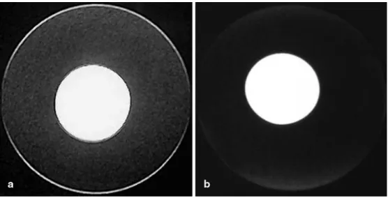
Figure 1 illustrates the results of the phantom measure-ments with the gadopentate dimeglumine container in the center surrounded by olive oil. The fat-saturated T1-weighted image in Fig. 1a obtained with the modified three-point Dixon technique reveals a homogeneous fat suppression comparable in quality to the T1-weighted 2D image (Fig. 1b) obtained with the conventional pre-saturation technique on the high-field scanner.
The two thin but distinct crescent-shaped bright rings seen in the periphery surrounding the olive oil container on the modified three-point Dixon image represent a dou-ble vision artifact. The doudou-ble vision artifact as a signal contamination is observed at the edge of the compart-ments because of a difference in geometric distortion be-tween the in-phase RF echo image and the two out-of-phase gradient-echo images [10]. This artifact was not observed in any of our clinical studies, but typically oc-curs on sharp edges in the periphery of the image.
Patients
Distinction between fat and gadolinium. The distinction between fat and gadolinium dimeglumine was rated as very good in 139 (89%) (Fig. 2) or good in 17 (11%)
Fig. 1 MR images of the
gadolinium (inner container)/ oil (outer container) phantom acquired applying the WFE (gradient-echo) sandwich sequence for the fat separation images. a Modified three-point Dixon sequence on the 0.35-T open magnet revealing a homogenous fat suppression comparable in quality to the fat-suppressed spin-echo sequence of a 1.5-T unit b.
Two thin, crescent-shaped, bright rings surround the olive
oil container and represent a double vision artifact of the modified three-point Dixon image a
Fig. 2 Axial MR images of the
cervical spine in a 23-year-old patient with pain in the upper right neck (marker indicated by
curved arrow). a Pre-contrast
spin-echo T1-weighted image showing several round masses (solid arrows), b which are not suppressed in a STIR sequence.
c The three-point Dixon image
reveals a homogeneous fat suppression, and the left-sided enhancing mass representing swollen lymph nodes in the course of metastatic disease. Discrete susceptibility artifacts can be recognized on the air/soft tissue interface (arrowhead)
axial slices (Fig. 3). No image was rated as difficult or not possible.
Frequency of motion and phase artifact. In 117 (75%) images, displacement artifacts were completely absent. Minor displacement artifacts that did not hamper the reading occurred in 35 (22%) images. Major displace-ment artifacts seen in four (3%) images were due to chest movement in a lower cervical spine exam.
Usefulness for routine examination. Thirty-six scanned disc levels, out of a total of 37 covered in axial planes, were useful for routine reporting. The single unanalyz-able scan was due to the chest movement artifact men-tioned above.
Discussion
Suppression of the relatively high signal intensity of fat in T1-weighted images leads to a more efficient use of the dynamic range of gray levels used to display the tis-sue contrast of MR images enhanced by paramagnetic agents. Therefore, the conspicuity of enhancing tissues after injection of T1-shortening gadolinium chelates is increased on T1-weighted images by using fat suppres-sion, because the images devoid of the high fat signal are rescaled [2–4].
The STIR and the frequency selective presaturation pulse techniques are routine sequences on every
stan-dard, up-to-date high-field MR imaging unit and are effective fat suppression techniques [2, 11, 12]. Intra-venous gadolinium offers an additional diagnostic ad-vantage [13, 14]. Because intravenous paramagnetic con-trast material shortens the T1-relaxation of enhancing tissues to the same range as that of fat, the STIR se-quence is not applicable in fat-suppressed contrast-enhanced MR imaging, as fat and enhancing tissues are equally suppressed [15]. Also, average image signal is frequently small in comparison to the high noise levels in STIR imaging, which is a major drawback for the utility of this sequence in routine imaging.
Another fat suppression technique applied on high-field scanners is based on the chemical shift between water and fat using a frequency-selective presaturation technique [11, 12, 16]. The main magnetic field of low-field open magnets is considerably poorer in homogene-ity and uniformhomogene-ity than that of conventional supercon-ductive MR scanners, particularly at the periphery of the gantry. The main obstacle to achieving an acceptable fat suppression on low-field magnets is the fact that the spectral separations between the fat and water proton pools are too narrow to allow selective excitation of a single resonance. Frequency-selective fat suppression techniques are therefore not applicable on a low-field open scanner [6].
An alternative approach is the chemical shift imaging as first introduced by Dixon. It offers the opportunity to apply appropriate post-processing, which results in con-trast-enhanced fat-suppressed images even on a low-field open magnet [9]. In the original version, two images (two Point Dixon technique) were acquired with fat and water in-phase and out-of-phase, respectively.
In this study, we applied a single RF echo, single-scan method for water and fat separation first described by Zhang et al. [10]. By adding a third acquisition (in our study an out-of-phase image), off-resonance phase accu-mulations can be achieved on a pixel-by-pixel basis and provide the possibility to calculate a B0 field map. This map is used to compensate for phase shifts caused by B0 inhomogeneities in each pixel and therefore allows a more accurate decomposition into water-specific and fat-specific images [10].
Various three-point Dixon methods have been applied in a broad variety of clinical applications. One potential problem in previous studies was the relatively long acquisition time needed to acquire multiple in-phase and out-of-phase images [17]. The major advantage of the modified three-point Dixon sandwich technique applied in this study is the greatly reduced scan time. The use of a relatively long TR provided an acceptable trade-off be-tween complete anatomic coverage and sufficient T1-weighting. By combining three acquisitions in a single scan, the scan time of the “sandwich sequence” is only one-third of that of a single-echo method [18]. This is a very important feature in a clinical setting, where scan
Fig. 3 Axial MR images of a lumbar spine demonstrating a fat
suppression artifact in a 56-year-old patient without a pathological finding. Modified three-point Dixon sequence revealing a non-uniform fat suppression with a paradoxical water saturation in the right psoas muscle region (arrowhead). This artifact can be found on high-field and low-field scanners because the three-point Dixon technique is like the presaturation pulse technique on high-field magnets, a frequency-dependent fat saturation method rely-ing on magnetic field homogeneity
time is relevant from an economical point of view as well as for the patients’ comfort. The sandwich tech-nique is not as prone to image displacement artifacts as the multiple echo methods because of the shorter acqui-sition time of the complete scan (4.5–6.4 min in our study for a lumbar or cervical spine) and the shorter lag between the three acquisitions. We encountered major displacement artifacts that interfered with our reading in only four (3% of all) images. This artifact on one single disc level was due to chest movement in a lower cervical spine exam. Movement artifacts, however, can signifi-cantly deteriorate the image quality of all MR sequences that are in routine use today. Still, in comparison to prior versions of the three-point Dixon sequence, the sandwich technique contributes to the reduction of movement arti-facts by shortening the imaging time and the time lag be-tween acquisitions of the multiple image layers.
The phantom measurements on the open low-field magnet (applying the modified three-point Dixon tech-nique) revealed a very effective distinction between homogeneous fat suppression of the gadolinium chelate, comparable in quality to the presaturation pulse tech-nique on the high-field unit. The chemical shift artifact in the frequency encoding direction was not as pro-nounced on the low-field unit as the high-field scanner. However, a double vision artifact (Fig. 1a) was noticed on the modified three-point Dixon image on the low-field open scanner. This artifact was detected on the sharp edges at the periphery of the phantom and was caused by a difference in the geometric distortion be-tween the in-of-phase and out-of-phase images. The artifact was not noted on any of the clinical images, even after a thorough review. An algorithm to correct the double vision artifact has been created [10] and is in the process of undergoing clinical validation for application to routine imaging.
The results of the present clinical study indicate that the modified three-point Dixon chemical shift imaging sandwich technique offers a good distinction between fat and paramagnetic contrast media in all exams of the
cer-vical and lumbar spine. The capability of acquiring im-ages with and without fat saturation before and after an intravenous gadolinium administration on a low-field open magnet is a valuable development. This practical new sequence might be an advantageous addition to ex-isting imaging protocols in diagnostic situations in which the anatomic structures of interest are enhanced only modestly with paramagnetic contrast.
The proposed sequence may have implications on MR-guided interventional procedures, for example, for percutaneous abdominal and skeletal biopsies, which combine the advantages of an open magnet with the ability to apply contrast agents and fat saturation at low-field strength [19].
Further studies using the modified three-point Dixon sandwich technique and its correction algorithms are necessary, and additional trials directly comparing the standard fat suppression techniques applied on high-field scanners should be performed. This study, however, shows that this new sequence is valuable for routine re-porting and that further research should be pursued to improve this sequence type.
Conclusion
The modified three-point Dixon sandwich technique pro-vides a method of gaining acceptable, fat-suppressed contrast-enhanced images of cervical and lumbar spine exams on a 0.35-T open MR system. Combining three echoes in a single-scan offers the added advantages of acquiring fast and reliable contrast-enhanced fat-sup-pressed images even when B0 homogeneity is limited. Displacement artifacts occur in a very small fraction of the images. The first clinical application appears promis-ing.
Acknowledgments The authors would like to thank Weiguo
Zhang, PhD, and Yves Miaux, MD PhD, for their scientific sup-port as well as Victoria Vandenberg for editing the final version of this paper.
References
1. Daniel BL, Butts K, Glover GH, Cooper C, Herfkens RJ (1998) Breast cancer: gadolinium-enhanced MR imaging with a 0.5-T open imager and three-point Dixon technique. Radiology 207:183–190
2. Hirsch JA, Loevner LA, Yousem DM, Siegelman ES, Keiper MD, Marquis RP, Grossman RI (1998) Gadolinium-enhanced fat-suppressed T1-weighted imaging of the head and neck: comparison of gradient and conven-tional SE sequences. J Comput Assist Tomogr 22:771–776
3. Battolla L, Zampa V, Mascalchi M, Falaschi F, Bagnolesi P, Tessa C, Paolicchi A, Bartolozzi C (1994) Gadolinium-DTPA combined with a fat suppression technique in studies with magnetic resonance at 0.5 T in soft-tissue neoplasms. Radiol Med (Torino) 88:353– 358
4. Schreiber WG, Brix G, Knopp MV, Hess T, Lorenz WJ (1996) Improved visualization of breast lesions with gadolinium-enhanced magnetization transfer MR imaging. Magn Reson Med 35:861–869
5. Longo M, Granata F, Ricciardi K, Gaeta M, Blandino A (2003) Contrast-enhanced MR imaging with fat suppression in adult-onset septic spondylodiscitis. Eur Radiol 13:626– 637
6. Peterfy CG, Roberts T, Genant HK (1998) Dedicated extremity MR imaging: an emerging technology. Magn Reson Imaging Clin N Am 6:849–870
7. Wohlgemuth WA, Roemer FW, Bohndorf K (2002) Short tau inversion recovery and three-point Dixon water-fat separation sequences in acute trau-matic bone fractures at open 0.35 tesla MRI. Skeletal Radiol 31:343–348 8. Bredella MA, Losasso C, Moelleken
SC, Huegli RW, Genant HK, Tirman PF (2001) Three-point Dixon chemical-shift imaging for evaluating articular cartilage defects in the knee joint on a low-field-strength open magnet. AJR Am J Roentgenol 177:1371–1375 9. Dixon WT (1984) Simple proton spectroscopic imaging. Radiology 153:189–194
10. Zhang W, Goldhaber DM, Kramer DM (1996) Separation of water and fat MR images in a single scan at 0.35 T using “sandwich” echoes. J Magn Reson Imaging 6:909–917
11. Mao J, Yan H, Bidgood WD Jr (1992) Fat suppression with an improved selective presaturation pulse. Magn Reson Imaging 10:49–53
12. Keller PJ, Hunter WW Jr, Schmalbrock P (1987) Multisection fat-water imag-ing with chemical shift selective pre-saturation. Radiology 164:539–541 13. Tang Y, Yamashita Y, Arakawa A,
Namimoto T, Mitsuzaki K, Abe Y, Katahira K, Takahashi M (1999) Detection of hepatocellular carcinoma arising in cirrhotic livers: comparison of gadolinium- and ferumoxides-enhanced MR imaging. AJR Am J Roentgenol 172:1547–1554 14. Spencer JA, Ward J, Guthrie JA,
Guillou PJ, Robinson PJ (1998) Assessment of resectability of pancre-atic cancer with dynamic contrast-enhanced MR imaging: technique, surgical correlation and patient out-come. Eur Radiol 8:23–29
15. Rubin DA, Kneeland JB (1994) MR imaging of the musculoskeletal system: technical considerations for enhancing image quality and diagnostic yield. AJR Am J Roentgenol 163:1155–1163
16. Bobman SA, Atlas SW, Listerud J, Grossman RI (1991) Postoperative lumbar spine: contrast-enhanced chemical shift MR imaging. Radiology 179:557–562
17. Hardy PA, Hinks RS, Tkach JA (1995) Separation of fat and water in fast spin-echo MR imaging with the three-point Dixon technique. J Magn Reson Imaging 5:181–185
18. Glover GH, Schneider E (1991) Three-point Dixon technique for true water/ fat decomposition with B0 inhomo-geneity correction. Magn Reson Med 18:371–383
19. Adam G, Bucker A, Nolte-Ernsting C, Tacke J, Gunther RW (1999) Interven-tional MR imaging: percutaneous abdominal and skeletal biopsies and drainages of the abdomen. Eur Radiol 9:1471–1478