ORIGINAL PAPER
Gómez
–López-Hernández syndrome: reappraisal
of the diagnostic criteria
Biayna Sukhudyan&Varsine Jaladyan&
Gayane Melikyan&Jan Ulrich Schlump&
Eugen Boltshauser&Andrea Poretti
Received: 6 July 2010 / Revised: 10 July 2010 / Accepted: 12 July 2010 / Published online: 23 July 2010 # Springer-Verlag 2010
Abstract Gómez–López-Hernández syndrome (GLHS) is a rare and possibly underdiagnosed condition. So far, 21 patients have been reported and all of them were sporadic observations. We report six additional patients. The hallmark triad of GLHS, also named cerebellotrigeminal dermal dysplasia, consists of rhombencephalosynapsis, trigeminal anesthesia (often giving rise to corneal opacities), and bilateral parietal or parieto-occipital alopecia. Our patients had rhombencephalosynapsis and alopecia, but none had trigeminal dysfunction. In this respect, the term cerebellotrigeminal dermal dysplasia is potentially misleading. In conclusion, only rhombencephalo-synapsis and alopecia are consistently present in GLHS and are required diagnostic criteria, while trigeminal anesthesia, dys-morphic features, and ataxia are inconsistent findings. A high index of suspicion is required to diagnose GLHS, particularly as alopecia tends to be hidden by surrounding scalp hair.
Keywords Gómez–López-Hernández syndrome .
Alopecia . Ataxia . Trigeminal anesthesia . Rhombencephalosynapsis
Introduction
Gómez–López-Hernández syndrome (GLHS; MIM 601853), also called cerebellotrigeminal dermal dysplasia, is a rare syndrome characterized by the triad of rhombencephalosy-napsis, trigeminal anesthesia, and bilateral parietal or
parieto-occipital alopecia [4, 15]. Rhombencephalosynapsis is
defined by absence of the cerebellar vermis, fusion of the cerebellar hemispheres across the midsagittal plane (best seen as abnormal horizontal orientation of the cerebellar folia on posterior coronal images), dentate nuclei appearing as an almost single structure consisting of contributions by left and right hemispheres, and fusion of the superior cerebellar peduncles. Inconsistent addi-tional features of GLHS include craniofacial dysmorphic signs (such as a brachy-turricephaly (without craniosy-nostosis), midface hypoplasia because of underdeveloped malar and maxillar bones (giving rise to downward slanted palpebral fissures and relative prognathism), hypertelorism, low-set, posteriorly rotated ears, ptosis, broad nasal tip, thin upper vermillion, and downturned corners of the mouth), strabismus, short stature, and corneal opacities secondary to trigeminal anesthesia. Cognitive functions are usually impaired, but four patients without cognitive impairment have been described
[11,15,17,23].
Recently, we reported four patients calling attention to
the observation that the diagnosis is easily missed [15].
Currently, we are aware of 21 published patients [2–6,10,
11,15,17,18,20,23,25]. The aim of this study is (1) to
describe six additional patients, four were diagnosed in Armenia following a teaching course on cerebellar malfor-mations, and (2) to reappraise the value of the traditional (diagnostic) triad.
B. Sukhudyan
:
V. JaladyanDepartment of Pediatric Neurology,“Arabkir” Joint Medical
Center and Institute of Child and Adolescent Health, Yerevan, Armenia
G. Melikyan
Division of Neurology and Epileptology, “Erebouni” Medical Center,
Yerevan, Armenia
J. U. Schlump:E. Boltshauser (*)
:
A. PorettiDepartment of Pediatric Neurology,
University Children’s Hospital,
Steinwiesstrasse 75, 8032 Zurich, Switzerland
e-mail: Eugen.Boltshauser@kispi.uzh.ch DOI 10.1007/s00431-010-1259-7
T able 1 Clinical and neuroimaging findings in six patients with Gómez –López-Hernández syndrome Patients 1 2 3 4 5 6 Sex Male Male Male Male Male male Nationality Armenian Armenian Armenian Armenian Swiss Swiss-Polish Age at first signs and symptoms Neonate Infant Infant Infant Neonate Infant First signs and symptoms Feeding problems Global dev . delay Floppy child V entriculomegaly Hydrocephalus Motor dev . delay Age at study (years) 12 7 2 20 1.5 12 Brain MRI R h o m b en ce p h al o sy n ap si s + + + + + + Other abnormalities −− − Mild vm, cleft b Hydrocephalus − Central nervous system T runcal ataxia + − n.a. − n.a. − Dysmetria + − n.a. − n.a. − Muscular hypotonia + + + − + − Head nodding − ++ + − + T rigeminal anesthesia −− − − − − Corneal opacities −− − − − − Cognitive functions Perf. IQ = 5 9 Perf. IQ = 7 8 Normal development Full-scale IQ = 9 0 E Q 9 0 Full-scale IQ = 1 14 Craniofacial Alopecia T emporal a Parietal right, temporal left Parietal T emporal T emporal T emporal Brachycephaly −− ++ + − Midface hypoplasia + + − ++ − Hypertelorism −− − − + − Low-set, posteriorly rotated ears ++ + − + − Growth Short stature (P <3 ) + + −− − − Additional signs Nasal voice, mild ptosis, strabismus, cryptorchidism, relative prognathism Strabismus, epileptic seizures, agenesis of the right kidney Strabismus Mild ptosis − Nasal V oice Dev . developmental, EQ developmental age, IQ intelligence quotient, vm ventriculomegaly aAdditional small areas of alop ecia in the ver tex area b Cleft in the left brainste m –ce rebella r junction
Patients and methods
Patients
Between April and November 2009, we diagnosed GLHS
in six new patients. In four of them (patients 1–4), the
diagnosis was made after a teaching course in Armenia. Patients 5 and 6 are of Swiss and Swiss-Polish origin, respectively. All patients in this series are males. There was no parental consanguinity. Two patients presented neona-tally with feeding problems and hydrocephalus. In the other four children, the first symptoms appeared in infancy as global or motor developmental delay, marked muscular hypotonia, and ventriculomegaly. At the time of this study, the median age was 9.5 years (range 1.5 to 20 years).
Methods
Cognitive impairment was defined as an IQ of <70. Cognitive functions were tested in four patients according to age with the non-verbal part of the Kaufman Assessment Battery for Children-II (n=2); currently, there is no validated Armenian
verbal part of this test [9], Hamburg-Wechsler-Intelligenztest
für Kinder III (n=1) [21], and Wechsler Intelligenztest für
Erwachsene III (n=1) [24]. In one infant (patient 5), the
developmental age was tested using the third edition of the Griffith mental developmental scales. Patient 3 was not formally tested: Information about his development has been obtained from history and clinical observations.
Results
The relevant clinical and neuroimaging data are summarized
in Table1.
Typical signs of rhombencephalosynapsis were present in all patients. Additionally, the MRI of patient 4 showed a
cleft at the left brainstem–cerebellar junction (Fig.1). Other
neuroimaging abnormalities included ventriculomegaly and hydrocephalus due to stenosis of the Sylvian aqueduct in one patient.
Ataxia (truncal and limb) was found only in one patient, while muscular hypotonia and horizontal side-to-side head nodding were present in four patients.
Cognitive functions could be assessed in four patients. In two patients, full-scale IQ was measurable and was normal in both cases (90 and 114, respectively). In patient 6, verbal IQ was 106 and performance IQ 120. In patients 1 and 2, only performance IQ could be assessed (59 and 78, respectively). Due to his young age, in one patient, only developmental age, not IQ, could be assessed and this was normal (EQ 90). Cognitive development of patient 3 could not be formally tested, but information obtained from history and clinical observations showed normal development at the age of 2 years.
Bilateral alopecia was present from birth in all patients.
It was bitemporal in four cases (Figs. 2b, 3b, and 4a, b),
temporal on one side and parietal on the other in one, and biparietal in one boy. Patient 1 had additional small areas of
alopecia frontally and in the region of the vertex (Fig.2a).
In three patients, the alopecia could be perfectly hidden by the surrounding hairs, and in three cases, it was asymmetric. Interestingly, patient 4 had an assisted birth with forceps, and bilateral alopecia has been interpreted as residual after forceps delivery.
Trigeminal sensation was normal in all patients, corneal reflexes could be elicited in all cases, and corneal opacities were not detectable in any patient.
Craniofacial dysmorphic signs were present in five of
six patients: midface hypoplasia (Figs.2band3b) and
low-set, posteriorly rotated ears (Fig.2b) in four, brachycephaly
Fig. 1 Brain MRI of patient 4 at the age of 20 years. a Axial T2-weighted MRI showing fusion of the cerebellar hemispheres with no intervening vermis and a cleft in the left
brainstem–cerebellar junction
(white arrows). b Posterior coronal T1-weighted MRI demonstrating fused cerebellar hemispheres, abnormal horizontal orientation of cerebellar folia, and ventriculomegaly
(Fig. 3b) and strabismus in three, bilateral mild ptosis in
two (Figs. 2a and 3a), and hypertelorism and relative
prognathism (Fig.2b) in one.
Discussion
Gómez–López-Hernández syndrome is a rare, sporadic syndrome and is likely to be underdiagnosed. Parental
consanguinity was reported only in one of 27 patients [6].
Males are markedly more likely to be affected than females (19 males, eight females), but in terms of disease severity, we did not found a gender-related difference. So far, genetic analyses (karyotype, CGH arrays) have not yielded abnormal results for GLHS, or for isolated rhombencephalosynapsis either. All patients (GLHS and rhombencephalosynapsis) are sporadic observations. Therefore, either spontaneous domi-nant mutations or de novo chromosomal rearrangements are
possible explanations [15]. From this point of view, GLHS is
an excellent candidate to be used in a total exome sequencing project. It is not certain whether GLHS and (isolated) rhombencephalosynapsis are different entities, or whether they represent a disease spectrum. Therefore, at this point, both rhombencephalosynapsis and GLHS need to be further defined by neuroimaging and other clinical features, respectively.
According to MIM catalogue and literature reports, GLHS is characterized by a triad including rhombencepha-losynapsis, bilateral parietal or parieto-occipital alopecia, and trigeminal anesthesia. Typical additional findings include several craniofacial dysmorphic signs (brachy-turricephaly, midface hypoplasia, hypertelorism, and low-set, posteriorly rotated ears) and neurological findings (ataxia, muscular hypotonia, cognitive impairment, and strabismus).
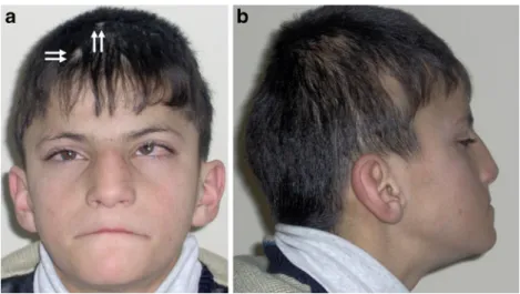
Rhombencephalosynapsis constitutes a consistent key feature of GLHS and was reported in all but one case (not reported in the first description by Gómez because neuro-Fig. 2 Patient 1 at the age of
12 years. a Front view showing broad forehead, mild bilateral ptosis, strabismus, prominent nose, and small areas of alopecia frontally and in the vertex area (white arrows). b Right side view revealing low-set, posteriorly rotated ears, relative prognathism, midface hypoplasia, and temporal alopecia
Fig. 3 Patient 4 at the age of 20 years. a Front view showing broad, high forehead, mild bilateral ptosis, and slight synophris. b Left side view demonstrating brachy-turricephaly, midface hypoplasia, and temporal alopecia
imaging studies had not been performed). Rhombencepha-losynapsis mostly occurs in isolation but may be associated with other brain anomalies such as hydrocephalus, aplasia of the septum pellucidum, hypoplasia of the corpus callosum, hippocampal abnormalities, and fusion of the
fornices [14,22]. In this series, we found a cleft at the left
brainstem–cerebellar junction in one patient. There are no previous reports of a similar neuroimaging finding in rhombencephalosynapsis, and its pathogenesis and clinical
significance are unknown (Fig.1).
Alopecia has also been found consistently in all but one case (not described in the case reported by Truwit et al.). In GLHS, alopecia is focal, bilateral, congenital, and
predom-inantly located in the parieto-occipital region [4,11,14,18,
19]. However, alopecia can be markedly asymmetric [15]
or, as in one case, even unilateral [4]. Less frequent
localizations are the temporal (as in most cases of this series), frontoparietal, frontotemporoparietal, or temporo-parietal regions. One patient in this report had bitemporal alopecia with additional small areas of alopecia anteriorly
and on the region of the vertex (Fig.2). Similar additional
alopecia areas have not been previously reported. In some cases, alopecia may be easily concealed by the surrounding scalp hair and must be carefully looked for.
All the other clinical and neurological signs and findings do not occur consistently in published GLHS cases, as well as in the cases reported here.
Trigeminal anesthesia most often affects the ophthalmic branch, causing abnormal sensation of the forehead and cornea, and may result in frontal scars and corneal opacities. Trigeminal anesthesia was absent in four of 21
cases [2,6,15, 17]. In the present series, all patients had
normal forehead sensation and corneal reflexes.
Ataxia (truncal and/or limb) and muscular hypotonia are the most common neurological manifestations of
rhomben-cephalosynapsis and GLHS [14,22]. These symptoms can
vary considerably in severity but are mostly mild, and only a few patients are partly restricted in daily life activities. So far, ataxia was present in 17 reported cases, muscular hypotonia in ten. However, only one patient in this report presented with mild truncal and limb ataxia, while muscular hypotonia was detectable in four of six cases. Therefore, both ataxia and muscular hypotonia are frequent, but inconsistent, findings in GLHS.
Head nodding stereotypies consist of side-to-side “no”
movements, up-and-down “yes” movements, or
shoulder-to-shoulder resembling an infinite sign [1,7]. The resulting
disruption may severely interfere with social skills, partic-ularly in children with normal cognitive functions. Head
nodding has been reported in six patients with GLHS [3,
15,20, 25] and was present in four of six children in this
report. It is probably underrecognized. Head nodding has also been reported in nonsyndromic rhombencephalosynapsis
[14] and other cerebellar abnormalities such as unilateral
cerebellar hypoplasia [8,16].
Cognitive impairment to a varying degree has been found in most (13 of 21) previously reported patients. Fig. 4 Patient 6 at the age of
12 years. a Right side view showing temporal alopecia. b Left side view revealing less pronounced temporal alopecia. Ears and midface were normal
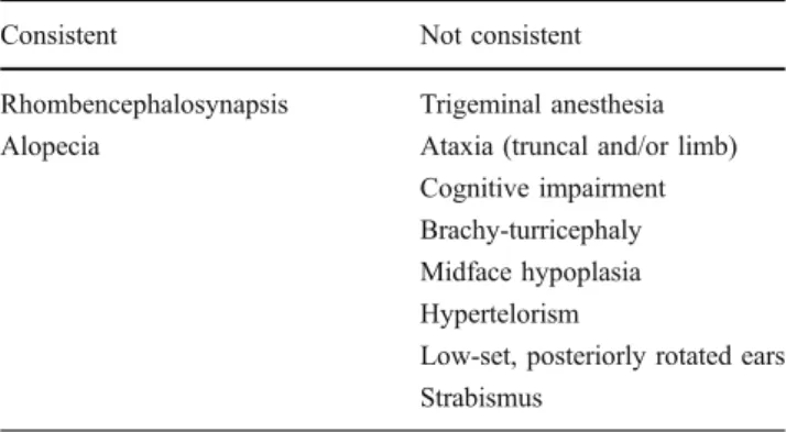
Table 2 Clinical and neuroimaging findings in Gómez
–López-Hernández syndrome
Consistent Not consistent
Rhombencephalosynapsis Trigeminal anesthesia
Alopecia Ataxia (truncal and/or limb)
Cognitive impairment Brachy-turricephaly Midface hypoplasia Hypertelorism
Low-set, posteriorly rotated ears Strabismus
However, four patients [11,15,17,23] as well as three of this report showed normal cognitive functions. Long-term cognitive outcome is also variable in nonsyndromic rhombencephalosynapsis, but cognitive functions are usually
impaired [14]. Nevertheless, both nonsyndromic
rhombence-phalosynapsis and GLHS are compatible with a favorable cognitive outcome in some cases.
Craniofacial phenotype in GLHS typically includes midface hypoplasia, turri- and/or brachycephaly, low-set and posteriorly rotated ears, hypertelorism, and strabismus. These dysmorphic signs are present in most, but not all, cases (as in patient 6 of this report).
Most reported patients with GLHS are not associated with other diseases. In our previous series, one patient suffered from neurogenic bladder dysfunction and another
from esophageal atresia [15]. In this series, one patient had
cryptorchidism and another agenesis of the right kidney. Renal agenesis has been also reported by Pasquier et al. in
one fetal case of rhombencephalosynapsis [13].
In conclusion, only RS and alopecia are consistently present in all GLHS cases and are crucial for the diagnosis
(Table 2). Both the term cerebellotrigeminal dermal
dysplasia and the admission of incomplete forms without rhombencephalosynapsis or alopecia (as proposed by
Fernandez-Jaen et al. [4]) are potentially misleading.
Rhombencephalosynapsis can be diagnosed by fetal MRI
[12, 20]. However, GLHS can only be diagnosed after
birth, as alopecia is only detectable postnatally. In all patients with rhombencephalosynapsis, alopecia must be painstakingly sought (can easily be hidden), and rhomben-cephalosynapsis must be sought if congenital alopecia is present. Otherwise, the diagnosis of GLHS can easily be missed.
Acknowledgments We thank the families for their cooperation and
permission to publish clinical photos.
Conflict of interest None of the authors have any conflict of
interest.
References
1. Bonnet C, Roubertie A, Doummar D et al (2010) Developmental and benign movement disorders in childhood. Mov Disord epub June 18, 2010
2. Bowdin S, Phelan E, Watson R et al (2007) Rhombencephalosy-napsis presenting antenatally with ventriculomegaly/hydrocephalus in a likely case of Gomez–Lopez-Hernandez syndrome. Clin Dysmorphol 16:21–25
3. Brocks D, Irons M, Sadeghi-Najad A et al (2000) Gomez–Lopez-Hernandez syndrome: expansion of the phenotype. Am J Med
Genet 94:405–408
4. Fernandez-Jaen A, Fernandez-Mayoralas DM, Calleja-Perez B et
al (2009) Gomez–Lopez-Hernandez syndrome: two new cases and
review of the literature. Pediatr Neurol 40:58–62
5. Gomez MR (1979) Cerebellotrigeminal and focal dermal dysplasia: a
newly recognized neurocutaneous syndrome. Brain Dev 1:253–256
6. Gomy I, Heck B, Santos AC et al (2008) Two new Brazilian
patients with Gomez–Lopez-Hernandez syndrome: reviewing the
expanded phenotype with molecular insights. Am J Med Genet A 146A:649–657
7. Harris KM, Mahone EM, Singer HS (2008) Nonautistic motor stereotypies: clinical features and longitudinal follow-up. Pediatr Neurol 38:267–272
8. Hottinger-Blanc PM, Ziegler AL, Deonna T (2002) A special type of head stereotypies in children with developmental (?cerebellar) disorder: description of 8 cases and literature review. Eur J
Paediatr Neurol 6:143–152
9. Kaufman AS, Kaufman LN (2004) Kaufman assessment battery for children: manual. AGS, Circles Pines
10. Lopez-Hernandez A (1982) Craniosynostosis, ataxia, trigeminal anaesthesia and parietal alopecia with pons-vermis fusion anomaly (atresia of the fourth ventricle). Report of two cases. Neuropediatrics
13:99–102
11. Munoz RM, Santos AC, Graziadio C et al (1997)
Cerebello-trigeminal-dermal dysplasia (Gomez–Lopez-Hernandez syndrome):
description of three new cases and review. Am J Med Genet 72:34– 39
12. Napolitano M, Righini A, Zirpoli S et al (2004) Prenatal magnetic resonance imaging of rhombencephalosynapsis and associated brain anomalies: report of 3 cases. J Comput Assist Tomogr
28:762–765
13. Pasquier L, Marcorelles P, Loget P et al (2009) Rhombencepha-losynapsis and related anomalies: a neuropathological study of 40
fetal cases. Acta Neuropathol 117:185–200
14. Poretti A, Alber FD, Burki S et al (2009) Cognitive outcome in children with rhombencephalosynapsis. Eur J Paediatr Neurol
13:28–33
15. Poretti A, Bartholdi D, Gobara S et al (2008) Gomez
–Lopez-Hernandez syndrome: an easily missed diagnosis. Eur J Med
Genet 51:197–208
16. Poretti A, Limperopoulos C, Roulet-Perez E et al (2010) Outcome of severe unilateral cerebellar hypoplasia. Dev Med Child Neurol
52:718–724
17. Purvis DJ, Ramirez A, Roberts N et al (2007) Gomez–Lopez-Hernandez syndrome: another consideration in focal congenital alopecia. Br J Dermatol 157:196–198
18. Schell-Apacik CC, Cohen M, Vojta S et al (2008) Gomez–Lopez-Hernandez syndrome (cerebello-trigeminal-dermal dysplasia): description of an additional case and review of the literature.
Eur J Pediatr 167:123–126
19. Tan GM, Arnone D, McIntosh AM et al (2009) Meta-analysis of magnetic resonance imaging studies in chromosome 22q11.2 deletion syndrome (velocardiofacial syndrome). Schizophr Res
115:173–181
20. Tan TY, McGillivray G, Goergen SK et al (2005) Prenatal magnetic
resonance imaging in Gomez–Lopez-Hernandez syndrome and
review of the literature. Am J Med Genet A 138:369–373
21. Tewes U, Rossmann P, Schallberger U (1999) Hamburg-Wechsler-Intelligenztest für Kinder III (HAWIK-III). Hans Huber, Bern 22. Toelle SP, Yalcinkaya C, Kocer N et al (2002)
Rhombencepha-losynapsis: clinical findings and neuroimaging in 9 children.
Neuropediatrics 33:209–214
23. Truwit CL, Barkovich AJ, Shanahan R et al (1991) MR imaging of rhombencephalosynapsis: report of three cases and review of
the literature. AJNR Am J Neuroradiol 12:957–965
24. von Aster M, Neubauer A, Horn R (2006) WIE, Wechsler Intelligenztest für Erwachsene. Harcourt Test Services, Frankfurt am Main
25. Whetsell W, Saigal G, Godinho S (2006) Gomez–Lopez-Hernandez