Dopaminergic Neurons from Oxidative Stress
Peter Bou Dib1,2¤, Bettina Gna¨gi1, Fiona Daly1,3, Virginie Sabado3, Damla Tas3, Dominique A. Glauser4, Peter Meister1, Emi Nagoshi1,3,5*
1 Institute of Cell Biology, University of Bern, Bern, Switzerland, 2 Graduate School for Cellular and Biomedical Sciences, University of Bern, Bern, Switzerland, 3 Department of Genetics and Evolution, University of Geneva, Sciences III, Geneva, Switzerland, 4 Department of Biology/Zoology, University of Fribourg, Chemin du Muse´e, Fribourg, Switzerland,5 PRESTO, Japan Science and Technology Agency, Saitama, Japan
Abstract
Parkinson’s disease (PD) is the most common neurodegenerative movement disorder characterized by the progressive loss of dopaminergic (DA) neurons. Both environmental and genetic factors are thought to contribute to the pathogenesis of PD. Although several genes linked to rare familial PD have been identified, endogenous risk factors for sporadic PD, which account for the majority of PD cases, remain largely unknown. Genome-wide association studies have identified many single nucleotide polymorphisms associated with sporadic PD in neurodevelopmental genes including the transcription factor p48/ptf1a. Here we investigate whether p48 plays a role in the survival of DA neurons in Drosophila melanogaster and Caenorhabditis elegans. We show that a Drosophila p48 homolog, 48-related-2 (Fer2), is expressed in and required for the development and survival of DA neurons in the protocerebral anterior medial (PAM) cluster. Loss of Fer2 expression in adulthood causes progressive PAM neuron degeneration in aging flies along with mitochondrial dysfunction and elevated reactive oxygen species (ROS) production, leading to the progressive locomotor deficits. The oxidative stress challenge upregulates Fer2 expression and exacerbates the PAM neuron degeneration in Fer2 loss-of-function mutants. hlh-13, the worm homolog of p48, is also expressed in DA neurons. Unlike the fly counterpart, hlh-13 loss-of-function does not impair development or survival of DA neurons under normal growth conditions. Yet, similar to Fer2, hlh-13 expression is upregulated upon an acute oxidative challenge and is required for the survival of DA neurons under oxidative stress in adult worms. Taken together, our results indicate that p48 homologs share a role in protecting DA neurons from oxidative stress and degeneration, and suggest that loss-of-function of p48 homologs in flies and worms provides novel tools to study gene-environmental interactions affecting DA neuron survival.
Citation: Bou Dib P, Gna¨gi B, Daly F, Sabado V, Tas D, et al. (2014) A Conserved Role for p48 Homologs in Protecting Dopaminergic Neurons from Oxidative Stress. PLoS Genet 10(10): e1004718. doi:10.1371/journal.pgen.1004718
Editor: Bingwei Lu, Stanford University School of Medicine, United States of America Received April 10, 2014; Accepted August 29, 2014; Published October 23, 2014
Copyright: ß 2014 Bou Dib et al. This is an open-access article distributed under the terms of the Creative Commons Attribution License, which permits unrestricted use, distribution, and reproduction in any medium, provided the original author and source are credited.
Data Availability: The authors confirm that all data underlying the findings are fully available without restriction. All relevant data are within the paper and its Supporting Information files.
Funding: This work was supported by the Swiss National Science Foundation (31003A_130387 to EN, PP00P3_133744 to PM, PZ00P3_131943 to DAG; http:// www.snf.ch/en/Pages/default.aspx), the European Commission (ERC-StG-311194 to EN, PCIG10-GA-2011-302077 to DAG; http://ec.europa.eu/index_en.htm), the Swiss University Conference to EN (SUK/CUS, BIO-BEFRI; http://www.cus.ch/wEnglisch/index.php), JST PRESTO Program to EN (4129; http://www.jst.go.jp/kisoken/ presto/en/), and the Swiss Foundation for Muscle Diseases Research (http://www.fsrmm.ch/home/) and the University of Bern to PM. The funders had no role in study design, data collection and analysis, decision to publish, or preparation of the manuscript.
Competing Interests: The authors have declared that no competing interests exist. * Email: [email protected]
¤ Current address: University Medical Center Go¨ttingen, Institute for Cellular Biochemistry, Go¨ttingen, Germany
Introduction
Dopaminergic (DA) neurons play critical roles in motor control, cognition and motivation and are affected in many neurological and psychiatric disorders [1,2,3,4]. The progressive degeneration of DA neurons in the substantia nigra pars compacta (SNc) is a principal pathological feature of Parkinson’s disease (PD). PD is the most prevalent neurodegenerative movement disorder, for which no preventive or restorative therapies are available [5,6]. The discovery of the genes associated with the rare familial forms of PD has led to the development of many animal models and advanced the understanding of PD pathogenesis. However, the majority of PD cases are sporadic and likely caused by a combination of environmental factors, such as pesticide exposure, and endogenous risk factors. These endogenous risk factors remain
largely unknown. A recent meta-analysis on genome-wide association studies (GWAS) for PD showed that SNPs in the genes involved in multiple aspects of neural development are highly represented in sporadic PD patients [7], suggesting that genetic variations in these pathways may contribute to PD susceptibility. Indeed, several studies in mammals have shown the critical roles of developmental genes, such as Engrailed1, foxa2 and Nurr1, in the survival of DA neurons in old age [8,9,10,11]. The identification and characterization of such genes may yield a better molecular understanding of adult-onset neurodegeneration in PD.
The nervous system in invertebrate model organisms such as Drosophila and C.elegans shares many features with its mamma-lian counterpart and offers a powerful tool to study neural development and neurodegeneration. Drosophila DA neurons
comprise multiple subclasses, some of which play roles similar to those played by the DA neurons in mammals, such as reward signaling and sleep regulation [12,13]. The nematodeC.elegans has 8 DA neurons, which are thought to have mechanosensory functions and have been shown to play a role in the modulation of locomotion [14]. Despite advances in anatomical and functional characterization, the mechanisms underlying the development and maintenance of DA neurons in flies and worms are poorly understood. Drosophila Fer2, a homolog of mammalian p48/ ptf1a, belongs to the bHLH-transcription factor family, which is often involved in neurogenesis and neural subtype specification. The mammalianp48 gene is a critical regulator for neural tube development [15], in which a candidate causal SNP for PD has been detected [7,16, The database of Genotypes and Phenotypes (dbGaP; NCBI)]. Previously, we showed thatFer2 is required for the development of a subclass of circadian clock neurons, ventral Lateral Neurons (LNvs) [17]. Here we characterized additional roles of Fer2 to better understand the genetic mechanisms of neuronal subtype development and maintenance. We unexpect-edly found that Fer2 is required for the development and maintenance of a subclass of DA neurons important for locomotion. Fer2 exerts its neuroprotective role in adulthood in the oxidative stress response, and loss of Fer2 expression in adulthood causes adult-onset progressive degeneration of these DA neurons. We further demonstrated that theC. elegans homolog of p48, hlh-13, is also required for the survival of DA neurons in adult worms under oxidative stress. Collectively, our results established a conserved role ofp48 homologs in protecting DA neurons from oxidative stress and degeneration.
Results
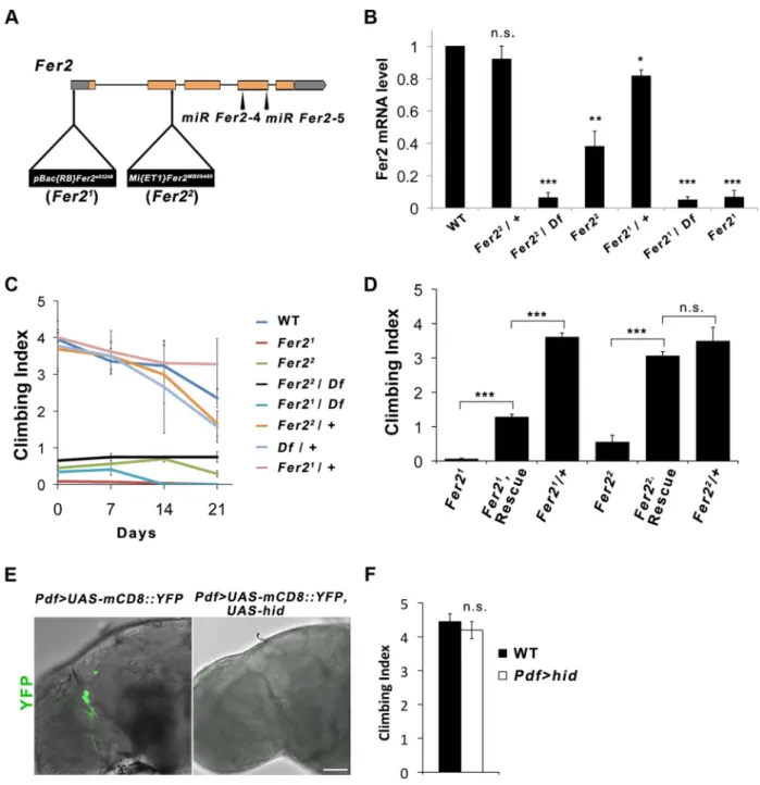
Fer2 is required for the startle-induced climbing ability Drosophila Fer2e03248(henceforth referred to as Fer21) muta-tion was induced by the insermuta-tion of PBac{RB} into the Fer2 59UTR [18]. TheFer2MB09480(henceforth calledFer22) allele has aMi{ET1} transposon insertion in the 39 end of the second exon [19] (Fig. 1A). To molecularly characterize the Fer2 mutant
alleles, we determined theFer2 mRNA levels using quantitative real-time PCR (qPCR). Consistent with our previous results,Fer2 mRNA expression of theFer21 homozygotes was approximately 5% of the wild-type level [17]. In theFer22homozygous flies,Fer2 mRNA expression was reduced to about 40% of the wild-type level. Thus, Fer21 is an extreme hypomorphic allele, whereas Fer22 is a milder hypomorph. Fer2 mRNA expression of both Fer21/+ andFer22
/+ flies was only slightly reduced relative to the wild-type level, suggesting that the loss of one copy of a Fer2 gene is compensated at the mRNA level by transcriptional or post-transcriptional mechanisms (Fig. 1B). Compensation of gene dose has been observed widely in both Drosophila and mammals [20,21,22]. Therefore, Fer2 is a haplosufficient gene andFer2 heterozygous mutants are expected to be phenotypically wild-type.
We noticed that Fer2 mutant flies tend to climb up the walls poorly when tapped down to the bottom of the vials. We quantified this behavior using a startle-induced climbing assay. Wild-type and heterozygous Fer2 mutants showed similar climbing abilities at least during the first 3 weeks of the adult life. In contrast, all Fer2 homozygous or hemizygous mutants displayed severely impaired climbing abilities throughout adult-hood (Fig. 1C). We generated a driver fly line expressingGAL4 under the control of theFer2 promoter (Fer2-GAL4) and a UAS line expressing FLAG-taggedFer2 cDNA (UAS-Fer2-FLAG). The expression ofFer2-FLAG with Fer2-GAL4 partially but signifi-cantly rescued the decreased climbing ability of theFer21flies and
restored the climbing ability of theFer22flies to the control level (Fig. 1D). These data indicate thatFer2 is necessary for the startle-induced climbing ability.
Fer2 is required for the development and survival of dopaminergic neurons
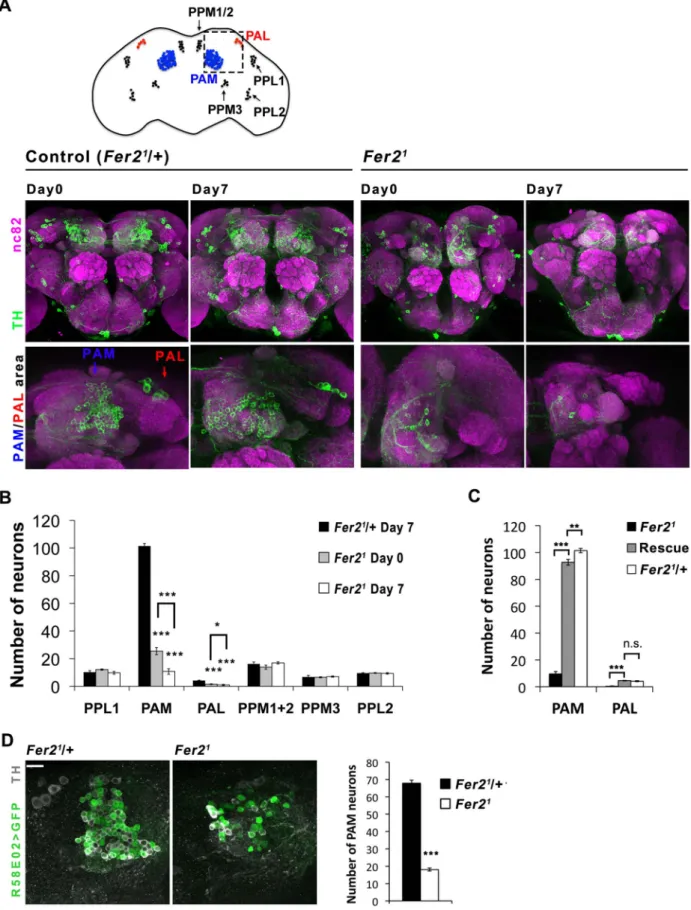
We have previously shown that Fer21 mutation impairs the development of LNvs, which express the neuropeptide pigment-dispersing factor (PDF) [17]. Although it has been shown that PDF is necessary for the normal negative geotaxis behavior [23], whether PDF or LNvs are necessary for startle-induced climbing has not been documented. We found that the expression of UAS-hid with Pdf-GAL4, which selectively ablates LNvs [24], does not impair the startle-induced climbing ability (Fig. 1E, F). Thus, the decrease in startle-induced climbing ability inFer2 mutant flies is due to the deficits other than the lack of LNvs. Because loss of climbing ability is often associated with impaired CNS integrity [25] and the available transcriptome data indicate that Fer2 is almost exclusively expressed in the brain (modENCODE Tissue Expression Data, FlyAtlas [26]), we examined the integrity of major neuron types in the brains of adultFer21mutants. We did not find any obvious differences in the overall morphology of the cholinergic, glutamatergic and serotonergic neurons between Fer21 and controls, although we cannot exclude the possibility that there are subtle differences in the number of these neurons (Fig. S1). Interestingly, we found an evident reduction of dopaminergic (DA) neurons inFer21mutants. Seven DA neuron clusters were detected by anti-tyrosine hydroxylase (TH) staining in the Fer21 heterozygouse flies, which were very similar in number and morphology to those in wild-type flies [27]. In contrast, there were markedly fewer DA neurons in the PAM and PAL clusters in homozygous Fer21 flies on the first day after eclosion (day 0); there were even fewer of them in 7-day-old flies (Fig. 2A). In addition, we expressedUAS-GFP under the control of theHL9-GAL4 driver to label several clusters of DA neurons [28] and found a similar dramatic reduction of PAM and PAL neurons in the homozygousFer21 flies (Fig. S2A). This indicates Author Summary
Parkinson’s disease is a common movement disorder with no known cure. Its characteristic motor symptoms are primarily caused by the progressive loss of midbrain dopaminergic neurons. Although studies have shown that various environmental and genetic factors both contribute to the development of the disease, the underlying mechanisms remain unknown. Here we use powerful invertebrate model organisms, fruit flies and nematode worms, and identify a new gene required for the survival of dopaminergic neurons. We show that homologs of the p48/ptf1-a gene in both flies and worms are expressed in dopaminergic neurons and mutations in p48 increase the susceptibility of dopaminergic neuron death when animals are under oxidative stress. Importantly, genetic variations in p48 in humans have been detected in the sporadic Parkinson’s disease patients, indicating the possibility that similar mechanism might play a role in the death of dopaminergic neurons in humans. Oxidative stress has been regarded as a major pathogenic factor for Parkinson’s disease. Our results add evidence to the link between oxidative stress and neurodegeneration, and suggest that p48 mutant flies and worms can be used to study mechanisms of neurodegeneration in Parkinson’s disease.
that PAM and PAL neurons were reduced in number inFer21 mutants, rather than merely having reduced TH expression. Quantification of the HL9 . GFP-positive neurons revealed a 75% reduction in PAM neuron counts already at day 0 and a 90% reduction at day 7 in Fer21 compared to the heterozygous
controls. Four out of 5 PAL neurons were undetectable at day 0 in theFer21flies, and most brains had no PAL neurons at day 7. The
numbers of other DA neuron clusters were not different between Fer21and controls at both ages (Fig. 2B). To further verify the loss
of DA neurons in the Fer21 flies, we expressed GFP using the R58E02-GAL4 driver, which is derived from the promoter of the dopamine transporter gene and expressed almost exclusively in PAM neurons [29]. There were significantly fewer R58E02-GAL4-labeled PAM neurons in the Fer21 flies compared to the
control, supporting the finding that a large fraction of PAM neurons were absent inFer21 (Fig. 2D). The expression of
Fer2-FLAG by Fer2-GAL4 restored the loss of PAM and PAL neurons in theFer21flies to quasi wild-type levels (Fig. 2C).
Figure 1.Fer2loss-of-function mutation impairs the startle-induced climbing ability. (A) Schematic of the Fer2 mutant alleles and the Fer2 miRNA target sites (miR Fer2-4, miR Fer2-5). Gray boxes are 59 and 39 UTRs, orange boxes are Fer2 gene exons. (B) Fer2 mRNA levels in the fly heads normalized to the level in w1118(WT). Mean 6 SEM from 3 independent experiments. *p,0.05, ** p,0.01, *** p,0.001. n.s., no statistical significance. (C) Climbing abilities of Fer2 mutants and control flies tested weekly. Mean Climbing Index from 6 SEM. (D) Fer2. Fer2-FLAG (Rescue) significantly improved the impaired climbing ability of the Fer21and Fer22 flies (7-day-old). Mean 6 SEM. ***p,0.001. (E) PDF-positive LNvs visualized by expressing UAS-mCD8::YFP with Pdf-GAL4 in the wild-type (w1118) and in the flies expressing UAS-hid. Error bar, 50 mm. (F) Climbing ability of the wild-type and LNv-ablated flies (Pdf . hid) at day 4. Mean 6 SEM.
Figure 2. Selective loss of PAM and PAL cluster DA neurons in theFer2extreme hypomorphic mutants. (A) Top, a diagram of DA neuron clusters in the adult fly brain. Bottom, representative images of the brains stained with anti-TH antibodies. Central brain and anterior brain region where PAM and PAL neurons are located are shown. (B) Quantification of DA neurons per hemisphere detected by TH-staining. For PAM and PAL neurons, HL9 . GFP and TH double positive neurons were quantified. Fer21/+, n = 17. Fer21
: day 0, n = 12; day 7, n = 24. Mean 6 SEM, * p,0.05, *** p, 0.001, comparing Fer21and Fer21/+ or Fer21day 0 vs. day 7. No significant differences were found between Fer21and Fer21/+ where no asterisks are
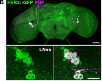
To examine ifFer2 is expressed in PAM and PAL neurons, we monitored the expression of a GFP-tagged Fer2 genomic transgene in the brain by GFP/TH double staining. FER2::GFP expression was observed in all the PAM and PAL neurons and in a few other clusters of cells. As expected, GFP/PDF double staining confirmed the expression of FER2::GFP in the LNvs, consistent with the previous RNA analysis results [17] (Fig. 3A, B). Fer2-GAL4 showed a more widespread expression pattern than FER2::GFP, as is often the case with promoter-GAL4s. Nonethe-less,Fer2-GAL4 was also expressed in all PAM neurons and 4 out of 5 PAL neurons (Fig. S2B). Having validated the expression of
Fer2-GAL4 in PAM and PAL neurons, we next used it to express UAS-TH in the Fer21 flies and immunostained the brains with anti-TH antibodies.Fer2 . TH slightly increased the number of neurons detected by TH-staining in the PAM and PAL clusters but not to the control level, which demonstrates again the absence of these cells (Fig. S2C). These results suggest that Fer2 is expressed in PAM and PAL neurons and further support that Fer21mutation selectively reduces the number of these neurons.
To examine the possibility that the dopaminergic neurotrans-mitter identity of PAM and PAL neurons is changed inFer21and
thus they are undetectable, we analyzed the cell lineage derived
shown. (C) Quantification of the PAM and PAL neurons in the 7-day-old Fer21(n = 8), Rescue (Fer21, Fer2 . Fer2-FLAG; n = 10) and control (Fer21 /+; n = 17) flies. (D) Left, representative confocal images of the brains of the 0-day-old flies expressing UAS-GFP with R58E02-GAL4 double stained for GFP and TH. Right, quantification of the GFP/TH double positive neurons. Mean 6 SEM, ***p,0.001. Fer21/+, n = 7. Fer21, n = 14.
doi:10.1371/journal.pgen.1004718.g002
Figure 3. FER2 expression in the brain. FER2 is expressed in the PAM and PAL DA neurons and the LNvs. Brains of the Fer2::GFP flies were stained for GFP and TH (A) or GFP and PDF (B). Scale bars in the whole brain images are 50 mm, 10 mm in the insets.
from theFer2-GAL4-expressing cells. By combining Fer2-GAL4, UAS-FLP and UbiP63.stop. EGFP, the Fer2-GAL4-express-ing lineage was marked with GFP in the control andFer21flies (Fig. S2D) [30]. While most of the PAM and 4 PAL neurons were GFP-positive in the heterozygous control flies, the majority of these neurons were not present and no ectopic GFP-positive cells were observed in Fer21 (Fig. S2E, F). Therefore, the reduction of PAM and PAL neurons in Fer21 is not due to a cell-fate switch.
To learn more about the developmental impairments of PAM and PAL neurons in Fer21 mutants, we next examined DA
neurons in the pupal brains with anti-TH staining, because these neurons are not present in the larval brain and some of the PAM neurons are known to be born during pupal stages [27,31]. In the Fer21 heterozygous controls, approximately 80% of PAM and
PAL neurons were clearly detectable within 5 days after puparium formation (APF). Whereas inFer21homozygotes, PAM and PAL neurons gradually increased in number but were significantly fewer than in the controls throughout pupal development. These observations indicate that the majority of PAM and PAL neurons were not formed or died before maturation into DA neurons in Fer21 mutants (Fig. S3A, B). Taken together with the
observation that the loss of PAM/PAL neurons progresses at least up to 7 days into adulthood (Fig. 2B), these results indicate that Fer21mutation impairs the development of the DA neurons in the PAM and PAL clusters and also causes their rapid degeneration in adulthood.
Fer2 is a survival factor for PAM neurons in aging flies We next asked whether the integrity of DA neurons is affected in the milder hypomorphic mutant Fer22 as well. We focused our
analysis on the PAM cluster DA neurons and monitored their number in theFer22flies by anti-TH staining. At day 0, the number of PAM neurons was slightly reduced inFer22 compared to the control, but to a much lesser extent as inFer21. Remarkably, the
loss of PAM neurons continued progressively at least up to 28 days inFer22 (Fig. 4A). Lineage tracing usingHL9-GAL4, UAS-FLP andUbiP63.stop . EGFP flies verified the loss of PAM neurons in Fer22 (Fig. S4A). The loss of PAM neurons was rescued by
expressingFer2-FLAG with Fer2-GAL4 (Fig. S4B).
The contrasting results between Fer21 and Fer22 mutants
suggest that a moderate reduction ofFer2 expression has only a minor effect on the development of DA neurons but is sufficient to deteriorate PAM neurons in aged flies. To test this, we generated UAS-transgenic lines to express 2 independent microRNAs (miRNAs) that targetFer2 (miR Fer2-4 and -5) and one negative control miRNA (miR Fer2-N) that contains a sequence of 19 random nucleotides unrelated to any Drosophila gene (see Fig. 1A). When expressed with Fer2-GAL4, both miR Fer2-4 and-5 reduced the Fer2 mRNA levels to approximately 35% of the wild-type level, whereasmiR Fer2-N had no effect on the Fer2 mRNA (Fig. S5A). As expected, constitutive expression of miR Fer2-N by Fer2-GAL4 at 25uC did not alter the number of PAM neurons. In the flies expressingmiR Fer2-4 or miR Fer2-5, the number of PAM neurons remained stable until 35 days of age. However, at 49 days, many flies in either of the knockdowns had a reduced number of PAM neurons, and the reduction was significant inFer2 . miR Fer2-5 flies (Fig. 4B). The constitutive knockdown bymiR Fer2-4 or miR Fer2-5 at 25uC with R58E02-GAL4 or HL9-R58E02-GAL4 resulted in a similar trend, with a significant reduction of PAM neurons in the flies aged over several weeks old (Fig. 4C, S5B). The PAL and other DA neuron clusters were not affected by any of theFer2 knockdowns, although Fer2-GAL4 and HL9-GAL4 are expressed in most of the DA clusters including
PAL neurons. While there were subtle differences in the onset of degeneration that were likely due to the differences in GAL4 expression levels, the data nevertheless illustrate that Fer2 knockdown causes adult-onset PAM neuron degeneration.
The foregoing observations indicate that a moderate reduction of Fer2 expression either by a hypomorphic mutation or by knockdown has little effect on DA neuron development but mainly affects the survival of PAM neurons in adults. This further suggests that the role of Fer2 in the survival of adult PAM neurons is independent of its role in development. To test this more directly, we knocked-downFer2 only during adulthood using a combina-tion of UAS-miR Fer2s, Fer2-GAL4 and temperature-sensitive GAL80 expressed under the tubulin promoter (tub-GAL80ts). These flies were reared at 18uC (a permissive temperature for GAL80ts) until eclosion, and then the temperature was shifted to 29uC (a restrictive temperature for GAL80ts
) to allow for transcriptional activation by GAL4 throughout adulthood [32]. We found that the adult-specific knockdown ofFer2 induced the adult-onset progressive degeneration of PAM neurons without affecting PAL neurons (Fig. 4D). Notably, the loss of PAM neurons was more evident in these flies than in flies with constitutive knockdown (Fig. 4B), which is consistent with the greater GAL4 activity at 29uC than at 25uC [33]. These results clearly distinguish the role ofFer2 in developing and adult DA neurons and demonstrate thatFer2 expression is required for the survival of adult PAM neurons in aging flies.
Loss of PAM neurons leads to the locomotor deficits We next asked whether the loss of PAM neurons, which is the most prominent cellular phenotype inFer2 mutants, is the cause of their locomotor impairment. To test the role of PAM neurons in the startle-induced climbing ability more directly, we knocked-down Fer2 in PAM neurons by HL9-GAL4 and performed a climbing assay. Knocking-downFer2 with either of the miRNAs resulted in significant declines in climbing ability after 49 days (Fig. 5A). This is consistent with the observation thatHL9 . miR Fer2s induce the adult-onset degeneration of PAM neurons only after several weeks (Fig. S5B).
To further assess the contribution of PAM neurons in the climbing ability, we sought to rescue the loss of PAM neurons using a PAM neuron-specific driver in Fer2 mutant flies. Expression of UAS-Fer2-FLAG using HL9-GAL4 or R58E02-GAL4 did not rescue the loss of PAM neurons in Fer21. This is
most likely because the majority of PAM neurons fail to form or die inFer21 before these DA neuron-specific drivers start to be expressed, consistent with the fact thatR58E02-GAL4 has little expression in the larval brain (H. Tanimoto and A. Thum, personal communication). Thus, we reasoned that PAM-neuron specific rescue might be possible inFer22flies, which show little impairments in DA neuron development but display progressive PAM neuron degeneration. Indeed, R58E02 . Fer2-FLAG suppressed the degeneration of PAM neurons in the Fer22 mutants in adulthood (Fig. 5B). A climbing assay revealed that R58E02 . Fer2-FLAG significantly improves the climbing impairments in the Fer22 flies (Fig. 5C). The rescue of the climbing ability was partial. This may be because some of the PAM neurons that failed to develop inFer22were not rescued, or because other unknown cell types affected inFer22contribute to
the climbing ability. Nevertheless, these observations together with the results of the PAM neuron targeted-knockdown indicate that PAM neurons are necessary, although may not be sufficient, for the normal climbing ability of the flies.
The dopaminergic system is critically involved in the control of locomotion in both vertebrates and invertebrates [34,35].
The motor symptoms of PD arise mainly from the loss of DA neurons in the SNc [5]. L-dopa, a dopamine biosynthesis precursor, remains the gold standard for treatments of PD motor symptoms. We found that the locomotor deficit of theFer21flies
was partially but significantly rescued by feeding with L-dopa (Fig. 5D). By anti-TH staining, we observed no significant rescue of the number of DA neurons by L-dopa, which is consistent with a previous study [36]. Since L-dopa has to be converted to dopamine in the DA neuron terminals to exert its therapeutic effect, the partial rescue of the locomotion by L-dopa is also consistent with the marked loss of PAM neurons observed inFer21
mutants.
Fer2 plays a critical role in oxidative stress response and protection of PAM neurons against oxidative insults
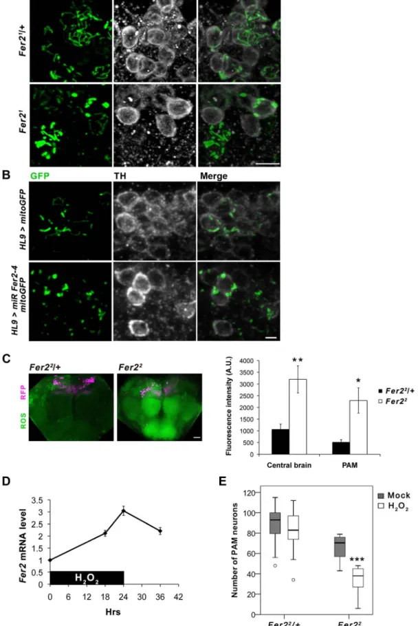
Accumulating evidence suggests that dysfunctions in multiple aspects of mitochondrial biology are associated with the DA neurodegeneration in PD and pathogenesis of other neurodegen-erative disorders [37,38]. To examine whether mitochondrial dysfunction is involved in the loss of DA neurons caused by the loss ofFer2 expression, we visualized mitochondria in the adult PAM neurons by expressing mitochondria-targeted GFP (mitoGFP) [39] withHL9-GAL4. The majority of visible mitochondria in the cell bodies of the remaining PAM neurons inFer21 mutants was in enlarged aggregations and did not form tubular networks as in the
Figure 4. Partial loss ofFer2expression impairs the viability of PAM neurons in aging flies. Quantification of PAM neurons in the flies of different genotypes detected by anti-TH staining at indicated ages. Box boundaries are the 25th and 75th percentiles, the horizontal line across the box is the median, and the whiskers are the lowest and highest values that are not outliers. The open circles represent outliers and closed circles are extreme cases. *p,0.05, **p,0.01, *** p,0.001, comparing Fer22/+ and Fer22
at the same age (A), or between miR-Fer2-N and miR-Fer2-4/-5 at the same age (B-D). Sample sizes are indicated in the Materials and Methods. (A) PAM neurons in Fer22and the control (Fer22
/+). (B) Constitutive expression of miR Fer2s by Fer2-GAL4. (C) Constitutive expression of miR Fer2s by R58E02-GAL4. (D) Adult-specific Fer2 knockdown and the control. Fer2-GAL4, tub-gal80ts: control without UAS-transgene.
control flies (Fig. 6A). Similarly, DA neuron-selective Fer2 knockdown by HL9-GAL4 and Fer22 mutation lead to the accumulation of abnormally enlarged mitochondria in PAM neurons (Fig. 6B, S6A). Mitochondrial morphology in some of the TRH-GAL4-positive serotonergic neurons was indistinguish-able between Fer21 homozygotes and heterozygotes, suggesting that mitochondria in PAM neurons are particularly vulnerable to loss ofFer2 expression (Fig. S6B).
Since mitochondria are the major source of ROS, mitochon-drial dysfunction leads to an excessive ROS production and oxidative damages to various macromolecules. Oxidative stress causes rapid depolarization of mitochondrial inner membrane and inhibits complex I activity, exacerbating ROS production. Thus, mitochondrial defects and elevated ROS levels are interdependent and are thought to have prominent roles in PD pathogenesis [40]. We therefore asked whether ROS levels are increased in theFer2 mutant brains and if it has a causative role on PAM neuron degeneration. We monitored intracellular ROS levels in the brains of Fer22 mutant and the heterozygous control flies using 29,79-dichlorofluorescein (H2DCF), which produces green fluorescence
upon reacting with ROS. Interestingly, ROS levels were significantly elevated throughout the brain in 5-day-old Fer22 flies compared with the age-matched controls. Although there was no regional specificity of ROS accumulation, ROS levels within the PAM neurons were also significantly elevated in Fer22 (Fig. 6C). These suggest that loss ofFer2 expression leads to a systemic increase in oxidative stress in the brain.
The surprisingly dramatic increase of ROS levels in Fer22 mutants prompted us to further examine if Fer2 is involved in oxidative stress response. We first tested ifFer2 expression levels can be altered upon oxidative stress-challenge by feeding flies with non-lethal dose of hydrogen peroxide (H2O2, 5%) for 24 hr. We
found that H2O2 treatment significantly increases Fer2 mRNA
levels (Fig. 6D). We next examined whether oxidative stress-challenge aggravates the degeneration of PAM neurons in Fer2 mutants by anti-TH staining. The H2O2treatment did not affect
PAM neurons inFer22
heterozygous flies, whereas the number of PAM neurons was significantly decreased inFer22
homozygotes after the treatment (Fig. 6E). DA neuron counts in other clusters were unchanged by the same H2O2treatment inFer22 mutants
Figure 5. Loss of PAM neurons leads to the locomotor deficits. (A) Climbing abilities of the flies expressing Fer2 miRNAs or negative control miRNA with HL9-GAL4. Mean from 3 independent experiments 6 SEM. * p,0.05, ** p,0.01, ***p,0.001. (B and C) PAM neuron-targeted expression of Fer2-FLAG by R58E02-GAL4 significantly rescued the degeneration of PAM neurons and climbing ability in the Fer22mutants. Sample sizes are in the Materials and Methods. (B) PAM neuron counts. Fer22
/+: positive control. Fer22, R58E02-gal4: negative control. Fer22, R58E02 . Fer2: rescue. * p,0.05, *** p,0.001. (C) Mean Climbing Index from 3 independent experiments 6 SEM. * p,0.05, *** p,0.001. (D) Climbing ability of the 3-day-old wild-type (WT) and Fer21mutant flies fed with or without L-dopa. Mean 6 SEM. ***p,0.001.
Figure 6.Fer2protects PAM neurons from oxidative stress. (A-B) Mitochondrial morphology in PAM neurons visualized by expressing mitoGFP with HL9-GAL4 and double stained for GFP and TH. (A) 1-day-old control (Fer21/+) and Fer21
mutant flies. Scale bar, 5 mm. (B) DA neuron-targeted Fer2 knockdown by HL9 . miR Fer2-4 and control (14-day-old). Scale bar, 2 mm. (C) ROS levels in the 5-day-old control (Fer22
/+) and Fer22flies monitored using H2DCF dye. Left, representative confocal images of the ROS production in the brain. PAM neurons were RFP-labeled with R58E02-GAL4. Scale
(Fig, S6C), indicating that loss of Fer2 expression renders PAM neurons selectively more vulnerable to increased oxidative stress. Taken together, these results point toward a role for Fer2 in oxidative stress response and suggest thatFer2 contributes to the protection of PAM neurons against oxidative stress.
C.elegans hlh-13 is required for the survival of DA neurons under oxidative stress
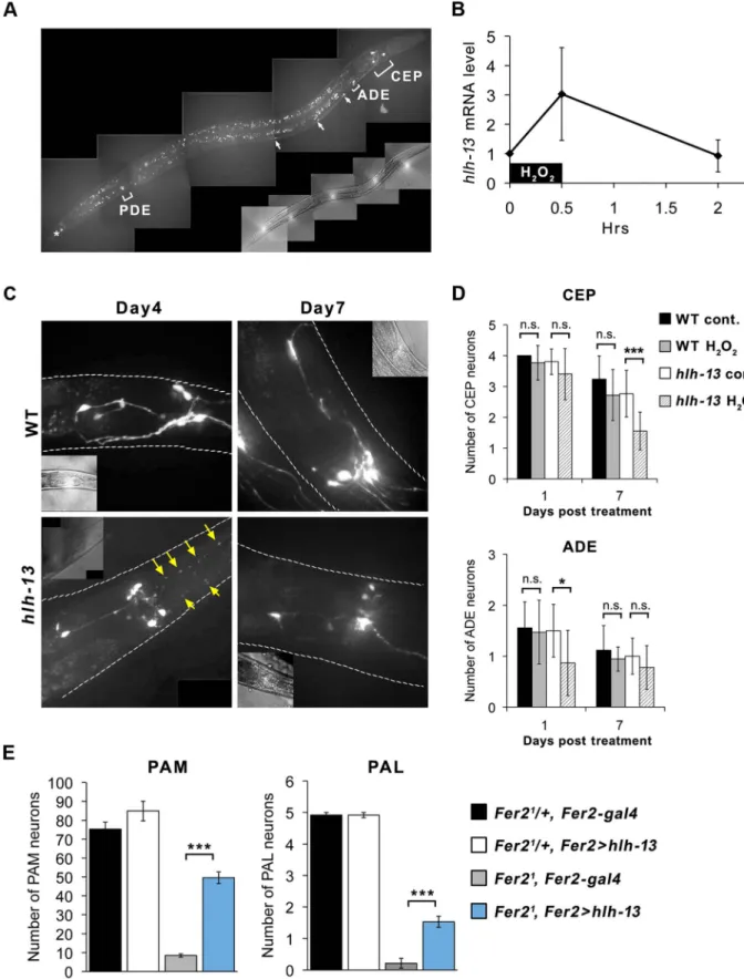
Fer2 homologs are found from nematodes to vertebrates [41]. hlh-13 is predicted to be the single homolog of Drosophila Fer2 and mammalian p48/ptf1a in C.elegans [42]. Consistent with a previous study [42], a GFP::hlh-13 genomic transgene was expressed in all DA neurons (named CEP (4 cells), ADE (2 cells) and PDE (2 cells)) and in a tail neuron in developing and adult worms. GFP::hlh-13 expression was also observed in several unidentified ventral nerve cells from L2 to L4 stages (Fig. 7A).
Since both fly Fer2 and worm hlh-13 are expressed in DA neurons, we next asked whetherhlh-13 has a comparable function asFer2 in DA neuron development or survival. We used the hlh-13 knockout mutant hlh-hlh-13(tm2279) to test the effect of hlh-hlh-13 loss-of-function on the number of DA neurons and on a dopamine-dependent behavior, the basal slowing response. The basal slowing response is a slowing of locomotion rate when worms encounter bacteria and has been shown to require dopamine signaling [43] (Text S1). The knockout mutant showed no differences in the number of DA neurons and basal slowing response compared to wild-type worms (Fig. 7D control, Fig. S7A). Therefore, unlike Fer2, hlh-13 is not required for the development, survival or function of DA neurons under normal growth conditions.
Next, to test ifhlh-13 is involved in the survival of DA neurons under oxidative stress, we treated wild-type andhlh-13(tm2279) mutant adult worms with 1 mM H2O2for 30 min and analyzed
thehlh-13 mRNA levels and DA neuron integrity at subsequent time points.hlh-13 mRNA levels were upregulated by approxi-mately 3-fold immediately after the H2O2treatment and returned
to the non-treated levels after 2 hrs (Fig. 7B). To monitor DA neurons in the wild-type orhlh-13(tm2279) background, we used the dat-1::gfp reporter driving GFP expression in DA neurons. Since it was difficult to reliably detect PDE neurons, we focused our analysis on CEP and ADE neurons located in the head. In wild-type animals, at least until 7 days after the H2O2treatment,
there were no significant differences in the number or morphology of the CEP and ADE neurons between treated and untreated groups. By contrast, H2O2- treated hlh-13(tm2279) mutants
showed fragmentation of the CEP neuron projections starting from day 4 after treatment, followed by the loss of cell bodies. Similarly, the number of ADE neurons was also reduced in the H2O2-treated mutants (Fig. 7C, D). Despite the apparent change
in DA neuron numbers, the basal slowing response was not different between the control and stressed worms in either genotype (Fig. S7B), which is consistent with the previous observation that basal slowing response is defective only when all 4 CEPs are ablated [43]. These results indicate that, similar to fly Fer2, hlh-13 is likely to be involved in the oxidative stress response and required for the protection of DA neurons under oxidative stress in adult worms.
To examine the extent to whichhlh-13 shares the function with Fer2, we next sought to test cross-species complementation of the Fer2 loss-of-function mutation in flies by the hlh-13 gene. We generated a UAS-hlh-13 construct and expressed it with Fer2-GAL4 in the Fer21mutant background. We found that the loss of PAM and PAL neurons inFer21 was partially but significantly
rescued by the expression ofhlh-13 (Fig. 7E). Collectively, these results confirm thathlh-13 is the C.elegans ortholog of Fer2 and suggest that the protection of DA neurons against oxidative insults is a conserved role between these orthologs.
Discussion
Many neurodegenerative disorders are multi-factorial, in which interactions between environmental and genetic factors play important causal roles. Oxidative stress has emerged as a major pathogenic factor for common neurodegenerative diseases, yet how such a ubiquitous phenomenon leads to the loss of selective neuronal populations remains unclear [44]. Here we presented evidence that loss-of-function inp48 homologs in Drosophila and C.elegans renders DA neurons susceptible to degeneration under oxidative stress in adult animals. Interestingly, genome-wide association studies for PD have identified candidate causal SNPs in p48/ptf1a [7,16], suggesting the possibility that p48 loss-of-function may represent an as-yet-unknown genetic risk factor that increases susceptibility of DA neurons to environmental toxins also in mammals. Many familial PD-associated genes are widely expressed; nevertheless, mutations in these genes result in a selective loss of SNc DA neurons, suggesting that cell-type-specific factors, those similar toFer2 and hlh-13, might contribute to the DA neuron vulnerability even in the familial PD cases. The identification of Fer2 and hlh-13 upstream and downstream pathways may thus shed light on the common mechanisms underlying the selective loss of DA neurons in diverse PD cases.
The major cellular phenotype in Fer21 mutants was the developmental defects in 2 subsets of DA neurons, in addition to the developmental loss of LNvs [17], although we cannot exclude the possibility that other neuronal types are also affected (Fig 2, S1). Judging from the results of the lineage-tracing experiments and the observation of DA neurons in the pupal brain,Fer2 is not a selector gene for dopaminergic phenotype in PAM/PAL neurons but is required for neurogenesis or survival of postmitotic neurons before phenotypic maturation (Fig. S2E, F and S3). The notion that genes required for the development of DA neurons confer important roles in adult DA neuron survival has been postulated by several studies in mammals [8,9,10,11]. Although the molecular mechanisms underlying their roles in adult neurons remain elusive, these developmental genes may actively control the genetic programs required for the maintenance of cell identity in adults [45]. Our findings on the Fer2’s dual roles extend this notion to invertebrate nervous systems and underscore its significance.
PAM neuron-targetedFer2 knockdown induces PAM neuron degeneration (Fig. 4C) and mitochondrial dysfunction within PAM neurons (Fig. 6B, S6A). These results indicate that mitochondrial dysfunction and cell death can be induced by a cell-autonomous reduction of Fer2 expression within the PAM cluster. On the other hand, ROS levels are increased brain-wide in
bar, 30 mm. Right, quantification of the ROS levels. Mean 6 SEM. Control, n = 5. Fer22, n = 5. Two independent experiments were performed with similar results. * p,0.05, ** p,0.01. (D) Relative Fer2 mRNA levels in wild-type flies during and after 24-hr 5% H2O2treatment (5-day-old at the onset of the treatment). Data are shown as Fer2 mRNA levels of the H2O2-treated flies relative to the mock-treated controls. Mean 6 SEM from 2–3 independent experiments. (E) PAM neuron counts in the Fer22heterozygous and homozygous flies with or without H2O2treatment. Fer22/+: control, n = 40; H2O2, n = 14. Fer2
2
: control, n = 10; H2O2, n = 14. *** p,0.001. doi:10.1371/journal.pgen.1004718.g006
Figure 7.C.elegans hlh-13andDrosophila Fer2share a role in DA neuron protection under oxidative stress. (A) GFP::hlh-13 is expressed in DA neurons (CEP, ADE and PDE), unidentified ventral nerve cells (arrows), and in a tail neuron (asterisk). Fluorescence signal that is not labeled is the gut autofluorescence. A representative image of a L4 worm. (B) hlh-13 mRNA levels in wild-type worms after a brief 1 mM H2O2treatment quantified by qPCR relative to the mock-treatment value. Mean from 4 independent experiments. Error bar, SEM. (C) Representative images of GFP-labeled DA neurons in the head in wild-type and hlh-13(tm2279) worms after H2O2treatment. Arrows indicate fragmented CEP neuron projections. (D) Quantification of CEP and ADE neurons in the H2O2-treated and non-treated worms. Mean 6 SEM. * p,0.05, *** p,0.001. Between 14 and 18 worms were examined for each condition. (E) Loss of PAM and PAL neurons in the Fer21flies was significantly rescued by expressing hlh-13 with Fer2-GAL4. Fer21/+, Fer2-gal4: n = 13. Fer21
/+, Fer2 . hlh-13: n = 13. Fer22
, Fer2-gal4: n = 16. Fer21, Fer2 . hlh-13: n = 22. 1-day-old flies. Mean 6 SEM. *** p,0.001. doi:10.1371/journal.pgen.1004718.g007
theFer22flies, despite the fact thatFer2 expression is restricted to several clusters of cells in the brain (Fig. 3, 6C). Thus, loss ofFer2 expression leads to both cell-autonomous and non-cell-autono-mous consequences to the animal’s well-being. How does the brain-wide ROS increase occur byFer2 mutation although Fer2 is not expressed ubiquitously? An intriguing recent study in C. elegans demonstrated that mitochondrial perturbation in neuronal cells modulates mitochondrial stress response in distal tissues autonomously [46]. Flies might exhibit similar non-cell-autonomous mitochondrial stress response that causes systemic ROS production. Systemic increase in oxidative stress is a clinical feature common to many aging-related neurological diseases including PD [47]. Studies in mammals have documented that inflammation is a major factor mediating excessive ROS production and PD pathology. Activated microglia produces ROS and mediates DA neuron death. Dying DA neurons stimulate microglia, exacerbating the ROS production and DA neurodegeneration [48]. As CNS glia inDrosophila are thought to possess immune-like function [49], similar mechanisms via inflammatory responses might mediate global elevation of ROS production inFer2 mutants.
Are the abnormal mitochondria in PAM neurons a cause or a consequence of the ROS upregulation? Because mitochondrial defects and excessive ROS production are inter-dependent, it is not possible to clarify the causality in the current study. However, becauseFer2 expression is upregulated upon H2O2treatment and
the same acute H2O2treatment triggers PAM neuron death in the
absence of Fer2 (Fig. 6D, E), we favor the hypothesis that Fer2 provides protection against oxidative stress rather than directly acting on mitochondria (Fig. 8). These phenomena are remark-ably similar in C.elegans; an acute H2O2 treatment upregulates
hlh-13 expression and triggers DA neuron degeneration in hlh-13 null mutants (Fig. 7B–D). These data suggest that the oxidative stress response is an ancestral role ofp48 homologs. Alternatively, hlh-13’s roles in neural development in worms might have been taken over by other genes. Either way, these findings suggest that loss-of-function in Fer2 and hlh-13 can be used to study pathophysiology of DA neuron degeneration under oxidative stress. Interestingly,Fer2 mRNA levels remain upregulated at least up to 12 hr after the 24-hr H2O2 treatment, whereas hlh-13
mRNA levels return to the non-treated levels 2 hr after a brief H2O2treatment (Fig. 6D, 7B). This difference in gene expression
kinetics may reflect the duration of the H2O2 treatment, RNA
stability, or difference in signal transduction mechanisms. Various stress response genes show highly restricted temporal expression upon stress, as the continuous activation of these genes are often detrimental to the cell [50]. Initial upregulation of hlh-13 immediately after an acute oxidative stress might be necessary and sufficient to trigger the downstream genetic programs that continue to scavenge ROS and repair the cellular damages during the following days. Identification of the downstream genetic programs controlled by Fer2 and hlh-13 will be a key toward understanding the evolutionarily conserved mechanisms of neu-roprotection.
Mild loss ofFer2 expression by Fer22mutation or knockdown leads to a progressive loss of PAM neurons associated with mitochondrial dysfunction, increase in ROS production and progressive locomotor deficits, all of which are reminiscent of the pathological characteristics of PD. Unlike other fly PD models that are derived from genetic modifications of human PD-associated genes or their homologs, Fer2 is not an ortholog of known familial PD-associated genes. Yet, the magnitude of the DA neuron degeneration caused by the loss of Fer2 expression is markedly greater than in existing fly PD models [51]. We demonstrated that the loss of PAM neurons is at least partly responsible for the impaired climbing ability caused byFer2 loss-of-function (Fig 5A–C). Because rescue of the PAM neuron counts in Fer21 mutants to quasi wild-type level does not restore the climbing ability to the control level (compare Fig. 1D and 2C), it is likely that some cells other than PAM and PAL neurons are somehow affected in Fer21 and contribute to the locomotor deficits. Nonetheless, our results are in agreement with the recent study by S. Birman and colleagues, which demonstrates that the progressive motor deficits in the flies expressing human a-synuclein, a transgenic model of PD, derives from the dysfunction of a subset of PAM neurons [52]. Because the selective degeneration of DA neurons within the SNc is the principal cause of the motor manifestations of PD, theDrosophila PAM neurons parallel the DA neurons of the human SNc with regard to function and vulnerability. Thus, Fer2 loss-of-function may serve as a model to better understand the mechanisms by which the loss of specific subsets of DA neurons leads to locomotor deficits in PD. Materials and Methods
Fly strains
PBac {RB} Fer2e03248 (Fer21) has been previously
character-ized [17]. The following lines were obtained from the Blooming-ton Stock Center: Df(3R)Exel7328 (referred to herein as Df), Mi{ET1}Fer2MB09480(Fer22),UAS-mitoGFP and the strain used forFer2-GAL4 flip-out assay (w; RedStinger}4, P{UAS-FLP1.D}JD1, P{Ubi-p63E(FRT.STOP)Stinger}9F6/CyO) [30]. HL9-GAL4 [28] was a gift from G. Miesenbo¨ck. dvglutCNSIII -GAL4 and Cha--GAL4 [53] were gifts from A. DiAntonio. TRH-GAL4 [54] was from O. Alekseyenko. DILP2-TRH-GAL4 [55] was a gift from P. Le´opold.R58E02-GAL4 [29] was from H.Tanimoto and UAS-TH [56] was from J.True. Fer2::GFP genomic transgene FlyFos022529 was derived from a fosmid clone including approximately 5 kb upstream and 20 kb downstream of the Fer2 gene. FlyFos022529 (pRedFlp-Hgr) (Fer2[16092]:: 2XTY1-SGFP-V5-preTEV-BLRP-3XFLAG)dFRT) was generat-ed and generously providgenerat-ed by the project ‘‘A reverse genetic toolkit for systematic study of gene function and protein localization in Drosophila’’ (M.IF.A.MOZG8070).
To generateFer2-GAL4, 2359 bp upstream of Fer2 ATG were amplified using the following primers: EagFer2upF,
59-Figure 8. A model for the role of Fer2in the survival of DA neurons. Mitochondrial dysfunction and oxidative stress are interde-pendent and are both known to mediate DA neuron death. Fer2 expression is upregulated in response to oxidative stress and counteracts PAM neuron degeneration.
TTTCGGCCGTGGATTTGCTCTGGTTTGGATGC -39 and XhoFer2upR, 59- TTTCTCGAGTTTTACGCACTTCCGCT-GTCC -39. The amplified fragment was cloned into a pENTR 3c gateway vector (Invitrogen), verified by sequencing and cloned into to the transformation vector containing GAL4 (pBPGal4.2 Uw2; Addgene) using the gateway system (Invitrogen). The Fer2-GAL4 transgene was inserted into the attP16 landing site [57] on the second chromosome by PhiC31-mediated recombination by a commercial transformation service (BestGene, Inc.). UAS-miR Fer2 constructs were generated as described in [58]. Four different miRNA coding sequences were generated along with a negative control, which does not target any sequence in the Drosophila melanogaster genome. From these 4, 2 miRNAs (lines 4 and 5) were used for further experiments. The miRNA sequence of line 4 was 59-TGAGCAAGATCGACACTCTGC-39, the miRNA se-quence of line 5 was 59-TCAAAGCGGATAGGGCTAATT-39 and the sequence of the negative control miRNA was 59-TACCCGTATCGGGTTAATCGA -39. The stem loop structure was predicted using the RNAfold Webserver of the institute of Theoretical Chemistry at the University of Vienna (http://rna.tbi. univie.ac.at/cgi-bin/RNAfold.cgi). UAS-miR Fer2 constructs were inserted into the attP40 landing site on the second chromosome (BestGene, Inc.). To generate the UAS-Fer2-FLAG construct,Fer2 cDNA with a 3xFLAG tag coding sequence at its 39 end was amplified and cloned into pCR II Topo vector (Invitrogen) and verified by sequencing. A DNA fragment containing Fer2-FLAG coding sequence was then cloned into a UAS-containing transformation vector (pUAST-UAS-Stringer attB). The UAS-Fer2-FLAG construct was integrated into the attP40 landing site. The UAS-hlh-13 construct was generated by cloning a PCR-amplified hlh-13 full-length cDNA into the pBid-UASC-G vector [59] by Gateway cloning, and integrated into the attP40 landing site.
Throughout the text and in the figures, genotype ‘‘X.Y’’ indicates a combination of GAL4 (X) and UAS-effector (Y). Worm strains
C.elegans were cultured using standard protocol unless other-wise indicated. The following strains were obtained from the Caenorhabditis Genetic Center: wild-type (N2), BZ555 egIs1[dat-1::gfp] and IU189 rwls1[hlh-13p::GFP::hlh-13,mec-7::RFP]. IU129hlh-13(tm2279) was a gift from S. Lee [42].
Climbing assay
To assay the startle-induced locomotion of the flies, we used a negative geotaxis assay with modifications [60]. Twenty flies were anesthetized with CO2 and placed in a vertical glass column
(25 cm length, 1.5 cm diameter) with a conical bottom. The columns were divided into 5 equally spaced zones and graded from 1 to 5 from the bottom to the top. After a 1-hr recovery period from CO2 exposure, the flies were gently tapped to the
bottom. The flies were then allowed to climb the wall for the subsequent 20 seconds. The experiments were video recorded and the videos were manually analyzed using VLC software. The numbers of flies that climbed up to each zone within 20 seconds were counted. Flies that remained at the bottom were defined to be in zone 0. A climbing index (CI) was calculated using the following formula: CI = (06n0+ 16n1+ 26n2+ 36n3+ 46n4+
56n5) / ntotal, where ntotalis the total number of flies and nxis the
number of flies that reached zone X. One experiment consisted of 3 trials performed at 5 min intervals. Two or three independent experiments were performed for each condition, and the mean climbing indexes of independent experiments are shown. All
climbing assays were performed 2 hr after lights on (ZT2) to avoid any circadian variation in locomotor activities.
L-dopa treatment
The feeding of the flies with L-dopa feeding was performed as described previously with minor modifications [61]. Flies were raised on fresh medium made from instant food (formula 4–24) containing the antioxidant ascorbic acid (25 mg/100 ml), the antifungal agent Nipagin and L-dopa (1 mM) (Sigma Aldrich, D9628). Control vials contained only ascorbic acid and Nipagin.
Immunostaining
For the co-immunostaining of fly brains with anti-GFP and nc82 antibodies, flies were decapitated and the heads were fixed with 4% paraformaldehyde +0.3% Triton X-100 for 1 hour on ice and washed twice with PBST-0.5 (PBS, 0.5% Triton X-100). Subsequently, the head cuticle was partly removed and the heads were washed twice more and blocked in blocking solution (5% normal goat serum, PBS, 0.5% Triton X-100) for 1 hr at room temperature and incubated with the primary antibodies overnight at 4uC. After 2 washes, the heads were incubated with secondary antibodies (Alexa-conjugated) for 2 hours at room temperature. Cuticles and tracheas were removed, and the brains were mounted in Vectashield mounting medium. For staining with anti-TH antibodies with or without other antibodies, 0.3% Triton X-100 was added instead of 0.5% in PBST and in blocking solution. Primary antibodies were incubated over 2 nights at 4uC, and secondary antibodies were incubated at 4uC overnight. The primary antibodies and concentrations used in this study were as follows: rat monoclonal anti-GFP (GF090R) (Nacalai Tesque, Inc.) 1:500, rabbit polyclonal anti-tyrosine hydroxylase (ab152) (Milli-pore) 1:100, mouse monoclonal antibody nc82 (Developmental Studies Hybridoma Bank) 1:100, mouse monoclonal anti-RFP (AKR-021) (Cell BioLabs, INC.) 1:500, mouse monoclonal anti-tyrosine hydroxylase (22941) (Immunostar) 1:50 and polyclonal rabbit anti-GFP (A6455) (Invitrogen) 1:200.
Microscopy and image analysis
Leica TCS SP5 confocal microscope was used to image fly brains. Quantification was performed using ImageJ software (NIH). To count the number of DA neurons, anti-TH-positive neurons or anti-TH, anti-GFP double-positive neurons were counted manually through confocal Z-stacks. To image the expression of hlh-13-GFP a Leica DM5500 B microscope was used. TILL Phototonics iMIC digital microscope was used to image DA neurons in worms and DA neurons were counted manually on each Z-stack using ImageJ software.
qRT-PCR
Total RNA isolation from fly heads, cDNA synthesis and quantitative-PCR (qPCR) analysis were performed as described previously [17]. mRNA levels ofFer2 were normalized to those of the housekeeping gene elongation factor 1b (Ef1b). For qPCR analysis of worm mRNAs, worms were collected from the plates with M9 buffer, placed into the Falcon tubes and left to settle for 15 min to remove bacteria in their guts. Worms were then washed twice by spinning at 3000 rpm for 1 min. Total RNAs were isolated using TRIZOL (Life Technologies), and mRNAs were reverse-transcribed and used as templates for qPCR. hlh-13 mRNA levels were normalized to theits-1 levels.
Statistical analyses
Statistical analyses were performed using StatPlus (AnalystSoft) and SPSS software (IBM). For normally distributed data sets, two-tailed Student’s t-tests were used to compare the means of two groups. The data that were not normally distributed were analyzed with non-parametric statistics (Mann–Whitney U test). For all experiments, the level of significance was set at p,0.05.
Sample sizes
The numbers of brain hemispheres examined in Figure 4A-D are as follows. (A) Control (Fer22/+): day 0, n = 6; day 1, n = 9; day
21, n = 8.Fer22: day0, n = 6; day1, n = 6; day 21, n = 9. (B)Fer2 .miR Fer2-N: day 0, n = 6; day 35, n = 9; day 49, n = 17. Fer2 . miR Fer2-4: day 0, n = 8; day 35, n = 7; day 49, n = 19. Fer2 . miR Fer2-5: day 0, n = 8; day 35, n = 7; day 49, n = 21. (C) R58E02 . miR Fer2-N: day 0, n = 10; day 21, n = 8; day 35, n = 12; day 63, n = 14.R58E02 . miR Fer2-4: day 0, n = 8; day 21, n = 12; day 35, n = 16; day 63, n = 16.R58E02 . miR Fer2-5: day 0, n = 10; day 21, n = 8; day 35, n = 14; day 63, n = 17. (D) Control without UAS-transgene (Fer2-GAL4, tub80): day 0, n = 15; day 28, n = 7; day 35, n = 18. Control miRNA (Fer2 . miR Fer2-N): day 0, n = 15; day 28, n = 13; day 35, n = 24. Fer2 . miR Fer2-4, tub80: day 0, n = 19; day 28, n = 14; day 35, n = 18.Fer2 . miR Fer2-5, tub80: day 0, n = 13; day 28, n = 20; day 35, n = 13.
Sample sizes in Figure 5B are as follows. Positive control (Fer22/ +): day 0, n = 8; day 14, n = 14. Negative control (Fer22, R58E02-gal4): day 0, n = 15; day 14, n = 16. Rescue (Fer22, R58E02 . Fer2): day 0, n = 17; day 14, n = 13.
H2O2treatment
5-day-old flies were transferred into the empty vials for 6 hrs, then placed onto the food prepared from instant food (Formula 4– 24 Instant Drosophila Medium, Carolina(R)) containing 5% H2O2
for 24 hrs. Control food contained only dH2O. The vials were
placed in a humid box and kept at 25uC. Flies were subsequently collected for RNA analysis or placed on the normal food for 24 hrs prior to the dissection and TH staining.
Worms were synchronized by bleaching and stressed as young adults 2 days later (22.5uC). After washing worms off the plates with M9 medium, they were spun down gently at 130 g and washed once with M9 to remove bacteria in the solution. The worms were transferred in 5 ml M9 to an empty petri dish and H2O2-containing M9 (5 ml) was added to the final concentration
of 1 mM. The worms were kept shaking for 30 min at room temperature. Worms were then washed 3 times with M9 to remove H2O2and were placed back on the normal NGM plates
for recovery. ROS detection
In vivo detection of ROS production in fly brains was performed using 2979-dichlorofluorescein (H2DCF) as detailed in Owusu-Ansah et al. (Protocol Exchange, 2008, doi:10.1038/ nprot.2008.23). Brains of theR58E02-GAL4, UAS-mCherry flies in the Fer22 heterozygous or homozygous background were
imaged by confocal microscopy and the fluorescence intensity was measured using the FIJI software [62]. Since there was no regional specificity in the H2DCF signal, the central brain area (entire brain except for the optic lobe) was manually defined and the signal intensity in the defined region across Z-stacks was measured by performing a Z-SUM projection. To quantify the H2DCF signal within PAM neurons, PAM neurons were defined by
thresholding the RFP signal and the total H2DCF signal within the defined volume was measured by a Z-SUM projection. Supporting Information
Figure S1 Fer21 mutation does not affect the apparent
morphology of major neuron types.UAS-GFP was expressed in the control (Fer21/+) and Fer21 flies in cholinergic neurons with
Cha-GAL4, glutamatergic neurons with Dvglut-GAL4, and serotonergic neurons withTRH-GAL4. Brains of the 7-day-old flies were stained with anti-GFP and nc82 antibodies. Represen-tative confocal z-projection images are shown. At least 10 brains per genotype were examined and showed no apparent morpho-logical differences between the control andFer21flies. Scale bar, 50mm.
(TIF)
Figure S2 Loss of PAM and PAL neurons inFer21mutants. (A) Brains of the control (Fer21/+) and Fer21 flies expressing UAS-GFP with HL9-GAL4 were stained for UAS-GFP and TH. Represen-tative images of anterior brain regions including the PAM and PAL clusters are shown. Scale bar, 10mm. (B) Brains of the Fer2-GAL4, UAS-RFP flies stained for RFP and TH. Since PAM neurons are numerous and densely positioned, only a part of the PAM cluster is shown for clarity. Arrows indicate 5 PAL neurons. Scale bar, 10mm. (C) Quantification of the TH-immunoreactive PAM and PAL neurons inFer21/+ (n = 19), Fer21withGAL4 only
(n = 11) andFer21expressingUAS-TH with Fer2-GAL4 (n = 10).
Fer2. TH did not rescue the number of TH-positive neurons in Fer21, indicating the absence of PAM and PAL neurons in the Fer21flies (day 0). Mean 6 SEM, **p,0.01. ***p,0.001. (D) A schematic of the genetic tracing of a GAL4-expressing lineage, showingFer2-GAL4 as an example. (E) Representative images of theFer2-GAL4 lineage marked by GFP expression in 7-day-old control (Fer21/+) and Fer21 flies. InFer21, PAM neurons were largely undetectable and there were no ectopic GFP-positive neurons. (F) Quantification of PAM and PAL neurons examined by TH/GFP double staining in the Fer2-GAL4-expressing lineage. Mean 6 SEM. ***p,0.001, *p,0.05, comparing Fer21/+ and Fer21orFer21day 0 vs. day 7.
(TIF)
Figure S3 Fer21mutation impairs the development of PAM and PAL neurons. (A) Brains of theFer21/+ and Fer21pupae 2 and 5 days after puparium formation (APF) stained with anti-TH. Bottom panels are high-magnification images of the PAM neurons in the squares shown in the top panels. White arrows indicate the examples of matured PAM neurons having cytoplasmic TH expression. Yellow arrowheads point to unknown cells weakly expressing TH. Because these cells were found also in other brain areas and only at day 2 APF, they were excluded from the analysis. Scale bars, 50mm (top) and 5mm (bottom). (B) Number of matured PAM and PAL neurons in theFer21/+ and Fer21pupae. Mean 6 SEM. ***p,0.001.
(TIF)
Figure S4 Loss of PAM neurons in Fer22 mutants. (A) Quantification of PAM neurons in the HL9-GAL4-expressing lineage in theFer22/+ and Fer22background. Significantly fewer
PAM neurons were detected in Fer22. Mean 6 SEM. ***p, 0.001. Fer22/+, n = 12. Fer22, n = 12. (B) Fer2 . Fer2-FLAG genetic rescue restored the number of PAM neurons in theFer22 flies (7-day-old). Mean 6 SEM. ***p,0.001. Fer22/+, n = 7. Fer22, Fer2-gal4, n = 8. Fer22, Fer2. Fer2, n = 6.
Figure S5 Fer2 knockdown leads to a progressive loss of PAM neurons. (A)Fer2 knockdown efficiency analyzed by qPCR. Fer2 mRNA levels in the flies expressing miR Fer2s and negative control miR-Fer2-N using Fer2-GAL4 were normalized to the level inw1118. (B) Quantification of PAM neurons in the flies with a constitutive expression of miR Fer2s by HL9-GAL4. Box boundaries are the 25th and 75th percentiles, the horizontal line across the box is the median, and the whiskers are the lowest and highest values that were not outliers. The open circles represent outliers.HL9 . miR Fer2-N: day 0, n = 7; day 21, n = 9; day 63, n = 6.HL9 . miR Fer2-4: day 0, n = 6; day 21, n = 9; day 63, n = 6.HL9 . miR Fer2-5: day 0, n = 7; day 21, n = 7; day 63, n = 12. *p,0.05, comparingmiR Fer2-N and miR Fer2-4 or -5 at the same age. Although PAM neuron counts betweenmiR-Fer2-N and miR-Fer2-5 at day 63 were not significantly different (p = 0.08), miR-Fer2-5 between day 0 and day 63 were significantly different (p,0.01), suggesting a gradual loss of PAM neurons inHL9 . miR-Fer2-5.
(TIF)
Figure S6 Selective vulnerability of PAM neurons inFer2 loss-of-function mutants. (A) Mitochondrial morphology in PAM neurons visualized by expressing mitoGFP in the Fer22 and the
heterozygous control flies (14-day-old). Scale bar, 5mm. (B) Mitochondrial morphology in serotonergic neurons inFer21and Fer21/+ was monitored by expressingmitoGFP with TRH-GAL4
(7-day-old). Panels 1 and 2 are high magnification images of the areas 1 and 2 in the left panels. Scale bars, 50mm (left) and 5mm (panels 1 and 2). (C) Number of DA neurons except for PAM neurons in theFer22flies with or without 24-hr H2O2treatment.
Mock, n = 32. H2O2, n = 24. There were no statistically significant
differences between mock and H2O2-treated groups in any cell
type. (TIF)
Figure S7 hlh-13 is not required for the basal slowing response in worms. (A) The basal slowing response of wild-type and hlh-13(tm2279) worms scored by manually counting body bends. (in) and (out) indicate the worms in or out of the food, respectively. No significant differences were observed between the two genotypes at any age examined (Student’s t-test). Mean 6 SD. (B) The basal slowing response of wild-type and hlh-13(tm2279) worms after H2O2treatment. Worms were video-recorded and average speed
of approximately 30 worms per group was analyzed. Data are shown as a mean from 5-6 independent experiments 6 SD. No significant difference between the two genotypes was found by Student’s t-test.
(TIF)
Text S1 Materials and methods for basal slowing response in worms.
(DOCX)
Acknowledgments
We thank Gero Miesenbo¨ck, Aaron DiAntonio, Olga Alekseyenko, Pierre Leopold, Hiromu Tanimoto, John True, and the Bloomington Drosophila stock center for fly stocks, Michael Levine for pHB and pNE2 vectors, Lisa C. Schild, Edouard Jaumouille´ and Dirk Beuchle for technical assistance, Siu S. Lee and CGC for worm strains. We also thank Mihail Sarov, Pavel Tomancak, Frank Schnorrer, K. VijayRaghavan and Mani Ramaswami for theFer2::GFP flies. We are grateful to Jose Botella-Munoz and Serge Birman for helpful comments on the manuscript.
Author Contributions
Conceived and designed the experiments: EN PM. Performed the experiments: PBD BG FD VS DT. Analyzed the data: PBD BG FD VS DT PM EN. Contributed reagents/materials/analysis tools: DAG PM. Wrote the paper: EN.
References
1. Schultz W (2007) Behavioral dopamine signals. Trends Neurosci 30: 203–210. 2. Schultz W (2007) Multiple dopamine functions at different time courses. Annu
Rev Neurosci 30: 259–288.
3. Dunlop BW, Nemeroff CB (2007) The role of dopamine in the pathophysiology of depression. Arch Gen Psychiatry 64: 327–337.
4. Dichter GS, Damiano CA, Allen JA (2012) Reward circuitry dysfunction in psychiatric and neurodevelopmental disorders and genetic syndromes: animal models and clinical findings. J Neurodev Disord 4: 19.
5. Shulman JM, De Jager PL, Feany MB (2011) Parkinson’s disease: genetics and pathogenesis. Annu Rev Pathol 6: 193–222.
6. Obeso JA, Rodriguez-Oroz MC, Goetz CG, Marin C, Kordower JH, et al. (2010) Missing pieces in the Parkinson’s disease puzzle. Nat Med 16: 653–661. 7. Song GG, Lee YH (2013) Pathway analysis of genome-wide association studies
for Parkinson’s disease. Mol Biol Rep 40: 2599–2607.
8. Sonnier L, Le Pen G, Hartmann A, Bizot JC, Trovero F, et al. (2007) Progressive loss of dopaminergic neurons in the ventral midbrain of adult mice heterozygote for Engrailed1. J Neurosci 27: 1063–1071.
9. Kittappa R, Chang WW, Awatramani RB, McKay RD (2007) The foxa2 gene controls the birth and spontaneous degeneration of dopamine neurons in old age. PLoS Biol 5: e325.
10. Jiang C, Wan X, He Y, Pan T, Jankovic J, et al. (2005) Age-dependent dopaminergic dysfunction in Nurr1 knockout mice. Exp Neurol 191: 154–162. 11. Eells JB (2003) The control of dopamine neuron development, function and survival: insights from transgenic mice and the relevance to human disease. Curr Med Chem 10: 857–870.
12. Waddell S (2010) Dopamine reveals neural circuit mechanisms of fly memory. Trends Neurosci 33: 457–464.
13. Van Swinderen B, Andretic R (2011) Dopamine in Drosophila: setting arousal thresholds in a miniature brain. Proc Biol Sci 278: 906–913.
14. Chase DL, Koelle MR (2007) Biogenic amine neurotransmitters in C. elegans. WormBook: 1–15.
15. Meredith DM, Masui T, Swift GH, MacDonald RJ, Johnson JE (2009) Multiple transcriptional mechanisms control Ptf1a levels during neural development including autoregulation by the PTF1-J complex. J Neurosci 29: 11139–11148. 16. Pankratz N, Beecham GW, DeStefano AL, Dawson TM, Doheny KF, et al. (2012) Meta-analysis of Parkinson’s disease: identification of a novel locus, RIT2. Ann Neurol 71: 370–384.
17. Nagoshi E, Sugino K, Kula E, Okazaki E, Tachibana T, et al. (2010) Dissecting differential gene expression within the circadian neuronal circuit of Drosophila. Nat Neurosci 13: 60–68.
18. Thibault ST, Singer MA, Miyazaki WY, Milash B, Dompe NA, et al. (2004) A complementary transposon tool kit for Drosophila melanogaster using P and piggyBac. Nat Genet 36: 283–287.
19. Metaxakis A, Oehler S, Klinakis A, Savakis C (2005) Minos as a genetic and genomic tool in Drosophila melanogaster. Genetics 171: 571–581.
20. Stenberg P, Lundberg LE, Johansson AM, Ryden P, Svensson MJ, et al. (2009) Buffering of segmental and chromosomal aneuploidies in Drosophila melano-gaster. PLoS Genet 5: e1000465.
21. McAnally AA, Yampolsky LY (2010) Widespread transcriptional autosomal dosage compensation in Drosophila correlates with gene expression level. Genome Biol Evol 2: 44–52.
22. Guidi CJ, Veal TM, Jones SN, Imbalzano AN (2004) Transcriptional compensation for loss of an allele of the Ini1 tumor suppressor. J Biol Chem 279: 4180–4185.
23. Toma DP, White KP, Hirsch J, Greenspan RJ (2002) Identification of genes involved in Drosophila melanogaster geotaxis, a complex behavioral trait. Nat Genet 31: 349–353.
24. Stoleru D, Peng Y, Agosto J, Rosbash M (2004) Coupled oscillators control morning and evening locomotor behaviour of Drosophila. Nature 431: 862–868. 25. Lessing D, Bonini NM (2009) Maintaining the brain: insight into human neurodegeneration from Drosophila melanogaster mutants. Nat Rev Genet 10: 359–370.
26. Chintapalli VR, Wang J, Dow JA (2007) Using FlyAtlas to identify better Drosophila melanogaster models of human disease. Nat Genet 39: 715–720. 27. Mao Z, Davis RL (2009) Eight different types of dopaminergic neurons
innervate the Drosophila mushroom body neuropil: anatomical and physiolog-ical heterogeneity. Front Neural Circuits 3: 5.
28. Claridge-Chang A, Roorda RD, Vrontou E, Sjulson L, Li H, et al. (2009) Writing memories with light-addressable reinforcement circuitry. Cell 139: 405– 415.
29. Liu C, Placais PY, Yamagata N, Pfeiffer BD, Aso Y, et al. (2012) A subset of dopamine neurons signals reward for odour memory in Drosophila. Nature 488: 512–516.
30. Evans CJ, Olson JM, Ngo KT, Kim E, Lee NE, et al. (2009) G-TRACE: rapid Gal4-based cell lineage analysis in Drosophila. Nat Methods 6: 603–605. 31. Blanco J, Pandey R, Wasser M, Udolph G Orthodenticle is necessary for
survival of a cluster of clonally related dopaminergic neurons in the Drosophila larval and adult brain. Neural Dev 6: 34.
32. McGuire SE, Le PT, Osborn AJ, Matsumoto K, Davis RL (2003) Spatiotemporal rescue of memory dysfunction in Drosophila. Science 302: 1765–1768.
33. Brand AH, Manoukian AS, Perrimon N (1994) Ectopic expression in Drosophila. Methods Cell Biol 44: 635–654.
34. Riemensperger T, Isabel G, Coulom H, Neuser K, Seugnet L, et al. (2011) Behavioral consequences of dopamine deficiency in the Drosophila central nervous system. Proc Natl Acad Sci U S A 108: 834–839.
35. Kravitz AV, Freeze BS, Parker PR, Kay K, Thwin MT, et al. (2010) Regulation of parkinsonian motor behaviours by optogenetic control of basal ganglia circuitry. Nature 466: 622–626.
36. Coulom H, Birman S (2004) Chronic exposure to rotenone models sporadic Parkinson’s disease in Drosophila melanogaster. J Neurosci 24: 10993–10998. 37. Martin LJ (2011) Mitochondrial and Cell Death Mechanisms in
Neurodegen-erative Diseases. Pharmaceuticals (Basel) 3: 839–915.
38. Arduino DM, Esteves AR, Cardoso SM (2011) Mitochondrial fusion/fission, transport and autophagy in Parkinson’s disease: when mitochondria get nasty. Parkinsons Dis 2011: 767230.
39. Pilling AD, Horiuchi D, Lively CM, Saxton WM (2006) Kinesin-1 and Dynein are the primary motors for fast transport of mitochondria in Drosophila motor axons. Mol Biol Cell 17: 2057–2068.
40. Henchcliffe C, Beal MF (2008) Mitochondrial biology and oxidative stress in Parkinson disease pathogenesis. Nat Clin Pract Neurol 4: 600–609. 41. Ledent V, Vervoort M (2001) The basic helix-loop-helix protein family:
comparative genomics and phylogenetic analysis. Genome Res 11: 754–770. 42. Liachko N, Davidowitz R, Lee SS (2009) Combined informatic and expression
screen identifies the novel DAF-16 target HLH-13. Dev Biol 327: 97–105. 43. Sawin ER, Ranganathan R, Horvitz HR (2000) C. elegans locomotory rate is
modulated by the environment through a dopaminergic pathway and by experience through a serotonergic pathway. Neuron 26: 619–631.
44. Gandhi S, Abramov AY (2012) Mechanism of oxidative stress in neurodegen-eration. Oxid Med Cell Longev 2012: 428010.
45. Eade KT, Fancher HA, Ridyard MS, Allan DW (2012) Developmental transcriptional networks are required to maintain neuronal subtype identity in the mature nervous system. PLoS Genet 8: e1002501.
46. Durieux J, Wolff S, Dillin A (2011) The cell-non-autonomous nature of electron transport chain-mediated longevity. Cell 144: 79–91.
47. Serra JA, Dominguez RO, Marschoff ER, Guareschi EM, Famulari AL, et al. (2009) Systemic oxidative stress associated with the neurological diseases of aging. Neurochem Res 34: 2122–2132.
48. Peterson LJ, Flood PM (2012) Oxidative stress and microglial cells in Parkinson’s disease. Mediators Inflamm 2012: 401264.
49. Freeman MR, Doherty J (2006) Glial cell biology in Drosophila and vertebrates. Trends Neurosci 29: 82–90.
50. de Nadal E, Ammerer G, Posas F (2011) Controlling gene expression in response to stress. Nat Rev Genet 12: 833–845.
51. Munoz-Soriano V, Paricio N (2011) Drosophila models of Parkinson’s disease: discovering relevant pathways and novel therapeutic strategies. Parkinsons Dis 2011: 520640.
52. Riemensperger T, Issa AR, Pech U, Coulom H, Nguyen MV, et al. (2013) A single dopamine pathway underlies progressive locomotor deficits in a Drosophila model of Parkinson disease. Cell Rep 5: 952–960.
53. Daniels RW, Gelfand MV, Collins CA, DiAntonio A (2008) Visualizing glutamatergic cell bodies and synapses in Drosophila larval and adult CNS. J Comp Neurol 508: 131–152.
54. Alekseyenko OV, Lee C, Kravitz EA (2010) Targeted manipulation of serotonergic neurotransmission affects the escalation of aggression in adult male Drosophila melanogaster. PLoS One 5: e10806.
55. Geminard C, Rulifson EJ, Leopold P (2009) Remote control of insulin secretion by fat cells in Drosophila. Cell Metab 10: 199–207.
56. True JR, Edwards KA, Yamamoto D, Carroll SB (1999) Drosophila wing melanin patterns form by vein-dependent elaboration of enzymatic prepatterns. Curr Biol 9: 1382–1391.
57. Markstein M, Pitsouli C, Villalta C, Celniker SE, Perrimon N (2008) Exploiting position effects and the gypsy retrovirus insulator to engineer precisely expressed transgenes. Nat Genet 40: 476–483.
58. Haley B, Hendrix D, Trang V, Levine M (2008) A simplified miRNA-based gene silencing method for Drosophila melanogaster. Dev Biol 321: 482–490. 59. Wang JW, Beck ES, McCabe BD (2012) A modular toolset for recombination
transgenesis and neurogenetic analysis of Drosophila. PLoS One 7: e42102. 60. Friggi-Grelin F, Coulom H, Meller M, Gomez D, Hirsh J, et al. (2003) Targeted
gene expression in Drosophila dopaminergic cells using regulatory sequences from tyrosine hydroxylase. J Neurobiol 54: 618–627.
61. Pendleton RG, Parvez F, Sayed M, Hillman R (2002) Effects of pharmacological agents upon a transgenic model of Parkinson’s disease in Drosophila melanogaster. J Pharmacol Exp Ther 300: 91–96.
62. Schindelin J, Arganda-Carreras I, Frise E, Kaynig V, Longair M, et al. (2012) Fiji: an open-source platform for biological-image analysis. Nat Methods 9: 676– 682.