HAL Id: hal-03131286
https://hal.archives-ouvertes.fr/hal-03131286
Submitted on 4 Feb 2021
HAL is a multi-disciplinary open access
archive for the deposit and dissemination of
sci-entific research documents, whether they are
pub-lished or not. The documents may come from
teaching and research institutions in France or
abroad, or from public or private research centers.
L’archive ouverte pluridisciplinaire HAL, est
destinée au dépôt et à la diffusion de documents
scientifiques de niveau recherche, publiés ou non,
émanant des établissements d’enseignement et de
recherche français ou étrangers, des laboratoires
publics ou privés.
Developmental improvements in voluntary control of
behavior: Effect of preparation in the fronto-parietal
network?
Nadia Alahyane, Donald Brien, Brian Coe, Patrick Stroman, Douglas Munoz
To cite this version:
Nadia Alahyane, Donald Brien, Brian Coe, Patrick Stroman, Douglas Munoz. Developmental
im-provements in voluntary control of behavior: Effect of preparation in the fronto-parietal network?.
NeuroImage, Elsevier, 2014, 98, pp.103-117. �10.1016/j.neuroimage.2014.03.008�. �hal-03131286�
Developmental improvements in voluntary control of behavior:
Effect of preparation in the fronto-parietal network?
Nadia Alahyane
⁎⁎
, Donald C. Brien, Brian C. Coe, Patrick W. Stroman, Douglas P. Munoz
⁎
Centre for Neuroscience Studies, Queen's University, Kingston, Ontario K7L 3N6, Canadaa b s t r a c t
a r t i c l e i n f o
Article history: Accepted 9 March 2014 Available online 15 March 2014 Keywords: Saccade Cognitive control Task set fMRI Child development Frontal cortex
The ability to prepare for an action improves the speed and accuracy of its performance. While many studies indicate that behavior performance continues to improve throughout childhood and adolescence, it remains unclear whether or how preparatory processes change with development. Here, we used a rapid event-related fMRI design in three age groups (8–12, 13–17, 18–25 years) who were instructed to execute either a prosaccade (look toward peripheral target) or an antisaccade (look away from target) task. We compared brain activity with-in the core fronto-parietal network with-involved with-in saccade control at two epochs of saccade generation: saccade preparation related to task instruction versus saccade execution related to target appearance. The inclusion of catch trials containing only task instruction and no target or saccade response allowed us to isolate saccade preparation from saccade execution. Five regions of interest were selected: the frontal, supplementary, parietal
eyefields which are consistently recruited during saccade generation, and two regions involved in top down
executive control: the dorsolateral prefrontal and anterior cingulate cortices. Our results showed strong evidence that developmental improvements in saccade performance were related to better saccade preparation rather than saccade execution. These developmental differences were mostly attributable to children who showed reduced fronto-parietal activity during prosaccade and antisaccade preparation, along with longer saccade reaction times and more incorrect responses, compared to adolescents and adults. The dorsolateral prefrontal cortex was engaged similarly across age groups, suggesting a general role in maintaining task instructions
through the whole experiment. Overall, thesefindings suggest that developmental improvements in behavioral
control are supported by improvements in effectively presetting goal-appropriate brain systems.
© 2014 Elsevier Inc. All rights reserved.
Introduction
The ability to select and generate appropriate behaviors, according to goals or changes in the environment, undergoes important develop-mental changes between infancy and adulthood (Durston and Casey, 2006; Johnson, 2001; Luna et al., 2010). This cognitive control of behav-ior can be examined with the antisaccade task where participants are instructed to resist looking toward aflashed visual target and instead, to execute a saccade to the opposite side (Hallett, 1978; Munoz and Everling, 2004). The antisaccade task is often compared to a basic oculo-motor task, the prosaccade task, where participants are required to sim-ply look at the target when appearing on the screen. Many behavioral studies revealed that both prosaccade and antisaccade reaction times,
and errors in the antisaccade task (i.e., unwanted prosaccades made to-ward the target) which probe response inhibition, gradually decrease through development to stabilize around late adolescence or young adulthood (Fischer et al., 1997; Klein and Foerster, 2001; Kramer et al., 2005; Luna et al., 2004; Munoz et al., 1998; Ordaz et al., 2010; for review,Luna et al., 2008). Yet, it is only recently that a handful of studies combining functional magnetic resonance imaging (fMRI) and eye movement recording have emerged to examine the neural correlates underlying the developmental improvements in antisaccade control (Hwang et al., 2010; Velanova et al., 2008, 2009).
In the present study, we asked the question: which specific pro-cess that composes saccade generation changes over development (8–25 years) and contributes to age-related changes in saccade per-formance? Indeed, in most saccade experiments, an instruction is present at the start of the trial (conveyed for example by the color or shape of a central cue) that signals which task to perform (prosaccade or antisaccade). Then, a peripheral target appears, prompting the par-ticipants to execute the correct response (look toward target location, or away, respectively). Therefore, a saccade trial can be decomposed into an instruction-related epoch and a target-related epoch. The instruction-related epoch is crucial because it allows participants to
⁎ Correspondence to: D.P. Munoz, Centre for Neuroscience Studies, Queen's University, Botterell Hall, Room 234, 18 Stuart St., Kingston, Ontario K7L 3N6, Canada. Fax: +1 613 533 6840.
⁎⁎ Correspondence to: N. Alahyane, Laboratoire Vision Action Cognition, Université Paris Descartes Centre Henri Piéron - UFR Institut de Psychologie 71, avenue Edouard Vaillant 92774 Boulogne-Billancourt Cedex.
E-mail addresses:[email protected](N. Alahyane),[email protected] (D.P. Munoz).
http://dx.doi.org/10.1016/j.neuroimage.2014.03.008 1053-8119/© 2014 Elsevier Inc. All rights reserved.
Contents lists available atScienceDirect
NeuroImage
get ready, decide and prepare for the appropriate action in advance of target appearance (Connolly et al., 2002). Importantly, this ability to prepare for upcoming actions optimizes performance (Chikazoe et al., 2009; Meiran and Daichman, 2005; Monsell, 2003): responses are initi-ated faster and inappropriate responses are more efficiently inhibited. We hypothesize that improvements in saccade initiation and inhibition from childhood to adulthood (Luna et al., 2008) may be related to age-related enhancements in response preparation. A clue in favor of our hypothesis can be found in a behavioral study which showed that par-ticipants 8–31 years-old responded faster and more accurately in the antisaccade task when given more time to prepare during the instruc-tion period (Ordaz et al., 2010). This suggests that the basic preparatory processes, which are engaged during instruction to establish the antisaccade task set, are in place in children. However, because the beneficial effects on performance were similar across ages, and perfor-mance in children never reached adult levels, the authors suggested that‘developmental improvements in rates of inhibitory successes and latencies to inhibit a response are not supported by limitations in the time needed to prepare a response. Rather, persistent age-related differences in inhibitory control may instead reflect qualitative differ-ences in control aspects of preparatory processing’. Unfortunately, prosaccade trials were not tested to verify whether possible improve-ments in preparation were related to both saccade types, given that prosaccade performance also changes with development (Luna et al., 2008).
FMRI studies in adults have revealed that preparation of anti-saccades and proanti-saccades involves a core fronto-parietal cortical net-work, including the frontal eyefield (FEF), supplementary eye field (SEF), parietal eyefield (PEF), dorsolateral prefrontal cortex (DLPFC), and anterior cingulate cortex (ACC) (Brown et al., 2007; Connolly et al., 2002, 2005; Curtis and Connolly, 2008; Curtis and D'Esposito, 2003; DeSouza et al., 2003; Ford et al., 2005), consistent with neuro-physiological studies in monkey (Amador et al., 2004; Everling and Munoz, 2000; Johnston and Everling, 2006; Johnston et al., 2007). The FEF is an important node in the saccade network, carrying set-related signals for antisaccades and prosaccades to the superior colliculus and premotor circuitry (Bruce and Goldberg, 1985; Munoz and Everling, 2004; Schiller et al., 1980). Importantly, in both humans and monkeys, levels of preparatory activity in the FEF predict successful performance in the antisaccade task (Curtis and D'Esposito, 2003; Everling and Munoz, 2000; but see Ford et al., 2005), and how fast a correct prosaccade or antisaccade will be initiated (Connolly et al., 2005; Everling and Munoz, 2000). These same frontal and parietal regions are recruited in adults during saccade execution after target appearance (Brown et al., 2007; Curtis and Connolly, 2008; Curtis and D'Esposito, 2003; DeSouza et al., 2003; Ford et al., 2005). However, contrary to the FEF, SEF and PEF, significant differences in ACC and DLPFC activity between antisaccades and prosaccades were observed only during preparation (Brown et al., 2007). It was suggested that these two areas provide top down executive control by biasing the oculomotor circuitry for antisaccade performance before target appearance (Brown et al., 2007; Johnston et al., 2007). Finally, this fronto-parietal network is also recruited by children (N8 years-old) and adolescents when making correct antisaccades, although with different magnitudes compared to adults (Luna et al., 2001; Velanova et al., 2008, 2009). However, these previous studies did not dissociate activation related to antisaccade preparation and activation related to execution.
Here, we used fMRI in participants from 8 to 25 years old while they performed interleaved prosaccade and antisaccade tasks. Our approach is hypothesis-driven and focused on the core fronto-parietal network involved in saccade control and engaged in children, adolescents and adults. We tracked developmental changes in brain activity in the FEF, SEF, PEF, DLPFC and ACC during two separate epochs: 1) during instruc-tion to make a prosaccade or antisaccade while subjects were preparing for the task and 2) after target appearance when they executed the cor-rect saccade. To temporally dissociate saccade preparation and saccade
execution, some fMRI studies used long instruction periods (Connolly et al., 2002; Curtis and Connolly, 2008; Curtis and D'Esposito, 2003; DeSouza et al., 2003; Ford et al., 2005), which are necessary to avoid an overlap of the hemodynamic responses related to task instruction and target appearance. Although adults can maintain preparation for 6 to 14 s, this is not suitable for young participants (Okazaki et al., 2004; Olivier and Rival, 2002). Additionally, maintaining preparation for so long may require extra processes (e.g., prolonged memorization of in-structions,‘waiting’ strategies). To circumvent these disadvantages while being able to isolate saccade preparation from saccade execution, we chose another approach which consisted of intermixing catch trials containing the instructional cue to make a prosaccade or antisaccade, but no target presentation or saccade response (Brown et al., 2007; Geier et al., 2010). If, as we hypothesize, developmental improvements in saccade performance are related to more efficient preparatory pro-cesses, we predicted that brain activity in the fronto-parietal network should correlate with development primarily during task instruction. These developmental effects on preparatory activity should be found for both prosaccade and antisaccade tasks because behavioral perfor-mance improves with age in both tasks. Overall this would suggest that immaturities in behavioral control may be related to immaturities in effectively presetting goal-appropriate response pathways. Materials and methods
Subject recruitment and experimental procedures were reviewed and approved by the Health Sciences Human Research Ethics Board at Queen's University, and followed the principles of the Declaration of Helsinki.
Participant recruitment and selection
Originally, 161 subjects ranging from 8 to 25 years old (26 between 18 and 25 years-old) were recruited from the greater area of Kingston through advertisements in local newspapers andflyers around Queen's university campus. They were screened for history of psychiatric or neurological condition, education, medication and substance use, based on a custom-made questionnaire. The volunteers aged below 18 years came for an initial visit (30–45 min) to tour the MRI facility and familiarize themselves with the MRI settings, safety procedures, and eye movement tasks. Once they were comfortable with the task demands, they were trained to remain still in a mock 0 T scanner while performing one or two runs of the saccade tasks (5–10 min). Eye movements were not recorded and no feedback about their per-formance was given. After this session, 31 families declined to partic-ipate in the subsequent fMRI session. Three were unable to remain still in the mock scanner and six had a learning disability or neurodevelopmental disorder. Three other families did not come at the given appointment. In total, 92 young participants 8–17 years-old were enrolled in the fMRI study (90–120 min). The 26 adult vol-unteers, who reported no brain disorder or psychoactive medication, did not receive the training session (but were invited to do so if in-terested) and participated in the fMRI session only. Adult partici-pants and parents or guardians of minors signed informed written consent after the procedures were reviewed with the experimenter. Children also gave written assent to confirm that they understood the procedures and were willing to participate. All volunteers were given 5 min of practice of the saccade tasks prior to entering the MRI room. Vision was normal or corrected-to-normal by wearing contact lenses. Volunteers received $20 per hour compensation for their participation.
After the fMRI session, data from 24 participants were immediately excluded from subsequent analyses because of technical failure with the MRI or eye tracking equipment (n = 8; 9–15 years; 6 males), dental braces or a retainer causing artifacts in the functional images (n = 4; 9–13 years; 1 male), falling asleep in the scanner
(3 children: 9–11 years; 1 male; 3 adults: 20–23 years; 1 male), non-compliance with the task (n = 4; 9–12 years; 1 male), and withdraw-al by the volunteer after one functionwithdraw-al run (n = 2 mwithdraw-ales; 9, 14 years). Data from 15 additional participants (8–17 years) were excluded because they did not meet the quality assurance criteria for image artifacts and motion (seePreprocessingsection). In sum, data from 31 children, 25 adolescents and 23 adults are reported here (see
Table 1). All 79 participants were right-handed except two children who were left-handed. Finally, despite the relatively high attrition rate and practice in young participants compared to adults, it is impor-tant to note that we found similar saccade performance and similar developmental trends (seeResultssection for details) to previous behavioral studies (e.g.,Luna et al., 2004; Munoz et al., 1998). Behavioral paradigm
Participants were scanned while performing between 6 and 8 runs of saccade tasks depending on their motivation or tiredness in a rapid fMRI event-related design (Fig. 1), with rest breaks in be-tween (1 child performed 4 runs only, 1 child and 1 adult performed 5 runs). The mean number of collected runs was similar across children (6.6 +/− 1.1), adolescents (6.9 +/− 1.01) and adults (6.9 +/− 1.1; one-way ANOVA: F2, 76b 1). The visual stimuli and duration of each
run (4 min, 37.5 s) were chosen to make the experiment more enter-taining and enjoyable for the young participants and keep them engaged during the entire run.
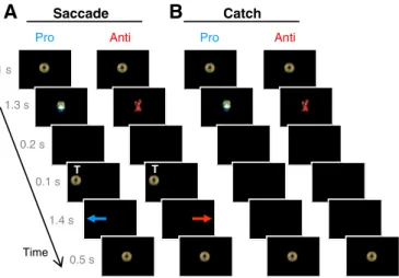
Each run comprised 64 trials: 16 prosaccade trials, 16 antisaccade trials, 8 procatch trials, 8 anticatch trials, and 16fixation trials. Each saccade trial (Fig. 1A) started with 1 s offixation of a central coin on a black screen. The coin then turned into a red crab or green turtle for 1.3 s, followed by a 200-ms gap with no stimulus. A gap was inserted because it is known to release activefixation and facilitate preparation of the upcoming saccade before target presen-tation, which leads to reduced saccade reaction times and higher propensity to make unwanted prosaccades in the antisaccade task (Dorris and Munoz, 1995; Everling et al., 1998; Munoz and Corneil, 1995). After the gap, a peripheral coin wasflashed for 100 ms to the right or the left, at eccentricity of either 6° or 7°, with equal proba-bility. Then the screen was blank for 1.4 s. During this period, partici-pants were required to execute a prosaccade or an antisaccade according to the instructional cue and then hold their gaze at that ec-centric location until a visual coin came back to the center for 500 ms in-dicating the start of the next trial. When the central cue was a green turtle (prosaccade task), participants were asked to direct their eyes to the peripheral coin when appearing on the screen. When the cue was a red crab (antisaccade task), they were asked to refrain from looking at the coin and instead, to direct their eyes away from it, to the opposite position. Participants were asked to respond as quickly and accurately as possible. They were encouraged to do the best they could and to correct their mistakes if they“thought” they made any (for example, they made a prosaccade instead of an antisaccade, and conversely). No feedback regarding their performance was given. They were also instructed that in some trials, the central cue (green tur-tle or red crab) was not followed by a peripheral coin (Fig. 1B). Instead, the instruction cue was followed by a black screen for 1700 ms during which participants were asked to maintainfixation at center until the appearance of the central coin indicating the end of the trial. These
catch trials provided an important measure of task preparation, isolated from task execution related to target presentation (Brass and von Cramon, 2002; Brown et al., 2007; Curtis et al., 2005; Geier et al., 2010; Ollinger et al., 2001a,b; Orr and Weissman, 2009). Indeed, be-cause all trials were randomly interleaved and the participants did not know during the presentation of the instructional cue whether it would be followed by a target or not, they had to prepare for the appropriate saccade task (‘look toward’ or ‘look away’) until the pe-ripheral target was presented, or not. A ratio of 2:1 between saccade trials (n = 16 per instruction type) and catch trials (n = 8 per in-struction type) was chosen to favor the tendency to respond and therefore, to increase the need to actively prepare for the upcoming saccade task. Finally, 16 neutralfixation trials were included (not il-lustrated) containing a centralfixation coin for three different dura-tions: 1.5 s (n = 8), 3 s (n = 4), and 4.5 s (n = 4). The variable length introduced temporal jitter necessary in rapid event-related designs to optimize the separation of the overlapping estimated blood oxy-genation level-dependent (BOLD) responses elicited by the different trial types (Burock et al., 1998; Dale, 1999; Ollinger et al., 2001a,b). Moreover these neutral trials served as a baseline condition in the fMRI analysis, in addition to a 16.5 s long neutralfixation period that ended each run. This latterfixation period also allowed for the hemodynamic response to return toward baseline. The order of trials, type of instruction and target location were randomly intermixed with-in each run.
Image acquisition
Imaging data were collected from a 3 T whole-body Siemens Magnetom Trio system with a receiver head coil. Padding was placed around the head to minimize head motion. At the start of the session, a high-resolution anatomical MP-RAGE 3D T1-weighted scan was
ac-quired (repetition time TR = 1760 ms, echo time TE = 2.2 ms,flip angle FA = 9°, 1 mm isovoxel resolution, 176 slices). During this scan, young participants watched a cartoon whereas adults had the choice to listen to music if they wanted. The purpose of cartoons was to help children to relax from the beginning while getting used to the scanner environment, and to enhance their motivation. Audio was pro-vided through high-quality stereo headphones (NordicNeuroLab NNL) which were also used to attenuate the scanner noise (35 dB sound pro-tection). Functional T2*-weighted echo-planar images (EPIs) sensitive
Table 1
Participant characteristics.
8–12 years 13–17 years 18–25 years
n 31 25 23
Mean age (SD) 10.4 (1.3) 14.9 (1.3) 21.7 (1.8)
Males 14 15 10
SD: standard deviation.
Pro Anti Pro Anti 1 s 1.3 s 0.2 s 1.4 s 0.1 s 0.5 s Time Saccade Catch T T
A
B
Fig. 1. Behavior paradigm. Participants had to execute two tasks: to look toward peripheral target T position (pro-saccade, blue arrow) upon its appearance, or to prevent from looking at T and instead to direct their eyes to the opposite side (anti-saccade, red arrow). A. In saccade trials, a central cue, presented 1.5 s in advance of T onset, instructed the subjects which task was to be performed. A green turtle indicated to execute a prosaccade, and a red crab indicated to make an antisaccade. B. In catch trials, only the in-structional cue was presented. The different types of trials were randomly interleaved.
to blood oxygen level dependent (BOLD) contrast were then acquired interleaved (TR = 1500 ms, TE = 30 ms, FA = 72°,field-of-view = 211 × 211 mm, matrix size = 64 × 64, 3.3 mm isovoxel resolution, 24 axial slices from the top of the brain). Six to eight functional runs were collected per participant (see beginning ofBehavioral paradigm
section), each with 185 volumes. For each run, thefirst two volumes (containing a centralfixation coin) allowed for signal equilibration and were subsequently discarded.
Eye tracking
Along with functional imaging, eye position was monitored at 120 Hz with an ISCAN video based eye tracking system (ISCAN Inc., Burlington, MA, USA) equipped with an infraredfiber-optic light-source that was attached to the head coil and directed to the right eye. Task performance was also watched online to verify that partic-ipants were performing the experiment while in the scanner bore. A nine-point calibration of eye position was performed before thefirst run and repeated between runs when necessary. The visual display and behavioral experimental design were controlled by a PC com-puter running E-prime (Psychology Software Tools, Pittsburgh, PA, USA). Visual images were back-projected via a NEC LT265 DLP pro-jector (Tokyo, Japan) with a refresh rate of 60 Hz on a high-contrast screen (DA-LITE) at the head of the bore and viewed by the participant through a mirror mounted on the head coil. Behavioral data analysis
Trials were scored offline by custom-made MatLab (version 7.9, MathWorks Inc.) programs and subsequent statistical analyses were performed with Statistica (version 9.0, StatSoft Inc.). Onset and termina-tion of eye movements were determined when velocity exceeded a threshold of 50°/s. Reaction time (RT) was calculated as the time from target appearance to saccade onset in saccade trials. Saccade trials were considered to be incorrect if subjects brokefixation during the in-struction cue presentation, made no saccade after target appearance, made a correct response with RT below 100 ms (i.e., anticipatory) or above 1000 ms, or made a direction error (i.e., made afirst eye move-ment toward the target location in antisaccade trials, or away from the target location in prosaccade trials). Neutralfixation trials and catch tri-als where participants failed to maintainfixation were scored as incor-rect. All these incorrect trials were discarded from the fMRI analyses.
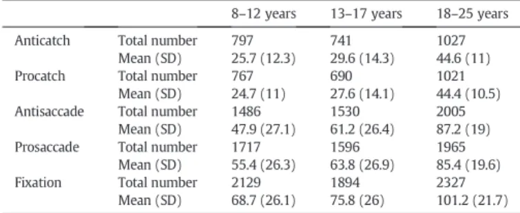
Table 2depicts the number of correctly performed trials per age group that wasfinally used in statistical analyses after both behavioral and fMRI preprocessing. Direction errors were however kept as a measure of task performance (i.e., accuracy;Dyckman et al., 2007; Ethridge et al., 2009; Hutton and Ettinger, 2006; Luna et al., 2008; McDowell et al., 2008; Munoz and Everling, 2004). Saccades directed to the right or left target were pooled together as there was no main effect of target location for RT or direction error rate (repeated measures ANOVAs, pN .05). One-way ANOVAs were performed to compare saccade RT
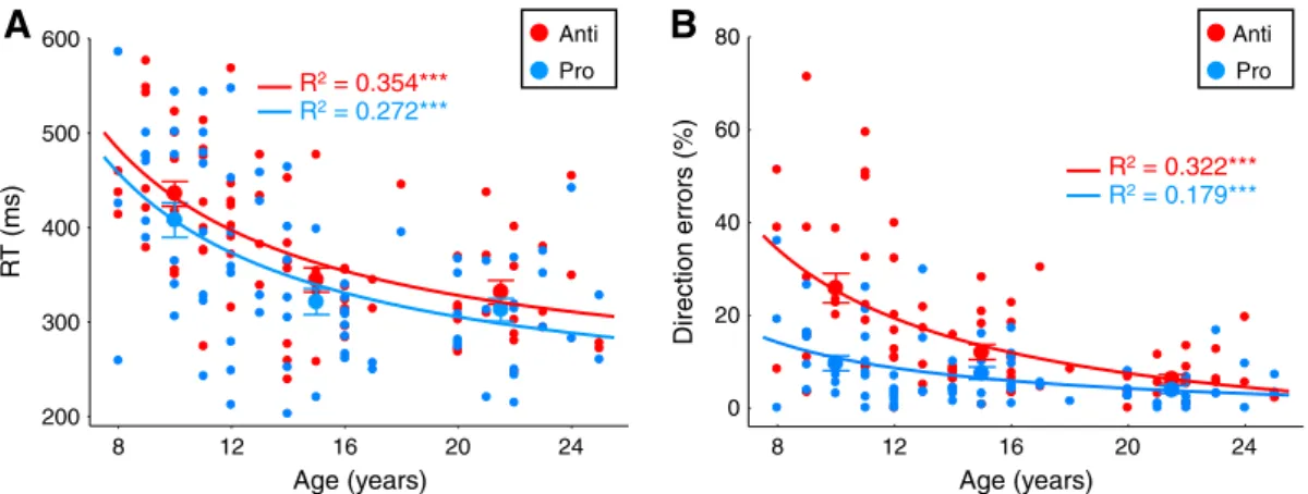
and direction errors across children, adolescents and adults. Significant main effects of age group were examined further by post-hoc Tukey HSD (honestly significant difference) tests. Age-related changes in saccade performance were also examined using an inverse best-fitting regression model (Fig. 2), consistent with previous studies (Luna et al., 2004; Ordaz et al., 2010).
fMRI data analysis
Data were analyzed using Brain Voyager QX (Version 2.3; Brain Innovation, Maastricht, The Netherlands).
Preprocessing
Each scan wasfirst visually inspected for quality assurance to de-tect any abrupt head motion and/or artifacts by watching time course movies. Based on the movies, data sets fromfifteen partici-pants (8 males, 2 females, aged 8–12 years; 4 males, 1 female, aged 13–17 years) were deemed not usable and thus eliminated from fur-ther analysis. Motion correction was applied to the remaining 416 time-series (148 in children, 126 in adolescents, 142 in adults) using a rigid body algorithm with trilinear interpolation within-run. Six move-ment parameters (3 translation parameters and 3 rotation parameters in x, y, z planes) were obtained from this realignment procedure. A root mean square value was calculated for each run across frames and across the three x, y, and z dimensions for each subject and for transla-tion and rotatransla-tion separately. Mean translatransla-tion ranged between 0.025 and 0.82 mm across all subjects and mean rotation ranged from 0.016 to 0.78°, which was below our a priori cutoff of 2 (~2/3 voxels). Root mean square values were averaged across runs within individuals and means were subjected to Pearson correlation analyses. There was no significant correlation between translation and age (r = −0.046, p = 0.684), or rotation and age (r =−0.193, p = 0.089). As a check, we looked back at thefiles from the 15 subjects we eliminated following data quality assurance (see above). Motion correction analysis con-firmed our decision: the output still showed evidence of large and abrupt motion, with movement exceeding 1 voxel. Next, slice scan time correction with cubic spline interpolation was applied to each run, followed by temporalfiltering (high-pass filter with cut-off of 2 cycles per run and linear trend removal). Functional data were superimposed on anatomical images and normalized to Talairach space. Importantly, this stereotactic space has been validated for use in children 7 years-old and older, and the feasibility of comparing BOLD responses directly across development in this common atlas space and across different vascular territories well established (Brown et al., 2005; Burgund et al., 2002; Church et al., 2010; Kang et al., 2003; Wenger et al., 2004). Finally, functional images were spatially smoothed using a Gaussian kernel of 4 mm full-width at half-maximum.
Statistical analysis
The preprocessed fMRI data for each run and each participant were analyzed using the deconvolution design matrix model in Brain Voyager. This general linear model does not make any assump-tion about the shape of the BOLD response, which has been success-fully validated in studies using rapid event-related designs (Miezin et al., 2000; Ollinger et al., 2001a,b). For each event type, a series of 13 predictors was defined covering a period of 20 s locked to trial onset, providing an estimate of the event-related BOLD response at each of the 13 time points, i.e., every ~ 1.54 s (corresponding to ~ 1 TR). This resulted in an estimated time course spanning 13 time points per voxel and per event type. Five event types were modeled: correct catch trials (pro, anti), correct saccade trials (pro, anti) and all incorrect trials, resulting in 65 regressors (5 trial types × 13 time points) entered into the design matrix. Importantly, this analy-sis allowed us to examine correctly performed trials separately from incorrect trials, and thus determine developmental differences in
Table 2
Correctly performed trials used infinal fMRI analyses.
8–12 years 13–17 years 18–25 years
Anticatch Total number 797 741 1027
Mean (SD) 25.7 (12.3) 29.6 (14.3) 44.6 (11)
Procatch Total number 767 690 1021
Mean (SD) 24.7 (11) 27.6 (14.1) 44.4 (10.5) Antisaccade Total number 1486 1530 2005
Mean (SD) 47.9 (27.1) 61.2 (26.4) 87.2 (19) Prosaccade Total number 1717 1596 1965
Mean (SD) 55.4 (26.3) 63.8 (26.9) 85.4 (19.6)
Fixation Total number 2129 1894 2327
Mean (SD) 68.7 (26.1) 75.8 (26) 101.2 (21.7) SD: standard deviation.
BOLD responses when the same behavioral response (i.e., correct) was being generated by the three age groups. The total and mean numbers of correct trials for children, adolescents, and adults are presented inTable 2. Incorrect trials were excluded from the analyses but were separately modeled as a predictor of no interest to account for any variance in the BOLD signal associated with these errors (Brown et al., 2007; Murphy and Garavan, 2004). The six motion parameters were also included as nuisance factors in the model. The es-timated BOLD response associated with neutralfixation trials was used as implicit baseline. Data were processed using a Z-transformation. The event-related parameter estimates were submitted to group analyses using a random-effects general linear model.
Regions of interest analyses. Our study is hypothesis-driven and did not include exploratory analyses across the whole brain. We focused on re-gion of interest (ROI) analyses infive core regions known to be recruited by children, adolescents and adults (for reviews,Luna et al., 2008, 2010; McDowell et al., 2008): the frontal eyefield (FEF), a key oculomotor area, the supplementary eyefield (SEF) and the parietal eye field (PEF) that are two other regions consistently recruited during saccade generation, and the dorsolateral prefrontal cortex (DLPFC) and the ante-rior cingulate cortex (ACC) that are both implicated in top down execu-tive control. ROIs were defined using both anatomical criteria and functional constraints.
Anatomical boundaries. The FEF was located around the junction of the precentral sulcus and the superior frontal sulcus on each hemi-sphere (Curtis et al., 2005; Manoach et al., 2007; Paus, 1996). The SEF was located in the dorsomedial frontal cortex in and around the paracentral sulcus, superior to the cingulate sulcus (Curtis et al., 2005; Manoach et al., 2007). The PEF was localized to the intraparietal sulcus defined as dividing the superior and inferior parietal lobules (Curtis and Connolly, 2008; Curtis and D'Esposito, 2003; Geier et al., 2010; Manoach et al., 2007). The DLPFC was localized in the middle frontal gyrus (Brown et al., 2007; Pierrot-Deseilligny et al., 2004; Velanova et al., 2008). The ACC was defined as the region in or below the cingulate sulcus, anterior to the anterior commissure (Curtis et al., 2005; Paus et al., 1993; Pierrot-Deseilligny et al., 2004).
Functional constraints. First, for each group (children: 8–12 years, adolescents: 13–17, adults: 18–25), we performed a general contrast all correct saccade trialsN fixation baseline at time points 5, 6, and 7, across participants. This time window was chosen from a prelimi-nary inspection of the event-related time courses related to saccade trials because it encompassed maximal activation from trial start and accounted for both preparation and execution time intervals across various brain regions. The three age group-level contrast maps
were thresholded at pb 0.05, corrected at the p b 0.05 level for mul-tiple comparisons using a Monte Carlo simulation that required a minimum of 12 voxels. This general contrast highlighted the brain network involved in saccade generation while being both unbiased to differences between task types (pro, anti) and independent of our contrasts of interest (i.e., task preparation and task execution) (Kriegeskorte et al., 2009; Poldrack and Mumford, 2009). It con-firmed that all three age groups showed similar spatial activation, in agreement with previous developmental studies (Luna et al., 2001; Velanova et al., 2008). Second, to select the ROIs independently of the effect of age (Kriegeskorte et al., 2009; Luna et al., 2010; Poldrack and Mumford, 2009), the individual contrast maps of all 79 participants were combined. Beta contrast images were obtained for each individual and then averaged to obtain a group beta contrast map, with an extent threshold of 12 voxels. In this combined group map (Fig. 3), within the predetermined anatomical boundaries described above, each ROI was selected consisting in a cubic cluster of activation containing the most significant 125 voxels. This resulted in two labels, one in each hemi-sphere, for the FEF, PEF, DLPFC and ACC, and one cluster for the SEF (Table 3).
From these functional clusters, we extracted the mean BOLD re-sponse estimates averaged across the 125 voxels, for each partici-pant treated as a random effect, and for each of the four events of interest (catch: pro, anti; saccade: pro, anti). These averaged esti-mated BOLD responses were then used to study the age-related changes in instruction-related activation, reflecting prosaccade or antisaccade preparation, and in target-related activation, reflecting prosaccade or antisaccade execution. Saccade trials contained both preparation and execution epochs (Fig. 1A). In catch trials, a target was never presented (Fig. 1B). These catch trials were thus used to assess preparatory activation, without being contaminated by any execution-related process. Specifically, preparatory activation was measured with the contrast catch trialsN fixation baseline. Execution-related activation was isolated by subtracting the activation associated with the catch trials (catchN fixation baseline) from the activation associated with the saccade trials (saccadeN fixation baseline). All the activation contrasts were performed for prosaccade and antisaccade tasks separately. Given that catch trials did not contain a peripheral target, preventing from assessing preparatory activation according to target location (right or left) and saccade direction, BOLD responses in the FEF, PEF, DLPFC and ACC were averaged across both hemispheres.
Effects of age across development. In afirst step, the estimated BOLD response time courses associated with task preparation and task execu-tion derived from the FEF, SEF, PEF, DLPFC and ACC were analyzed by
8 12 16 20 24 200 300 400 500 600 R T (ms) Direction errors (%) Age (years) R2= 0.272*** R2= 0.354*** Age (years)
A
B
Pro Anti 8 12 16 20 24 0 20 40 60 80 R2= 0.179*** R2= 0.322*** Pro AntiFig. 2. Age-related changes in task performance in saccade trials. A. Mean reaction times (RTs) for correctly performed prosaccades (blue trace) and antisaccades (red trace). B. Percentage of direction errors in the antisaccade task (red trace) and prosaccade task (blue trace). Errors in the antisaccade task corresponded to saccades directed toward target position instead of away from it. Errors in the prosaccade task represented saccades directed away from target location. A, B. Small circles represent mean performance for each of the 79 participants. Large circles represent mean (+/−SE) performance across participants within three age groups: 8–12 years, 13–17 years, and 18–25 years. The lines depict the best curve-fits (i.e., inverse) on saccade parameters by age; ***pb 0.001.
repeated measures ANOVAs with time (1–13 time points) as a within-subject factor in each age group separately (children, adolescents, adults). This approach was similar to Brown et al.'s developmental study and was a crucial initial step to determine whether BOLD signal modulated significantly in the 5 ROIs during task preparation and exe-cution for each age group. A significant effect of time indicated that the hemodynamic response was not aflat line across the 13 time points and that there was significant activation (Brown et al., 2005; Geier et al., 2010). In other words, a given ROI showing a main effect of time was interpreted as being recruited. All ANOVAs were corrected for sphericity using Greenhouse–Geisser correction and the levels of significance reported were therefore corrected values.
In a second step, one-way ANOVAs across the three age groups were performed to examine developmental differences in BOLD re-sponse during task preparation vs. task execution, for prosaccade and antisaccade tasks separately. Significant main effects of age group were then examined by post-hoc Tukey HSD tests. Mean of parameter estimates from time points 5 to 6 relative to trial onset was used to examine the ROI activation associated specifically with task preparation during task instruction (Fig. 4A;Fig. 6, left panels). This epoch was chosen a priori because it best represented peak activation during catch trials across ROIs and age groups. Mean of pa-rameter estimates from time points 6 to 7 relative to trial onset was used to examine the ROI activation associated specifically with task execution after target appearance (Fig. 4B;Fig. 6, middle panels). The execution-related epoch was shifted by 1.5 s later relative to the preparation-related epoch because the peripheral target that triggered the prosaccade or antisaccade response in saccade trials appeared 1.5 s (~ 1 TR) after the cue instruction (Fig. 1A). This
approach is similar to previous saccade studies using two separate epochs to examine preparation vs. execution (Brown et al., 2007, 2008; Cameron et al., 2012; Hakvoort Schwerdtfeger et al., 2013).
Adults had significantly more correct trials than both adolescents and children who did not differ from each other (one-way ANOVAs, psb 0.001;Table 2), which may contribute to developmental differ-ences in activation. Significant effects of age group were thus further explored by ANOVAs covarying the number of trials used for the ac-tivation contrasts (catch andfixation trials for preparation-related activation; saccade, catch andfixation trials for execution-related activation; see end ofRegions of interest analysessection).
For all analyses, effects were considered significant at an alpha of 0.05.
Results Task performance
Regression analyses (Fig. 2) revealed that reaction times for both cor-rect prosaccades (F1, 78= 28.8, pb 0.001, R2= 0.272) and antisaccades
(F1, 78= 42.1, pb 0.001, R2= 0.354), as well as direction errors
(i.e., unwanted prosaccades) in the antisaccade task (F1, 78= 36.5,
pb 0.001, R2= 0.322), decreased steeply from childhood to late
ado-lescence, while performance tended to stabilize through adulthood. One-way ANOVAs for reaction times revealed main effects of age group for both prosaccade (F2, 76= 11.9, pb 0.0001) and antisaccade
(F2, 76= 20.2, pb 0.000001) tasks. Post-hoc Tukey HSD tests indicated
that children showed longer prosaccade and antisaccade reaction times compared to their elders (psb 0.001), while performance in adoles-cents and adults did not differ significantly (ps N 0.78). There was also a main effect of age group for direction errors in the antisaccade task (F2, 76= 18.6, pb 0.000001), children making more errors than
adolescents and adults (post-hoc Tukey HSD tests: psb 0.001). As expected, participants were overall slower to respond correctly in the antisaccade task than the prosaccade task (main effect of task: F1, 78= 15.1, pb 0.001). Moreover, most direction errors (75%)
were corrected by a corrective saccade to the appropriate location. Importantly, the rate of correction did not covary with age (Pearson correlation r = 0.041, p = 0.723), suggesting that the higher percentage of direction errors in younger participants (Fig. 2B) was not due to a misunderstanding or non-compliance of the task instructions for the antisaccade task, but rather to an inability to efficiently inhibit the automatic response to the target.
For comparison, we also examined the rate of errors (i.e., unwanted antisaccades) in the prosaccade task (Fig. 2B). As expected, participants on average made fewer errors in the prosaccade task than the
FEF SEF PEF DLPFC ACC Z = 51 2.5 0.5 b 3.0 b 1.0 Y = 34 Y = 7
Fig. 3. Saccade brain network. Group beta map (pb 0.05, N12 voxels) of the general contrast all correct saccade trials minus fixation baseline, across all participants, overlaid on a repre-sentative brain (female, age of 25 years). The contrast was performed using time points 5 to 7. Five regions known to show consistent activation in participants from 8 years-old were selected a priori to examine task preparation vs. task execution through development: FEF (frontal eyefield), SEF (supplementary eye field), PEF (parietal eye field), DLPFC (dorsolateral prefrontal cortex), and ACC (anterior cingulate cortex). Right side of image corresponds to left brain.
Table 3
Regions of interest (ROIs).
Region x y z Beta Right FEF 28 −12 48 3.38 Left FEF −28 −13 49 3.55 SEF −2 −4 54 3.58 Right PEF 15 −71 48 3.57 Left PEF −20 −65 48 3.87 Right DLPFC 35 31 33 1.14 Left DLPFC −34 31 36 1.16 Right ACC 5 7 40 2.05 Left ACC −8 7 40 1.79
ROIs corresponded to cubic clusters containing the 125 most significant voxels centered around peak activation in the general contrast all correct saccadesN fixation baseline across all participants (depicted inFig. 3). Talairach coordinates (x, y, z) and beta value at peak activation are shown.
antisaccade task (7.34 ± 0.83% vs. 15.8 ± 1.7%, main effect of task: F1, 78= 23.6, pb 0.001). The rate of errors observed on prosaccade
trials was not surprising given that prosaccade and antisaccade trials were interleaved in our fMRI design, and not performed in separate blocks of trials (Cherkasova et al., 2002; Ethridge et al., 2009). There was a main effect of age group (F2, 76= 4.28, pb 0.05), children
making more errors in the prosaccade task than adults (Tukey HSD test, p = 0.013).
In sum, these fMRI behavioral results successfully replicate previ-ous behavioral studies (Fischer et al., 1997; Klein and Foerster, 2001; Kramer et al., 2005; Luna et al., 2004; Munoz et al., 1998; Ordaz et al., 2010), allowing the examination of the role of task preparation in these behavioral improvements.
Brain activation during task preparation vs. execution
Fig. 3depicts the brain network associated with saccade generation (prosaccades and antisaccades pooled together) across all participants, replicating many previous studies (for review,McDowell et al., 2008). Within this saccade network,five core regions of interest (ROIs) were selected for quantitative analysis: the frontal eyefield (FEF), the supplementary eyefield (SEF), the parietal eye field (PEF), the dorsolateral prefrontal cortex (DLPFC) and the anterior cingulate cortex (ACC). These ROIs were selected because of consistent recruit-ment by children, adolescents and adults from past studies (for reviews,
Luna et al., 2008, 2010; McDowell et al., 2008).Table 3provides the loca-tion of peak voxels within the selected ROIs, which is consistent with prior adult (e.g.Agam et al., 2010; Brown et al., 2007; Connolly et al., 2002; DeSouza et al., 2003; Ettinger et al., 2008; Ford et al., 2005; Luna et al., 1998) and developmental (Geier et al., 2010; Padmanabhan et al., 2011; Velanova et al., 2008) fMRI studies. The main question we asked here was whether brain activity in these regions, as measured by the blood oxygen level dependent (BOLD) response estimate, was different across developmental age groups when making correct prosaccades and antisaccades during two separate epochs of saccade generation: 1) the presentation of the instructional cue, reflecting task preparation required before target onset and 2) the appearance of the peripheral target, reflecting execution of the prosaccade or antisaccade in the cor-rect dicor-rection. Preparatory activation was measured with catch trials that contained only the task instruction, and no target presentation (Fig. 1B). Execution-related activation was assessed by subtracting the activation associated with catch trials from the activation associated with saccade trials (seeRegions of interest analysessection).
FEF
Relationship between FEF activation and age.Table 3provides the location of maximal activation (125 voxels) for the left (Talairach coordinates: peak at−28, −13, 49) and right FEF (28, −12, 48). The averaged event-related BOLD response estimates were extracted from these two FEF clusters for each participant and then averaged across both
B
D
Age group (years)
FEF par ameter estimate Pro Anti
E
Time pointA
FEF par ameter estimateC
Anti
8-12 y 18 -25 y 13 -17 yPro
Preparation ExecutionPreparation Execution Saccade trials
Age group (years)
*
**
8-12 13-17 18-25 0.0 0.2 0.4 0.6 0.8 8-12 13-17 18-25 0.0 0.2 0.4 0.6 0.8 0.0 0.4 0.8 1.2 1 2 3 4 5 6 7 8 9 10 11 12 13 1 2 3 4 5 6 7 8 9 10 11 12 13 1 2 3 4 5 6 7 8 9 10 11 12 13 0.0 0.4 0.8 1.2 0.0 0.4 0.8 1.2 0.0 0.5 1.0 0.0 0.5 1.0 0.0 0.5 1.0Fig. 4. Age-related changes in FEF activation. A. FEF activation time course associated with catch trials to assess task preparation. B. FEF activation time course associated with the contrast saccade trials minus catch trials to isolate target-related activation, i.e. task execution. C. FEF activation time course associated with saccade trials containing both preparation and execu-tion. Time courses were derived from the right and left FEF functional clusters indicated inTable 3, then averaged across both hemispheres. D, E. Effect of age group on FEF preparatory activation and execution-related activation. Mean of parameter estimates across time points 5 and 6 was taken in panel A (gray bar) for preparation during cue instruction (D), and mean of parameter estimates across time points 6 and 7 in panel B (gray bar) was taken for saccade execution associated with target appearance (E). Large circles represent mean (+/−SE) data across participants within three age groups: children aged 8–12 years, adolescents aged 13–17 years, and adults aged 18–25 years. Black asterisks represent significant differences between children and the two other groups for both saccade tasks (post-hoc Tukey HSD tests following one-way ANOVAs). *pb 0.05; **p b 0.01; others: non-significant.
hemispheres. The time courses of FEF activation locked to trial onset (13 time points = 20 s) are illustrated for the three age groups (8–12 years, 13–17 years, 18–25 years) inFigs. 4A–C. For the three age groups, sep-arate repeated measures ANOVAs with time (1–13 time points) as within-subject factor revealed that the FEF showed significant activa-tion for both task preparaactiva-tion (Fig. 4A) and task execution (Fig. 4B), as indicated by main effects of time (psb 0.005 for preparation, ps b 0.05 for execution). In other words, children, adolescents, and adults recruit-ed the FEF to prepare and execute the appropriate prosaccade or antisaccade response.
A notable result was that for both prosaccade and antisaccade tasks, execution-related activation was indistinguishable between the three age groups (Fig. 4B), while in sharp contrast, FEF prepara-tory activation heightened from children to adults (Fig. 4A). To fur-ther explore these developmental effects, we performed one-way ANOVAs across age groups, using mean activation across time points 5 and 6 for instruction-related preparation (Fig. 4A, gray box), and across time points 6 and 7 for target-related execution (Fig. 4B, gray box; for details seeEffects of age across developmentsection). One-way ANOVAs revealed main effects of age group for preparatory activation for both prosaccade (F2, 76 = 8.01, p b 0.001) and
antisaccade (F2, 76= 11.6, pb 0.0001) tasks (Fig. 4D). Post-hoc
Tukey HSD comparisons indicated that preparatory activity was lower in children compared to adolescents (psb 0.05) and adults (psb 0.01) whereas no significant difference was found between ad-olescents and adults (psN 0.6). The significant effect of age group for preparatory activation remained when covarying the number of catch trials andfixation trials for both prosaccade (p b 0.01) and antisaccade (pb 0.005) tasks, with no significant effect of the covar-iates (psN 0.08). On the contrary, no main effects of age group were found for execution-related activation (Fig. 4E) in the prosaccade (F2, 76= 1.11, p = 0.333) or antisaccade (F2, 76b 1) task.
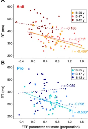
In sum, FEF activation reflecting task preparation, not task execu-tion, was most sensitive to developmental age groups, with children showing reduced activation compared to adolescents and adults. Relationship between FEF preparatory activation and task performance. Past adult fMRI (Connolly et al., 2005) and monkey single-neuron re-cording (Everling and Munoz, 2000) studies revealed that higher preparatory activity in the FEF correlates with shorter saccade reac-tion times. We tested here whether we would reproduce these re-sults. We found that across all participants both prosaccade RT (r = −0.312, p = 0.005) and antisaccade RT (r = −0.512, p b 0.001) de-creased significantly with increasing preparatory activation. Partial correlations controlling for age indicated that these relationships were maintained for the antisaccade task (r =−0.354, p = 0.001), or reduced to a trend for the prosaccade task (r =− 0.186, p = 0.102). When considering each age group separately (Fig. 5A), the negative relationship between antisaccade RT and FEF preparatory activation was significant in adults (r = −0.469, p = 0.024), mar-ginally significant in adolescents (r = −0.371, p = 0.068), but lack-ing in children (r =−0.186, p = 0.316). For prosaccade preparation (Fig. 5B), a significant negative correlation between RT and FEF activation was observed in adults (r =− 0.503, p = 0.014) but not in adolescents (r =−0.298, p = 0.148) or children (r = −0.089, p = 0.633).
Everling and Munoz (2000)also found a correlation between levels of preparatory activity in FEF neurons at the location where the target was represented and the occurrence of direction errors in the antisaccade task in monkey. Although our protocol was not ideal to dis-sociate preparatory activation related to incorrect antisaccade trials from correct antisaccade trials, we checked whether we could neverthe-lessfind a correlation between preparatory activation as measured by catch trials and the rate of direction errors on antisaccade trials in our study. The frequency of direction errors across all participants decreased with increasing FEF preparatory activation (r =−0.33, p = 0.003).
However, this relationship was lost after controlling for age (r = −0.119, p = 0.299). Moreover, no correlation was observed in each age group separately (psN 0.1).
In short, our data suggest overall that levels of FEF preparatory ac-tivation reflecting task preparation can predict how fast participants initiate correct saccades.
SEF and PEF
SEF and PEF are two other brain regions consistently recruited during saccade generation and interconnected with the FEF.Table 3
provides the location of maximal activation for these two brain areas. The activation times courses associated with saccade prepara-tion and execuprepara-tion in the SEF and PEF are depicted inFigs. 6A and B, respectively. Both ROIs showed significant activation for both task preparation and execution in the three age groups (main effect of time: psb 0.05; p = 0.056 in adolescents for PEF activation during prosaccade task preparation). Thus, children, adolescents and adults re-cruited the SEF and PEF during both task preparation and execution.
Here again (Figs. 6A, B), the magnitude of BOLD responses looked similar across age groups for task execution, whereas it heightened from children to adults for task preparation. For the SEF (Fig. 7A), there was indeed a main effect of age group for preparatory activa-tion for both prosaccade (F2, 76= 4.68, p = 0.012) and antisaccade
(F2, 76= 6.83, p = 0.002) tasks. Preparatory activation in children
was reduced compared to adolescents and adults, while adolescents and adults did not differ from each other. These developmental dif-ferences remained when covarying the number of catch andfixation
-0.4 0.0 0.4 0.8 1.2 1.6 200 300 400 500 600 -0.4 0.0 0.4 0.8 1.2 1.6 200 300 400 500 600 R T (ms) r = -0.186
A
B
Anti
Pro
8-12 y 18-25 y 13-17 y 8-12 y 18-25 y 13-17 y R T (ms)FEF parameter estimate (preparation) r = -0.371&
r = -0.469*
r = 0.089
r = -0.298 r = -0.503*
Fig. 5. Relationship between task performance and FEF preparatory activation. Preparatory activation derives from the mean across time points 5 and 6 in the activation time courses associated with catch trials inFig. 4A (gray box). A. Correlation across participants within each age group (children, adolescents, adults) between reaction time (RT) and FEF prepa-ratory activation for the antisaccade task. B. Correlation across participants within each age group (children, adolescents, adults) between reaction time (RT) and FEF preparatory activation for the prosaccade task. *pb 0.05; &p = 0.068; others: non-significant.
trials for both prosaccade (p = 0.052) and antisaccade (pb 0.05) tasks, with no significant effect of the two covariates (ps N 0.5). For the PEF (Fig. 7C), the magnitude of preparatory activation also increased with
age group for both prosaccade (F2, 76= 9.88, pb 0.001) and antisaccade
(F2, 76= 10.8, pb 0.0001) tasks. Both children and adolescents showed
reduced preparatory activation compared to adults but did not differ
Preparation Execution Saccade trials
Parameter estimate Time point 8-12 y 18-25 y 13-17 y 8-12 y 18-25 y 13-17 y
Anti
Pro
Parameter estimate Parameter estimate Parameter estimateA
D
C
B
PEF
DLPFC
SEF
1 2 3 4 5 6 7 8 9 10 11 12 13 1 2 3 4 5 6 7 8 9 10 11 12 13 1 2 3 4 5 6 7 8 9 10 11 12 13FC
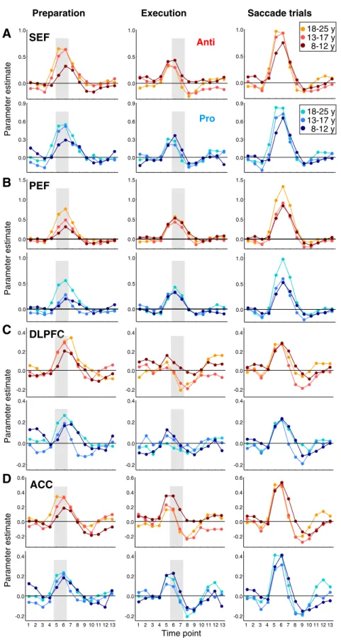
0.0 0.5 1.0 0.0 0.5 1.0 0.0 0.5 1.0 0.0 0.3 0.6 0.9 0.0 0.3 0.6 0.9 0.0 0.3 0.6 0.9 0.0 0.5 1.0 1.5 0.0 0.5 1.0 1.5 0.0 0.5 1.0 1.5 0.0 0.5 1.0 0.0 0.5 1.0 0.0 0.5 1.0 -0.2 0.0 0.2 0.4 -0.2 0.0 0.2 0.4 -0.2 0.0 0.2 0.4 -0.2 0.0 0.2 0.4 -0.2 0.0 0.2 0.4 -0.2 0.0 0.2 0.4 -0.2 0.0 0.2 0.4 0.6 -0.2 0.0 0.2 0.4 0.6 -0.2 0.0 0.2 0.4 -0.2 0.0 0.2 0.4 -0.2 0.0 0.2 0.4 0.6ACC
-0.2 0.0 0.2 0.4Fig. 6. Time course of activation related to task preparation (left panels), task execution (middle panels), and to saccade trials containing both preparation and execution (right panels) in the SEF (A), PEF (B), DLPFC (C) and ACC (D) for each age group: 8–12 years, 13–17 years, and 18–25 years. Time courses were derived from the functional clusters indicated inTable 3, then averaged across the right and left for bilateral regions. Same format asFigs. 4A–C. Hot colors represent the antisaccade task; cold colors represent the prosaccade task. The gray box in the left and middle panels represents the epoch used to assess the effects of age group on preparatory activation (time points 5–6) and execution-related activation (time points 6–7), respectively.
from each other. This developmental increase in preparatory activation remained when covarying the number of catch andfixation trials for both prosaccade (pb 0.05) and antisaccade (p b 0.01) tasks, the covar-iates showing no significant effect (ps N 0.3). On the contrary, no sig-nificant effect of age group was found for prosaccade execution in the SEF (F2, 76 = 1.17, p = 0.314) or PEF (F2, 76 b 1), or for
antisaccade execution in the SEF (F2, 76= 1.44, p = 0.242) or PEF
(F2, 76= 1.08, p = 0.344;Figs. 7B, D).
In sum, similarly to the FEF, SEF and PEF activation reflecting task preparation, not task execution, heightened with development.
DLPFC and ACC
Next, we interrogated the DLPFC and ACC, two areas involved in executive control and critical for antisaccade performance.Table 3 pro-vides the location of maximal activation for these two brain areas. The activation times courses associated with saccade preparation and exe-cution in DLPFC and ACC are depicted inFigs. 6C and D, respectively. For the DLPFC (Fig. 6C), a main effect of time (1–13 time points) for pre-paratory activation was observed within each age group (psb 0.05), suggesting that children, adolescents and adults recruited the DLPFC during saccade preparation. For task execution, results were 8-12 13-17 18-25 0.0 0.2 0.4 0.6 0.8 8-12 13-17 18-25 0.0 0.2 0.4 0.6 0.8 8-12 13-17 18-25 0.0 0.2 0.4 0.6 0.8 8-12 13-17 18-25 0.0 0.2 0.4 0.6 0.8 8-12 13-17 18-25 0.0 0.2 0.4 0.6 8-12 13-17 18-25 -0.2 0.0 0.2 0.4 0.6 8-12 13-17 18-25 -0.2 0.0 0.2 0.4 0.6 8-12 13-17 18-25 0.0 0.2 0.4 0.6
G
Age group (years) Age group (years)
Preparation Execution
H
E
F
C
D
A
Pro AntiB
PEF
ACC
DLPFC
SEF
P a rameter estimate*
&*
*
***
*
*
*
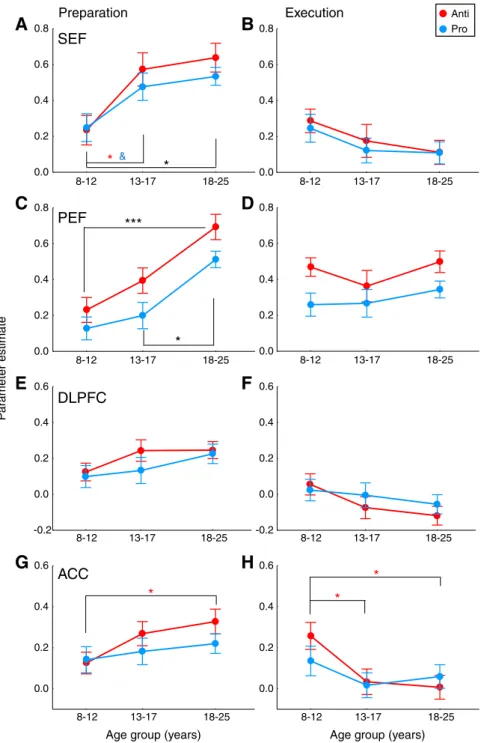
Fig. 7. Age-related changes in SEF, PEF, DLPFC and ACC activation. Effect of age group on preparatory activation (A, C, E, G) and execution-related activation (B, D, F, H). Same format as Figs. 4D–E. Values of preparatory and execution-related activation derive from the epoch illustrated by the gray box inFig. 6(left and middle panels, respectively). Large circles represent mean (+/−SE) data across participants within three age groups: 8–12 years, 13–17 years, and 18–25 years. Black asterisks depict significant differences in activation between age groups for both saccade tasks, whereas blue ampersand and red asterisks depict significant differences in activation between age groups for the prosaccade or antisaccade task specifically (post-hoc Tukey HSD tests). *pb 0.05; **p b 0.01; ***p b 0.001; &p = 0.061; others: non-significant.
rather mixed. For the antisaccade task, activation was significant in adults (main effect of time: F12, 264= 8.35, pb 0.001) and marginally
significant in adolescents (F12, 288 = 2.13, p = 0.057). For the
prosaccade task, only a marginally significant effect of time was ob-served in children (F12, 360= 2.17, p = 0.055). These mixed results
were not surprising given the superior involvement of DLPFC in sac-cade preparation rather than execution according to human fMRI studies (Brown et al., 2007). One-way ANOVAs revealed no significant differences in DLPFC activation among age groups for prosaccade prep-aration (F2, 76= 1.02, p = 0.367) or antisaccade preparation (F2, 76=
1.87, p = 0.16;Fig. 7E), or for prosaccade execution (F2, 76b 1) or
antisaccade execution (F2, 76= 2.51, p = 0.09;Fig. 7F).
The ACC (Fig. 6D) exhibited significant activation for both task preparation and execution within each age group (main effect of time: psb 0.05), except in the adolescent group where the effect of time for prosaccade execution-related activation was reduced to a trend (F12, 288= 1.96, p = 0.096). Thus the ACC was reliably
re-cruited by children, adolescents and adults during saccade prepara-tion. Antisaccade preparatory activation differed across age groups (F2, 76= 3.46, pb 0.05), children showing lower levels of activation
than adults (Fig. 7G). This effect of age group was lost when covary-ing the number of catch andfixation trials (p = 0.145), although the covariates showed no significant effect (ps N 0.4). Antisaccade execution-related activation also differed across age groups (F2, 76=
5.01, pb 0.01), children showing increased activation compared to both adolescents and adults (Fig. 7H). This decrease in activation from children to adults did not remain when covarying the number of saccade, catch and fixation trials (p = 0.142), these covariates showing no significant effect (ps N 0.08). No age group effects were observed in the prosaccade task (Figs. 7G, H) for preparatory activation (F2, 76b 1) or execution-related activation (F2, 76b 1).
In sum, the DLPFC and ACC were consistently recruited during saccade preparation. Preparatory activation in the DLPFC did not differ among age groups. From children to adults, ACC activation increased for antisaccade preparation whereas it decreased for antisaccade execution. These opposite developmental effects in the ACC may be partly accounted for the number of trials included in fMRI analyses as indicated by the additional analyses that used the number of trials as a covariate.
Discussion
We examined fronto-parietal activity at two stages of saccade control (task preparation during instruction vs. task execution after target appearance) to test the hypothesis that preparatory processes contribute to the improvements in prosaccade and antisaccade per-formance from childhood to adulthood. The presentfindings provide strong support in favor of our hypothesis. Preparatory activation heightened from children to adults in the core oculomotor network composed of the FEF, SEF and PEF for both prosaccade and antisaccade tasks. The ACC also showed higher preparatory activa-tion with increasing age for the antisaccade task, reinforcing its crit-ical role in antisaccade cognitive control. In contrast, execution-related activation did not change across age groups, except in the ACC where activation decreased from children to adults in the antisaccade task. Changes in ACC activation across age groups may be also partly due to the number of trials included per participant (seeDLPFC and ACCsection) and thus, cautions should be taken in the interpretations of these specific changes. At last, the DLPFC showed no age-related changes during saccade preparation or exe-cution. Three important implications can derive from these results. 1) The data reinforce the crucial role of the core fronto-parietal network in saccade preparation, and extend its involvement during development. 2) Developmental improvements in both prosaccade and antisaccade performance are related to enhancements in prepa-ratory processes associated with task instruction rather than
execution processes associated with target appearance. In other words, developmental improvements in behavior control are sup-ported by improvements in the ability to effectively preset goal-appropriate brain systems. 3) Children may use compensatory execution-related processes to correctly perform the tasks.
Improvements in task preparation across development
Our neuroimagingfindings provide new and direct evidence that developmental improvements in saccade performance are related to saccade preparation prior to target appearance, confirming and ex-tending a behavioral study that manipulated the length of the prepa-ratory period to study the role of preparation in development of antisaccade control (Ordaz et al., 2010). We found that the fronto-parietal network including the FEF, SEF, PEF, DLPFC and ACC that is critical for saccade preparation before target appearance in adults (for review,McDowell et al., 2008) is also recruited by children and adolescents, but in a‘suboptimal’ manner. Indeed, ANOVAs revealed that the magnitude of preparatory activation within these different areas (except the DLPFC) increased with development, suggesting that the core preparatory processes that are in place in children con-tinue to undergo gradual enhancements through childhood and ado-lescence. Importantly, the age-related differences in preparatory activation were unlikely to be due to differences infixation and/or stimuli properties. Fixation baseline activation did not significantly differ across age groups within the different ROIs, and no age-related changes in activation were found during catch trials in pri-mary visual cortex (not illustrated). A subset of electrophysiological studies supports ourfindings. The contingent negative variability, an electrophysiological index of task preparation, was reduced in chil-dren below around the age of 12, and was related to a delayed re-cruitment of anterior-central areas including frontal motor areas (Bender et al., 2005; Flores et al., 2009; Jonkman, 2006; Klein and Feige, 2005). Moreover, a number of developmental fMRI studies using tasks requiring cognitive control revealed immature patterns of activation in children and adolescents compared to adults in the fronto-parietal cortical network including lateral prefrontal cortex, parietal cortex, anterior cingulate cortex, and pre-supplementary/ supplementary motor area (e.g.,Bunge et al., 2002; Crone et al., 2006; Luna et al., 2001; Rubia et al., 2006; Velanova et al., 2008, 2009). Similarly to previous studies (Crone et al., 2006; Velanova et al., 2008, 2009), we found patterns of activation that were mature by adolescence in parallel to adult-like behavior performance, but also still immaturities in adolescence (here the PEF), which suggests continuing refinements into adulthood. Overall, we propose that a component of cognitive control that continues to improve through childhood and adolescence is the representation of task set: the abil-ity to prospectively configure an upcoming task based on arbitrary rules (Bunge et al., 2005; Sakai, 2008).
The DLPFC was the only studied region that exhibited no age-related changes in preparatory activation. Structural maturation of the pre-frontal cortex through childhood and adolescence (Gogtay et al., 2004) is thought to support maturation of cognitive control into ad-olescence (Luna and Sweeney, 2004). Actually, fMRI studies reported different developmental trajectories of DLPFC activation during inhi-bition tasks, which may depend on the type of task or design used. Some showed an increase in DLPFC activation from children to adults (Tamm et al., 2002), others a decrease (Casey et al., 1997; Durston et al., 2002; Velanova et al., 2008), and others showed non-linear changes (Luna et al., 2001). Our rapid interleaved design may have required higher vigilance and increased cognitive demands during the presentation of the instruction cue, such as enhanced reliance on inhibitory control, increased working memory for rules and for task set maintenance, and increased rule-based response selection demands. These processes have been shown to be associated with the DLPFC (e.g.,Brown et al., 2007; Bunge, 2004; Crone et al., 2006;