HAL Id: hal-01991334
https://hal.sorbonne-universite.fr/hal-01991334
Submitted on 23 Jan 2019
HAL is a multi-disciplinary open access
archive for the deposit and dissemination of
sci-entific research documents, whether they are
pub-lished or not. The documents may come from
teaching and research institutions in France or
abroad, or from public or private research centers.
L’archive ouverte pluridisciplinaire HAL, est
destinée au dépôt et à la diffusion de documents
scientifiques de niveau recherche, publiés ou non,
émanant des établissements d’enseignement et de
recherche français ou étrangers, des laboratoires
publics ou privés.
Selective vulnerability of the primitive meningeal layer
to prenatal Smo activation for skull base meningothelial
meningioma formation
Julien Boetto, Caroline Apra, Matthieu Peyre, Franck Bielle, Michel
Kalamarides
To cite this version:
Julien Boetto, Caroline Apra, Matthieu Peyre, Franck Bielle, Michel Kalamarides. Selective
vulnera-bility of the primitive meningeal layer to prenatal Smo activation for skull base meningothelial
menin-gioma formation. Oncogene, Nature Publishing Group, 2018, 37 (36), pp.4955-4963.
�10.1038/s41388-018-0328-7�. �hal-01991334�
Selective vulnerability of the primitive meningeal layer to prenatal
Smo activation for skull base meningothelial meningioma formation
Julien Boetto1●Caroline Apra1●Franck Bielle1,2●Matthieu Peyre1,3●Michel Kalamarides1,3
Abstract
Somatic activating mutations of smoothened (SMO), a component of the embryonic sonic hedgehog (SHH) signaling pathway, are found in 3–5% of grade I meningiomas, most of them corresponding to meningothelial meningiomas located at the anterior skull base. By generating different developmental stage-specific conditional activations in mice, we define a restricted developmental window during which conditional activation of Smo in Prostaglandin D2-synthase-positive mesoderm-derived meningeal layer of the skull base results in meningothelial meningioma formation. We show a selective vulnerability of the arachnoid from the skull base to Smo activation to initiate tumor development. This prenatal period and specific topography are correlated to the timing and location of SHH signaling involvement in the formation of craniofacial and meninges patterning, strongly corroborating the hypothesis of a developmental origin for Smo-activated meningiomas. Finally, we provide preclinical in vitro evidence of the efficacy of the SMO-inhibitor Sonidegib, supporting further preclinical and clinical evaluation of targeted treatment for refractory SMO-mutant meningiomas.
Introduction
Meningiomas are the most common primary central nervous system tumors in adults [1]. Treatment algorithms are based on surveillance, surgery, and radiotherapy/radiosurgery [2]. Meningiomas are classified into three prognostic histologi-cal groups following the WHO classification: grade I (65–80%), grade II (20–35%, atypical), and grade III (<3%, anaplastic) [1,2]. Recent advances in the molecular genetics of WHO grade I meningioma have led to the discovery of mutations in several oncogenes, including AKT1, KLF4,
SMO, PIK3CA, POLRA2, and TRAF7, all mutually exclu-sive to mutations in the NF2 tumor suppressor gene involved in at least 50% of meningiomas [3–6]. Discovery of this mutational landscape enabled the establishment of histological and/or anatomical correlations with specific gene mutations [3, 7, 8]. In a recent series of 775 menin-giomas, Clark et al. described Sonic Hedgehog (SHH) pathway-related meningiomas harboring 42 SMO gene mutations (recurrent mutations L412F and W535L in 76% of cases), 5 SUFU mutations and 3 PRKAR1A mutations [4]. These tumors were mostly WHO grade I, menin-gothelial, and located at the anterior skull base, suggesting a possible link between this potential driver mutation and anatomical site of origin. Indeed, SMO mutations alter the SHH pathway, which plays a critical role in craniofacial patterning during embryonic development, and conse-quently in the development of meninges at this location [9]. However, it is not clear whether meningioma tumorigenesis is induced by early prenatal deregulation of meningeal embryogenesis or by late postnatal dedifferentiation of mature cells into a stem-like state [4]. In fact, very little is known about the origin and regulation of meningeal development in humans and mice. By generating different tissue and developmental stage-specific conditional knock-out mice with neural crest and mesoderm specific pro-moters, we and others have characterized the embryonic
* Michel Kalamarides michel.kalamarides@aphp.fr
1 CRICM INSERM U1127 CNRS UMR 7225, Institut du Cerveau et de la Moelle Epinière, Sorbonne Université, Paris F-75013, France
2 Department of Pathology, AP-HP, Hôpital Pitié-Salpêtrière, Paris F-75013, France
3 Department of Neurosurgery, AP-HP, Hôpital Pitié-Salpêtrière, Paris F-75013, France
Electronic supplementary materialThe online version of this article
(https://doi.org/10.1038/s41388-018-0328-7) contains supplementary
origin of the meninges with those covering the tele-ncephalon being of neural crest origin, and those at the level of the skull base deriving from the cephalic mesoderm [10– 12]. We showed that prostaglandin D2 synthase (PGDS) gene is a specific marker of arachnoidal cells and menin-giomas in humans and mice and that PGDS-positive cells of the primitive meningeal layer developed during embry-ogenesis are the progenitor cells of origin capable of gen-erating meningiomas [12]. Here we demonstrate that mouse meningiomas induced by Smo activation in PGDS-positive cells have the same histological—meningothelial—and anatomical—skull base—characteristics as their human SMO-mutant tumors counterparts. In addition, we have identified a specific temporal window of activation for Smo-related meningioma tumorigenesis, providing clues to the developmental origin of this subgroup of meningiomas. These findings provide new insights in the molecular mechanisms of meningioma formation and support the use of targeted medical therapy for those rare skull base SMO-mutant meningiomas that are refractory to surgery and radiotherapy/radiosurgery with high morbidity [13].
Results
Identi
fication of a restricted developmental
temporal window for
Smo activation and
meningioma vulnerability
To investigate the role of Smo mutation in meningeal tumorigenesis, we generated genetically engineered SmoM2 mice in which activation of the Shh pathway was achieved by Cre-mediated conditional expression of an activated Smoothened allele (SmoM2) [14]. Prenatal activation of Smo was induced by generating 42 PGDSCre;SmoM2 mice where SmoM2 was expressed early (starting at E12.5) at the skull base level in the meningeal precursor cells identified as PGDS-positive cells [12]. After a mean survival of 14.2 months, 9 of 42 (21%) PGDSCre;SmoM2 mice developed meningiomas and 32 (76%) presented foci of meningothelial proliferation, the first manifestation of meningioma tumorigenesis in mice [15] (Table 1). All tumor lesions were localized at the skull base level ventral to the brainstem. Histologically, they appeared as
meningothelial grade I meningiomas showing the typical aspect of tumoral meningothelial cells with oval nuclei and nuclear clearing disposed in lobules (Fig.1a–c).
To analyze the effect of postnatal Smo activation, a cohort of 53 SmoM2 mice was injected with adCre at postnatal day 2 (25 at the convexity-s.d. (subdural)- and 28 at the skull base-t.o.(trans orbitary)-). Forty-nine mice were analyzed after a mean survival of 14.2 months: only one meningothelial meningioma (2%) and 23 (43%) menin-gothelial proliferations were observed at the skull base (Fig. 1d). All meningothelial proliferations were located at the skull base level even after injection at the convexity. Interestingly, the four other mice in the cohort died shortly after a mean survival of 2.8 months. All presented typical large cerebellar medulloblastomas composed by sheets of poorly differentiated small cells (Fig. 1e), without menin-gothelial proliferation. Our hypothesis is that after sub-dural injection the adCre virus transduced cells in the neuraxis by diffusion through the cerebrospinal fluid. This induced SmoM2 expression in neural progenitors of developing cerebellar cortex, the cell of origin of Shh-activated medulloblastoma representing 25% of this tumor type [16, 17]. Activation of the Hedgehog pathway was con-firmed in vivo in both models by immunohistochemical expression of Gli-1 in Smo-related meningothelial pro-liferations and meningiomas (Fig.1f, g). No meningothelial proliferation, meningioma, or medulloblastoma were found in the control cohort of 8 adLacZ-SmoM2 mice. These results defined a restricted developmental window during which conditional activation of Smo in PGDS mesoderm-derived meningeal layer of the skull base results in meningioma formation.
Selective vulnerability of the skull base compared to
convexity arachnoidal cells to
SmoM2 expression on
proliferation and Shh pathway activation
To analyze the effect of Smo activation in the meninges according to their location and embryologic origin, we established primary arachnoidal cell cultures from meninges of the skull base or the convexity of SmoM2 mice. After adCre infection of primary arachnoidal cells in vitro, SmoM2 activation was assessed by detection of YFP fluorescence (Fig.2a, b). Nofluorescence was expressed in
Table 1 Summary of the phenotypic consequences of Smo activation in meningeal cells in vivo Mouse genotype Phenotypic abnormality PGDSCre-SmoM2 n = 42 AdCre-injected SmoM2 n =
53 AdLacZ-injected SmoM2 n = 8 Meningothelial meningioma 9 (21%) 1 (2%) 0 p = 0.003 Meningothelial proliferation 32 (76%) 21 (43%) 0 p < 0.001 Medulloblastoma 0 4 (8%) 0 p = 0.14
the adLacZ-infected control SmoM2 cells (data not shown). Proliferation assays showed that SmoM2 expression induced a slight proliferative advantage in skull base ara-chnoidal cells at day 7 (p = 0.009, t-test, Fig.2c), whereas no significant effect was seen in convexity arachnoidal cells (p = 0.85, t-test, Fig. 2d). WST1 activity was not sig-nificantly modified by SmoM2 expression in skull base arachnoidal cells compared to convexity arachnoidal cells (p = 0.46, t-test, Fig. 3b). Contrary to expectations, apop-tosis was significantly increased when SmoM2 was expressed (16.7 vs 8.1%, p < 0.001, Fig.2g–i). On the other hand, cellular senescence was not activated by SmoM2 expression as demonstrated by the SAβ-galactosidase assay
(data not shown). In skull base arachnoidal cells, SmoM2 expression induced a significant increase in Gli1 (p = 0.005, t-test) and Ptch1 (p = 0.03, t-test) mRNA levels (Fig. 2e), the two main target genes of the canonical Shh pathway [18]. In contrast, no difference in Gli1 (p = 0.37, t-test) or Ptch1 (p = 0.77, t-test) mRNA levels was found in the convexity arachnoidal cells (Fig. 2f). The differences in arachnoid from skull base and convexity region are not intrinsic as growth pattern and mRNA levels of Gli1 and Ptch1 were similar in control/wild-type cells (infected with adLacZ). These results clearly showed a differential response and thus vulnerability to Smo activation according to the location and embryological origin of the meninges,
Fig. 1 Pathological
characterization of Smo-induced tumors in PGDSCre;SmoM2 mice and adCre;SmoM2 mice. a H&E stained sections showing meningothelial meningioma overlying the brain (arrowhead, ×200). b Another
meningothelial meningioma composed of typical tumoral arachnoidal cells with oval nuclei at higher magnification (×400). c H&E stained section showing normal arachnoidal layer (c, arrowhead ×200) and a meningothelial proliferation (d, arrowhead ×200). e H&E-Safran stained section of a large medulloblastoma (star, ×200) invading the cerebellum). Gli-1 immunohistochemistry in a meningothelial proliferations (f) and a meningothelial
meningioma (g) demonstrating positivity of meningioma cells. Br brain, tg trigeminal nerve, cer Cerebellum
those of the skull base being exquisitely sensitive to Shh pathway activation.
LDE-225 (Sonidegib) inhibits Shh pathway and cell
proliferation in skull base
SmoM2 arachnoidal cells
To obtain preclinical evidence of the efficacy of SMO inhibitors in the treatment of meningiomas, we analyzed the effect of LDE-225 (Sonidegib) on primary arachnoidal cells in culture. Since the pro-proliferative effect of SmoM2 expression was found only in arachnoidal cells of the skull base, all the experiments with Sonidegib were carried out with those cells. Skull base adCre-infected SmoM2 ara-chnoidal cells were treated with Sonidegib (10 or 100 nM) 1 day after adCre infection. Short-term proliferation assays showed that Sonidegib at 10 nM was able to inhibit the proliferation of adCre-infected SmoM2 cells in terms of cell count (p = 0.02, t-test, Fig.3a) although WST1 activity was not significantly decreased (p = 0.41, t-test, Fig.3b). There was a significant reduction in Gli1 (p = 0.005, t-test) mRNA expression compared to vehicle (DMSO) treatment, but no difference was observed for Ptch1 mRNA levels (p = 0.71, t-test) (Fig.3c, d).
Discussion
Among the nine different histological subtypes of Grade I meningiomas in humans, the unique histological subtype— meningothelial- of SMO-mutant meningiomas supports the view that this oncogenic pathway is critical to the ara-chnoidal meningeal lineage at the origin of this subtype compared to thefibroblastic subtype. Previous embryologic studies have shown that PGDS-positive primordial
meningeal cells arising at E12.5 give rise to meningeal cells of both arachnoid and dura-mater layers at E14.5 (Fig. 4) [12]. We previously showed using gentically engineered mouse models that the two major histological subtypes of WHO Grade I meningiomas were inducible by Nf2 inacti-vation and found in 38% of PGDSCre;Nf2flox/flox2 mice: Meningothelial tumors were originating from the arachnoid layer (arachnoid border cells) and fibroblastic tumors were emanating from the dura-mater (dural border cell layer) [12] (Fig. 4). In contrast, for Smo-related meningeal tumor-igenesis, only meningothelial and no fibroblastic menin-giomas were found in PGDSCre;SmoM2 mice, thus implying that, unlike PGDS-positive arachnoidal cells, PGDS-positive dura-mater cells are not vulnerable to Smo activation for meningothelial meningioma development (Fig.4). All meningiomas found in PGDSCre;SmoM2 mice displayed histological features strikingly similar to human SMO-mutant meningiomas. To explore the postnatal developmental window for tumor development, we have previously reported a model with adCre injection in the subdural space of newborn mice and shown that Nf2 loss in very early postnatal meningeal cells was sufficient to induce meningioma formation (WHO Grade I meningiomas of different histological subtypes in 23% of mice) [12]. In contrast to Nf2-related tumorigenesis, the absence of meningioma in adCre;SmoM2 mice, corresponding to an early postnatal activation, and the selective finding of meningothelial (WHO Grade I) meningiomas at the skull base level of PGDSCre;SmoM2 mice suggest that embryonic activation of Smo in the arachnoidal meningeal layer is necessary for Smo-related meningioma tumorigen-esis (Fig.4). The temporal developmental window in which Smo mutation culminates in meningioma formation is more restrictive for Smo compared to Nf2 where the window for tumor initiation is wider, encompassing the embryonic and early postnatal periods [12]. The benign nature and unique spatial location of these meningiomas is also reflected by the slight in vitro proliferative advantage and selective sensitivity to Sonidegib of SmoM2-expressing arachnoidal cells of the skull base versus the convexity and by the presence of meningothelial proliferation restricted to the skull base level regardless of the injection site of adCre, respectively.
Taken together, these results indicate that Smo activation is necessary and sufficient to induce meningioma develop-ment at the skull base level in a restricted temporal window during the prenatal period starting at E12.5, strongly arguing in favor of the developmental nature of this sub-group of meningiomas and supporting the hypothesis that the developmental stage of arachnoidal cells determine their susceptibility to tumor-initiating alterations, This hypothesis and the predominance of human SMO-mutated meningio-mas at the median skull base level are also sustained by the
Fig. 2 Proliferation assays and SHH-pathway activation study show-ing a spatial restriction of Smo activation effect dependshow-ing on topo-graphic origin of arachnoidal cells. Morphologies of primary skull base SmoM2 arachnoidal cells in culture after adCre infection (a), showing SmoM2 activation assessed by expression of YFP in fluor-escence microscopy (b). Proliferation assays on skull base (c) and convexity (d) arachnoidal cells: SmoM2 expression induces a pro-liferation advantage among skull base arachnoidal cells at day 7 (p = 0.009, t-test) but not among convexity cells (p = 0.85, t-test). Com-parison of Gli1 and Ptch1 mRNA levels among skull base (e) and convexity cells (f): SmoM2 expression induces a significant increase in Gli1 (p = 0.005, t-test) and Ptch1 (p = 0.03, t-test) among skull base arachnoidal cells but not among convexity cells (p = 0.37 and 0.77, t-test). Apoptosis assay: annexin V versus propidium iodide (PI) cell sorting for skull base SmoM2 arachnoidal cells infected with adLacZ virus (g) or adCre virus (h). The percentage of Annexin V-positive/PI-negative cells (early stage of apoptosis, Q4) and Annexin V-positive/ PI-positive cells (late stage of apoptosis, Q2) were separated and measured using flow cytometry. Late apoptosis was significantly higher in cells expressing SmoM2 (i) (16.7 versus 8.1%, p < 0.001). All experiments were performed in triplicate
fact that SHH is the main embryonic signaling pathway implicated in ventral craniofacial development by promot-ing rapid cell expansion of the facial mesenchyme, quickly followed by a transition to a terminally differentiated state of the connective tissue of the face and skull [16,19,20]. It has already been shown that targeted disruption of Smo in mice affect appropriate growth and patterning in the early facial primordial, while its constitutive activation (Wnt1-Cre;SmoM2) produces hyperplasia of the facial processes and gross disorganization, with a failure of most craniofa-cial skeletogenesis [9]. Identically, in ameloblastoma, a locally destructive tumor occurring during tooth develop-ment, a relation between anatomical site and driver gene mutation has also been recently shown: Mutations in SMO are common in ameloblastomas arising from the maxilla whereas BRAF mutations are predominant in tumors of the mandible [21]. This anatomical specificity for odontogenic development in the upper or lower dentition is likely to reflect distinctive developmental pathways based on spatial temporal and/or cell type-specific cues. Finally, similar to our approach and results, Ohli et al.[22] have observed in a Math1-CreERT2;SmoM2 medulloblastoma mouse model that post-natally induced activation of SmoM2 lead to a different tumor burden compared to embryonically
induced activation of SmoM2. Their results were in line with the distribution of SHH medulloblastomas in human and indicate that the localization of SHH medulloblastomas was dependent on the time-restricted susceptibility of granule cell precursors in defined cerebellar compartments.
Curiously, no PGDSCre;SmoM2 mouse suffered from symptomatic meningioma before 15 months of age. This is likely due to slow, indolent tumor growth with no clinical impact until late in life, like in humans, where SMO-mutant meningiomas are also diagnosed later in life (mean age of 55 years) [13]. In a recent paper, an integrated model of the growth dynamics of benign meningiomas using radiocarbon retrospective birth dating was provided for the first time [23]. In their series, the mean lifetime of WHO grade I meningiomas was 22.1 ± 6.5years (whereas atypical WHO grade II meningiomas originated 1.5 ± 0.1years) prior to surgery. Assuming an additional period of time between the first initiating mutation in a single meningeal cell and the time a microscopic lesion can be measured, in addition to the fact that olfactory groove meningiomas (like most SMO-mutated meningiomas [13]) can remain silent for a long time despite a very large volume, then we should reconsider our concepts on the natural history of grade I
Fig. 3 Effect of Sonidegib (LDE-225) on proliferation and SHH pathway in skull base SmoM2-induced cells. Sonidegib at 10 nM reversed the proliferation advantage of SmoM2-induced cells at day 7 when counting the cells and in WST1 assay (a, p = 0.02, t-test and b,
p = 0.41, t-test), and Gli1 mRNA expression (c, p = 0.005, t-test) compared to vehicle (DMSO). No significant effect was found on Ptch1 mRNA levels (d, p = 0.71, t-test). All experiments were per-formed in triplicate
meningiomas. In contrast, other developmental malignant tumors of the central nervous system, such as neuro-blastomas, SMO-mutant or SHH-activated medullo-blastomas, and malignant rhabdoid tumors generally occur during infancy or childhood [16, 24]. The restricted early developmental time window for Smarcb1 loss to promote malignant rhabdoid tumor formation and vulnerability to additional Nf2 tumor suppressor gene mutations resulting in benign schwannoma formation later in life, provides another striking example of close interrelationship between timing of driving mutations, tumor tissue of origin, and clinical behavior [25].
From a prognostic and therapeutical point of view, despite their bland histologic appearance, we have shown that SMO mutation is associated with poorer prognosis within the olfactory groove meningiomas subgroup with several patients showing serious clinical morbidity (blind-ness, pituitary gland dysfunction) and late recurrences [13]. These recurrences were observed despite optimal available treatments, suggesting that SMO inhibitors would be clinically useful in daily practice. SMO inhibitors are under
clinical evaluation for SHH-mutated medulloblastoma [26] and routinely used in metastatic basal cell carcinoma [27]. Our study provides the first preclinical in vitro evidence of the efficiency of Sonidegib for the treatment of SMO-mutant meningiomas. Sonidegib significantly inhibited both Shh pathway activation and growth in SmoM2 activated skull base arachnoidal cells at nanomolar con-centrations. Thus, we provide a rationale for clinical trials evaluating SMO inhibitors in the treatment of refractory SMO-mutant meningiomas. A multicenter phase II study of Vismodegib is now recruiting patients with progressive or recurrent SMO-mutant meningiomas (NCT02523014, clinicaltrials.gov).
Materials and methods
Mice
Rosa26-lox-STOP-lox-SmoM2 mice (called SmoM2 mice in this paper) were purchased from the Jackson Laboratory. In this model, a conditional allele of SmoM2 with a C-terminal YFP tag is targeted into the ubiquitously expressed Rosa26 locus, after a lox-Pflanked polyadenylation stop sequence cassette. In presence of Cre recombinase, SmoM2 gene is expressed and encodes for a mutant form of protein Smo previously identified in human basal cell carcinoma [28]. In this mutant allele, an activating mutation (W535L) in the seventh transmembrane domain results in ligand-independent constitutive activation of the Shh pathway in target tissue [14].
To induce postnatal SmoM2 activation in meningeal cells, wefirst performed injections in the sub-dural space of 3–4 μL of adenoviruses Ad5CMV-Cre (REF #VVC-U of Iowa-5, University of Iowa, Viral Vector Core, Iowa City, IA, USA), called further adCre, in SmoM2 pups (postnatal day 2) as previously described (trans-orbitary (t.o.) to target the skull base and subdurally (s.d.) to target the meninges at the convexity) [15]. A control group of SmoM2 pups were injected with Ad5CMVntLacZ (REF #VVC-U of Iowa-1917, University of Iowa, Viral Vector Core, Iowa City, IA, USA), called adLacZ, with the same methodology.
To induce prenatal activation of Shh in meningeal pre-cursor cells, SmoM2 mice were bred with homozygous PGDSCre mice to obtain hemizygous PGDSCre;SmoM2 mice [12]. In this model, Cre recombinase is expressed selectively in PGDS-positive cells and is able to induce the expression of the SmoM2 gene. All animal care and experimentation reported herein were conducted in com-pliance with the guidelines and with the specific approval of the Institutional Animal Care and Use Committee of the French Department of Agriculture.
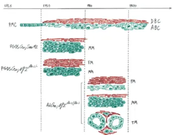
Fig. 4 Illustration of the mouse Grade I skull base meningiomas in relation with the meningeal cell of origin and the mutation of the Smo or Nf2 genes: A restricted developmental temporal and spatial window for Smo activation for meningioma vulnerability in comparison with Nf2 inactivation. In the normal meninges of skull base, primordial meningeal cells (PMC) (starting at E12.5) undergo lineage commit-ment to form both dural border cells (DBC) and arachnoid barrier cells (ABC) at E15.5. Early mutation of Smo at the embryonic cell stage leads only to the development of meningothelial meningioma (MM) in PGDSCre;SmoM2 mice. In contrast, mutation of Smo at a later developmental stage, following early postnatal (PN1) adenoviral Cre injection (AdCre;SmoM2 mice), has no effect on tumorigenesis. For Nf2-related meningioma tumorigenesis, early Nf2 loss in embryonic primordial meningeal cells (PGDSCre;Nf2flox2/- mice) induces the development offibroblastic meningiomas (FM) in the dura mater and meningothelial meningiomas (MM) in the arachnoid layer. All histo-logical subtypes, including transitional (TM), were observed following early postnatal (PN1-10) Nf2 inactivation by adenoviral Cre injection (AdCre;Nf2flox2/flox2mice)
Histopathology and immunohistochemistry
Mice were monitored closely and killed when seriously ill or at 15 months. To preserve the arachnoidal layer, the head was fixed in formalin in toto, decalcified, and sliced cor-onally before embedding in paraffin as previously described [29]. The terminology used for the description of the meningothelial lesions in the mouse models is based on the WHO classification of human tumors with adaptations [30]. The term “meningothelial hyperplasia” is not used, as in humans where it refers to the proliferation of reactive, normal arachnoidal cells. “Meningothelial proliferation” refers to very small (microscopic) lesions composed of meningothelial cells that represent early tumor formation. “Meningioma” refers to a larger meningothelial lesion, with features similar to a WHO grade I meningioma in humans. Immunohistochemical staining was performed on 5-μm paraffin sections. For antigen retrieval, after pressure boiling in 1 mM EDTA (pH 8.0) for 30 min, sections were blocked in 10% serum followed by incubation with primary anti-body. Biotinylated secondary antibodies were used at 1:200 (Gli-1, ab151796, Abcam). Stainings were developed using Vectastain ABC system and DAB substrate kit (Vector Labs). Normal mouse arachnoid was used as positive control.
Arachnoidal cells culture
Primary arachnoidal cells cultures were established from SmoM2 mice. The arachnoidal tissue was surgically removed at the ventral surface of the brainstem (skull base arachnoid) and at the convexity (convexity arachnoid) after killing and plated in Petri dishes after 30 min digestion with Collagenase III (Wothington, Lakewood, NJ, USA). Ara-chnoidal cells were then cultured in Dulbecco’s modified Eagle’s medium (DMEM; Gibco/Invitrogen, Carlsbad, CA, USA) containing 10% Fetal Calf Serum (FCS), insulin 0.05%, Penicillin/streptomycin 1%, and Epidermal Growth Factor 0.004%. After 15 days of primary culture, cells were passaged in 12-well plates. Cells were infected after 48 h either with adCre (group called “SmoM2-adCre treated”) or with adLacZ (control group, called “SmoM2-adLacZ trea-ted”) at 50 pfu/cells for 24 h. Expression of YFP was ver-ified using a fluorescence microscope (Nikkon, IHC software) to assess SmoM2-YFP expression.
In vitro functional assays
Proliferation assay
After infection, arachnoidal cells were passaged, seeded at a concentration of 15,000 cells/mL in triplicate in 12-well plates, and counted manually using a Malassez cell counting
chamber 1, 4, and 7 days after plating. Arachnoidal cells were seeded at a concentration of 3000 cells/100 µL in tri-plicate in 96-well plates. WST1 assay was performed by adding 10% WST1 reagent (Roche, Indianapolis, Indiana, USA) in the medium and measuring absorbance after 3 h using a Spectromax M4 for wavelengths 450–620 nm. Senescence assay
SA β-galactosidase staining was performed on infected arachnoidal cells [31].
Apoptosis assay
After infection, arachnoidal cells were passaged, stained according to a standardized Annexin V FITC—Propidium Iodide protocol (Thermo Fisher Scientific, Waltham, MA, USA) and sorted using flow cytometry analysis (FACS-Diva, BD Biosciences, San Jose, CA, USA).
RNA extraction and RT-qPCR
After infection, cells were passaged and seeded in 12-well plates. RNA was extracted 48 h after passage using a Nucleospin RNA kit (Macherey-Nagel, Germany) and quantified using Nanodrop (Thermo Fisher, Villebon sur Yvette, France). RNA was retrotranscribed using the Maxima First Strand cDNA Synthesis Kit (Thermo Fischer). The cDNA obtained was used as a template for the determination of Gli1 and Ptch1 mRNA expression by qPCR on a Lightcycler® 96 (Roche, Bâle, Switzerland) using a Quantifast assay (Qiagen, Hilden, Germany). The ΔCt method was applied to normalize the mRNA levels using the expression of glucuronidase-β. The primer sets for qPCR were designed with the Roche design center (www. universalprobelibrary.com) (Supplementary Table S2). LDE-225 (Sonidegib) treatment
Sonidegib, a selective inhibitor of SMO, was provided by Novartis (MTA 43219). Proliferation assays and Gli1 and Ptch1 expression analyses were performed with Sonidegib at different concentrations (10 and 100 nM) in triplicate.
Statistical analysis
Student’s t-tests were used in proliferation assays and mRNA level analyses. The distribution of categorical vari-ables was compared with Fisher’s exact test. All tests were two-sided and a p-value of ≤0.05 was considered to be statistically significant. Statistical analyses were performed using Statview version 5.0 software (SAS Institute, Cary, NC, USA).
Acknowledgements We are grateful to Marco Giovannini for critical reviewing of the manuscript. We are indebted to Marine Giry and Amithys Rahimian (Onconeurotek) for technical assistance, and Ver-onique Parietti and Martine Chopin for mouse handling.
Funding: This work was supported by a grant from the foundation ARC (PJA 20131200431), France. J.B. was funded by a grant from UM1 University, Montpellier, France.
Ethical approval: All procedures performed in studies involving ani-mals were in accordance with the ethical standards of the institution at which the studies were conducted, and all applicable national guide-lines for the care and use of animals were followed.
Compliance with ethical standards
Conflict of interest The authors declare that they have no conflict of interest.
References
1. Louis DN, Ohgaki H, Wiestler OD. WHO classification of tumours of the Central Nervous System, Revised.. Fourth Edition. Lyon: International Agency for Research On Cancer; 2016. 2. Rogers L, Barani I, Chamberlain M, Kaley TJ, McDermott M,
Raizer J, et al. Meningiomas: knowledge base, treatment out-comes, and uncertainties. A RANO review. J Neurosurg. 2015;122:4–23.
3. Clark VE, Erson-Omay EZ, Serin A, Yin J, Cotney J, Ozduman K, et al. Genomic analysis of non-NF2 meningiomas reveals mutations in TRAF7, KLF4, AKT1, and SMO. Science. 2013;339:1077–80.
4. Clark VE, Harmancı AS, Bai H, Youngblood MW, Lee TI, Bar-anoski JF, et al. Recurrent somatic mutations in POLR2A define a distinct subset of meningiomas. Nat Genet. 2016;48:1253–9. 5. Abedalthagafi M, Bi WL, Aizer AA, Merrill PH, Brewster R,
Agarwalla PK, et al. Oncogenic PI3K mutations are as common as AKT1 and SMO mutations in meningioma. Neuro-Oncology. 2016;18:649–55.
6. Kros J, de Greve K, van Tilborg A, Hop W, Pieterman H, Avezaat C, et al. NF2 status of meningiomas is associated with tumour localization and histology. J Pathol. 2001;194:367–72.
7. Reuss DE, Piro RM, Jones DTW, Simon M, Ketter R, Kool M, et al. Secretory meningiomas are defined by combined KLF4 K409Q and TRAF7 mutations. Acta Neuropathol. 2013;125:351–8.
8. Yuzawa S, Nishihara H, Yamaguchi S, Mohri H, Wang L, Kimura T, et al. Clinical impact of targeted amplicon sequencing for meningioma as a practical clinical-sequencing system. Mod Pathol. 2016;29:708–16.
9. Jeong J, Mao J, Tenzen T, Kottmann AH, McMahon AP. Hedgehog signaling in the neural crest cells regulates the pat-terning and growth of facial primordia. Genes Dev. 2004;18:937–51.
10. Yoshida T, Vivatbutsiri P, Morriss-Kay G, Saga Y, Iseki S. Cell lineage in mammalian craniofacial mesenchyme. Mech Dev. 2008;125:797–808.
11. McBratney-Owen B, Iseki S, Bamforth SD, Olsen BR, Morriss-Kay GM. Development and tissue origins of the mammalian cranial base. Dev Biol. 2008;322:121–32.
12. Kalamarides M, Stemmer-Rachamimov AO, Niwa-Kawakita M, Chareyre F, Taranchon E, Han Z-Y, et al. Identification of a progenitor cell of origin capable of generating diverse menin-gioma histological subtypes. Oncogene. 2011;30:2333–44.
13. Boetto J, Bielle F, Sanson M, Peyre M, Kalamarides M. SMO mutation status defines a distinct and frequent molecular subgroup in olfactory groove meningiomas. Neuro-Oncology. 2017;19:345–51.https://doi.org/10.1093/neuonc/now276
14. Mao J, Ligon KL, Rakhlin EY, Thayer SP, Bronson RT, Rowitch D, et al. A novel somatic mouse model to survey tumorigenic potential applied to the Hedgehog pathway. Cancer Res. 2006;66:10171–8.
15. Kalamarides M, Niwa-Kawakita M, Leblois H, Abramowski V, Perricaudet M, Janin A, et al. Nf2 gene inactivation in arachnoidal cells is rate-limiting for meningioma development in the mouse. Genes Dev. 2002;16:1060–5.
16. Marshall GM, Carter DR, Cheung BB, Liu T, Mateos MK, Meyerowitz JG, et al. The prenatal origins of cancer. Nat Rev Cancer. 2014;14:277–89.
17. Ellison DW, Dalton J, Kocak M, Nicholson SL, Fraga C, Neale G, et al. Medulloblastoma: clinicopathological correlates of SHH, WNT, and non-SHH/WNT molecular subgroups. Acta Neuro-pathol. 2011;121:381–96.
18. Varjosalo M, Taipale J. Hedgehog: functions and mechanisms. Genes Dev. 2008;22:2454–72.
19. Xavier GM, Seppala M, Barrell W, Birjandi AA, Geoghegan F, Cobourne MT. Hedgehog receptor function during craniofacial development. Dev Biol. 2016;415:198–215.
20. Eberhart JK, Swartz ME, Crump JG, Kimmel CB. Early Hedge-hog signaling from neural to oral epithelium organizes anterior craniofacial development. Dev Camb Engl. 2006;133:1069–77. 21. Sweeney RT, McClary AC, Myers BR, Biscocho J, Neahring L,
Kwei KA, et al. Identification of recurrent SMO and BRAF mutations in ameloblastomas. Nat Genet. 2014;46:722–5. 22. Ohli J, Neumann JE, Grammel D, Schüller U. Localization of
SHH medulloblastoma in mice depends on the age at its initiation. Acta Neuropathol. 2015;130:307–9.
23. Huttner HB, Bergmann O, Salehpour M, El Cheikh R, Nakamura M, Tortora A, et al. Meningioma growth dynamics assessed by radiocarbon retrospective birth dating. EBioMedicine. 2017;27:176–81.
24. Kool M, Jones DTW, Jäger N, Northcott PA, Pugh TJ, Hovestadt V, ICGC PedBrain Tumor Project. et al. Genome sequencing of SHH medulloblastoma predicts genotype-related response to smoothened inhibition. Cancer Cell. 2014;25:393–405.
25. Vitte J, Gao F, Coppola G, Judkins AR, Giovannini M. Timing of Smarcb1 and Nf2 inactivation determines schwannoma versus rhabdoid tumor development. Nat Commun. 2017;8:300. 26. Samkari A, White J, Packer R. SHH inhibitors for the treatment of
medulloblastoma. Expert Rev Neurother. 2015;15:763–70. 27. Migden MR, Guminski A, Gutzmer R, Dirix L, Lewis KD,
Combemale P, et al. Treatment with two different doses of soni-degib in patients with locally advanced or metastatic basal cell carcinoma (BOLT): a multicentre, randomised, double-blind phase 2 trial. Lancet Oncol. 2015;16:716–28.
28. Xie J, Murone M, Luoh SM, Ryan A, Gu Q, Zhang C, et al. Activating smoothened mutations in sporadic basal-cell carci-noma. Nature. 1998;391:90–92.
29. Giovannini M, Robanus-Maandag E, van der Valk M, Niwa-Kawakita M, Abramowski V, Goutebroze L, et al. Conditional biallelic Nf2 mutation in the mouse promotes manifestations of human neurofibromatosis type 2. Genes Dev. 2000;14:1617–30. 30. Peyre M, Stemmer-Rachamimov A, Clermont-Taranchon E,
Quentin S, El-Taraya N, Walczak C, et al. Meningioma progres-sion in mice triggered by Nf2 and Cdkn2ab inactivation. Onco-gene. 2013;32:4264–72.
31. Le Roux I, Konge J, Le Cam L, Flamant P, Tajbakhsh S. Numb is required to prevent p53-dependent senescence following skeletal muscle injury. Nat Commun. 2015;6:8528.