HAL Id: tel-01870042
https://tel.archives-ouvertes.fr/tel-01870042
Submitted on 7 Sep 2018HAL is a multi-disciplinary open access
archive for the deposit and dissemination of sci-entific research documents, whether they are pub-lished or not. The documents may come from teaching and research institutions in France or
L’archive ouverte pluridisciplinaire HAL, est destinée au dépôt et à la diffusion de documents scientifiques de niveau recherche, publiés ou non, émanant des établissements d’enseignement et de recherche français ou étrangers, des laboratoires
Gammarus sp. as a valuable non-vertebrate model?
Kahina Mehennaoui
To cite this version:
Kahina Mehennaoui. Understanding the impact of engineered nanoparticles Gammarus sp. as a valuable non-vertebrate model?. Ecotoxicology. Université de Lorraine, 2017. English. �NNT : 2017LORR0388�. �tel-01870042�
AVERTISSEMENT
Ce document est le fruit d'un long travail approuvé par le jury de
soutenance et mis à disposition de l'ensemble de la
communauté universitaire élargie.
Il est soumis à la propriété intellectuelle de l'auteur. Ceci
implique une obligation de citation et de référencement lors de
l’utilisation de ce document.
D'autre part, toute contrefaçon, plagiat, reproduction illicite
encourt une poursuite pénale.
Contact : [email protected]
LIENS
Code de la Propriété Intellectuelle. articles L 122. 4
Code de la Propriété Intellectuelle. articles L 335.2- L 335.10
http://www.cfcopies.com/V2/leg/leg_droi.php
Laboratoire Interdisciplinaire
des Environnements Continentaux
LIEC – UMR 7360 CNRS, Metz
THESE
Pour l’obtention du titre de:
DOCTEUR DE L’UNIVERSITE DE LORRAINE
Mention: Ecotoxicologie, Biodiversité, Ecosystèmes
Kahina MEHENNAOUI
Understanding the impact of engineered nanoparticles
Gammarus sp. as a valuable non-vertebrate model?
Soutenue publiquement Le 20 décembre 2017 devant la commission d’examen :
Laure Giamberini Université de Lorraine – UMR 7360 CNRS - LIEC Arno Gutleb Luxembourg Institute of Science and Technology Catherine Mouneyrac Université Catholique de l’Ouest – MMs - Angers Mélanie Auffan CNRS – CEREGE – Aix en Provence
Erik Ropstad Norwegian University of Life Sciences
Elise David Université Reims Champagne-Ardenne – UMR-I 02 SEBIO François Guérold Université de Lorraine – UMR 7360 CNRS - LIEC
Sebastien Cambier Luxembourg Institute of Science and Technology
Ecole Doctorale Sciences et Ingénierie
Ressources Procèdes Produits Environnement,
RP2E, ED Nº 410, Nancy
Environmental Research and Innovation (ERIN)
Department,
Luxembourg Institute of Science and Technology,
(LIST), Luxembourg
Directeur de thèse Directeur de thèse Rapporteur Rapporteur Examinateur Examinateur Examinateur ExaminateurABSTRACT
The potential toxicity of nanomaterials is of high societal and scientific interest due to the promise of ground-breaking innovations for many technical applications. However, toxicity can often not be related to the actual size, mass or surface area of the single nanoparticles (NPs) or the NP agglomerates. Therefore, it can be proposed that the toxicity is greatly influenced by other inherent and non-understood properties of the particles to which ions dissolving from the particle, surface or molecules adhering to the surface interfering with the uptake of NPs into cells, may have important contributions.
The PhD project “NANOGAM”, closely linked up to CORE2012 NANION project that aims to obtain knowledge to understand some of the processes and factors involved in NP uptake and toxicity as such understanding is a prerequisite for the development of nanomaterials following the safer-by-design philosophy.
This PhD project aims to investigate, based on known characteristics of the key physico-chemical parameters; as size and surface functionalities, of a well-chosen list of silver and gold NPs, the uptake, and dependent biological effects of different complexity (mortality, behavioural effects, physiological effects, transcriptomic effects, etc.), on a sensitive species; Gammarus fossarum (Crustacea Amphipoda), in order to understand to which extent toxicity of nanomaterials is due to intrinsic material properties or ion leaching. Such understanding will contribute to the prediction of toxicity based on material properties rather than repetitive testing of an indefinite number of new nanomaterials.
G. fossarum were exposed at low concentrations of AgNPs and AuNPs for 72h or 15 days in presence or absence
of food. The obtained results showed that (i) surface coating is the main factor governing AgNPs and AuNPs uptake by G. fossarum, (ii) both released ions and NPs themselves play a role in the potency of the studied AgNPs and AuNPs and (iii) chemical composition led to different effects at the sub-individual levels (target genes expression) and different tissue distribution as AgNPs were found in G. fossarum gills while AuNPs were found in the intestinal caeca. Additionally, this work shows that Gammarus sp. are valuable models for the study of the effects of AgNPs and AuNPs.
Keywords: Gammarus sp.; silver nanoparticles; gold nanoparticles; ions release; multi-biomarker approach;
transcriptomic
RESUM E
La toxicité potentielle des nanomatériaux présente un intérêt sociétal et scientifique élevé en raison de la promesse d'innovations pour de nombreuses applications techniques. Cependant, elle n’est pas forcément liée à la taille réelle, à la masse, à la surface des nanoparticules (NP) ou à leurs agglomérats. La toxicité des NPs pourrait être fortement influencée par d'autres propriétés inhérentes et encore incomprises telles que le relargage d’ions, de la particule elle-même, sa surface, ou des molécules adhérentes à la surface, qui interfèreraient avec l'absorption cellulaires des NPs.
Le projet « NANOGAM» étroitement lié au projet « FNR CORE2012 NANION », vise à définir certains processus et facteurs impliqués dans l'absorption des NPs et leur toxicité. Une telle compréhension est une condition préalable au développement des nanomatériaux, fondement de la philosophie « safer-by-design ».
Les objectifs de ce projet de thèse sont multiples. En tenant compte des caractéristiques des principaux paramètres physico-chimiques tels que la taille et l’aspect de la surface, l’étude a porté sur l'absorption de NPs d'argent et d'or, et leurs effets biologiques via une approche multi-biomarqueurs (mortalité, effets comportementaux, effets physiologiques, effets transcriptomiques, etc.) sur une espèce sensible, Gammarus
fossarum (Crustacea Amphipoda). Le but de cette investigation est de comprendre si la toxicité des nanomatériaux
est inhérente aux propriétés intrinsèques des NPs ou plutôt aux ions relargués, ce qui contribuera à la prédiction de la toxicité des NPs en rapport avec leurs propriétés physico-chimiques et ce afin de limiter le nombre d’essais répétitifs sur de nouveaux nanomatériaux.
G. fossarum ont été exposés à de faibles concentrations d'AgNPs et AuNPs pendant 72h à jeun et 15 jours nourris.
Les résultats obtenus ont montré que (i) la nature de l’enrobage de surface est le principal facteur responsable de l'absorption d'AgNPs et d'AuNPs par G. fossarum ; (ii) les ions libérés et les NPs elles-mêmes jouent un rôle dans la toxicité des AgNPs et AuNPs étudiées ; (iii) la composition chimique des NPs a conduit à des effets différents aux niveaux sub-individuels (transcriptomique), ainsi qu’à une distribution différente dans les tissues selon la nature métallique de la NP. Les AgNPs ont été localisées dans les branchies de G. fossarum tandis que les AuNPs ont été observées dans les caeca intestinaux. Cette étude a également révélé que Gammarus sp. est un excellent modèle pour l'étude de la toxicité et des effets des AgNPs et des AuNPs.
multi-The research performed within the project” NanoGAM” have been funded by the National Fund for Research (FNR Luxembourg- AFR-PhD-9229040). This project was closely linked up to CORE2012 NANION and FP7 FUTURENANONEEDS (FNN). These two projects allowed the funding of ICP-MS and NanoSIMS analyses. Additionally, NanoGAM project were closely linked to PhD projects at Université de Lorraine (ANR P2N MESONNET and ANR nanoSALT) and PhD programs at Oslo University (NanoZebra).
ACKNOWLEDGEMENTS
These last four years were such a rewarding and meaningful experience. I would never have succeeded in this work without the support of colleagues, relatives and friends.
First of all, I would like to thank Arno Gutleb and Pr. Laure Giamberini. Thank you for giving me the opportunity to make this PhD and research possible. Your advices, ideas and inputs helped me throughout this process. You also taught me how to face the difficulties encountered during this work. Thank you, Laure, for your trust and for accepting to be my supervisor. Thank you for your availability, your precious advices and the good moments shared during the different conferences over Europe. Thank you, Arno, for giving me the opportunity to make a “PRE-doc” in the TOX group that resulted in this present PhD project. Thanks a lot for your availability, trust and optimism. Thank you also for keeping my ideas structured and realistic. Working with you was such an amazing experience.
I also would like to thank Pr. François Guérold and Sebastien Cambier for accepting to be my co-supervisors. Thank you, François, for your advices and our discussions about the Gammarus. Sebastien, thank you for your availability, for teaching me molecular biology, for your help in the lab even on Sundays, for criticizing my work, and helping me finding solutions. Thanks also for all the discussions we had during these last 4 years.
I would like to address my sincere acknowledgments to the opponents who accepted to evaluate my present work: Catherine Mouneyrac, Mélanie Auffan, Elise David and Erik Ropstad. I am also deeply grateful to the Luxembourg Institute of Science and Technology, Lucien Hoffmann, the Fond National de la Recherche (Luxembourg) and the Laboratoire Interdisciplinaire des Environnements Continentaux at Lorraine University for making this research possible.
This work would have been impossible without the help of colleagues. Tommaso, thank you for correcting my works, for your help and support, especially during these last very stressful months. I learned a lot with you. I am also grateful to Natasa, who supervised my work during the internship and who kept helping me from NIVA Oslo where she is now. Working with you was a great experience. I will never be thankful enough to Aline, Boris and Sylvain. Thank you all for the support. Sylvain thank you for your precious help and advices, for all the discussions we had on the way back to Metz. Thanks also to Aurélie for her support during difficult moments. Thanks, the both of you for your presence and support. Aline you’ve been a precious colleague and friend. I’ll be always grateful to you. You’ve always been here and it was so nice to work with you. Thank you very much for your support. I am also grateful to Boris for his good spirit, optimism, and enthusiasm. You’ve always made the days funnier in the lab… there’re so many things to say that I do not even know from where to start… Thank you for being here. I have been very lucky to share the office with you girls; Anouk, Joanna and Blandine. I wish you all the best for your future careers. I also thank all my PhD fellows: Marc, Marie B, Sebastien L, Rodolphe and Benoit, I wish you the best and I’m sure you will succeed. I am also indebted to all the LISRA group and the nice moments shared at SETAC. Thank you, Enrico, for your
availability. I am also grateful to all the ERIN and MRT colleagues: Gea, Xavier, Magda, Christelle, Elisa,
Vincent R, and Brahim.
This work would not be possible without the help of all the ERIN support. Thank you Servane, Marie F,
Delphine and Lionel for helping me in the lab and in the field for Gammarus sampling. Servane, it was so
nice to work with you. Thank you also Johanna, Sebastien P, Audrey L, Audrey J, Aude, Laurent, Francois,
Marine, Celine, Cecile, Cyril, Jeff, Cedric and Jenny. Thank you, Alain, for all your help in the beginning of my
PhD, for helping me finding the nice “spot” for the Gammarus sampling. I am also greatly thankful to Anaïs
C for your support during difficult moments and availability and for helping me in the lab. Thank you also Jean-Nicolas, Nathalie, Esther, Gaëlle, Jean-Sebastien and Patrick for your help and fruitful collaboration. I
learned a lot with you concerning new techniques. I also thank Maryline and Vincent for your trust and for the collaboration.
I also would like to thank my colleagues from LIEC; Maël Garaud, Jennifer Andreï, Vanessa Koehle-Divo and
Alice Gossiaux for your help during the lab work. Thank you, Carole, Sandrine, Vincent and Simon, for your
advices these last years.
I would like to dedicate this work to the late Pr. Stephen J Klaine who inspired me for many years. I am so grateful to have known you.
I am also deeply grateful to my friends in France and Algeria. Thank you all for being here. I thank also Hanne for all the great moments and very interesting discussions we had. I also thank Greg for your support, your help your advices and kindness. Thank you for keeping me in the right behaviour to achieve my objectives. Last but not least, I would like to thank my family; my parents, my brother and his wonderful wife; Mehdi and Fairouz for your great support and all the good moments spent with you each time I came back home. My parents, Smaïl and Fatima, thank you for allowing me to achieve this work. Thank you for giving me this opportunity despite the very hard moments 7 years ago when I left home. I am deeply grateful to you, you always supported me, helped me in the difficult moments. I learned so much with you and I hope that I make you proud. I love you.
THESIS ACHIEVEMENTS
Articles in the thesis frame
Gançalo Vale, Kahina Mehennaoui, Sébastien Cambier, Giovanni Libralato, Stéphane Jomini, Rute F. Domingos. Manufactured nanoparticles in the aquatic environment – biochemical responses on freshwater organisms: a critical overview. 2016. Aquatic toxicology 170, 162-174.
Kahina Mehennaoui, Anastasia Georgantzopoulou, Vincent Felten, Jennifer Andreï, Maël Garaud, Sébastien Cambier, Tommaso Serchi, Sandrine Pain-Devin, François Guérold, Jean-Nicolas Audinot, Laure Giamberini, Arno C. Gutleb. 2016. Gammarus fossarum (Crustacea, Amphipoda) as a model organism to study the effects of silver nanoparticles. Science of the Total Environment 566, 1649-1659.
Kahina Mehennaoui, Sébastien Cambier, Sylvain Legay, Tommaso Serchi, François Guérold, Laure Giamberini, Arno C. Gutleb. Identification of reference genes for RT-qPCR data normalization in Gammarus fossarum (Crustacea Amphipoda). Submitted to Scientific reports
Kahina Mehennaoui, Sébastien Cambier, Tommaso Serchi, François Guérold, Johanna Ziebel, Jean-Sebastien Thomann, Nathalie Valle, Laure Giambérini, Arno C. Gutleb. Influence of size and surface coating on silver and gold nanoparticles uptake and their molecular effects on Gammarus fossarum (Crustacea Amphipoda).
Submitted to Science of the Total Environment
Kahina Mehennaoui, Sébastien Cambier, Tommaso Serchi, François Guérold, Laure Giambérini, Arno C. Gutleb. Sub-chronic effects of silver and gold nanoparticles on Gammarus fossarum (Crustacea Amphipoda): from molecular to behavioural responses. In prep.
Book chapter
Arno C. Gutleb, Sébastien Cambier, Teresa Fernandes, Anastasia Georganztopoulou, Thomas A.J. Kuhlbusch, Iseult Lynch, Ailbhe Macken, Kahina Mehennaoui, Ruth Moeller, Carmen Nickel, W. Peijnenburg, Tommaso Serchi. 2016. Chapter 4: Environmental Fate and Effects of Nanomaterials in aquatic Freshwater Environments. 96-114. Nanomaterials: A guide to fabrication and applications. Edited by Sivashankar
Krishnamoorthy. CRC Press, Taylor & Francis Group.
Articles in the nanotoxicology topic
Jennifer Andreï, Sandrine Pain-Devin, Vincent Felten, Simon Devin, Laure Gaimberini, Kahina Mehennaoui, Sébastien Cambier, Arno C. Gutleb, François Guérold. 2016. Silver nanoparticles impact the functional role of
Gammarus roeseli (Crustacea Amphipoda). Environmental Pollution 208, part B, 608-618.
Articles out of the topic
Vincent Rogé, Anastasia Georgantzopoulou, Kahina Mehennaoui, Ioana Fechete, François Garin, Aziz Dinia, Arno C. Gutleb, Damien Lenoble. 2015. Tailoring the optical properties of ZnO nano-lazers and their effect on
Oral presentations
Effets de différentes tailles de nanoaprticles d’argent sur le comportement et la physiologie de Gammarus sp. Mehennaoui K., Georgantzopoulou A., Felten V., Garaud M., Andreï J., Cambier S., Serchi T., Contal S., Balachandran Y.L., Pain-Devin S., Giamberini L., Gutleb A.C. Colloque ARET-SFTG, Paris France, 3-4.06.2014. Influence of size and surface coating on silver and gold nanoparticles uptake by Gammarus fossarum. Mehennaoui K., Cambier S., Serchi T., Ziebel J., Chauvière A., Lentzen E., Valle N., Thomann J.S., Guérold F., Giamberini L., Gutleb A.C., 32nd international conference on environmental geochemistry and health,
Brussels, Belgium. 4-8.07.2016.
Influence of size and surface coating on silver and gold nanoparticles uptake by Gammarus fossarum. Mehennaoui K., Cambier S., Serchi T., Ziebel J., Chauvière A., Lentzen E., Valle N., Thomann J.S., Guérold F., Giamberini L., Gutleb A.C., ES1205 final conference, Aveiro, Portugal. 7-8.02.2017.
Do size and surface coating of AgNPs and AuNPs influence their uptake and molecular effects on Gammarus
fossarum? Mehennaoui K., Cambier S., Serchi T., Ziebel J., Chauvière A., Lentzen E., Valle N., Thomann J.S.,
Guérold F., Giamberini L., Gutleb A.C., PRIMO19 international conference, Matsuyama, Ehime, Japan. 30.6-3.07.2017.
Posters
Effects of different sizes of silver nanoparticles on physiological and behavioural responses of Gammarus
fossarum. Mehennaoui K., Georgantzopoulou A., Felten V., Garaud M., Andreï J., Cambier S., Serchi T., Contal
S., Balachandran Y.L., Pain-Devin S., Giamberini L., Gutleb A.C. Geel, Belgium. 4.12.2014.
Surface and Size-dependent effects of silver nanoparticles on Gammarus fossarum: Link between physiological and behavioural responses. Mehennaoui K., Georgantzopoulou A., Felten V., Garaud M., Andreï J., Cambier S., Serchi T., Contal S., Ziebel J., Guignard C., Balachandran Y.L., Pain-Devin S., Giamberini L., Gutleb A.C. Nanoposter Virtual conference. 04.2015.
Surface and Size-dependent effects of silver nanoparticles on Gammarus fossarum: Link between physiological and behavioural responses. Mehennaoui K., Georgantzopoulou A., Felten V., Garaud M., Andreï J., Cambier S., Serchi T., Contal S., Ziebel J., Guignard C., Balachandran Y.L., Pain-Devin S., Giamberini L., Gutleb A.C. 25th SETAC Europe Annual meeting, Barcelona Spain. 3-7.05.2015.
Effects of Ag nanoparticles on two aquatic invertebrates. S. Cambier, K. Mehennaoui, E.J. Keuzenkamp, A. Georgantzopoulou, T Serchi, L Giamberini, A.C. Gutleb. SOT Annual meeting, New-Orleans, Louisiana, United States. 13-17.03.2016.
Surface and size-dependent effects of silver nanoparticles on behavioural and physiological responses of
Gammarus fossarum and uptake evaluation with NanoSIMS50. Mehennaoui K., Georgantzopoulou A., Felten
V., Garaud M., Andreï J., Cambier S., Serchi T., Contal S., Balachandran Y.L., Pain-Devin S., Giamberini L., Gutleb A.C. SOT Annual meeting, New-Orleans, Louisiana, United States. 13-17.03.2016.
Influence of Size and Surface coating on silver nanoparticles uptake by Gammarus fossarum. K. Mehennaoui, S. Cambier, J. Ziebel, N. Valle, J.S. Thoamnn, F. Guérold, L. Giamberini, A.C. Gutleb. 26th SETAC Europe Annual
meeting, Nantes, France. 22-26.05.2016.
Influence of Size and Surface coating on silver and gold uptake by Gammarus fossarum. K. Mehennaoui, S. Cambier, J. Ziebel, N. Valle, J.S. Thoamnn, F. Guérold, L. Giamberini, A.C. Gutleb. Beltox Annual meeting, Louvain-La-Neuve, Belgium, 12.2016.
Viualisation of the assimilation of nanoaprticles in biological tissues by SIMS nano-analyses. E. Lentzen, J.N. Audinot, N. Valle, A. Georgantzopoulou, K. Mehennaoui, S. Cambier, A.C. Gutleb. 6th international NanoSIMS
user meeting, Utrecht, The Netherlands. 26-27.09.2016.
Influence of Size and Surface coating on silver and gold uptake by Gammarus fossarum. K. Mehennaoui, S. Cambier, J. Ziebel, N. Valle, J.S. Thoamnn, F. Guérold, L. Giamberini, A.C. Gutleb. Nanosafety 2017, Saarbrucken, Germany, 10-13.10.2017.
Identification of reference genes for RT-qPCR data normalization in Gammarus fossarum (Crustacea Amphipoda). K. Mehennaoui, S. Legay, T. Serchi, F. Guérold, L. Giamberini, A.C. Gutleb, S. Cambier. Beltox Annual Meeting, Leuven, Belgium, 01.12.2017.
Lectures
Utilisation des biomarqueurs en écotoxicologie : effets de différentes tailles de nanoparticules d’argent sur le comportement et la physiologie de Gammarus sp. K. Mehennaoui. Mentouri University, Constantine, Algeria. 2.01.2015.
Utilisation des biomarqueurs en écotoxicologie : effets de différentes tailles de nanoparticules d’argent sur les organismes aquatiques. K. Mehennaoui. Mentouri University, Constantine, Algeria. 3.01.2016.
TABLE DES MATIERES/ TABLE OF CONTENT
ABSTRACT ... 4
RESUME ... 4
AVANT PROPOS/ FOREWORD... 6
ACKNOWLEDGEMENTS ... 7
THESIS ACHIEVEMENTS ... 10
ARTICLES IN THE THESIS FRAME ... 10
BOOK CHAPTER ... 10
ARTICLES IN THE NANOTOXICOLOGY TOPIC ... 10
ARTICLES OUT OF THE TOPIC ... 10
ORAL PRESENTATIONS ... 11 POSTERS ... 11 KEYNOTE LECTURES ... 12 LIST OF FIGURES... 19 LIST OF TABLES ... 23 ABBREVIATIONS ... 25 GENERAL INTRODUCTION ... 27 CHAPTER 1 ... 30
PART 1: STAT OF THE ART BASED ON THE PUBLISHED REVIEW: ... 30
ABSTRACT ... 32
1. INTRODUCTION ... 32
2. NPS TRANSFORMATIONS IN AQUATIC SYSTEMS ... 33
3. NANOTOXICITY TOWARD AQUATIC ORGANISMS ... 36
3.1. GENERATION OF ROS ... 36
3.2. OMICS ENDPOINTS ... 38
4. NPS TOXICITY ON FRESHWATER ORGANISMS ... 40
4.1. SILVER NPS (AGNPS) ... 40
4.2. GOLD NANOPARTICLES (AUNPS) ... 44
5. GENERAL REMARKS AND CONCLUSIONS ... 46
ACKNOWLEDGEMENTS ... 48
REFERENCE ... 48
CHAPTER 1 ... 57
PART 2: G. FOSSARUM AS A MODEL ORGANISM IN NANOTOXICOLOGY: ... 57
1. SYSTEMATIC AND IDENTIFICATION OF GAMMARUS FOSSARUM ... 58
1.1. SYSTEMATIC POSITION ... 58
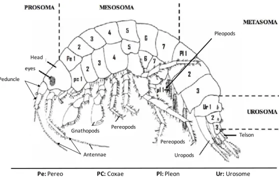
2. MORPHOLOGY... 62
2.1. PROSOMA ... 63
2.2. MESOSOMA ... 63
2.3. METASOMA ... 64
2.4. UROSOMA ... 64
3. ANATOMY OF GAMMARUS FOSSARUM ... 64
3.1. NERVOUS SYSTEM AND CIRCULATORY ORGANS ... 64
3.2. INTESTINAL CAECA AND DIGESTION SYSTEM ... 65
3.3. GAMMARUS GILLS AND RESPIRATION ... 66
3.4. REPRODUCTION ORGANS, LIFE CYCLE AND DEVELOPMENT ... 67
3.4.1. ANATOMY OF THE REPRODUCTION ORGANS ... 67
3.4.2. REPRODUCTION AND LIFE CYCLE ... 67
3.4.3. DEVELOPMENT ... 68
4. GAMMARUS SP. ECOLOGY ... 69
5. FORAGING PLASTICITY ... 69
6. GAMMARUS FOSSARUM AS A MODEL ORGANISM IN ECOTOXICOLOGY ... 70
REFERENCES ... 71
CHAPTER 2 ... 77
GAMMARUS FOSSARUM (CRUSTACEA AMPHIPODA) AS A MODEL ORGANISM FOR NANO-ECOTOXICOLOGY ... 77
AIM OF THE STUDY ... 78
EXPERIMENTAL DESIGN ... 78
KEY FINDINGS ... 78
ARTICLE 1 ... 79
COMPLEMENT 1... 91
SUPPLEMENTARY MATERIAL ... 92
PARTICLE DISPERSION AND CHARACTERISATION IN VOLVIC® WATER ... 92
ENERGY RESERVES, DETOXIFICATION AND LPO LEVEL MEASUREMENTS ... 92
CHAPTER 3 ... 96
G. FOSSARUM MOLECULAR RESPONSES ... 96
AIM OF THE STUDY ... 97
EXPERIMENTAL DESIGN ... 97
KEY FINDINGS ... 97
ARTICLE 2 ... 98
2. RESULTS ... 101
2.1. STABILITY OF THE CANDIDATE REFERENCE GENES IN G. FOSSARUM ... 101
2.2. OPTIMAL NUMBER OF REFERENCE GENE FOR DATA NORMALIZATION IN G. FOSSARUM USING GENORM ... 103
2.3. VALIDATION OF THE SELECTED REFERENCE GENES FOR G. FOSSARUM ... 104
3. DISCUSSION ... 105
4. MATERIALS AND METHODS ... 107
4.1. ORGANISMS SAMPLING AND ACCLIMATION ... 107
4.2. AGNO3, AGNPS AND AUNPS CONTAMINATION ... 108
4.3. GENE IDENTIFICATION AND QPCR PRIMER DESIGN ... 108
4.3.1. DNA EXTRACTION ... 108
4.3.2. LIBRARY PREPARATION AND SEQUENCING ... 108
4.3.3. DE NOVO ASSEMBLY... 108
4.3.4. GENE IDENTIFICATION ... 109
4.3.5. PRIMER DESIGN ... 110
4.4. RNA EXTRACTION, CDNA AND RT-QPCR ... 110
4.5. STABILITY OF THE CANDIDATES’ REFERENCE GENES ... 110
4.6. STATISTICAL ANALYSES ... 111 5. CONCLUSIONS ... 111 ABBREVIATIONS ... 112 REFERENCES ... 113 COMPLEMENT 2... 117 CHAPTER 4 ... 118
AIM OF THE STUDY ... 119
EXPERIMENTAL DESIGN ... 119
KEY FINDINGS ... 119
ARTICLE 3 ... 120
1 INTRODUCTION ... 123
2 MATERIALS AND METHODS ... 124
2.1 PARTICLES AND CHEMICALS ... 124
2.2 PARTICLE CHARACTERIZATION ... 124
2.3 ORGANISM SAMPLING AND ACCLIMATION ... 125
2.4 ACUTE TOXICITY TEST ... 125
2.4.1 Total and dissolved silver and gold measurements ... 126
2.4.2 Silver and gold bioaccumulation ... 126
2.4.4 Molecular responses ... 128
2.5 STATISTICAL ANALYSES ... 132
3 RESULTS ... 132
3.1 NANOPARTICLE CHARACTERIZATION ... 132
3.2 ACUTE TOXICITY TEST ... 133
3.2.1 Survival ... 133 3.2.2 AgNP exposure ... 133 3.2.3 AuNP exposure ... 136 3.2.4 Particle uptake ... 137 3.2.5 Molecular responses ... 140 4 DISCUSSION ... 144 5 CONCLUSION ... 148 COMPLEMENT 3... 157 SUPPLEMENTARY MATERIAL ... 158
1- COATING OF AGNPS AND AUNPS BY WET CHEMISTRY ... 158
2- PARTICLE CHARACTERIZATION ... 159
3- PRELIMINARY TEST: DAPHNIA MAGNA STRAUS MOBILITY INHIBITION TEST (ISO 6341:1996) ... 163
3.1-TEST ORGANISMS ... 163
3.2-BIOLOGICAL ASSAY ... 163
3.2.1- Reference test ... 163
3.2.2- Final test ... 164
3.2.3- Results ... 164
4- ACUTE TOXICITY OF AGNO3 ON GAMMARUS FOSSARUM ... 165
CHAPTER 5 ... 167
AIM OF THE STUDY ... 168
EXPERIMENTAL DESIGN ... 168
KEY FINDINGS ... 168
1. INTRODUCTION ... 171
2. MATERIALS AND METHODS ... 172
2.1. PARTICLES AND CHEMICALS ... 172
2.2. PARTICLE CHARACTERIZATION ... 173
2.3. ALDER LEAVES CONDITIONING ... 173
2.4. ORGANISMS SAMPLING AND ACCLIMATION... 173
2.5. TROPHIC EXPOSURE ... 174
2.5.1. EXPERIMENTAL DESIGN ... 174
2.5.2. TOTAL AND DISSOLVED SILVER AND GOLD MEASUREMENTS ... 174
2.5.3. SILVER AND GOLD BIOACCUMULATION ... 175
2.5.4. PARTICLES UPTAKE:CYTOVIVA ® ANALYSES ... 175
2.5.4.1. SAMPLE PREPARATION ... 175
2.5.4.2. CYTOVIVA® DARK FIELD HYPERSPECTRAL IMAGING ... 175
2.5.5.1. RNA EXTRACTION AND CDNA SYNTHESIS ... 176
2.5.5.2. PRIMER DESIGN AND QUANTITATIVE REAL-TIME PCR ... 176
2.5.6. BEHAVIOURAL RESPONSES AND OSMOREGULATION ... 177
2.5.6.1. LOCOMOTOR ACTIVITY ... 177
2.5.6.2. VENTILATION ACTIVITY... 177
2.5.6.3. OSMOREGULATION ... 177
2.5.6.3.1. HAEMOLYMPH SAMPLING ... 177
2.5.6.3.2. HAEMOLYMPH NA+,CL-AND CA2+ CONCENTRATIONS MEASUREMENTS ... 178
2.6. STATISTICAL ANALYSES ... 178
3. RESULTS ... 178
3.1. PARTICLE CHARACTERIZATION ... 178
3.2. SURVIVAL ... 178
3.3. SILVER AND GOLD BIOACCUMULATION... 179
3.4. CYTOVIVA® DARK FIELD HYPERSPECTRAL IMAGING... 181
3.5. MOLECULAR EFFECTS ... 182
3.6. BEHAVIOURAL RESPONSES AND OSMOREGULATION ... 182
3.6.1. LOCOMOTION AND VENTILATION ACTIVITY ... 182
3.6.2. OSMOREGULATION ... 185 4. DISCUSSION ... 185 4.1. PARTICLE CHARACTERIZATION ... 186 4.2. BIOLOGICAL ENDPOINTS ... 186 5. CONCLUSIONS ... 190 CHAPTER 6 ... 198
1. NPS SIZE, SURFACE COATING AND CHEMICAL COMPOSITION INFLUENCE THE UPTAKE ... 203
2. SYNTHESIS METHODS AND CHEMICAL COMPOSITION INFLUENCE AGNPS AND AUNPS EFFECTS ... 205
3. CONTRIBUTION OF SOLUBLE IONS TO THE EFFECTS ... 206
4. LIMITS OF THE MULTI-BIOMARKERS APPROACH ... 207
5. CONCLUDING REMARKS AND FUTURE PERSPECTIVES ... 208
REFERENCES ... 209
LIST OF FIGURES
Chapter 1 Part 1
Figure P1. 1. Representative chemical and physical transformations of NPs when entering in natural aquatic systems: dissolution, phosphatization, sulfidation, homo- and hetero-aggregation, and sedimentation. Important constituents
with which NPs can interact governing their fate and transport includes hardness cations (e.g., Ca2+, Mg2+), alkalinity,
phosphate and sulfide anions, pH, dissolved organic carbon (DOC), organic matter (OM) and mineral surfaces (such as iron and manganese oxides, and clays). Legend: blue circles: Engineered NPs; yellow circles: humic substances (HM); brown circles: natural inorganic colloids; blue lines: rigid biopolymers; gray surroundings: representing sulfidation; Mz+:
free metal ion. Adapted from(Domingos et al., 2015b) ... 34
Figure P1. 2. Antioxidant defense system in an animal cell (Cossu et al., 1997; Sroda, 2011; Garaud, 2015). CAT:
catalase; G6PD: glucose-6-phospho dehydrogenase; SeGPx: selenium dependent glutathione peroxidase, GPx: glutathione peroxidase; GR: glutathione reductase; GSH/GSSG: reduced/oxidised glutathione; NAPD+/NAPDH: oxidised/reduced nicotinamide dinucleotide phosphate; NOS: nitric oxide synthase; SOD: superoxide dismutase... 37
Figure P1. 3. Potential routes for the generation of ROS due to the presence of NPs. 1) Internalization of NPs ROS
generation could occur due to the NPs dissolution inside the cells and/or due to the NPs photocatalytic activity. 2) Dissolution of the NPs leads to an increase concentration of metal ions in the media; some of these metals can also be uptake by the organisms. 3) NPs and/or their surrounding coatings can adsorb/complex other metals present in the media, being taken up by the cells. 4) Photocatalytic activity of the NPs in the presence of UV and/or natural light. ... 38
Chapter 1 Part 2
Figure P2. 1. (A) Tree of life of the order Amphipoda (Yellow arrow) and its constituents’ suborders Gammaridea, Senticaudata and Hyperiidea. (B) Tree of life of Gammarus fossarum (Yellow arrow) (Lifemap)... 58 Figure P2. 2. Geographical distribution of Gammarus fossarum in (A) Europe and (B) Luxembourg (Dohet et al., 2008; Fauna Europaea). ... 59 Figure P2. 3. Determination key [in French] of Gammarus fossarum (Felten, 2003) ... 60 Figure P2. 4. Distribution of Gammarus fossarum cryptic species in Europe (Adapted from Westram et al., 2011). Grey shaded zone indicates the contact zone between Type A and B. ... 62 Figure P2. 5. General morphology of Gammarus sp. Adapted from Felten 2003. ... 63 Figure P2. 6. Lateral view of the anatomy of Gammarus sp. Illustrating the principal organs (Schmitz, 1992). ... 65 Figure P2. 7. Cross section of (A) Gammarus fossarum illustrating the main organs. (B) midgut and intestinal caeca and (B) gills of G. fossarum observed with optical microscope at 60x magnification. MG: midgut, IC: intestinal caeca
(Pictures: Chauvière A. and Mehennaoui K.) ... 66
Figure P2. 8. Reproduction organs of a male Gammarus and a female (Trapp, 2015). cmu: mucus cells, GA: androgen
gland, ci: incubation chamber, mt: non-differentiated mesenchymal tissue, ov1: primer vitellogenese oocyte, ov2: secondary vitellogenese oocyte, ovd: oviduct, ovd vst: vestigial oviduct, spc: spermatocyte, spg: spermatogonium, spz: spermatozoid, vd: spermiduct ... 67
Figure P2. 9. Gammarus fossarum male and female forming a prepopulate pair (Picture: Untereiner B. and Mehennaoui
Chapter 2 Article 1
Figure 1. Experimental design (adapted from(Arce Funck et al., 2013). CAT: Catalase; GPx: Glutathione
Peroxidase; TAC: Total Antioxidant Activity; ACP: Acid Phosphatase; GST: Glutathione S-Transferase; CHOL: Cholesterol; TRIG: Triglyceride: PROT: Proteins: ETS: Electron Transport System; LDH: Lactate dehydrogenase; LPO: Lipid peroxidation; CASP-3: Caspase 3………..82
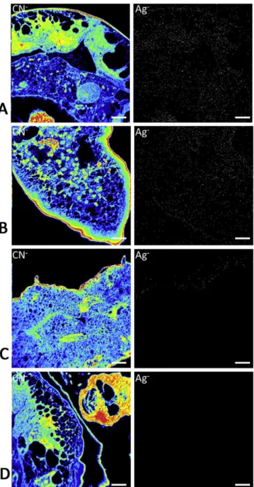
Figure 2. Elemental distribution of 12C14N- cluster and 109Ag- ion in 300 nm cuts of gills. G. fossarum were exposed
to AgNPs (A) 23 nm, (B) 27nm, (C) AgNO3 and (D) Control (Volvic® water). Scale bar is 5 µm………..85 Figure 3. G. f2 survival rates (Mean ± SD) after 72h of exposure to Ag, AgNPs 23 nm, AgNPs 27 nm, AgNPs 20 nm. Data were arcsin root square transformed. Letters (a-c) illustrate significant differences (One-way ANOVA +
Fisher LSD post hoc test at P<0.05 level of significance, n=15). ………86
Figure 4: G .f2 haemolymph osmolality (Mean ± SD) after 72h exposure to AgNO3, AgNPs 20 nm, AgNPs 23 nm
and AgNPs 27 nm. Different letters illustrate significant differences between different treatments (One-way
ANOVA + LSD Fisher post hoc test at P < 0.05 level of significance, n=15)………87
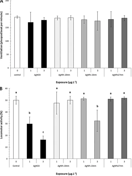
Figure 5: Behavioural responses of G.f2 after 72h of exposure to AgNO3, AgNPs 20 nm, AgNPs 23 nm, AgNPs 27 nm. (A) Ventilation (Mean pleopod beat frequency ± SD). No significant differences were detected for any of exposure conditions (One-way ANOVA + Fischer LSD post hoc test, P < 0.05, n=10). (B) Locomotor activities (Mean percentage of moving G.f2 ± SD). Letters illustrate significant differences. (One-way ANOVA + Fisher LSD
post hoc test at P < 0.05 level of significance, n=10)……….88
Chapter 3 Article 2
Figure 3. 1. Global ranking of candidate reference genes in G. fossarum. A number (from 1 to 6) was assigned to each stability coefficient. A mean rank was generated and error bars ... 103 Figure 3. 2 Determination of the optimal number of reference gens for data normalization in G. fossarum exposed to
AgNO3, AgNPs 40 nm and AuNPs 40 nm. The pairwise variation (Vn/Vn+1) was calculated between normalization factors
NF/NFn+1. The recommended cut-off threshold of 0.15 was applied in this study... 104
Figure 3. 3 HSP90 expression analysis using different normalization strategies. Error bars indicate the standard errors
of the means (n=4). Different letter (a-c) indicate significant differences at P < 0.05. ... 105
Chapter 4 Article 3
Figure 4. 1. G. fossarum Ag bioconcentration (mean ± SD) after 72h exposure to CIT and PEG-AgNPs 20, 40 and 80 nm.
... 135
Figure 4. 2. G. fossarum au bioconcentration (mean ± SD) after 72h exposure to CIT and PEG AuNPs 20, 40 and 80 nm.
... 137
Figure 4. 3. A) 300 nm cross section of G. fossarum observed under optical microscope. Red square represents the area
observed with NanoSIMS 50. Scale bar is 250 µm. B) Elemental distribution of 12C14n- clusters. C) 109Ag- ions in 300 nm
cross sections of G. fossarum gills. Animals were exposed to 10 µg. L-1 of CIT-AgNPs 40 nm and PEG-AgNPs 40 nm in
Volvic water. Scale bar is 2 µm. ... 137
Figure 4. 4. A) 300 nm cross section of G. fossarum observed under optical microscope. Red square represents the area
observed with NanoSIMS 50. Scale bar is 250 µm. B) Elemental distribution of 12C14N- clusters. C) 109Au- ions in 300 nm
cross sections of G. fossarum intestinal caeca. Animals were exposed for 72h to 10 µg. L-1 of CIT-AuNPs 40 nm and
PEG-AuNPs 40 nm in Volvic water. Scale bar is 2 µm ... 138
Figure 4. 5. A) 300 nm cross section of G. fossarum observed under optical microscope. Red square represents the area
observed with Cytoviva darkfield hyperspectral microscope. Scale bar is 250 µm. B) G. fossarum gills viewed with
Animals were exposed for 72h to 10 µg. L-1 of CIT-AgNPs 40 nm and PEG-AgNPs 40 nm in Volvic water. Scale bar is 6 µm
... 139
Figure 4. 6. A) 300 nm cross section of G. fossarum observed under optical microscope. Red square represents the area
observed with Cytoviva darkfield hyperspectral microscope. Scale bar is 250 µm. B) G. fossarum intestinal caeca viewed
with Cytoviva (60x oil immersion magnification). C) AuNPs accumulation in G. fossarum intestinal caeca (green spots
and white arrows). Animals were exposed for 72h to 10 µg. L-1 of CIT-AgNPs 40nm and PEG-AgNPs 40 nm in Volvic water.
Scale bar is 6 µm ... 140
Chapter 5 Article 4
Figure 5. 1. Survival rates (mean ± SD) of G. fossarum exposed for 15 days to 0.5 and 5 µg. L-1 of CIT and PEG-AgNPs
and PEG-AuNPs 40 nm ... 179 Figure 5. 2 A) 300 nm cross section of G. fossarum observed under optical microscope. Red square represents the area
observed with cytoviva darkfield hyperspectral microscope. Scale bar is 250 µm. B) G. fossarum gills or intestinal caeca
viewed with Cytoviva (60x oil immersion magnification). C) AgNPs accumulation in G. fossarum gills (red spots and
white arrows). Animals were exposed for 72h to 10 µg. L-1 of CIT-AgNPs 40nm and PEG-AgNPs 40 nm in Volvic water.
Scale bar is 6µm. ... 181
Figure 5. 3. A) 300 nm cross section of G. fossarum observed under optical microscope. Red square represents the area
observed with CytoViva darkfield hyperspectral microscope. Scale bar is 250 µm. B) G. fossarum intestinal caeca viewed
with CytoViva (60x oil immersion magnification). C) AuNPs accumulation in G. fossarum caeca (red spots and white
arrows). Animals were exposed for 72h to 10 µg. L-1 of CIT-AgNPs 40nm and PEG-AgNPs 40 nm in Volvic water. Scale bar
is 6µm. ... 182
Figure 5. 4. Behavioural responses of G. fossarum exposed for 15 days to CIT and PEG-AgNPs and AuNPs 40 nm. A) Locomotor activity (mean percentage of moving G. fossarum ± SD) and b) Ventilation (mean pleopods beat frequency
± SD). Different letters (a-f) indicates significant differences (one-way ANOVA + Tukey HSD post hoc test at P < 0.05 level of significance, n = 10). ... 184
Chapter 6
Figure 6. 1. Physiological and behavioural effects of synthetic and biological AgNPs on G. fossarum exposed for 72h: summary of results presented in Chapter 2. G. fossarum were exposed for 72h to synthetic AgNPs 20 and 200 nm and
biologically synthetized from plant leaf extract AgNPs 23 and 27 nm. Effects on antioxidant responses (GPx, TAC, CAT), defense mechanism (GST, ACP), cellular damage (LDH, CASP3, LOOH), energy reserves (Prot, Chol, Trig, ETS), osmoregulation and behaviour (Locomotion and ventilation) were assessed... 200
Figure 6. 2. Influence of size (20, 40 and 80 nm) and surface coating (CIT and PEG) of AgNPs and AuNPs on their uptake, tissue distribution and molecular effects of G. fossarum exposed for 72h: summary of the results presented in Chapter 4. G. fossarum were exposed for 72h to up to 50 µg. L-1 of AgNPs and AuNPs in absence of food. Influence of size and
surface coating on bioaccumulation was assessed using ICP-MS, internal distribution of AgNPs and AuNPs were evaluated using NanoSIMS50 and Cytoviva, and molecular effects were assessed using RT-qPCR. A set of stress-related genes expression including genes implied in cytoskeleton trafficking (Actin, TUB, UB), exoskeleton cuticle (Chitinase), antioxidant defence (CAT, MnSOD, CuZnSOD, GPx7), general stress (GST, HSP90, GAPDH), DNA damage and repair (Gadd45, NfkB, c-jun, P53), lysosomes (Cathepsin L), osmoregulation and respiration (Na+K+ATPase, HEM) was used.
... 201
Figure 6. 3. Sub-chronic toxicity of CIT and PEG-AgNPs and CIT and PEG-AuNPs 40 nm on molecular, physiological and behavioural responses of G. fossarum: summary of the results presented in Chapter 5. G. fossarum were exposed for
15 days to 0.5 and 5 µg.L-1 of CIT and PEG-AgNPs and AuNPs 40 nm in presence of food. Bioaccumulation was assessed
using ICP-MS, internal distribution of AgNPs and AuNPs were evaluated using Cytoviva and molecular effects were assessed using a set of stress-related genes expression including genes implied in cytoskeleton trafficking (Actin, TUB,
UB), exoskeleton cuticle (Chitinase), antioxidant defence (CAT, MnSOD, CuZnSOD, GPx7), general stress (GST, HSP90, GAPDH), DNA damage and repair (Gadd45, NfkB, c-jun, P53), lysosomes (Cathepsin L), osmoregulation and respiration (Na+K+ATPase, HEM). ... 202
Supplementary materials
Figure S 1. Size distribution of Ag NPs 23 nm (A), 27 nm (B), 20 nm (C) and 200 nm (D) in Volvic® water expressed as particle concentration x 106.mL-1. The red error bars indicate the ± SD of the mean of triplicate measurements... 96
Figure S 2 PEG coating on metal Nanoparticles (here silver NPs). SEM shows clearly the conformal silver coating with
the PEG layer. NTA analysis shows a small size increasing for the 40 and 80 nm silver nanoparticles as a consequence of their coating with the PEG layer. The sensitivity of NTA was not enough to resolve the size increase for 20 nm nanoparticles. ... 158
Figure S 3. Size distribution of CIT-AgNPs 20 nm (A), CIT-AgNPs 40 nm (B) and CIT-AgNPs 80 nm (C) in Volvic Water (T0h) and CIT-AgNPs 20 nm (D), CIT-AgNPs 40 nm (E) and CIT-AgNPs 80 nm (F) in Volvic water after 24h of incubation.
Size distribution is expressed as particle concentration E6.mL-1. The red errors bars indicate ±SD of the mean of triplicate
measurements ... 159
Figure S 4. Size distribution of PEG-AgNPs 20 nm (A), PEG-AgNPs 40 nm (B) and PEG-AgNPs 80 nm (C) in Volvic Water (T0h) and PEG-AgNPs 20 nm (D), PEG-AgNPs 40 nm(E) and PEG-AgNPs 80 nm (F) in Volvic water after 24h of incubation. Size distribution is expressed as particle concentration E6.mL-1. The red errors bars indicate ±SD of the mean
of triplicate measurements ... 160
Figure S 5. Size distribution of CIT-AuNPs 20 nm (A), CIT-AuNPs 40 nm (B) and CIT-AuNPs 80 nm (C) in Volvic Water (T0h) and CIT-AuNPs 20 nm (D), CIT-AuNPs 40 nm (E) and CIT-AuNPs 80 nm (F) in Volvic water after 24h of incubation.
Size distribution is expressed as particle concentration E6.mL-1. The red errors bars indicate ±SD of the mean of triplicate
measurements ... 161
Figure S 6. Size distribution of PEG-AuNPs 20 nm (A), PEG-AuNPs 40 nm (B) and PEG-AuNPs 80 nm (C) in Volvic Water (T0h) and PEG-AuNPs 20 nm (D), PEG-AuNPs 40 nm(E) and PEG-AuNPs 80 nm (F) in Volvic water after 24h of incubation. Size distribution is expressed as particle concentration E6.mL-1. The red errors bars indicate ±SD of the mean
of triplicate measurements ... 162
Figure S 7. Effects AgNO3 on survival of Gammarus fossarum collected in A) June, B) September and C) November after
LIST OF TABLES
Chapter 1 Part 2
Table P2. 1. Classification of the species Gammarus fossarum ... 59
Chapter 2 Article 1
Table 2.1. Particles size distribution (mode ± SD, 3 replicates) and ζ potential of AgNPs in Volvic® water………..84 Table 2.2: LC50 values with 95% confidence intervals for G.f1 and G.f2 exposed to AgNO3 and AgNPs20 nm,
AgNPs 23 nm, AgNPs 27nm and AgNPs 200 nm for 72h………84
Table 2.3: Total and dissolved Ag concentrations (mean ± SD) and dissolution rates of AgNO3 and AgNPs after
72h of exposure in Volvic water. Different letters illustrate significant differences between different treatments
(Two-way ANOVA + Tukey HSD post-hoc test at P < 0.05 level of significance, n=3)……….85
Table 2.4: G.f2 Ag Bioconcentration (Mean± SD) after 72h exposure to AgNO3, AgNPs 20 nm, AgNPs 23 nm,
AgNPs 27 nm. Different letters illustrate significant differences between different treatments (Kruskal-Wallis
ANOVA + Mann-Whitney U test at P < 0.05 level of significance, n=3)………86
Table 2.5: Mean values (±SD) of biomarkers measured in Gf2 exposed to AgNO3, AgNPs 20nm, AgNPs 23nm and
AgNPs 27 nm for 72h. Biomarkers in italic were analysed using One-way ANOVA and Tukey post hoc test, whereas
the others were analysed using Kruskal-Wallis ANOVA and Mann-Whitney U test. No significant differences were detected for any of the exposure conditions. a: mg. g fresh weight-1; b: µmol O
2. g proteins-1. h-1.; c: µmol
p-nitrophenol. g protein-1.h-1; d: µmol CDNB.min-1.g-1 proteins; e: µmol NADPH.g protein-1.min-1; f: mmol Trolox
equivalent.g protein-1; g: mmol H
2O2.g proteins-1.min-1; µmol NADH. g proteins-1. h-1; h: µmolpNA. g proteins-1.h-1;
j: nmol TBH. g proteins-1………..87
Chapter 3 Article 2
Table 3. 1.Ranking of candidate reference genes according to the five algorithms used ... 102 Table 3. 2 Identification of Gammarus fossarum gene sequences ... 108 Table 3. 3 List of primers of the candidate reference genes and target gene HSP90 ... 109
Chapter 4 Article 3
Table 4. 1. Specific primer pairs used for RT-qPCR analyses on Gammarus fossarum exposed for 72h to AgNPs and AuNPs (coated with CIT or PEG, sizes: 20, 40 and 80 nm, F: forward sequence, R: reverse sequence) ... 130 Table 4. 2. Size distribution of particles (mode ± SD, 3 replicates) and ζ potential of AgNPs and AuNPs in Volvic water (exposure medium) at T0h and T24h. ... 133
Table 4. 3. Total ag concentrations (mean ± SD) and recovery rates (mean ± SD) of AgNO3, CIT-AgNPs and PEG-AgNPs
in Volvic water (exposure medium). ... 134 Table 4. 4. Relative gene expression of G. fossarum exposed for 72h to CIT-AgNPs and PEG-AgNPs 20, 40 and 80 nm
... 142
Table 4. 5. Relative gene expression of G. fossarum exposed for 72h to CIT-AuNPs and PEG-AuNPs 20, 40 and 80 nm
Chapter 5 Article 4
Table 5. 1. G. fossarum Ag and Au uptake (mean ± SD) after 15 days of exposure to CIT and PEG-AgNPs and AuNPs 40 nm. ... 180 Table 5. 2. Relative gene expression of G. fossarum exposed for 15 days to CIT- and PEG-AgNPs 40 nm and CIT and PEG-AuNPs 40 nm ... 183 Table 5. 3. Haemolymph [Cl-], [Na+] and [Ca2+] of G. fossarum exposed for 15 days to CIT and PEG-AgNPs 40 nm and
CIT and PEG-AuNPs 40 nm. ... 185
Supplementary materials
Table S 1. Physico-chemical parameters of Volvic® water ... 95 Table S 2 D. magna reference test ... 163 Table S 3. EC50 values obtained after 48h exposure of D. magna to AgNPs and AuNPs ... 164
ABBREVIATIONS
ATP Adenosine triphosphate
cDNA Complementary DNA CYP450 Cytochrome P450
DTT Dithioreitol
EF1 Elongation factor 1 alpha
EMBL European Molecular Biology Laboratory
EST Expressed Sequence Tag
FC Fold change
FNR National Research Funds – Luxembourg
MIQe Minimum information for publication of qPCR experiments
mRNA Messenger ribonucleic acid
PCR Polymerase Chain Reaction
qPCR Quantitative real-time polymerase chain reaction
RIN RNA integrity number
RNA Ribonucleic acid
RNA-seq RNA sequencing
SI Supporting information
TEM Transmission electron microscope
TF Transcription factor
UV Ultraviolet
AgNPs Silver nanoparticles AuNPs Gold nanoparticles
AgNO3 Silver nitrate
Ag2S Silver sulphide
DMSO Dimethyl Sulfoxide
NTA Nanoparticles tracking analysis
DLS Dynamic light scattering
HIM-SIMS Helium Ion microscopy – secondary
ion mass spectrometer
EDTA Ethylenediaminetetraacetic acid HNO3 Nitric acid
TAC Total antioxidant capacity
HCl Chloride acid
CIT Citrate
PEG Polyethylene glycol
NanoSIMS 50 Nano secondary ion mass spectroscopy
ICP-MS Inductively coupled plasma – mass spectrometer
ACP Acid phosphatase
CAT Catalase
OS Oxidative stress
NPs Nanoparticles
ENPs Engineered nanoparticles
NMs Nanomaterials
HM Humic acid
DOC Dissolved organic carbon
TOC Total organic carbon
OM Organic matter
FPOM Fine particle organic matter
CASP Caspase
PEC Predicted environmental concentration
L(E)C 50 Lethal or effective concentration
that impact 50% of the exposed population
ETS Electron transport system
GR Glutathione reductase
GSH Reduced glutathione
GST Glutathione S-transferase
NOEC Non-observable effects concentrations
H2O2 Hydrogen peroxide
HSP Heat shock protein
MDA Malondialdehyde
ROS Reactive oxygen species
SOD Superoxide dismutase
LPO Lipid peroxidation
GENERAL INTRODUCTION
In the recent decades, nanotechnology has emerged as a fast-growing sector impacting key economical fields and providing new engineered nano-enabled products, constituted by nanoparticles (NPs), with novel and unique functions that reach the market every day. NPs are defined as compound presenting at least one dimension less than 100 nm (Klaine et al., 2008).
The use of nanomaterials is increasingly and continuously growing as they are used in different areas such as electronics, medicine, environmental technology, etc. Silver nanoparticles (AgNPs) are among the most promising group of NPs. AgNPs are used in different kind of daily-life products such as textiles, food packaging, healthcare products, etc., mostly for their antibacterial properties. AuNPs are being investigated for their unique optical properties and are used as contrast agents in electron microscopy, optical sensors, catalysts, and for therapeutic uses. The increasing use of AgNPs and AuNPs lead to their inevitable release in the environments and may reach the aquatic ecosystems where they may represent a threat for aquatic organisms. The potential toxicity of nanomaterials is of high societal and scientific interest and can be related to many properties such as the size, the mass, surface area and characteristics, aggregation and agglomeration (Georgantzopoulou et al., 2013). Their toxicity could also be linked to less known or less understood parameters like released ions and adhering molecules which could have an influence on their toxicity by interfering with their uptake and fate on living organisms.
The PhD project “NANOGAM” is a FNR funded project closely linked up to the “CORE2012 NANION” and to “FP7 FUTURENANONEEDS (FNN)” projects that aim to obtain knowledge to understand some of the processes and factors involved in NPs uptake and toxicity. The specific objectives of NANION are:
• Leaching ions from NPs have their own spectrum of toxicity and effects can be clearly separated between free ions in the exposure media and the NPs (Georgantzopoulou, 2015).
• Within NANION, a set of AgNPs and AuNPs comprising different size classes and coated with various surfaces (citrate and polyethylene glycol) are used. Prior to any testing, these NPs were carefully characterized (zeta-potential, agglomeration, aggregation, etc.) in the relevant exposure media. The ion release from uncoated and coated NPs (Au does not release ions) were studied in dependency of exposure media and related to toxicity endpoints ranging from molecular to organism level.
• Biomolecules attach easily and quickly to the surface of NPs. This so-called corona influences uptake, kinetics, distribution within the organism and thereby finally impact endpoints such as biochemical biomarkers but also reproduction, behaviour of the organism, and other relevant whole organism endpoints. The uptake kinetics, distribution of the NPs within the organism and toxicity for a range of relevant endpoints were studied and related to physico-chemical properties of the NPs.
Generally, NANOGAM aims at contributing to the understanding in how far surface properties of NPs (physical parameters, ion leaching, biological molecules attached, etc.) affect and interfere with the toxicity of NPs in a very relevant freshwater model organism, the crustacean Gammarus fossarum. G. fossarum was selected as test species due to its large distribution in Europe (Barnard and Barnard, 1983), high abundance (Felten et al., 2008; Kunz et al., 2010), clear sexual dimorphism, easiness of identification to the species level, collection and handling, the high sensitivity to a large range of toxicants and their major functional role in ecosystems. Indeed, as shredders, they play a key role in the litter breakdown process and thus in freshwater food chain and nutrient cycling (Forrow and Maltby, 2000; Vellinger et al., 2012a). Consequently, Gammarus sp. could provide valuable information about the potential effects of studied contaminants on some other taxa in aquatic ecosystem communities (Vellinger et al., 2012b) To our knowledge, only few studies have been done on the ecotoxicity of AgNPs and AuNPs in these species (Andreï et al., 2016; Baudrimont et al., 2017; Bundschuh et al., 2011; Park et al., 2015).
The “NanoGAM” project aims at investigating the characteristics of the key physico-chemical parameters and surface functionalities of a well-chosen list of AgNPs and AuNPs that control uptake, and dependent biological effects of different complexity (using a battery of biomarkers) on G. fossarum. Starting from a set of commercially available NPs that will form the baseline for the studies proposed within NANOGAM. The following questions have been raised within the present project:
• Is G. fossarum a valuable model to study the effects of nanoparticles?
• How do particle size, surface coating, synthesis method and chemical composition contribute to the toxicity and effects of AgNPs and AuNPs?
• To what extent does leaching ions play a role in the toxicity of AgNPs?
• What are the acute effects of AgNPs and AuNPs, with different sizes (20, 40 and 80 nm) and two different coatings (CIT and PEG), on G. fossarum?
• What are the sub-chronic effects of AgNPs and AuNPs on molecular, physiological and behavioural responses of G. fossarum?
These research questions are addressed in the following chapters:
• Chapter 1 is a critical overview on biochemical effects of AgNPs and AuNPs on aquatic organism (Part 1) with a focus on G. fossarum, the model organism used in the present PhD project (Part 2)
• In Chapter 2, the effects of a well-characterized and well-studied set of AgNPs are used in order to evaluate their effects on G. fossarum through a multi-biomarker approach. This study allows also the assessment of the contribution of leaching ions on the toxicity and effects of AgNPs. Furthermore, the internal distribution of AgNPs in G. fossarum is discussed.
• As G. fossarum is to our best knowledge a non-sequenced species, it was necessary to identify target stress-related genes for RT-qPCR experiments. For accurate analyses, it was necessary to identify a set of reference genes for RT-qPCR normalization and results are presented in Chapter 3.
• Chapter 4 provides evidence of the influence of AgNPs and AuNPs size and surface-coating on their uptake and internal tissue distribution in G. fossarum. Furthermore, the acute effects on the molecular responses are investigated.
• Chapter 5 deals with the sub-chronic effects of AgNPs and AuNPs on G. fossarum. A multi-biomarker approach including molecular, physiological and behavioural responses was applied to assess the effects of AgNPs and AuNPs. Additionally, tissues distribution of NPs is evaluated
• Finally, Chapter 6 discuss the knowledge acquired in the present work and the relevance of the current findings. Conclusions and future perspectives are also addressed.
CHAPTER 1
Part 1: Stat of the art based on the published review:
Gançalo Vale, Kahina Mehennaoui, Sébastien Cambier, Giovanni Libralato, Stéphane Jomini, Rute F.
Domingos. Manufactured nanoparticles in the aquatic environment – biochemical responses on
freshwater organisms: a critical overview. 2016. Aquatic toxicology 170, 162-174.
The published review is presented in Annexe 1
CHAPTER 1
Part 1: Stat of the art based on the published review:
Gançalo Vale, Kahina Mehennaoui, Sébastien Cambier, Giovanni Libralato, Stéphane Jomini,
Rute F. Domingos. Manufactured nanoparticles in the aquatic environment – biochemical
responses on freshwater organisms: a critical overview. 2016. Aquatic Toxicology 170, 162-174.
Manufactured nanoparticles in the aquatic environment –Biochemical responses on freshwater
organisms: A critical overview
1Gonçalo Vale
a,b*, Kahina Mehennaoui
c,e, Sebastien Cambier
c, Giovanni Libralato
d, Stéphane
Jomini
e, Rute F. Domingos
a,fa) Centro de Química Estrutural, Instituto Superior Técnico, Universidade de Lisboa, Torre Sul, Av. Rovisco Pais,
1049-001 Lisboa, Portugal. Gonçalo Vale:
[email protected]
b) Department of Molecular Genetics, University of Texas Southwestern Medical center, Harry Dallas, TX 75390, USA (present address)
c) Luxembourg Institute of Science and Technology, Environmental Research and Innovation (ERIN) Department,
Belvaux, Luxembourg.
Kahina Mehennaoui:
[email protected]
; Sebastien Cambier:
[email protected]
d) Department of Environmental Sciences, Informatics and Statistics, University Ca’ Foscari Venice, Via Torino, 155,
30172, Mestre, Venice, Italy. Giovanni Libralato:
[email protected]
e) Laboratoire Interdisciplinaire des Environements Continentaux (LIEC), Université de Lorraine, UMR 7360,
Campus Bridoux rue du Général Delestraint, 57070 Metz, France. Stéphane Jomini:
[email protected]
f) Institut de Physique du Globe de Paris, Sorbonne Paris Cité, UMR CNRS 7154, Université Paris Diderot, 75205
Paris Cedex 05, France. Rute F. Domingos:
[email protected]
(present address)*Corresponding author
Email:
[email protected]
1 Vale, G., Mehennaoui, K., Cambier, S., Libralato, G., Jomini, S., Domingos, R.F., 2016. Manufactured
nanoparticles in the aquatic environment-biochemical responses on freshwater organisms: A critical overview. Aquat. Toxicol. 170, 162–174. doi:10.1016/j.aquatox.2015.11.019
ABSTRACT
The enormous investments in nanotechnology have led to an exponential increase of new manufactured nano-enabled materials whose impact in the aquatic systems is still largely unknown. Ecotoxicity and nanosafety studies mostly resulted in contradictory results and generally failed to clearly identify biological patterns that could be related specifically to nanotoxicity. Generation of reactive oxygen species (ROS) is one of the most discussed nanotoxicity mechanism in literature. ROS can induce oxidative stress (OS), resulting in cyto- and genotoxicity. The ROS overproduction can trigger the induction of anti-oxidant enzymes such as catalase (CAT), superoxide dismutase (SOD) and glutathione peroxidases (GPx), which are used as biomarkers of response. A critical overview of the biochemical responses induced by the presence of NPs on freshwater organisms is performed with a strong interest on indicators of ROS and general stress. A special focus will be given to the NPs transformations, including aggregation, and dissolution, in the exposure media and the produced biochemical endpoints.
Keywords: nanoparticles, transformations, freshwater organisms, nanotoxicity, reactive oxygen species
(ROS), anti-oxidant enzymes
1. INTRODUCTION
Nanotechnology has emerged as a fast growing sector impacting key economical fields and providing new engineered nano-enabled products, constituted by nanoparticles (NPs), with novel and unique functions that reach the market every day (Bour et al., 2015). NPs are defined as materials with a size between 1 and 100 nm on at least one dimension, having unique physico-chemical properties differing from their bulk forms due to their greater surface area to volume ratio (Hood, 2004; The European Commision, 2011). The size related-properties results in larger reactivity and higher mobility, leading to numerous applications in medical diagnostics, electronics, computers, cosmetics and environmental remediation (Rauscher et al., 2015). The worldwide consumption of NPs is expected to grow from 225,060 metric tons in 2014 to nearly 584,984 metric tons in 2019 representing an annual growth rate of 21.1 % (BCC RESEARCH, 2017). Although impressive, these numbers are in fact “expected” values obtained by estimation or modelling. The lack of legislation for nanotechnologies gives the manufacturers no onus to reveal the real figures, thus, indeed, these predicted values are most probably significantly higher. The absence of real numbers hinders the prediction of the NPs amount that are actually being released into the environment (Piccinno et al., 2012). Even though several studies have been performed with the goal of modelling NPs environmental concentrations (Gottschalk et al., 2009, 2013; Yang et al., 2016), they should only be considered as guidelines, since they derive from uncertain data about the NPs production (often obtained by surveys to producers) and extrapolations used to scale up regional to worldwide amounts (Gottschalk et al., 2013; Keller et al., 2010; Piccinno et al., 2012).
When released in natural media NPs will be subjected to a dynamic physical and chemical environment that consequently results in different and unknown endpoints far from their pristine or as released state. Therefore, environments and living organisms are not facing pristine manufactured NPs but rather transformed nano-enabled products, which is factually accepted but so far neglected. In fact, the large majority of the physico-chemical and toxicity data obtained so far was focused on simple nanoscale particles and not on relevant nano-enabled products. This includes not only the NP embedded in the manufactured matrix but also the materials resulting from the interaction with biotic and abiotic (bio)molecules composing the natural systems (Nasser and Lynch, 2016). To further complicate the interpretation of the NPs studies, there are two distinct mechanisms that should be considered but are not easily differentiated:
• Chemical toxicity by the release of possible ions and/or formation of reactive oxygen species (ROS) (Fu et al., 2014)
• Physical stress or stimuli caused by NPs size, shape and surface properties (Libralato, 2014; Vale et al., 2014).
These materials are generally associated with cellular perturbations such as ROS generation, gene expression and proteome profiles alterations. For these reasons, the NPs escalating production and applications has raised concerns about their environmental and human safety, which have led to large investments in nanosafety-related projects resulting in a considerable amount of data assessing their potential hazard (Savolainen et al., 2013). However, the establishment of relationships between bioavailable NP-containing species and the specific bioadverse or biocompatible endpoints is still lacking, mainly since the effects are NP-dependent and also specie-dependent (Burić et al., 2015).
This work provides an overview of the latest studies on the impact of NPs onto aquatic environment with a focus on freshwater ecosystems, considered by many as the ultimate sink of these particles, with a special focus:
• NPs transformations and characterization in the different test media,
• Toxicological effects such as generation of ROS, genotoxicity, transcriptomic and proteomic changes. This survey is focused on metallic NPs including silver nanoparticles (AgNPs) and gold nanoparticles (AuNPs), mostly due to the great number of studies dedicated to these particles.
2. NPs TRANSFORM ATIONS IN AQU ATIC SYS TEMS
NPs can enter in an aquatic compartment from: • Wastewater treatment plants effluents,
• Direct use (e.g., application of NPs-containing paintings on boats), • Deposition from the air compartment.
When entering aquatic compartment, NPs will be exposed to a highly dynamic physical and chemical environment that leads to several transformations that will change their pristine or as released physico-chemical properties (Figure P1. 1). These transformations, including dissolution, aggregation and sedimentation, are dependent on both physico-chemical properties of the NPs and those of the environment into which they were released (Auffan et al., 2012).
Colloidal particles, including organic and inorganic matter, are ubiquitous in the aquatic environment and can be originated from both natural and anthropic sources. These colloids can strongly interact with NPs, thereby determining their forms over space and time (dynamic speciation), and greatly affecting their bioavailability. Thus, the NPs will have a specific speciation in each environmental compartment, and this speciation is always dynamic with reaction rates that depend upon the chemical nature and physical sizes of the engineered and natural colloids (Levard et al., 2012). Although it is clear that dynamic speciation must be considered in order to make relevant predictions of NPs fate, toxicity and risk, was mostly neglected (Domingos et al., 2015b).
Figure P1. 1. Representative chemical and physical transformations of NPs when entering in natural aquatic systems: dissolution, phosphatization, sulfidation, homo- and hetero-aggregation, and sedimentation.
Important constituents with which NPs can interact governing their fate and transport includes hardness cations
(e.g., Ca2+, Mg2+), alkalinity, phosphate and sulfide anions, pH, dissolved organic carbon (DOC), organic matter
(OM) and mineral surfaces (such as iron and manganese oxides, and clays). Legend: blue circles: Engineered NPs; yellow circles: humic substances (HM); brown circles: natural inorganic colloids; blue lines: rigid biopolymers; gray