HAL Id: hal-02666097
https://hal.inrae.fr/hal-02666097
Submitted on 31 May 2020
HAL is a multi-disciplinary open access
archive for the deposit and dissemination of
sci-entific research documents, whether they are
pub-lished or not. The documents may come from
teaching and research institutions in France or
abroad, or from public or private research centers.
L’archive ouverte pluridisciplinaire HAL, est
destinée au dépôt et à la diffusion de documents
scientifiques de niveau recherche, publiés ou non,
émanant des établissements d’enseignement et de
recherche français ou étrangers, des laboratoires
publics ou privés.
Differential effect of dietary antioxidant classes
(carotenoids, polyphenols, vitamins C and E) on lutein
absorption
Emmanuelle Reboul, Sinay Thap, Franck Tourniaire, Marc André, Christine
Juhel, Sophie Morange, Marie Josephe Amiot, Denis Lairon, Patrick Borel
To cite this version:
Emmanuelle Reboul, Sinay Thap, Franck Tourniaire, Marc André, Christine Juhel, et al.. Differential
effect of dietary antioxidant classes (carotenoids, polyphenols, vitamins C and E) on lutein
absorp-tion. British Journal of Nutrition, Cambridge University Press (CUP), 2007, 97 (3), pp.440-446.
�10.1017/S0007114507352604�. �hal-02666097�
Differential effect of dietary antioxidant classes (carotenoids, polyphenols,
vitamins C and E) on lutein absorption
Emmanuelle Reboul
1,2,3,4, Sinay Thap
1,2,3,4, Franck Tourniaire
1,2,3,4, Marc Andre´
1,2,3,4, Christine Juhel
5,
Sophie Morange
6, Marie-Jose`phe Amiot
1,2,3,4, Denis Lairon
1,2,3,4and Patrick Borel
1,2,3,4*
1INSERM, U476, 27 Bd Jean Moulin, Marseille, F-13385, France 2INRA, UMR1260, Marseille, F-13385, France
3Univ Aix-Marseille 2, Marseille, F-13385, France 4IPHM-IFR 125, Marseille, F-13385, France
5Avantage Nutrition, 116 Chemin des Sables Jaunes, Marseille, F-13012, France 6
Clinical Investigation Center, 270 Bd Sainte Marguerite, Marseille, F-13009, France
(Received 27 June 2006 – Revised 9 October 2006 – Accepted 16 October 2006)
Lutein is assumed to protect the human retina from blue light and oxidative stress and diminish the incidence of age-related macular degeneration. This antioxidant is commonly ingested with other dietary antioxidants. The aim of the present study was to assess whether the main dietary anti-oxidants, i.e. carotenoids, polyphenols and vitamins C and E, affect lutein absorption. We measured the effect of adding a mixture of antioxidants (500 mg vitamin C, 67 mg (100 IU) vitamin E and 1 g polyphenols) to a lutein-containing meal (18 mg) on the postprandial lutein response in the chylomicron-rich fraction in eight healthy men. Lutein response was weakest (2 23 %; P¼ 0·07) after ingestion of the meal containing antioxidants
(21·9 (SEM4·6) v. 28·4 (SEM7·2) nmol £ h/l). To assess the effect of each class of antioxidants and potential interactions, we subsequently
eval-uated the effect of various combinations of antioxidants on lutein uptake by human intestinal Caco-2 TC-7 cells. A full factorial design showed that both a mixture of polyphenols (gallic acid, caffeic acid, (þ )-catechin and naringenin) and a mixture of carotenoids (lycopene plus b-carotene) significantly (P, 0·05) impaired lutein uptake by (2 10 to 2 30 %), while vitamins C and E had no significant effect. Subsequent experiments showed that the aglycone flavanone naringenin was the only polyphenol responsible for the effect of the polyphenol mixture, and that the caro-tenoid effect was not carocaro-tenoid species-dependent. Taken together, the present results suggest that lutein absorption is not markedly affected by physiological concentrations of vitamins C and E but can be impaired by carotenoids and naringenin.
Bioavailability: Uptake: Enterocytes: Naringenin: Gallic acid: Caffeic acid
Lutein is a food microconstituent that belongs to the carotenoid family, specifically the xanthophyll subclass characterised by oxygenated groups. In the human diet, lutein is mainly found in dark-green leafy vegetables and egg yolk (Chug-Ahuja et al. 1993; Sommerburg et al. 1998). It cannot yet be considered as a nutrient (Hendrich et al. 1994) because there is not sufficient evi-dence that it sustains or enhances physiological functions and/or prevents disease. Nevertheless, this xanthophyll is attracting growing interest since it selectively accumulates in the retina (Bone et al. 1997; Chan et al. 1998), where it may protect photo-receptors against light-initiated oxidative damage (Rapp et al. 2000; Junghans et al. 2001; Krinsky, 2002), and intake of caro-tenoids has been associated with a lower incidence of age-related macular degeneration (Snodderly, 1995; Landrum & Bone, 2001).
In Western diets, lutein is mostly recovered as non-esteri-fied lutein. Lutein metabolism begins in the stomach where lutein-containing foods are subjected to acidic processing and gastric enzymes. It has been shown that lutein is partially released from spinach and transfers to the fat phase of the
meal (Tyssandier et al. 2003). Digestive enzymes in the duo-denum continue to release lutein from its food matrix, and it is assumed that lutein is transferred to dietary lipids found in the duodenum and then into mixed micelles. Pancreatic lipase appears to facilitate the transfer of lutein from lipid droplets to mixed micelles (Borel et al. 1996). This transfer mainly depends on pH, bile lipid concentration and carotenoid hydro-phobicity (Tyssandier et al. 2001). Mixed micelles carry lutein to the enterocyte where it is at least partly absorbed through the scavenger-receptor class B type I (Reboul et al. 2005a). After absorption, lutein is incorporated into chylomicrons and transported to the bloodstream, via the lymphatics.
In a normal meal containing plant-derived foods, lutein is necessarily ingested with other antioxidants, including caro-tenoids, polyphenols and vitamins C and E. Since lutein is an antioxidant and is therefore probably subject to oxidative degradation (Siems et al. 1999), it follows that other antioxi-dants found in the diet may protect it from degradation in the upper gastrointestinal tract, thereby enhancing its absorption efficiency. In contrast, it is also possible that dietary
* Corresponding author: Dr Patrick Borel, fax þ 33 4 91 78 21 01, email Patrick.Borel@medecine.univ-mrs.fr
antioxidants may compete with lutein for absorption. Indeed, several studies have shown that carotenoids compete for their absorption (van den Berg, 1999; Tyssandier et al. 2002). The aims of the present study were (i) to assess whether a mixture of the main classes of dietary antioxidants (caroten-oids, polyphenols, vitamin C and vitamin E) affect the bio-availability of lutein in human subjects, and (ii) to assess the individual effect of each class of antioxidant microconstituent on lutein uptake using an intestinal cell-culture model.
Material and methods Chemicals
Lutein (96 % pure), lycopene (95·5 % pure), b-carotene (95·6 % pure) and echinenone (98 % pure) were kindly provided by DSM Ltd (formerly F. Hoffmann-La Roche, Basel, Switzerland). (R,R,R)-a-tocopherol ($99 % pure) and (R,R,R)-g-tocopherol ($97 % pure) were purchased from Fluka (Vaulx-en-Velin, France). Quercetin, naringin, hesperidin, hesperetin, 2-oleoyl-1-palmitoyl-sn-glycero-3-phosphocholine (phosphatidylcholine), 1-palmitoyl-sn-glycero-3-phosphocholine (lysophosphatidylcho-line), monoolein, non-esterified cholesterol, oleic acid, sodium taurocholate and pyrogallol were purchased from Sigma-Aldrich (St Quentin Fallavier, France). Naringenin, (þ)-catechin, gallic acid, caffeic acid and eriodictyol were purchased from Extra-synthe`se (Genay, France).
Dulbecco’s modified Eagle medium containing 4·5 g/l glucose and trypsin-EDTA (at 500 mg/l and 200 mg/l, respect-ively) were purchased from Bio Whittaker (Fontenay-sous-Bois, France). Fetal bovine serum was purchased from Biomedia (Issy-les-Moulineaux, France), and non-essential amino acids, and penicillin and streptomycin were purchased from Gibco BRL (Cergy-Pontoise, France).
Postprandial experiment to assess the effect of an antioxidant mixture on lutein bioavailability in healthy human subjects Since there were no available literature data on the effect of a mixture of antioxidants on lutein absorption, we were unable to perform a power analysis to calculate the number of subjects required to observe a significant effect. We therefore used a number of eight subjects because this number has allowed find-ing significant differences in carotenoid absorption in previous studies (Cardinault et al. 2003; Riso et al. 2003; Tesoriere et al. 2004; Blum et al. 2005; Reboul et al. 2005b). Eight male sub-jects were selected by the local Clinical Investigation Centre (Sainte Marguerite, Marseille, France). They should be healthy, non-smoking, aged 20 – 40 years and with a BMI , 24 kg/m2. They should not take medication and have no history of gastro-intestinal disease, diabetes or disorders of lipid metabolism. To verify the latter points, serum glucose, TAG and cholesterol were measured by using enzymic procedures (Trinder, 1969; Fossati & Prencipe, 1982; Siedel et al. 1983). Subject character-istics and their daily nutrient intakes are reported in Table 1. Dietary intake was assessed by a 5 d food diary and analysed using Score-AN software (configured by Avantage Nutrition, Marseilles, France). The software’s nutrient database was obtained from Ciqual and Souci’s nutrition tables (Souci et al. 2000). The study was approved by the regional ethics commit-tee in Marseilles. The objectives and requirements of the study
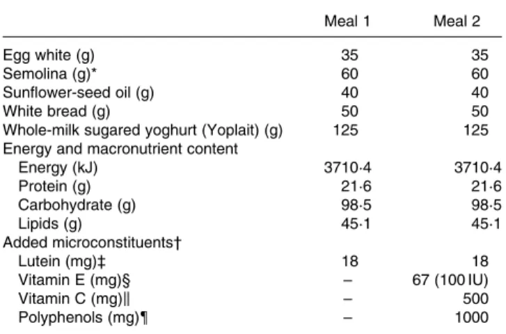
were fully explained to the participants, and informed written consent was obtained for each subject. Each subject received each meal in a random order at a 1-month interval (cross-over design with a 1-month wash-out period). On the evening before the experiment, the subjects were asked to consume a light meal and to fast overnight. In the morning, each subject had an intravenous catheter inserted into a forearm. A first blood sample was obtained at fasting (baseline sample), and the volunteers then consumed either meal 1 (containing 18 mg lutein) or meal 2 (containing 18 mg lutein plus 500 mg vitamin C, 67 mg (100 IU) natural vitamin E, 1 g polyphenols (a mixture of grapeseed extract and citrus bioflavonoids)). Antioxidant micronutrients were from Holland & Barrett (Nuneaton, War-wickshire, UK) and were given as pills which were swallowed by the subjects during the meal. No other food was consumed during the postprandial experiment. The amount of lutein in the test meal was chosen to accurately detect lutein in the chy-lomicron-rich fraction. This amount of carotenoid remains nutritional as it is close to the daily total carotenoid intake, which is estimated at 14 mg/d in Europe (O’Neill et al. 2001). The amount of other antioxidant was chosen to be close to the relative proportions found in the diet. Foods were purchased from a local supermarket. Meal compositions and microconsti-tuent characteristics are given Table 2. Additional blood samples were drawn 1, 2, 3, 4, 6 and 8 h after the beginning of the meal. Blood samples were immediately stored at 48C and rapidly centrifuged (at 610 g for 10 min at 48C) to isolate plasma fractions. Large chylomicrons (Sf. 400) were isolated
as described by Luchoomun & Hussain (1999). Chylomicron-rich fraction TAG were measured using the PAP 150 Biome´r-ieux kit (Charbonnie`re-les-Bains, France).
Preparation of microconstituent-rich media for cell experiments
For delivery of fat-soluble microconstituents (i.e. lutein, b-caro-tene, lycopene, (R,R,R)-a- and (R,R,R)-g-tocopherol) to cells, mixed micelles separately enriched with each of these microcon-stituents were prepared as previously described (Reboul et al. 2005a) to obtain the following final concentrations: 0·04 mM
-phosphatidylcholine, 0·16 mM-lysophosphatidylcholine, 0·3 m
M-monoolein, 0·1 mM-non-esterified cholesterol, 0·5 mM-oleic
acid, 5 mM-taurocholate and 0·1 to 5 mMof the relevant microcon-stituent. Microconstituent concentrations in the micellar solutions
Table 1. Characteristics and nutrient intakes of the volunteers 5 d before the beginning of the study
(Mean values with their standard errors)
Item Mean SEM Minimum Maximum Age (years) 30·3 3·2 20 40 Height (m) 1·80 0·03 1·73 1·88 Weight (kg) 72·1 3·4 63·0 87·0 Glucose (mmol/l) 4·64 0·14 4·01 5·34 TAG (mmol/l) 0·76 0·04 0·62 0·91 Cholesterol (mmol/l) 4·63 0·27 3·90 5·56 Energy (kJ/d)* 9345 652 7077 11 516 Carbohydrate (% energy) 53·0 2·7 43·8 63·6 Protein (% energy) 15·9 0·9 11·7 18·3 Fat (% energy) 31·1 2·3 24·2 42·1 * Energy intake was assessed by a 5 d food diary analysed using Score-AN
soft-ware (Avantage Nutrition, Marseille, France).
were checked before each experiment. The different stock sol-utions of micellar microconstituents were mixed to obtain the final mixtures required. Vitamin C was directly dissolved in the culture medium. Polyphenols were added to the media in dimethylsulfoxide (an identical volume of dimethylsulfoxide alone was added when there were no polyphenols). The microcon-stituent concentrations used are summarised in Table 3.
Measurement of lutein uptake by intestinal cells
Cell culture. Caco-2 clone TC-7 cells (Salvini et al. 2002) were kindly provided by Dr M. Rousset (U178 INSERM, Villejuif, France). Cells were cultured in the presence of Dulbecco’s modi-fied Eagle medium supplemented with 20 % heat-inactivated fetal bovine serum, 1 % non-essential amino acid and 1 % antibiotics (complete medium), as previously described (Reboul et al. 2005a). For each experiment, cells, passage
40 – 60, were seeded and grown on semi-permeable filters as previously described (Reboul et al. 2005a) to obtain differen-tiated confluent cell monolayers. The medium used in the apical and basolateral chambers was changed to a serum-free complete medium 12 h before each experiment. During prelimi-nary tests, the integrity of the cell monolayers was checked by measuring transepithelial electrical resistance using a volt-ohm-meter fitted with a ‘chopstick’ electrode (Millicell ERS; Milli-pore, Saint-Quentin-en-Yvelines, France).
Experiment design. At the beginning of each experiment, cell monolayers were washed with PBS with 1 ml at the apical side and 2 ml at the basolateral side. The apical side of the cell monolayers received either 1 ml micellar lutein or 1 ml micellar lutein plus other micellar microconstituents, while the basolat-eral side received 2 ml fetal-bovine-serum-free medium. Cell monolayers were incubated at 378C for 30 min. Media from each side of the monolayer were harvested at the end of the incu-bation period. Cell monolayers were washed twice with 0·5 ml PBS containing 10 mM-taurocholate to eliminate potentially
adsorbed lutein, then scraped and collected in 500 ml PBS. Absorbed lutein was estimated as lutein in the scraped cells plus lutein in the basolateral chambers.
Identification of the antioxidant microconstituents that affect lutein uptake by Caco-2 cells
A full factorial design was used to identify the antioxidant classes of microconstituents that significantly affected lutein absorption. The design was constructed using Trial Rune 1·0 (SPSS Inc., Chicago, IL, USA) software. The design com-prised two levels for each factor, i.e. with or without antioxi-dant microconstituents. The first factor was carotenoids (we used a mixture of b-carotene and lycopene). The second factor was vitamin C. The third factor was vitamin E (we used an equimolar mixture of (R,R,R)-a-tocopherol and (R,R,R)-g-tocopherol). The fourth factor was polyphenols (we used a mixture of gallic acid, caffeic acid, (þ )-catechin and naringenin). The sixteen experiments generated by the design were performed in triplicate, and the full factorial design was performed twice. Data analysis was based on the general linear model using ANOVA.
After having identified which antioxidant classes signifi-cantly (P, 0·05) affected lutein uptake by Caco-2 cells and any potential interactions, a second series of experiments was performed to identify exactly which antioxidant micro-constituents in the mixtures tested significantly affected uptake by Caco-2 cells.
All the samples were stored at 2 808C under N2with 0·5 %
pyrogallol as a preservative (reducing agent) until extraction and HPLC analysis. Aliquots of cell samples without pyrogal-lol were used to determine protein concentrations using a bicinchoninic acid kit (BCA kit; Pierce, Montluc¸on, France).
Lutein extraction and high-performance liquid chromatography analysis
Lutein was measured in chylomicron-rich fractions and in samples taken from the cell experiments according to the follow-ing method. Lutein was extracted from 500 to 800 ml samples under darkness. The carotenoid echinenone used as internal
Table 3. Concentration of antioxidant micro-constituents in the Caco-2 cell experiments of factorial design Microconstituent Concentration (mM) Lutein 0·55 – 0·85* b-Carotene 0·14 – 0·23* Lycopene 0·28 – 0·46* (R,R,R)-a-tocopherol 3·01 – 5·5* (R,R,R)-g-tocopherol 2·82 – 3·02* Naringenin 25 (þ )-Catechin 25 Gallic acid 50 Caffeic acid 50 Vitamin C 75
* Range of the concentrations used in the different experiments.
Table 2. Meal composition
Meal 1 Meal 2
Egg white (g) 35 35
Semolina (g)* 60 60
Sunflower-seed oil (g) 40 40
White bread (g) 50 50
Whole-milk sugared yoghurt (Yoplait) (g) 125 125 Energy and macronutrient content
Energy (kJ) 3710·4 3710·4 Protein (g) 21·6 21·6 Carbohydrate (g) 98·5 98·5 Lipids (g) 45·1 45·1 Added microconstituents† Lutein (mg)‡ 18 18 Vitamin E (mg)§ – 67 (100 IU) Vitamin C (mg)k – 500 Polyphenols (mg){ – 1000 * With 180 ml hot water.
† Microconstituents were purchased from Holland & Barrett (Nuneaton, Warwick-shire, UK).
‡ Lutein was free lutein (made from marigold extract).
§ Vitamin E was a natural mixture of d-a-, d-b-, d-g- and d-d-tocopherols. k Vitamin C (ascorbic acid) was provided in a pill also containing 25 mg
bioflavonoids.
{ Polyphenols (naringin 85 mg, hesperidin 557 mg, eriodyctiol 4 mg, naringenin 3 mg, hesperetin 1 mg, plus 350 mg non-identified polyphenols) were derived from grape seeds and from citrus extracts.
standard was added to the samples in one volume of ethanol. The mixture was extracted twice with two volumes of hexane. The hexane phases obtained after centrifugation (at 500 g for 5 min at 48C) were completely evaporated under N2; the residue was
re-dissolved in 100 ml acetonitrile –dichloromethane (50:50, v/ v) and 80 ml was used for injection. Analysis was performed via a reverse-phase isocratic HPLC method as described by Lyan et al. (2001), using a 250 £ 4·6 mm internal diameter RP C18,
5 mm Zorbax column (Interchim, Montluc¸on, France) and a guard column. The mobile phase was 70 % acetonitrile, 20 % dichloromethane and 10 % methanol. The flow rate was 1·5 ml/ min and the column was thermostated at 258C. The HPLC system consisted in a Dionex separation module (P680 HPLC pump and ASI-100 automated sample injector) and a Dionex UVD340U photodiode array detector (Dionex SA, Voisins le Bretonneux, France). Lutein and echinenone were quantified by their absorption at 450 nm and identified according to their reten-tion time and absorpreten-tion spectra (between 300 and 500 nm) com-pared with pure standards. Quantification was performed using Chromeleon software (version 6.50 SP4 Build 1000, Dionex SA, Voisins le Bretonneux, France) comparing peak areas with standard reference curves. All solvents were HPLC grade obtained from SDS (Peypin, France).
Identification of polyphenols in the polyphenol-rich pills used in the clinical study
Total polyphenol content of the pills (containing a mixture of grapeseed extract and citrus bioflavonoids) was assessed using the Folin– Ciocalteu assay as previously described (George et al. 2005). The pills contained 211 mg polyphenols/g powder. Since each pill contained 411 mg powder, eleven pills were necessary to obtain 1 g polyphenols. In order to assess which polyphenol species were found in the pills, we subsequently analysed poly-phenol content by HPLC. Pill powder (6 mg) was extracted with 5 ml of a mixture of trifluoroacetic acid (TFA)-acidified water – acetonitrile (pH 4·6, 50:50, v/v) in a glass tube. Quercetin was used as an internal standard. After a 10 s vortexing step, the sus-pension was centrifuged at 2000 g for 10 min at 48C. Aple of 200 ml of the clear supernatant fraction was transferred into an HPLC vial for analysis. Samples were analysed by reverse-phase HPLC using a Waters 2690 Alliance system. Separation was achieved using two monolithic type columns (Chromolith Performance RP18 E 100 £ 4·6 mm; VWR, Fontenay-sous-Bois, France) in series, and maintained at 378C. Solvents A (5 % acetonitrile, 0·006 % TFA) and B (100 % acetonitrile) were run at a flow rate of 1 ml/min. 100 % solvent A was held iso-cratically for 9 min, followed by an increase to 12 % of solvent B for 27 min, then increased to 55 % of solvent B for 14 min. Sample injection volume was 10 ml, and the eluent was monitored at 280 nm. Identification and quantification of polyphenols was per-formed using absorption spectra and calibration curves obtained with authentic standards.
Calculations and statistical analysis
Results are expressed as mean values with their standard errors. Responses in chylomicron-rich fraction TAG and lutein were assessed by calculating the area under the postprandial curves. These calculations were performed over the 0 – 8 h period using the trapezoidal method. Calculations were performed by
subtracting the baseline concentration from the concentration value measured at each postprandial point.
For the clinical study, differences between the two groups of paired data (n 8) were tested using one-tailed paired t tests of the log-transformed data. Elaboration and data analysis of the full factorial design designed to identify which antiox-idant classes affect lutein uptake by Caco-2 cells were per-formed using Trial Rune 1·0 software (SPSS France SA, Paris, France ). Results obtained in the experiment designed to assess the effect of different polyphenols on lutein uptake were analysed using the Kruskal – Wallis test followed by the Mann – Whitney U test, used as a post hoc test. Statistical analyses were performed using Statview software, version 5.0 (SAS Institute, Cary, NC, USA). Differences with P, 0·05 were considered significant.
Results
Effect of a mixture of antioxidant microconstituents on postprandial chylomicron triacylglycerols and lutein responses
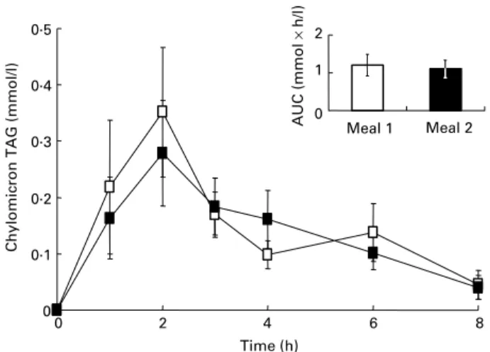
There was no significant difference between chylomicron-rich fraction TAG responses (expressed as area under the postpran-dial 0 to 8 h curves) following the ingestion of the two meals (Fig. 1). In contrast, the chylomicron-rich fraction lutein response was 23 % weaker (P¼ 0·07) after meal 2 (containing lutein plus the mixture of antioxidants) than after meal 1 (lutein alone) (21·9 (SEM 4·6) v. 28·4 (SEM 7·2) nmol £ h/l, respectively; Fig. 2). Individual chylomicron-rich fraction lutein responses were very variable, ranging from 8·8 to 73·5 nmol £ h/l after meal 1, but six subjects out of eight had a lower chylomicron-rich fraction lutein response after meal 2 than after meal 1, one had a similar response (28·5 and 27·2 nmol £ h/l) and one had a higher response (30·0 v. 15·9 nmol £ h/l). 0 0·1 0·2 0·3 0·4 0·5 0 2 4 6 8 Time (h)
Chylomicron TAG (mmol/l)
0 1 2 Meal 1 Meal 2 AUC (mmol × h/l)
Fig. 1. Plasma chylomicron-rich fraction TAG responses in healthy males after ingestion of meal 1 (A; 18 mg lutein) and meal 2 (B; 18 mg lutein þa mixture of antioxidant microconstituents). Data (change from fasting values) are means (n 8) with their standard errors represented by vertical bars. Inset: chylomicron-rich fraction TAG responses (area under the curve; AUC) after meal 1 and meal 2. Data are means (n 8) with their standard errors rep-resented by vertical bars.
Effect of antioxidant microconstituents on lutein uptake by Caco-2 cells
Before describing in detail the effect of antioxidant micronu-trients on lutein uptake by Caco-2 cells it should be mentioned that we found that there was no significant degradation of lutein during the duration of the experiments, whether or not there were other antioxidant micronutrients.
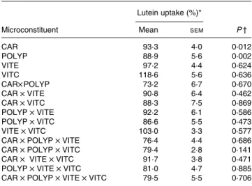
Identification of the antioxidant classes that affect lutein uptake. Data analysis of the results obtained in the factorial design showed that both the mixture of polyphenols (gallic acid, caffeic acid, (þ )-catechin and naringenin) and the mix-ture of carotenoids (lycopene plus b-carotene) significantly (P, 0·05) impaired lutein uptake. In contrast, vitamins C and E had no significant effect (Table 4). No interaction was observed between the different classes of microconstituents in terms of lutein uptake.
Effects of individual carotenoids. b-Carotene and lyco-pene, when separately co-incubated at physiological dietary con-centrations (up to 0·46 mM) with lutein for 30 min, were able to
decrease lutein uptake, but not significantly (data not shown). Effects of individual polyphenols. Experiments run separ-ately for each polyphenol showed that naringenin was the only polyphenol able to significantly impair lutein uptake (about 25 % for 25 mM- and 50 % for 150 mM-naringenin,
respect-ively; P, 0·05, Fig. 3). We observed that the mixture of poly-phenols containing 25 mM-naringenin had a similar effect to
25 mM-naringenin alone. This observation indicates that the other polyphenols tested (gallic acid, caffeic acid and (þ )-catechin) had no effect on lutein uptake.
Discussion
Given the aim of the present study was to assess the effect of diet-ary antioxidants on lutein absorption, we paid particular atten-tion to the choice of both type and relative proporatten-tions of the antioxidant microconstituents. The selected antioxidants belong to the main classes of dietary microconstituents, and the relative proportions of these microconstituents were very close to those observed in the standard Western diet. In the
clinical experiment, the amount of lutein contained in the test meal was set at 18 mg, i.e. about 8-fold higher than the average dietary intake, in order to accurately detect newly absorbed lutein in the chylomicron fraction (Tyssandier et al. 2002). Nevertheless, this amount remains close to the median daily total carotenoid intake (14 mg/d; O’Neill et al. 2001). The amounts of other antioxidant microconstituents added in the postprandial experiment were calculated in order to be close to the relative proportions found in the Western diet. We opted
0 4 8
0 2 4 6 8
Time (h)
Chylomicron lutein (nmol/l)
0 20 40 Meal 1 Meal 2 AUC (nmol × h /l) 6 6
Fig. 2. Plasma chylomicron-rich fraction lutein responses in healthy males after ingestion of meal 1 (W; 18 mg lutein) and meal 2 (
†
; 18 mg lutein þ a mixture of antioxidant microconstituents). Data (change from fasting values) are means (n 8) with their standard errors represented by vertical bars. Inset: chylomicron-rich fraction lutein responses (area under the curve; AUC) after meal 1 and meal 2. Data are means (n 8) with their standard errors rep-resented by vertical bars.Table 4. Effect of antioxidant microconstituents on lutein uptake in the Caco-2 cell experiments of factorial design
(Mean values with their standard errors)
Lutein uptake (%)*
Microconstituent Mean SEM P †
CAR 93·3 4·0 0·012 POLYP 88·9 5·6 0·002 VITE 97·2 4·4 0·624 VITC 118·6 5·6 0·636 CAR£ POLYP 73·2 6·7 0·670 CAR £ VITE 90·8 6·4 0·462 CAR £ VITC 88·3 7·5 0·869 POLYP £ VITE 92·2 6·1 0·586 POLYP £ VITC 86·6 5·5 0·473 VITE £ VITC 103·0 3·3 0·577 CAR £ POLYP £ VITE 76·4 4·4 0·686 CAR £ POLYP £ VITC 79·4 2·8 0·141 CAR £ VITE £ VITC 91·7 3·8 0·471 POLYP £ VITE £ VITC 81·0 4·7 0·885 CAR £ POLYP £ VITE £ VITC 79·5 5·5 0·706 CAR, mixture of the carotenoids b-carotene and lycopene; POLYP, mixture of the polyphenols naringenin, catechin, gallic acid and caffeic acid; VITE, mixture of the main vitamin E species a- and g-tocopherol; VITC, vitamin C.
* Lutein uptake was set at 100 % when lutein was provided without other antioxidant micronutrients.
† A P value of , 0·05 states that the compound, or the combination of compounds, significantly affected lutein uptake.
1 2 3 4 5 6 Experiment 7 8 9 10 0 20 40 60 80 100 120 140
Absorbed lutein (% of control)
*
Fig. 3. Effect of a range of individual polyphenols on lutein uptake by differen-tiated Caco-2 TC-7 monolayers. The apical sides of the cell monolayers were rinsed with PBS and then received fetal bovine serum-free medium containing either lutein-rich mixed micelles at 0·85 mMalone (1) or 0·85 mM-lutein-rich micelles supplemented with (2) 25 mM-naringenin, (3) 150 mM-naringenin, (4) 25 mM-(þ )-catechin, (5) 150 mM-(þ )-catechin, (6) 50 mM-gallic acid, (7) 150 mM -gallic acid, (8) 50 mM-caffeic acid, (9) 150 mM-caffeic acid and (10) a mixture of polyphenols (25 mM-naringenin, 25 mM-(þ)-catechin, 50 mM-gallic acid and 50 mM-caffeic acid). The basolateral sides received complete medium. Incu-bation time was 30 min. Data are means (for three assays) with their standard errors represented by vertical bars. * Mean value was significantly different from that of the control (assay performed without polyphenol) (P, 0·05).
not to add carotenoids to the antioxidant mixture used in the clinical study since it has been established that carotenoids com-pete with each other for absorption (van den Berg, 1999; Tyssan-dier et al. 2002), and we did not want this effect to mask an effect of other antioxidants. In the Caco-2 cell experiments, the con-centration of lutein in mixed micelles was set to about 0·7 mM
in order to accurately detect absorbed lutein. This represents the same concentration as found in the human duodenum after ingestion of a standard Western meal (Tyssandier et al. 2003). In the Caco-2 cell experiments, the competitive carotenoids chosen were b-carotene and lycopene, which are the main diet-ary carotenoids. The mixture of vitamin E contained equal amounts of a-tocopherol and g-tocopherol, which are the main dietary species of vitamin E. Finally, the selected polyphenols, i.e. naringenin, (þ )-catechin, gallic acid and caffeic acid, rep-resent the two main dietary classes of these microconstituents, i.e. flavonoids and non-flavonoids (mainly phenolic acids).
The postprandial experiment suggested that the mixture of antioxidant microconstituents diminished lutein bioavailability (2 23 %; P¼ 0·07). This difference was barely not significant probably due to the high inter-individual variability in chylo-micron lutein response. In fact, based on the data obtained, i.e. standard deviations and between-group differences, we calcu-lated that twenty-five subjects would be required to establish a significant decrease in chylomicron lutein response with a power efficiency of 80 %.
Since we provided a mixture of antioxidants, we could not reach definitive conclusions on the individual effect of each class of antioxidants or on possible synergies or antagonism between antioxidants. Furthermore, since it was unfeasible to run a clinical study to assess the effect of all the combi-nations of antioxidants on lutein absorption, we opted for an approach using the intestinal Caco-2 cell model in order to provide insight into the possible interactions between dietary antioxidants on lutein absorption. The Caco-2 cell line was selected because it is the most frequently used model for eval-uating the intestinal absorption and uptake of lipids and caro-tenoids (Garrett et al. 1999; Ferruzzi et al. 2001; Sugawara et al. 2001; During et al. 2002) and because it expresses the scavenger receptor class B type I, which facilitates lutein uptake (Reboul et al. 2003). We used the TC-7 clone because it is more homogeneous than the parental Caco-2 cell line (Gres et al. 1998) and possesses b-carotene 15,150 -monoxy-genase (During et al. 1998), which is a key enzyme in caroten-oid metabolism. The factorial design showed that carotencaroten-oids and polyphenols were the only two classes of antioxidants that had a significant effect on lutein uptake. The fact that vitamin C had no significant effect on lutein uptake apparently dis-agrees with a recent report stating that vitamin C facilitates lutein absorption (Tanumihardjo et al. 2005). However, it should be stressed that although vitamin C apparently enhanced lutein absorption rate in that study, it did not signifi-cantly affect blood lutein response, i.e. lutein bioavailability. The fact that both the mixture of carotenoids and the mixture of polyphenols significantly impaired lutein uptake raised the question as to which carotenoid(s) or polyphenol(s) were responsible for this effect. Therefore, we conducted a second series of experiments to measure the individual effect of each carotenoid and polyphenol on lutein uptake. The present results, as well as those obtained in another study (Reboul et al. 2005a), showed that the carotenoid effect was not
specific for b-carotene or lycopene but, rather, was dependent on the total concentration of carotenoids. This inhibitory effect of carotenoids on lutein uptake, which is in agreement with previous studies (van den Berg, 1999; Tyssandier et al. 2002), is probably the result of competition between lutein and other carotenoids for uptake through the scavenger recep-tor class B type I located at the enterocyte brush-border mem-brane (Reboul et al. 2005a). The second set of experiments also showed that naringenin was the only polyphenol that impaired lutein uptake. The specific effect exerted by narin-genin needs to be further elucidated by additional exper-iments, but it is nevertheless significant that narignenin is the most lipophilic of all the polyphenols tested (log P¼ 2·52 v. 0·86, 0·82 and 0·38 for gallic acid, caffeic acid and (þ )-catechin, respectively (Cooper et al. 1997)). It is possible that naringenin affects lutein uptake through an inter-action with scavenger receptor class B type I, which is known to transport lipophilic molecules with low substrate specificity. A second hypothesis might be related to an interaction between naringenin and membrane lipids (Tachibana et al. 2004) which may affect the invagination of lipid raft domains containing lutein receptors.
The moderate inhibitory effect of the antioxidant mixture observed in the clinical study, as compared with the effect observed in the Caco-2 experiments, can be explained by two factors: (i) the polyphenol supplement used in the clinical study contained only 4 mg aglycone flavanones (3 mg narin-genin þ 1 mg hesperitin), giving a lutein:aglycone flavanone ratio of 4·5 compared with 0·025 in the Caco-2 cell exper-iments; (ii) the flavanone glycosides (naringin and hesperidin) may not affect lutein absorption and are not very efficiently hydrolysed to their corresponding aglycones in the human digestive tract (Nemeth et al. 2003; Walle et al. 2005).
In conclusion, the results obtained show that some dietary antioxidants (vitamin C, vitamin E, flavones and phenolic acids) have no significant effect on lutein bioavailability, whereas it is apparently impaired by other antioxidants (caro-tenoids, naringenin and probably other aglycone flavanones). These findings should be taken into account when antioxidant supplements are consumed over long periods, as well as in the design of optimal human diets.
Acknowledgement
The present study was supported by INSERM and INRA (ATC Nutrition 2002, project no. A02256AS).
References
Blum S, Aviram M, Ben-Amotz A & Levy Y (2005) Effect of a Med-iterranean meal on postprandial carotenoids, paraoxonase activity and C-reactive protein levels. Ann Nutr Metab 50, 20 – 24. Bone RA, Landrum JT, Friedes LM, Gomez CM, Kilburn MD,
Menendez E, Vidal I & Wang W (1997) Distribution of lutein and zeaxanthin stereoisomers in the human retina. Exp Eye Res 64, 211 – 218.
Borel P, Grolier P, Armand M, Partier A, Lafont H, Lairon D & Azais-Braesco V (1996) Carotenoids in biological emulsions: solubility, surface-to-core distribution, and release from lipid dro-plets. J Lipid Res 37, 250 – 261.
Cardinault N, Tyssandier V, Grolier P, Winklhofer-Roob BM, Ribalta J, Bouteloup-Demange C, Rock E & Borel P (2003)
Comparison of the postprandial chylomicron carotenoid responses in young and older subjects. Eur J Nutr 42, 315 – 323.
Chan C, Leung I, Lam KW & Tso MOM (1998) The occurrence of retinol and carotenoids in human subretinal fluid. Curr Eye Res 17, 890 – 895.
Chug-Ahuja JK, Holden JM, Forman MR, Mangels AR, Beecher GR & Lanza E (1993) The development and application of a caroten-oid database for fruits, vegetables, and selected multicomponent foods. J Am Diet Assoc 93, 318 – 323.
Cooper DA, Webb DR & Peters JC (1997) Evaluation of the potential for olestra to affect the availability of dietary phytochemicals. J Nutr 127, S1699 – S1709.
During A, Albaugh G & Smith JC (1998) Characterization of
b-car-otene 15,150-dioxygenase activity in TC7 clone of human intestinal
cell line Caco-2. Biochem Biophys Res Comm 249, 467 – 474. During A, Hussain MM, Morel DW & Harrison EH (2002)
Caroten-oid uptake and secretion by Caco-2 cells: b-carotene isomer selec-tivity and carotenoid interactions. J Lipid Res 43, 1086 – 1095. Ferruzzi MG, Failla ML & Schwartz SJ (2001) Assessment of
degra-dation and intestinal cell uptake of carotenoids and chlorophyll derivatives from spinach puree using an in vitro digestion and Caco-2 human cell model. J Agric Food Chem 49, 2082 – 2089. Fossati P & Prencipe L (1982) Serum triglycerides determined
color-imetrically with an enzyme that produces hydrogen peroxide. Clin Chem 28, 2077 – 2080.
Garrett DA, Failla ML & Sarama RJ (1999) Development of an in vitro digestion method to assess carotenoid bioavailability from meals. J Agric Food Chem 47, 4301 – 4309.
George S, Brat P, Alter P & Amiot MJ (2005) Rapid determination of polyphenols and vitamin C in plant-derived products. J Agric Food Chem 53, 1370 – 1373.
Gres MC, Julian B, Bourrie M, Meunier V, Roques C, Berger M, Boulenc X, Berger Y & Fabre G (1998) Correlation between oral drug absorption in humans, and apparent drug permeability in TC-7 cells, a human epithelial intestinal cell line: comparison with the parental Caco-2 cell line. Pharm Res 15, 726 – 733. Hendrich S, Lee KW, Xu X, Wang HJ & Murphy PA (1994)
Defining food components as new nutrients. J Nutr 124, S1789 – S1792.
Junghans A, Sies H & Stahl W (2001) Macular pigments lutein and zeaxanthin as blue light filters studied in liposomes. Arch Biochem Biophys 391, 160 – 164.
Krinsky NI (2002) Possible biologic mechanisms for a protective role of xanthophylls. J Nutr 132, 540S – 542S.
Landrum JT & Bone RA (2001) Lutein, zeaxanthin, and the macular pigment. Arch Biochem Biophys 385, 28 – 40.
Luchoomun J & Hussain MM (1999) Assembly and secretion of chy-lomicrons by differentiated Caco-2 cells. Nascent triglycerides and preformed phospholipids are preferentially used for lipoprotein assembly. J Biol Chem 274, 19565 – 19572.
Lyan B, Azais-Braesco V, Cardinault N, Tyssandier V, Borel P, Alexandre-Gouabau MC & Grolier P (2001) Simple method for clinical determination of 13 carotenoids in human plasma using an isocratic high-performance liquid chromatographic method. J Chromatogr 751B, 297 – 303.
Nemeth K, Plumb GW, Berrin JG, Juge N, Jacob R, Naim HY, Williamson G, Swallow DM & Kroon PA (2003) Deglycosylation by small intestinal epithelial cell b-glucosidases is a critical step in the absorption and metabolism of dietary flavonoid glycosides in humans. Eur J Nutr 42, 29 – 42.
O’Neill ME, Carroll Y, Corridan B, Olmedilla B, Granado F, Blanco I, van den Berg H, Hininger I, Rousell AM, Chopra M, Southon S & Thurnham DI (2001) A European carotenoid database to assess carotenoid intakes and its use in a five-country comparative study. Br J Nutr 85, 499 – 507.
Rapp LM, Maple SS & Choi JH (2000) Lutein and zeaxanthin concentrations in rod outer segment membranes from perifoveal
and peripheral human retina. Invest Ophthalmol Vis Sci 41, 1200 – 1209.
Reboul E, Abou L, Mikail C, Ghiringhelli O, Andre M, Gleize B, Kaloustian J, Portugal H, Amiot M & Borel P (2003) Lutein is apparently absorbed by a carrier-mediated transport process in Caco-2 cells. Clin Nutr 22, S103.
Reboul E, Abou L, Mikail C, Ghiringhelli O, Andre M, Portugal H, Jourdheuil-Rahmani D, Amiot MJ, Lairon D & Borel P (2005a) Lutein transport by Caco-2 TC-7 cells occurs partly by a facilitated process involving the scavenger receptor class B type I (SR-BI). Biochem J 387, 455 – 461.
Reboul E, Borel P, Mikail C, Abou L, Charbonnier M, Caris-Veyrat C, Goupy P, Portugal H, Lairon D & Amiot MJ (2005b) Enrichment of tomato paste with 6 % tomato peel increases lycopene and b-carotene bioavailability in men. J Nutr 135, 790 – 794.
Riso P, Brusamolino A, Ciappellano S & Porrini M (2003) Compari-son of lutein bioavailability from vegetables and supplement. Int J Vitam Nutr Res 73, 201 – 205.
Salvini S, Charbonnier M, Defoort C, Alquier C & Lairon D (2002) Functional characterization of three clones of the human intestinal Caco-2 cell line for dietary lipid processing. Br J Nutr 87, 211 – 217. Siedel J, Hagele EO, Ziegenhorn J & Wahlefeld AW (1983) Reagent for the enzymatic determination of serum total cholesterol with improved lipolytic efficiency. Clin Chem 29, 1075 – 1080. Siems WG, Sommerburg O & van Kuijk FJ (1999) Lycopene and
b-car-otene decompose more rapidly than lutein and zeaxanthin upon exposure to various pro-oxidants in vitro. Biofactors 10, 105–113. Snodderly DM (1995) Evidence for protection against age-related
macular degeneration by carotenoids and antioxidant vitamins. Am J Clin Nutr 62, 1448S – 1461S.
Sommerburg O, Keunen JE, Bird AC & van Kuijk FJ (1998) Fruits and vegetables that are sources for lutein and zeaxanthin: the macular pigment in human eyes. Br J Ophthalmol 82, 907 – 910.
Souci SW, Fachmann W & Kraut H (2000) Food Composition and Nutrition Tables, 6th revised and completed edition, Boca Raton, FL: CRC Press/Medpharm Scientific Publishers.
Sugawara T, Kushiro M, Zhang H, Nara E, Ono H & Nagao A (2001) Lysophosphatidylcholine enhances carotenoid uptake from mixed micelles by Caco-2 human intestinal cells. J Nutr 131, 2921 – 2927. Tachibana H, Fujimura Y & Yamada K (2004) Tea polyphenol epi-gallocatechin-3-gallate associates with plasma membrane lipid rafts: lipid rafts mediate anti-allergic action of the catechin. Biofactors 21, 383 – 385.
Tanumihardjo SA, Li J & Dosti MP (2005) Lutein absorption is facili-tated with cosupplementation of ascorbic acid in young adults. J Am Diet Assoc 105, 114 – 118.
Tesoriere L, Allegra M, Butera D & Livrea MA (2004) Absorption, excretion, and distribution of dietary antioxidant betalains in LDLs: poten-tial health effects of betalains in humans. Am J Clin Nutr 80, 941–945. Trinder P (1969) Determination of blood glucose using 4-amino
phe-nazone as oxygen acceptor. J Clin Pathol 22, 246.
Tyssandier V, Cardinault N, Caris-Veyrat C, Amiot MJ, Grolier P, Bouteloup C, Azais-Braesco V & Borel P (2002) Vegetable-borne lutein, lycopene, and b-carotene compete for incorporation into chy-lomicrons, with no adverse effect on the medium- term (3-wk) plasma status of carotenoids in humans. Am J Clin Nutr 75, 526 – 534. Tyssandier V, Lyan B & Borel P (2001) Main factors governing the
transfer of carotenoids from emulsion lipid droplets to micelles. Biochim Biophys Acta 1533, 285 – 292.
Tyssandier V, Reboul E, Dumas JF, Bouteloup-Demange C, Armand M, Marcand J, Sallas M & Borel P (2003) Processing of vegetable-borne carotenoids in the human stomach and duodenum. Am J Physiol 284, G913 – G923.
van den Berg H (1999) Carotenoid interactions. Nutr Rev 57, 1 – 10. Walle T, Browning AM, Steed LL, Reed SG & Walle UK (2005) Fla-vonoid glucosides are hydrolyzed and thus activated in the oral cavity in humans. J Nutr 135, 48 – 52.