HAL Id: hal-02570114
https://hal.sorbonne-universite.fr/hal-02570114
Submitted on 11 May 2020
HAL is a multi-disciplinary open access
archive for the deposit and dissemination of
sci-entific research documents, whether they are
pub-lished or not. The documents may come from
teaching and research institutions in France or
abroad, or from public or private research centers.
L’archive ouverte pluridisciplinaire HAL, est
destinée au dépôt et à la diffusion de documents
scientifiques de niveau recherche, publiés ou non,
émanant des établissements d’enseignement et de
recherche français ou étrangers, des laboratoires
publics ou privés.
Neuroinflammatory Mechanisms in Alzheimer’s Disease
Harald Hampel, Filippo Caraci, A. Claudio Cuello, Giuseppe Mario Caruso,
Robert Nisticò, Massimo Corbo, Filippo Baldacci, Nicola Toschi, Francesco
Garaci, Patrizia Chiesa, et al.
To cite this version:
Harald Hampel, Filippo Caraci, A. Claudio Cuello, Giuseppe Mario Caruso, Robert Nisticò, et al.. A
Path Toward Precision Medicine for Neuroinflammatory Mechanisms in Alzheimer’s Disease. Frontiers
in Immunology, Frontiers, 2020, 11, pp.456. �10.3389/fimmu.2020.00456�. �hal-02570114�
Edited by: Sally Ann Frautschy, University of California, Los Angeles, United States Reviewed by: Maria Grazia Cifone, University of L’Aquila, Italy Maria F. Cano-Abad, Autonomous University of Madrid, Spain *Correspondence: Harald Hampel harald.hampel@med.uni-muenchen.de
†These authors have contributed
equally to this work and share senior authorship
Specialty section: This article was submitted to Multiple Sclerosis and Neuroimmunology, a section of the journal Frontiers in Immunology Received:02 November 2019 Accepted:27 February 2020 Published:31 March 2020 Citation: Hampel H, Caraci F, Cuello AC, Caruso G, Nisticò R, Corbo M, Baldacci F, Toschi N, Garaci F, Chiesa PA, Verdooner SR, Akman-Anderson L, Hernández F, Ávila J, Emanuele E, Valenzuela PL, Lucía A, Watling M, Imbimbo BP, Vergallo A and Lista S (2020) A Path Toward Precision Medicine for Neuroinflammatory Mechanisms in Alzheimer’s Disease. Front. Immunol. 11:456. doi: 10.3389/fimmu.2020.00456
A Path Toward Precision Medicine
for Neuroinflammatory Mechanisms
in Alzheimer’s Disease
Harald Hampel
1*, Filippo Caraci
2,3, A. Claudio Cuello
4,5,6,7, Giuseppe Caruso
3,
Robert Nisticò
8,9, Massimo Corbo
10, Filippo Baldacci
1,11,12,13, Nicola Toschi
14,15,16,
Francesco Garaci
14,17, Patrizia A. Chiesa
1,11,12, Steven R. Verdooner
18,
Leyla Akman-Anderson
18, Félix Hernández
19,20, Jesús Ávila
19,20, Enzo Emanuele
21,
Pedro L. Valenzuela
22, Alejandro Lucía
23,24,25, Mark Watling
26, Bruno P. Imbimbo
27,
Andrea Vergallo
1†and Simone Lista
1,11,12†, On behalf of the Alzheimer Precision
Medicine Initiative (APMI)
1Sorbonne University, GRC no. 21, Alzheimer Precision Medicine (APM), AP-HP, Pitié-Salpêtrière Hospital, Boulevard de
l’hôpital, Paris, France,2Department of Drug Sciences, University of Catania, Catania, Italy,3Oasi Research
Institute—IRCCS, Troina, Italy,4Department of Neurology and Neurosurgery, McGill University, Montreal, QC, Canada, 5Department of Anatomy and Cell Biology, McGill University, Montreal, QC, Canada,6Department of Pharmacology and
Therapeutics, McGill University, Montreal, QC, Canada,7Department of Pharmacology, University of Oxford, Oxford,
United Kingdom,8Laboratory of Neuropharmacology, EBRI Rita Levi-Montalcini Foundation, Rome, Italy,9School of
Pharmacy, Department of Biology, University of Rome Tor Vergata, Rome, Italy,10Department of Neurorehabilitation
Sciences, Casa Cura Policlinico, Milan, Italy,11Brain & Spine Institute (ICM), INSERM U 1127, CNRS UMR 7225, Boulevard
de l’hôpital, Paris, France,12Institute of Memory and Alzheimer’s Disease (IM2A), Department of Neurology, Pitié-Salpêtrière
Hospital, AP-HP, Paris, France,13Department of Clinical and Experimental Medicine, University of Pisa, Pisa, Italy, 14Department of Biomedicine and Prevention, University of Rome “Tor Vergata”, Rome, Italy,15Department of Radiology,
“Athinoula A. Martinos” Center for Biomedical Imaging, Boston, MA, United States,16Harvard Medical School, Boston, MA,
United States,17Casa di Cura “San Raffaele Cassino”, Cassino, Italy,18NeuroVision Imaging, Inc., Sacramento, CA,
United States,19Centro de Biología Molecular Severo Ochoa (CSIC-UAM), Madrid, Spain,20Network Center for Biomedical
Research in Neurodegenerative Diseases (CIBERNED), Madrid, Spain,212E Science, Robbio, Italy,22Systems Biology
Department, University of Alcalá, Madrid, Spain,23Faculty of Sport Sciences, Universidad Europea de Madrid, Madrid,
Spain,24Research Institute of the Hospital 12 de Octubre (“imas”), Madrid, Spain,25Centro de Investigación Biomédica en
Red Fragilidad y Envejecimiento Saludable (CIBERFES), Madrid, Spain,26TranScrip Partners, Reading, United Kingdom, 27Research & Development Department, Chiesi Farmaceutici, Parma, Italy
Neuroinflammation commences decades before Alzheimer’s disease (AD) clinical
onset and represents one of the earliest pathomechanistic alterations throughout
the AD continuum. Large-scale genome-wide association studies point out several
genetic variants—TREM2, CD33, PILRA, CR1, MS4A, CLU, ABCA7, EPHA1, and
HLA-DRB5-HLA-DRB1—potentially linked to neuroinflammation. Most of these genes
are involved in proinflammatory intracellular signaling, cytokines/interleukins/cell turnover,
synaptic activity, lipid metabolism, and vesicle trafficking. Proteomic studies indicate that
a plethora of interconnected aberrant molecular pathways, set off and perpetuated by
TNF-α, TGF-β, IL-1β, and the receptor protein TREM2, are involved in neuroinflammation.
Microglia and astrocytes are key cellular drivers and regulators of neuroinflammation.
Under physiological conditions, they are important for neurotransmission and synaptic
homeostasis. In AD, there is a turning point throughout its pathophysiological evolution
where glial cells sustain an overexpressed inflammatory response that synergizes with
amyloid-β and tau accumulation, and drives synaptotoxicity and neurodegeneration
in a self-reinforcing manner. Despite a strong therapeutic rationale, previous clinical
trials investigating compounds with anti-inflammatory properties, including non-steroidal
anti-inflammatory drugs (NSAIDs), did not achieve primary efficacy endpoints. It is
conceivable that study design issues, including the lack of diagnostic accuracy
and biomarkers for target population identification and proof of mechanism, may
partially explain the negative outcomes. However, a recent meta-analysis indicates
a potential biological effect of NSAIDs. In this regard, candidate fluid biomarkers of
neuroinflammation are under analytical/clinical validation, i.e., TREM2, 1β, MCP-1,
IL-6, TNF-α receptor complexes, TGF-β, and YKL-40. PET radio-ligands are investigated
to accomplish in vivo and longitudinal regional exploration of neuroinflammation.
Biomarkers tracking different molecular pathways (body fluid matrixes) along with
brain neuroinflammatory endophenotypes (neuroimaging markers), can untangle
temporal–spatial dynamics between neuroinflammation and other AD pathophysiological
mechanisms. Robust biomarker–drug codevelopment pipelines are expected to enrich
large-scale clinical trials testing new-generation compounds active, directly or indirectly,
on neuroinflammatory targets and displaying putative disease-modifying effects: novel
NSAIDs, AL002 TREM2 antibody), anti-Aβ protofibrils (BAN2401), and AL003
(anti-CD33 antibody). As a next step, taking advantage of breakthrough and multimodal
techniques coupled with a systems biology approach is the path to pursue for developing
individualized therapeutic strategies targeting neuroinflammation under the framework of
precision medicine.
Keywords: Alzheimer’s disease, neuroinflammation, microglia, neuroinflammatory pathways, biomarkers, anti-inflammatory therapy, systems biology, precision medicine
INTRODUCTION
Alzheimer’s disease (AD) is the most commoncause of
neurodegenerative dementia. According to current estimates,
17% of people aged 75–84 years in the United States have AD,
and the disease costs the country US$236 billion per year. The
prevalence is projected to triple by 2050 to >15 million, with
annual costs of >$700 billion (
1
). There is an urgent need
for developing pharmacological treatments with a
disease-modifying effect to halt the disease at its earliest preclinical
stage where brain and cognitive functions can still be preserved
(
2
,
3
). Indeed, drugs currently available on the pharmaceutical
market (i.e., acetylcholinesterase inhibitors and non-competitive
N-methyl-
D-aspartate antagonists) have been approved for a
symptomatic effect only and for the dementia stage of AD (
4
).
The
acknowledged
pathophysiological
hallmarks—(I)
extracellular deposition of amyloid beta (Aβ), (II) intracellular
aggregates of tau proteins, ultimately called neurofibrillary
tangles (NFT), and (III) neurodegeneration—have been
integrated in research diagnostic criteria (
5
–
8
).
Abbreviations: [11C]-PIB, [11C]-Pittsburgh compound-B; [18
F]-fluorodeoxyglucose-PET, [18F]-fluorodeoxyglucose-positron emission
tomography; Aβ, amyloid beta; Aβ1−40, 40-amino acid-long amyloid beta
peptide; Aβ1−42, 42-amino acid-long amyloid beta peptide; AD, Alzheimer’s
disease; ADAPT, Alzheimer’s disease anti-inflammatory prevention trial; ADCS, Alzheimer’s disease cooperative studies; APMI, Alzheimer precision medicine initiative; APMI-CP, Alzheimer precision medicine initiative cohort program; APS, Alzheimer progression score; ASC, apoptosis-associated speck-like protein containing a caspase recruitment domain; BDNF, brain-derived neurotrophic factor; CB2R, cannabinoid receptor type 2; CCE, cell cycle events; CD, cell surface cluster of differentiation; CDx, companion diagnostic; CNS, central
The hypothesis-free biomarker-guided “A/T/N” classification
scheme was introduced to categorize subjects based on core
AD hallmarks (
9
). The A/T/N scheme is anticipated to provide
consistent recruitment of individuals and target engagement
among various different sites in AD clinical trials. Even
though the A/T/N classification scheme provides crucial
pathophysiological insights, it offers a partial depiction
of the spectrum of pathomechanistic modifications of
AD (
10
,
11
).
The increasing animal and in-human evidence for the
upstream role that neuroinflammation may play in AD has
nervous system; COX, cyclooxygenase; COX-1, cyclooxygenase-1; COX-2, cyclooxygenase-2; CRP, C-reactive protein; CSF, cerebrospinal fluid; CSF-1R, colony stimulating factor 1 receptor; GWAS, genome-wide association studies; ILs, interleukins; IL-1Ra, interleukin-1 receptor antagonist; INTREPAD, impact of naproxen treatment in presymptomatic Alzheimer’s disease; JNK, c-Jun Kinase; K-ARPI, Korean AD research platform initiative based on immune-inflammatory biomarkers; LTP, long-term potentiation; MAPK, p38 mitogen-activated protein kinase; MCI, mild cognitive impairment; MCP-1, monocyte chemoattractant protein-1; MMP-9, metalloprotease-9; MMSE, Mini-Mental State Examination; mNGF, mature nerve growth factor; Nap1, Nck-associated protein 1; ND, neurodegenerative diseases; NF-κB, nuclear factor kappa-light-chain-enhancer of activated B cells; NFT, neurofibrillary tangles; NGF, nerve growth factor; NIH, National Institutes of Health; NSAIDs, non-steroidal anti-inflammatory drugs; P4M, predictive, preventive, personalized, and participatory medicine; PET, positron emission tomography; PMI, US precision medicine initiative; PMI-CP, US PMI cohort program; proNGF, precursor of the nerve growth factor; p-tau, hyperphosphorylated tau; RNS, reactive nitrogen species; ROS, reactive oxygen species; SCARA1, class A1 scavenger receptor; SphK1, sphingosine kinase 1; sTREM2, soluble TREM2; TGF-β1, transforming growth factor-beta1; TLRs, Toll-like receptors; TNF-α, tumor necrosis factor-alpha; TNF-Rs, TNF receptors; TNF-RI, TNF receptor I; TNF-RII, TNF receptor II; TREM2, triggering receptor expressed on myeloid cells 2; TSPO, translocator protein.
posed several conceptual therapeutic concerns and open up new
avenues for preventing AD cognitive decline.
The pathophysiological mechanisms of multifactorial and
polygenic AD are not limited to the neuronal tissue; they
are related to cerebral immunological responses (
12
). Indeed,
brains of patients with AD and other neurodegenerative diseases
(ND) show chronic inflammation (
13
). Neuroinflammation is
as an innate immunological response of the nervous system
that comprises microglia, astrocytes, cytokines, and chemokines,
which play a central role in an early phase of AD pathogenesis
(
12
,
14
). The key contribution of inflammation in the AD
pathophysiology has been hypothesized more than 20 years ago
(
12
,
15
–
17
). Recent studies demonstrate that this early
disease-aggravating central nervous system (CNS) inflammation starts
decades before the appearance of severe cognitive decay or AD
(
18
–
20
). Along this line, different longitudinal studies show
that inflammation and microglial activation occur years before
AD onset (
21
–
23
). Furthermore, there is a strong link between
neuroinflammation and amyloid and tau accumulation in the
human brain (
23
–
26
).
The
acknowledged
cell
mediators
of
inflammatory
mechanisms in AD are microglia and astrocytes (
12
). In
general, these cells play a substantial role in neural transmission
and synapse remodeling, as they facilitate the removal of
non-essential synapses by eradicating inadequate connections
(
27
,
28
). Thus, the efficiency of neuronal transmission
is increased.
NEUROINFLAMMATION AND CELL
MEDIATORS OF INFLAMMATORY
MECHANISMS IN ALZHEIMER’S DISEASE
The Role of Microglia and Astrocytes in
Alzheimer’s Disease Synaptic Dysfunction
Synapses exhibit a quad-partite arrangement that consists of an
axon terminal, a dendritic spine put in direct communication
with a microglial and an astrocytic process (
29
). Astrocytes and
microglia—the brain-resident macrophages—play a key role
in neural circuit development and synaptic homeodynamics
during adulthood. Astrocytes are essential for supporting
synaptogenesis (axonal and dendritic spines sprouting)
and regulating synaptic robustness (
30
–
32
). Astrocytes also
contribute to the spatiotemporal integration of several synaptic
signals and regulate the synaptic transmission (
33
,
34
). Microglial
cells play a key role in the immune surveillance of the presynaptic
microenvironment and also for the synaptic remodeling toward
axonal and dendritic terminals pruning by reshaping proteolytic
and phagocytic processes. Microglial cells are able to recruit
astroglia, or they can be recruited by the latter (
30
–
32
,
35
).
They are thought to drive the well-known age-related regional
synaptic vulnerability, as recently reported (
36
). Indeed, an
age-related ultrastructural and functional shift of microglia
cells is associated with increased synaptic susceptibility and
neurodegeneration (
35
).
Therefore, astrocytes and microglia express physiological
properties essential for synaptic transmission, the accurate
modulation of neural and synaptic plasticity, and both synaptic
adaptation and homeostasis (
30
–
32
).
In summary, it is well-established that microglia and
astrocytes
take
part
in
aberrant
molecular
pathways
that, ultimately, reflect AD pathomechanistic alterations,
i.e.,
brain
proteinopathies,
synaptic
failure,
loss
of
brain plasticity, neuroinflammation, axonal damage, and
neurodegeneration (
37
–
41
).
The Role of Microglia
Microglial cells, arising from the mesodermal (myeloid) lineage
(
42
), are the main category of macrophages in the CNS
parenchyma. They express a large assortment of receptors that
recognize exogenous or endogenous CNS insults and initiate
an immune response. Besides their typical immune cell role,
microglial cells protect the brain by stimulating phagocytic
clearance and providing trophic sustenance to preserve cerebral
homeostasis and support tissue repair. When circumstances
related to loss of homeostasis or tissue alterations occur, then
many dynamic microglial mechanisms are triggered, leading to
the “activated state” of microglia (
43
). These encompass cellular
morphology modifications, changes in the secretory profile
of molecular mediators, and increased proliferative responses
(
44
). A persistent homeodynamic imbalance, such as brain
accumulation of Aβ, can trigger a step further in activation,
referred to as “priming” (
37
). Priming of microglia is directed
by alterations in their microenvironment and the release of
molecules guiding their proliferation. Priming makes microglia
inclined to secondary inflammatory stimulating factors, which
can then elicit amplified inflammatory reactions (
37
).
Activated microglia is a typical pathophysiological feature of
AD and other ND (
12
,
43
,
45
). Two main types of microglia cells
are present in the brain, “resting” (or “quiescent”) and “active”
microglia. In particular, there is evidence for the high degree
of heterogeneity of microglial activation in the CNS, which can
be categorized into two opposite activation phenotypes: M1
and M2 (
43
,
46
,
47
). According to the phenotype activated,
microglia can generate either cytotoxic or neuroprotective effects
(
46
). The M1 or “proinflammatory” phenotype (classically
activated) displays proinflammatory cytokines and nitric oxide.
It decreases the release of neurotrophic factors, thus exacerbating
inflammation and cytotoxicity (
43
). In contrast, the M2 or
“inflammatory” phenotype (alternatively activated) displays
anti-inflammatory cytokines, increased expression of neurotrophic
factors, and several other signals involved in downregulation,
protection, or repair processes in response to inflammation (
43
).
Preliminary evidence from experimental studies suggests that the
phenotypic transformation of the activated M1/M2 functional
states (“phenotypic switching”) (
48
,
49
) can be determined by
both the stage and the severity of the disease. In preclinical
models, M1 microglia seems to prevail at the injury site, at the
end stage of disease, and once inflammation resolution and repair
processes of M2 microglia are diminished (
46
).
In light of the increasing evidence that the modality by which
microglia is activated is a continuum between proinflammatory
(M1) and anti-inflammatory (M2) phenotypes, the M1/M2
“dichotomy” (or “polarization” scheme) is still disputed. Actually,
it seems possible that the global process of microglia activation
represents a much larger heterogeneous spectrum of very
dissimilar responses (
43
).
Experimental models of AD demonstrate that microglia
cluster around plaques, likely via chemotactic mechanisms, and
may contribute both in Aβ (
39
,
44
) clearance and in limiting
the growth and further accumulation plaques (
39
,
44
). Moreover,
the dysregulation of microglia activity, including dystrophic
microglia, may be either a trigger, or a worsening factor, or both,
of the seeding of aberrant protein aggregates in the brain (
39
,
44
).
In AD, during inflammation, there is a transition from
the resting to the active functional state of microglia that,
at a general level, might be the consequence of stress or
depressive-like behavior (
50
). At a molecular level, inflammation
is promoted by the presence of Aβ aggregates, including
oligomers and fibrils (
51
–
54
). Indeed, microglia can bind to
soluble Aβ oligomers and insoluble Aβ fibrils through cell
surface receptors, including the class A1 scavenger receptor
(SCARA1), cell surface cluster of differentiation (CD) markers
(CD36, CD14, and CD47), the α6β1 integrin, and the
Toll-like receptors (TLRs) (
55
–
58
). A key point within the scientific
debate is represented by a recent evidence indicating that
microglia displays either beneficial or harmful effects throughout
the beginning and advancement of AD (
45
). This is strictly
related to the nature of the major activities: (I) clearance of
Aβ or (II) release of proinflammatory mediators. In early AD
pathogenesis, Aβ oligomers and fibrils gather in the extracellular
space and elicit a pathological cascade resulting in neuronal
apoptosis and depletion. Microglia eliminate Aβ peptides and
dying/dead cells through phagocytosis (
59
,
60
). Besides clearance
of Aβ oligomers and fibrils, microglia surrounds plaques and
fibrils likely creating a physical barrier that can prevent their
spreading and toxicity (
61
). Aβ clearance is also stimulated
by the release of numerous proteases participating in Aβ
degradation (
62
). In spite of the advantageous actions of
early activation of microglia cells, their chronic activation by
Aβ is detrimental and induces protracted inflammation and
disproportionate Aβ deposition, thus rushing neurodegeneration
(Figure 1). During AD pathogenesis, the production and
release of proinflammatory cytokines and other detrimental
components are intensified. In addition, the typical phagocytic
action of microglia is decreased. Moreover, the
microglial-dependent release of apoptosis-associated speck-like protein
containing a caspase recruitment domain (ASC) modulates the
diffusion of the pathology within and between cerebral areas
(
63
). Extracellular vesicles—constituted by microvesicles and
exosomes and released by reactive microglia—play a role in AD
pathogenesis (
64
) (Figure 1). Finally, microglial cells are able to
regulate AD pathogenesis via active interaction with neurons,
astrocytes, and oligodendrocytes. Indeed, activated microglial
cells induce altered astrocytes via proinflammatory cytokines
(Figure 1). These astrocytes can rush and aggravate neuronal and
oligodendrocytes death (
65
).
The still open question is to understand the specific
contributions of neuronal and glial cells in the early phase
of inflammation in preclinical AD. Aβ
1−42oligomers have
a major role in synaptic depletion and gradual cognitive
deterioration (
66
,
67
). They induce neuroinflammation
and neurodegeneration by stimulating the microglia to
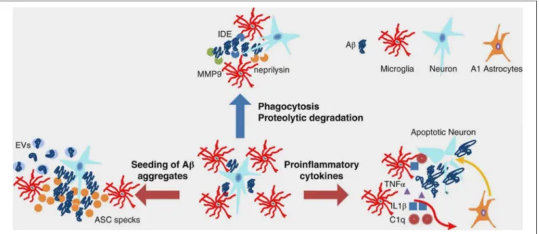
FIGURE 1 | Multifaceted functions of microglia during Aβ pathology. In healthy brain and early stages of AD, microglia clear small aggregates of Aβ peptides by phagocytosis and by secreting proteolytic enzymes, such as IDE, neprilysin, and MMP9. During advanced AD, microglia exacerbate AD pathology by releasing proinflammatory cytokines that induce neuronal cell death as well as A1 astrocytes, which, in turn, affect neuronal survival. Moreover, during advanced AD, microglia-derived ASC specks and EVs promote seeding of Aβ aggregates. Aβ, amyloid beta; AD, Alzheimer’s disease; ASC, apoptosis-associated speck-like protein containing a CARD; C1q, complement component 1q; EVs, extracellular vesicles; IDE, insulin degrading enzyme; IL-1β, interleukin-1 beta; MMP-9, metalloprotease-9; TNF-α, tumor necrosis factor-alpha. From Wang and Colonna (45). Copyright© 2019, Society for Leukocyte Biology. Reprinted with permission from Wiley.
FIGURE 2 | Role of neuroinflammation in AD pathogenesis: impairment of neurotrophin signaling. Aβ1−42oligomers promote neuroinflammation and neuronal death in
AD brain by eliciting the release of proinflammatory cytokines (IL-1β and TNF-α) from microglia and also interfering with the synthesis of anti-inflammatory cytokines such as TGF-β1. TNF-α inhibits microglia phagocytosis of Aβ and stimulates γ-secretase activity, thus facilitating Aβ accumulation and microglia-mediated neuroinflammation. Proinflammatory microglial activities promote neuronal death also through the formation of ROS and RNS. Neuroinflammatory phenomena can finally contribute to the pathogenesis of AD by impairing neurotrophin signaling function: (I) reducing the synthesis of BDNF and TGF-β1 and (II) causing an impairment of NGF metabolic pathway characterized by a reduced conversion of proNGF to biologically active mNGF and by an increased degradation of mNGF promoted by MMP-9. Aβ, amyloid beta; Aβ1−42, 42-amino acid-long amyloid beta peptide; BDNF, brain-derived neurotrophic factor; IL-1β, interleukin-1 beta; MMP-9,
metalloprotease-9; NGF, nerve growth factor; mNGF, mature nerve growth factor; proNGF, precursor of the nerve growth factor; RNS, reactive nitrogen species; ROS, reactive oxygen species; TGF-β, transforming growth factor-beta; TNF-α, tumor necrosis factor-alpha.
produce and release proinflammatory cytokines (
14
,
68
) and
also by interfering with the synthesis of anti-inflammatory
cytokines, for instance the transforming growth factor-beta
1 (TGF-β1) (Figure 2) (
69
–
71
). This early proinflammatory
process is characterized by neuronal and microglia-derived
cytokines and chemokines as well as by mobilization of
microglia toward Aβ-burdened neurons (Figure 2) (
19
,
72
).
In addition to Aβ, extracellular non-phosphorylated tau,
rather than hyperphosphorylated tau (p-tau), activates the p38
mitogen-activated protein kinase (MAPK) pathway, eliciting a
proinflammatory reaction (
73
).
The Role of Astrocytes
Astrocytes, differently from microglia and similarly to neurons
and oligodendrocytes, arise from the neuroectoderm (
74
). These
cells promotes synaptogenesis (axonal and dendritic spines
sprouting), regulates the synaptic strength, take part in the
spatial–temporal integration of multiple synaptic processes,
and modulate the neurotransmission. Hence, astrocytes execute
a variety of physiological activities, in both developing and
adult brain, that are essential for synaptic plasticity and a
solid and organized cognitive activity (
74
). Of note, astrocytes
modulate Ca
2+-dependent signaling pathways that are crucial
for hippocampal synaptic function and plasticity (
75
,
76
).
Indeed, depending on the fluctuations of intracellular Ca
2+concentrations, they release gliotransmitters, such as glutamate,
D-serine, and ATP, which have feedback actions on neurons (
77
).
Moreover, each astrocyte wraps several neurons, thus interacting
with hundreds of neuronal dendrites (
78
) and connecting with
up to two million synapses in the human cortex (
79
). This kind
of interconnectedness indicates that each astrocyte creates a hub
to facilitate the integration of the information (
74
). Moreover,
remodeling of astrocytes promotes neuroprotection and recovery
of injured neural tissue (
80
,
81
). Along with microglia activation,
hypertrophic reactive astrocytes gather around Aβ plaques as
reported in human postmortem studies (
82
) as well as in animal
models (
83
). Like microglia, astrocytes are also activated by tissue
injury, infection, and inflammation (
84
). In AD, after exposure
to Aβ, astrocytes release various proinflammatory molecules,
such as cytokines, interleukins (ILs), complement components
(
85
–
87
), nitric oxide, and other cytotoxic compounds, ultimately
amplifying the neuroinflammatory response.
Human neuropathological studies conducted on AD brains
report the presence of cytoplasmic inclusions of non-fibrillar
Aβ in astrocytes, supposed to reflect a phagocytic engulfment
from extracellular Aβ deposits (
86
). In addition, rodent models
of AD indicate the ability of astrocytes to uptake and clear
Aβ in subjects bearing cerebral fibrillar aggregates and diffuse
plaques (
16
,
17
,
33
,
86
). Conversely, the shutdown of
astrocyte-mediated homeodynamics is associated with increased Aβ plaque
burden and synaptic terminals dystrophy (
68
). This enhanced
phagocytic activity may represent a compensatory mechanism
to incipient increased Aβ accumulation to neutralize its
induced toxicity.
GENES MODULATING
NEUROINFLAMMATION IN ALZHEIMER’S
DISEASE
Genome-wide association studies (GWAS) allowed the detection
of more than 40 susceptibility gene variants associated with
a bigger risk of developing late-onset AD (
88
). These results
include genes associated with immune reaction (in particular,
ABCA7, CD33, CLU, CR1, EPHA1, HLA-DRB5-HLA-DRB1, and
MS4A). The relevance of neuroinflammation is further sustained
by recent large-scale GWAS showing that the risk of developing
late-onset AD is substantially more elevated in individuals with
rare variants of microglial immunoreceptors: TREM2, encoding
the triggering receptor expressed on myeloid cells 2 protein (
89
);
CD33 (transmembrane receptor CD33), expressed on cells of
myeloid lineage (
90
,
91
); and PILRA (paired
immunoglobulin-like type 2 receptor alpha) (
92
).
The receptor protein TREM2 enhances the rate of
phagocytosis in microglia and macrophages, modulates
inflammatory signaling, and controls myeloid cell number,
proliferation, and survival (
89
). Recent studies show that
triggering TREM2 receptor in microglial cells is closely associated
with the pathogenesis of AD (
93
). TREM2 modulates microglial
functions (e.g., stimulates the production of inflammatory
cytokines) in response to Aβ plaques and tau tangles (
94
,
95
).
TREM2 absence enhances amyloid pathology, during early
AD; however, this is exacerbated at later stages due to the
loss of phagocytic Aβ clearance (
94
). TREM2 variants cause
AD by decreasing the Aβ phagocytic ability of microglia and
through the dysregulation of the proinflammatory response
of these immune cells (
96
). Interestingly, the analysis of the
existing single-cell transcriptome datasets for human neurons
highlights the association of microglia with late-onset AD
(
97
). In addition, the study of regulatory networks of genes
showing differential expression in AD brains indicates that
immune- and microglia-specific gene modules primarily
contribute to AD pathophysiology (
98
). Finally, Tanzi and
colleagues, after exploring the potential role of the cross-talk
between CD33 and TREM2 in both neuroinflammation
and the cause of AD, propose that TREM2 is working
downstream of CD33 to modulate the neuroinflammatory
process (
99
).
ROLE OF NEUROINFLAMMATION IN
ADULT NEUROGENESIS AND
ALZHEIMER’S DISEASE
Besides the above-mentioned role of Aβ and tau in triggering
neuroinflammation, it is assumed that the presence of
extracellular tau plays a role in the transition from resting
to active microglia. In the resting microglia, the protein
fractalkine (CX3CL1), secreted by healthy neurons, binds to the
cell receptor (CX3CR1) present in the microglia allowing the
maintenance of microglia in the resting state. Tau pathology is
shown to be associated with neuroinflammatory processes. On
the other hand, microglia could be involved in tau propagation
in tauopathies. In this scenario, microglial CX3CR1 acts like
a receptor for extracellular tau, since the absence of CX3CR1
impairs the internalization of tau microglia (
100
). Thus,
extracellular tau can compete with CX3CL1 for a common
receptor. Microglia cells lacking CX3CR1 are deficient in
neuronal CX3CL1 signaling and are not in the resting state.
As a result, these active microglial cells could secrete some
compounds, such as cytokines, potentially affecting neuronal
functions like adult neurogenesis. The absence of the microglial
CX3CR1 impairs the synaptic integration of adult born
hippocampal granule neurons (
101
). Mice lacking CX3CR1
show modifications in both microglia and neurons of some
cerebral areas, like dentate gyrus. Adult-newborn neurons, in
CX3CR1–/– mice, show a deficient synaptic integration in the
neuronal network and exhibit a diminished amount of dendritic
spines. These display some morphological alterations, since mice
lacking CX3CR1 protein have a hyperactive, anxiolytic-like, and
depressive-like phenotype (
101
).
Mainly, all the previous remarks are observed in mouse
models, but little is known about the consequences of changes
in microglia in humans. Interestingly, CX3CL1 concentrations
are reduced in the cerebrospinal fluid (CSF) of AD patients
compared to control subjects, thus suggesting that variations
in CX3CL1 levels might represent a new target to use in
inflammation and AD (
102
). Two recent different publications
describe the consequences, in humans, of having homozygous
mutations in the colony-stimulating factor 1 receptor (CSF-1R)
gene expressing a cell receptor essential for the development and
maintenance of microglia. The consequences are the presence of
abnormalities not only in brain structures, like corpus callosum,
but also in bones that, in some cases, are overly dense and
malformed (
103
,
104
). In the future, it will be interesting to
explore possible changes in adult neurogenesis at the dentate
gyrus in the autopsy, in the cases of patients with biallelic
CSF-1R mutations.
CELLULAR AND MOLECULAR
NEUROINFLAMMATORY PATHWAYS IN
ALZHEIMER’S DISEASE
Neuroinflammatory pathways and microglial cells activation are
associated with neuronal ectopic cell cycle activation (
105
).
In particular, microglial activation induced by Aβ oligomers
promotes neuronal ectopic cell cycle events (CCEs) via the
tumor necrosis factor-alpha (TNF-α) and the c-Jun kinase (JNK)
signaling pathways. Hence, administering of non-steroidal
anti-inflammatory drugs (NSAIDs) in AD transgenic mice precludes
both microglial activation and stimulation of CCE (
105
,
106
).
Two analyses report the capability of ibuprofen to alter the
advancement of mild AD (
107
). However, forthcoming AD
clinical trials show no effectiveness in mild dementia individuals,
probably because these drugs are administered in a late phase
of CNS inflammation. Indeed, recent studies designate initial
CNS inflammation as an encouraging target to prevent the
advancement of the pathology (
19
).
Today, it is widely accepted that oxidative stress is strongly
associated with the inflammation observed in AD (
108
). In fact,
neuroinflammatory processes can act both as cause and as effect
of chronic oxidative stress (Figure 2). In this context, microglia
play a pivotal role. Proinflammatory microglial activities may
be detrimental in AD due to reactive oxygen and nitrogen
intermediate species—ROS and RNS, respectively—leading to
oxidative stress-induced neuronal death, which could be further
exacerbated by chronic stress (
109
,
110
). Cumulative evidence
suggests that microglial inflammation-induced oxidative stress
in AD is amplified. In contrast, microglial-mediated clearance
mechanisms are not functional (
43
,
110
).
TNF-α exerts a key role in this early proinflammatory
process observed in preclinical AD as emerges from preclinical
studies in animal models of AD (
111
–
114
) as well as from
human longitudinal studies (
21
,
113
–
115
). TNF-α is chronically
released during the course of AD pathology, likely by activated
microglia, neurons, and astrocytes stimulated by increased levels
of extracellular Aβ (
111
). Aβ oligomeric forms activate microglia
with anomalous TNF-α-mediated pathways in mouse models
(
68
). Such an atypical stimulation of cerebral innate immunity
is responsible for reduced serotonergic tonus, a primary event
in depression due to Aβ, a prodromal symptom of AD (
70
).
On the other hand, TNF-α can stimulate γ-secretase activity,
which results in an increased synthesis of Aβ peptides and a
further increase in TNF-α release (
113
,
116
). It is hypothesized
that this auto-amplified loop in the AD brain can contribute
to the maintenance of excessive levels of TNF-α, which could
then stimulate Aβ synthesis and neuronal loss, also inhibiting
microglia phagocytosis of Aβ (Figure 2) (
113
,
117
). Finally,
TNF-α significantly contributes to promote insulin resistance
and the following cognitive decline in AD (
118
,
119
). Aβ
oligomeric forms prompt peripheral glucose intolerance in
mice by activating TNF-α signaling in the hypothalamus (
120
).
Multiple studies detected elevated TNF-α levels in both mild
cognitive impairment (MCI) and AD (
21
,
113
). Interestingly,
Down syndrome cases with preclinical AD show significant links
among augmented levels of plasma TNF-α, Aβ accumulation,
and the following cognitive deterioration in the subsequent
years (
115
).
TNF-α exerts its activity by binding two distinct
high-affinity receptors (TNF-Rs) placed at the cell surface: TNF-RI,
ubiquitously expressed apart from erythrocytes, and TNF-RII,
whose expression is limited to myeloid cells, endothelial cells,
oligodendrocytes, microglia, astrocytes, and subpopulations of
neurons (
113
). The concentrations of the soluble forms of the
TNF receptors (sTNF-RI and sTNF-RII) are typically unaltered
in CSF and blood of AD patients compared to controls (
21
).
However, both TNF-α and TNF-RI concentrations are increased
in postmortem brains of early-stage AD patients (
113
). MCI
subjects present controversial data; longitudinal studies report
associations between TNF-R concentrations and the risk of
conversion from MCI to AD (
21
). Notably, the TNF-α receptor
complex and its functional proteins are assumed to play a
crucial role since they link neuroinflammatory pathways to
amyloid deposition process in a chronically damaging and
self-perpetuating way (
21
).
A strong neurobiological link is also found in the AD brain
between the deficit of anti-inflammatory cytokines, such as
TGF-β1, and the early proinflammatory process observed in preclinical
AD (
70
). TGF-β1 is a neurotrophic factor whose deficit exerts
a key role in AD. A selective impairment of TGF-β1 pathway
is present in early AD, both in the AD brain (
121
,
122
) and
in AD animal models (
71
,
123
,
124
). This deficit seems to
critically contribute to neuroinflammation in AD brain.
TGF-β1 displays both anti-inflammatory and neuroprotective actions
(
123
,
125
) and stimulates Aβ clearance by microglia (
126
).
Furthermore, it exhibits a primary role in synaptic plasticity and
memory creation processes, thus supporting the path from early
to late long-term potentiation (LTP) (
127
). We should reconsider
the relevance of TGF-β1 in neuroinflammation resulting from
microglia activation, contributing to reactivate the neuronal cell
cycle in the AD brain (
128
). According to this scenario, the
reactivation of the neuronal cell cycle might be assisted by the
disruption of Smad-dependent TGF-β1 pathways. Overall, these
studies suggest the potential contribution of the deficit of
Smad-dependent TGF-β1 pathway to neuroinflammation and cognitive
impairment (
70
).
Moreover,
neuroinflammatory
phenomena
might
impair neurotrophin signaling (Figure 2) and interfere
with brain-derived neurotrophic factor (BDNF)-induced
neuroprotection (
129
–
131
).
Finally, neuroinflammation can exert a primary function
in AD pathophysiology by interfering with nerve growth
factor (NGF) maturation and function. NGF is a neurotrophic
factor essential for the survival and homeostasis of basal
forebrain cholinergic neurons whose selective degeneration
critically contributes to cognitive decline in AD patients (
132
,
133
). Studies in transgenic animal models of AD indicate
that the proinflammatory process—initiated before plaque
deposition and promoted by soluble Aβ oligomers—leads to
an impairment of NGF metabolic pathway characterized by a
reduced conversion of the precursor proNGF to the mature NGF
(mNGF) as well as by an increased deprivation of mNGF (
18
,
132
,
134
). Neuroinflammatory processes promote an overactivation of
metalloprotease-9 (MMP-9), as observed in the brains of Down
syndrome patients (
132
), MCI subjects, and AD patients (
135
).
Increased MMP-9 activity would then facilitate the degradation
of mNGF, finally compromising mNGF activity in sustaining the
trophic dependence of the cholinergic neurons (
132
). Notably, a
strong correlation is present in Down syndrome cases showing
preclinical AD among the plasma TNF-α increase, a deficit
in NGF maturation (with grown concentrations of proNGF),
and an increased degree of cognitive impairment (
115
). This
study substantiates the key contribution of inflammatory markers
(i.e., TNF-α) in combination with plasma Aβ
1−42levels and
increased proNGF levels to better predict the worsening of
“latent” AD pathology with the consequential cognitive decline
in Down syndrome patients (
115
). The discovery of an imbalance
in the metabolic pathway controlling NGF maturation and
degradation in Down syndrome/AD patients provides a platform
for the identification of novel biomarker candidates as well for
the development of disease-modifying drugs. Therefore, drug
discovery processes should be directed in the future to develop
new drugs that are able to interfere with early CNS inflammation
and, at the same time, rescue neurotrophin signaling (e.g., BDNF,
NGF, TGF-β1) in the AD brain.
TARGETING NEUROINFLAMMATION IN
ALZHEIMER’S DISEASE: EVIDENCE FROM
ANIMAL MODELS
Among the different mediators of inflammation explored,
TNF-α
mediates proinflammatory processes in various ND including
AD (
136
). In normal conditions, TNF-α from glial cells
modulates homeostatic activity-dependent regulation of synaptic
connectivity (
137
). On the other hand, this cytokine mediates the
disrupting effects of Aβ on LTP in experimental AD. Accordingly,
mutant mice lacking TNF receptor type 1 exhibit normal
LTP following Aβ application and similar results are obtained
with the use of anti-TNF agents including the monoclonal
antibody infliximab and thalidomide, which also inhibits
TNF-α
production (
138
). Generally, several studies indicate that
blocking the TNF-α pathway in AD models is associated with:
(I) improvement in memory decline, as tested in different
behavioral tests evaluating cognitive function; (II) reduction
in immunohistochemical and histopathological markers like
formation of Aβ plaques and NFT; and (III) reduction in the
number of microglial cells in the AD brain (
139
).
Similarly to TNF-α, also the proinflammatory cytokine IL-1β
mediates the synaptotoxic effects of Aβ peptide (
140
). Indeed,
the interleukin-1 receptor antagonist (IL-1Ra) is able to reverse
synaptic plasticity alteration triggered by the administration of
the 40-amino acid-long Aβ peptide (Aβ
1−40) (
141
). However, the
role of ILs in AD pathogenesis is far more complex since some
exert proinflammatory while others exert anti-inflammatory
actions. In this frame, it is worth mentioning IL-12 and IL-23
which are increased in CSF in both AD and MCI (
142
,
143
).
Notably, genetic ablation of IL-12 and IL-23 or therapeutic
approaches directed against IL-12 and IL-23 signal reduce the
AD-like pathology, including histopathological and behavioral
changes, making them attractive targets for the treatment of
AD (
144
). On the other hand, IL-10 seems to play a protective
role since delivery of this cytokine via adeno-associated virus
leads to markedly decreased microgliosis and astrogliosis as well
as reversed cognitive impairment in transgenic AD mice (
145
),
although the use of a different adeno-associated virus approach
generates a different outcome (
146
).
There is a growing interest on the role of complement
and microglia in AD pathology (
147
). Microglia cells have
prominent functions in complement-mediated synaptic pruning,
in the postnatal period (
148
,
149
). It is hypothesized that an
inappropriate reactivation of this mechanism later in life could
result in synapse loss, thus facilitating the progression of ND
(
150
). In this frame, C1q, which mediates the toxic effects of Aβ
oligomers on LTP, is increased in synaptic connections before
plaque deposition, and inhibition of C1q, C3, or the microglial
complement receptor CR3 diminishes phagocytic microglia,
resulting in protection against synapse loss (
151
).
Investigations conducted in transgenic AD mice also address
the effects of NSAIDs on amyloid load and inflammation
(
152
). These studies suggest that NSAIDS not only exert
neuroprotection through the suppression of inflammatory events
but also reduce early amyloid pathology by mechanisms that
remain unclear (
153
). Of note, two selective cyclooxygenase-2
(COX-2) inhibitors are found to be effective in rescuing LTP
impairment by synthetic soluble Aβ
1−42, whereas the same effect
is not achieved with the cyclooxygenase-1 (COX-1) inhibitor
piroxicam (
154
). Similarly, ibuprofen prevents early memory
decline in AD model, and this effect is associated with activation
of hippocampal plasticity-related genes (
155
). Overall, these
studies indicate that NSAIDs exert neuroprotection and prevent
memory decline through the modulation of multiple neuronal
pathways (
156
).
BIOMARKERS OF NEUROINFLAMMATION
IN ALZHEIMER’S DISEASE
Most of the failed AD clinical trials—including trials
investigating anti-inflammatory compounds—did not assess any
biological in vivo identification of AD-related pathomechanistic
alterations, thus preventing proof of mechanisms (
157
)
and including a percentage of subject displaying non-AD
pathophysiology (
158
). Therefore, robust biomarkers–drug
codevelopment pipelines are strongly recommended for
next-generation clinical trials (
159
).
Fluid Biomarkers of Neuroinflammation in
Alzheimer’s Disease
Modifications of the concentrations of several cytokines (
160
–
164
) and other inflammatory biomarkers associated with
either microglia—e.g., soluble TREM2 (sTREM2), monocyte
chemoattractant protein-1 (MCP-1), and YKL-40 (
165
–
168
)—
or astroglia, e.g., YKL-40 (
161
), are extensively investigated
in AD patients. These alterations, potentially, reflect the
inflammatory mechanisms within the CNS coupled with the
neurodegenerative pathways (
11
,
166
). A recent meta-analysis
reports higher concentration of YKL-40, sTREM2, MCP-1,
and TGF-β in the CSF of AD patients compared to controls
(
160
). In particular, robust evidence from several studies focus
on CSF YKL-40 that shows a fair classificatory capability in
differentiating between AD individuals and controls as well
as in predicting the progression from the asymptomatic to
later prodromal and dementia stages (
166
–
168
). However, its
function in differentiating subjects with AD and other dementia
remains controversial since neuroinflammation seems to be
associated with neurodegeneration tout court and not with
specific neurodegenerative pathways (
12
,
169
).
The clinical meaning of inflammatory biomarkers in blood
needs to be elucidated, as they might represent low-invasive
and low-cost screening tools of cerebral inflammatory activity
during the early asymptomatic stages of AD (
170
–
172
).
The main issue concerning the peripheral measurements of
inflammatory biomarkers is that they may not directly reflect
brain neuroinflammation (
163
). Nonetheless, IL-6 and IL-1β
concentrations are significantly higher in AD compared to
cognitively normal controls in four meta-analysis (
160
–
163
).
IL-1β is a key molecule participating in the inflammatory
response, cell proliferation, differentiation, and apoptosis.
Some evidence suggest that IL-1β is produced and secreted
by microglia cells in response to Aβ deposition, thus resulting
in chronic neuroinflammation and, eventually, neuronal
disruption, dysfunction, and neurodegeneration (
173
,
174
).
A negative correlation between CSF concentrations of this
cytokine and cognitive scores has also been described in AD
(
175
). IL-6 levels are associated with the severity of cognitive
decline as assessed by Mini-Mental State Examination (MMSE)
scores (
161
). Notably, peripheral IL-6 concentrations positively
correlate with the cerebral ventricular volumes (
176
) and with
matched CSF samples (
177
) in AD. The peripheral modifications
of IL-6 levels could begin in the prodromal phase of AD; indeed,
a recent meta-analysis highlights the greater IL-6 concentrations
in MCI subjects compared to controls (
160
). In line with these
findings, a longitudinal study reports the association of elevated
plasma IL-6 levels with a greater risk of cognitive decay, at
2-year clinical follow-up. Other cytokines emerging as candidate
peripheral inflammatory biomarkers are IL-2, IL-12, IL-18, and
TGF-β (
160
–
163
).
Overall, these studies have several biases to consider. First,
the risk of misdiagnosis is high since AD and MCI diagnoses
are mainly clinical based in the majority of the studies lacking
the necessary biomarker information [e.g., cerebral
amyloid-positron emission tomography (PET) uptake or CSF Aβ
1−42measurements]. This means that at least 20–25% of the AD
patients and MCI subjects enrolled in the previous studies
do not have cerebral amyloid deposition (
6
). Moreover, these
studies are cross-sectional without an appropriate follow-up,
and this could lead to incorrect MCI diagnosis. Indeed, the
clinical picture of MCI is heterogeneous not only with a 10–15%
annual rate of developing AD (
178
) but also with a consistent
proportion of individuals who recover, remain stable, or develop
ND other than AD (
179
). In addition, the MCI classification
(e.g., amnestic or non-amnestic), which significantly impacts
clinical outcome (
8
,
179
,
180
), is inadequately specified in most
of the studies. Furthermore, data regarding comorbidities—such
as cerebrovascular diseases, coronary diseases, atrial fibrillation,
periodontitis, and diabetes or concomitant drugs (e.g.,
non-steroidal inflammatory medications, corticosteroids, statins) that
can significantly modify peripheral inflammatory biomarkers—
have been rarely reported. For instance, persistent higher
plasma levels of IL-1β and IL-6 are observed in relation to
cardiovascular diseases as well as atherosclerosis (
181
,
182
).
Other potential biases include technical issues: detection methods
(e.g., ELISA kits) for inflammatory biomarkers in biological
fluids are consistently different among studies as well as sample
handling approaches [e.g., measurements on different fluids
matrix (plasma or serum) and storage protocols].
In conclusion, neuroinflammation is certainly a relevant
pathophysiological mechanism of neurodegeneration in AD.
However, we still lack reliable inflammatory biomarkers to be
used in a screening context of use. In essence, sTREM2,
MCP-1, IL-6, TGF-β, and, particularly, YKL-40 are interesting novel
inflammatory CSF biomarkers, but they cannot be proposed
in detecting the early asymptomatic phases of AD, as it
would be altered with disease-modifying treatments. Prospective
observational studies enrolling large cohorts of participants
with accurate clinical and biomarker-based characterizations are
needed to identify potentially effective inflammatory blood-based
biomarkers of AD.
PET Radiotracers Targeting
Neuroinflammation in Alzheimer’s Disease:
State-of-the-Art on Human Studies
There are several genetic association studies highlighting a
key role of neuroinflammation in AD by demonstrating the
occurrence of specific genetic variations related to immune
response in patients with ND including AD (
183
). As a
direct consequence, the possibility of tracking the regional
evolution of neuroinflammation and imaging non-invasively
the neuroinflammatory process in AD patients opens up
exciting novel opportunities to monitor disease progression and,
eventually, to explore immune-therapeutic strategies to prevent
or decelerate disease progression.
It is interesting to note that it could be possible to
assess the neuroinflammatory status by conventional [
18F]-fluorodeoxyglucose (FDG)-PET, provided that the whole uptake
curve is studied (
184
). Neuroinflammation can be measured
more specifically using targeted radio-ligands for PET imaging—
that allow accomplishing the regional in vivo exploration of
neuroinflammation—like [
11C]-PK11195 (
185
). A number of
studies show alterations in [
11C]-PK11195 binding in AD and
several other ND (
186
–
189
), Parkinson’s disease (
190
), and
progressive supranuclear palsy (
189
,
191
), and the distributions
of [
11C]-PK11195 found in these studies are akin to the
well-known distribution of neurodegeneration (e.g., posterior cortical
regions in AD). However, one should also note that translocator
protein (TSPO) gene polymorphisms can greatly affect binding
affinity (
192
), and TSPO expression is not circumscribed to
activated microglia, which can also occur on astrocytes, or
endothelial cells (
193
). In this context, a number of novel
TSPO-specific PET radiotracers are currently available, both
carbon-11 (i.e., [
11C]-PK11195, [
11C]-PBR28) and fluoroine-18 labeled
[e.g., [
18F]-GE-180, [
18F]-DPA-714, and [
18F]-PBR06], typically
used in preclinical investigations (
194
,
195
). In addition, a very
recent tracer named [
18F]-FEPPA is able to provide a high
potential for TSPO-PET in humans (
192
).
Interestingly, microglia activation is only one (albeit
important) part of the chain of events that eventually lead to
neuroinflammation and that can potentially be imaged with
even more specific tracers. For example, protein misfolding,
aggregation, and accumulation may trigger glial response
and, therefore, neurotoxicity. To date, the causal relationships
between neuroinflammation and other pathogenetic mechanisms
of AD is not elucidated yet. PET radiotracers can represent a
suitable tool for untangling these dynamics along the roadmap
of discovering new targets for anti-inflammatory
disease-modifying strategies. In this context, a number of specific
PET tracers can target protein aggregates in the brain. For
example, the [
11C]-Pittsburgh compound-B ([
11C]-PIB) is
able to bind Aβ fibers (
186
,
190
). Aβ can also be imaged
through, e.g., (
18F)-labeled derivatives like [
18F]-Florbetaben,
[
18F]-Florbetapir, and [
18F]-Flutemetamol (
196
). In addition,
hyperphosphorylation and abnormal aggregation of tau, which
is crucial to neuronal activity, can be imaged using definite
tracers, using specific ligands: T807, Flortaucipir as well as the
phenyl/pyridinyl-butadienyl-benzothiazole/benzothiazolium
derivative PBB3 (
197
,
198
). Additionally, tracers [
18F]-FA and
[
18F]-EFA (analogs of 2-fluoroacetate, which can be utilized to
inhibit glial cell metabolism) are able to selectively enter the
metabolic compartment (
199
) and may, therefore, be promising
candidates for evaluating glial metabolism when thinking of
astrocytic response.
Finally, there are other molecular targets that can offer a more
exhaustive depiction of in vivo neuroinflammation (
200
). For
example, the cyclooxygenase (COX) enzyme is involved in both
inflammation and generation of proinflammatory mediators.
In this context, COX-1 radioligands, like [
11C]-KTP-Me, show
promising results in AD animal models (
201
). In addition, the
cannabinoid receptor type 2 (CB2R) is subject to upregulation in
activated microglia in various ND (
202
), possibly in conjunction
with a neuroprotective effect (
203
), and postmortem studies
emphasize the potential of compounds like [
11C]-RS-016, which
show high specific binding (
204
). This is emphasizing the role
of CB2R as an additional potential target for PET imaging
of neuroinflammation, in humans. Further encouraging targets
examined in preclinical examinations are the purinergic receptor
P2X7 ([
11C]-GSK1482160) (
205
) and the adenosine receptor
A2AR (i.e., [
11C]-TMSX).
WHY DID ANTI-INFLAMMATORY THERAPY
FAIL IN ALZHEIMER’S DISEASE?
Clinical Trials of Anti-inflammatory Drugs
in Alzheimer’s Disease
NSAIDs have long been hypothesized to play a protective role
in AD. This assumption is reinforced by several cohort analyses.
A recent meta-analysis including 16 investigations demonstrate
that present or previous utilization of NSAIDs is linked to a
decreased relative risk of AD (0.81; 95% confidence interval,
0.70–0.94) (
206
). Despite the observational epidemiological data
suggesting a protective effect of NSAIDs and the evidence for a
biologically plausible role for anti-inflammatory treatment, all
placebo-controlled trials of a wide range of anti-inflammatory
agents (NSAIDs, corticosteroids, and others) in both
mild-to-moderate AD patients (Table 1) and MCI subjects (Table 2)
are negative. Studies in cognitively normal subjects at risk
of developing AD are also negative (Table 3). The first,
large primary prevention study of naproxen and celecoxib
[Alzheimer’s Disease Anti-inflammatory Prevention Trial
(ADAPT)] has been prematurely interrupted for cardiovascular
safety concerns after the enrollment of 2, 528 subjects in
the study and their treatment for a median time of 2 years.
The study is not able to support the hypothesis that either
drugs could postpone AD beginning in adults with a family
history of dementia (
227
). A subsequent 2-year, primary
prevention trial [Impact of Naproxen Treatment in
Pre-symptomatic Alzheimer’s Disease (INTREPAD)] has been
used to compare the effects of naproxen and placebo on the
Alzheimer Progression Score (APS) in 195 cognitively normal
older persons with a positive family history of AD (
226
). Over
time, the APS scores progressively increase to a similar extent
in both study groups, thus suggesting that naproxen does not
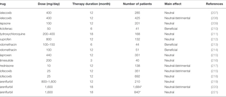
TABLE 1 | Double-blind, randomized, placebo-controlled trials using anti-inflammatory drugs in mild-to-moderate AD patients.
Drug Dose (mg/day) Therapy duration (month) Number of patients Main effect References
Celecoxib 400 12 285 Neutral (207) Celecoxib 400 12 425 Neutral/detrimental (208) Dapsone 100 12 201 Neutral (209) Diclofenac 50 6 41 Beneficial (210) Hydroxychloroquine 200–400 18 168 Neutral (211) Ibuprofen 800 12 132 Neutral (212) Indomethacin 100–150 6 44 Beneficial (213) Indomethacin 100 12 51 Beneficial (214) Naproxen 440 12 351 Neutral (215) Nimesulide 200 3 40 Neutral (216) Prednisone 10 12 138 Neutral/detrimental (217) Rofecoxib 25 12 351 Neutral/detrimental (215) Rofecoxib 25 12 692 Neutral (218) Tarenflurbil 800–1,600 12 210 Neutral (219) Tarenflurbil 1,600 18 1,684* Neutral/detrimental (220) Tarenflurbil 1,600 18 840* Neutral (221)
*Patients with mild AD. AD, Alzheimer’s disease.