Analysis of an Integrated Human Multiorgan
Microphysiological System for Combined
Tolcapone Metabolism and Brain Metabolomics
The MIT Faculty has made this article openly available. Please share how this access benefits you. Your story matters.
Citation Wang, Xin et al., "Analysis of an Integrated Human Multiorgan
Microphysiological System for Combined Tolcapone Metabolism and Brain Metabolomics." Analytical Chemistry 91, 13 (June 2019): p. 8667–75 doi. 10.1021/acs.analchem.9b02224 ©2019 Authors
As Published https://dx.doi.org/10.1021/ACS.ANALCHEM.9B02224
Publisher American Chemical Society (ACS)
Version Author's final manuscript
Citable link https://hdl.handle.net/1721.1/125910
Terms of Use Creative Commons Attribution-Noncommercial-Share Alike
1 1
2
3
Analysis of an Integrated Human Multi-organ Microphysiological
4
System for Combined Tolcapone Metabolism and Brain Metabolomics
5 6 7
Xin Wang,*,† Murat Cirit,† John S. Wishnok,† Linda G. Griffith,† and Steven R. Tannenbaum*,†, ‡
8 9 10
†Departments of Biological Engineering and ‡Chemistry, Massachusetts Institute of Technology,
11
Cambridge, Massachusetts 02139, United States
12 13 14 15 16 *Corresponding Author 17 Tel: (617) 253-3729. Fax: (617) 252-1787. 18
E-mail: srt@mit.eduor xinw@mit.edu;
19 20 21
2
ABSTRACT
1
Human-on-a-chip systems are rapidly advancing due to the availability of human stem cells from 2
a variety of tissues, but publications have utilized mostly simple methods of biochemical analysis. 3
Here, we apply mass spectrometry to a sophisticated multi-organ human-on-a-chip system for the 4
comprehensive study of tolcapone metabolite profiling and metabolomics. The developed human-5
on-a-chip includes seven interacting microphysiological systems (MPSs): brain, pancreas, liver, 6
lung, heart, gut, endometrium, with a mixer chamber for systemic circulation and tolcapone dose. 7
We investigated tolcapone metabolism by analyzing the circulating medium using mass 8
spectrometry. Twelve tolcapone metabolites were identified, three of which are newly reported. 9
These metabolites demonstrated that oxidation, reduction, and conjugation reactions were the most 10
important routes of tolcapone metabolism. In parallel, metabolomics in brain MPS evaluated the 11
tolcapone influences on endogenous pathways in human brain. Untargeted metabolomics 12
identified 18 key biomarkers significantly changed in human brain MPS after tolcapone dosing, 13
which were mainly associated with perturbation of tryptophan and phenylalanine metabolism (BH4 14
cycle), glycerophospholipid metabolism, energy metabolism, and aspartate metabolism. This is 15
the first example of successfully combining drug metabolism, metabolomics and cell engineering 16
to capture complex human physiology and the multi-organ interactions; the results we present here 17
could be a step toward using analytical chemistry to advance the utilization of human-on-a-chip 18
for testing both drug efficacy and toxicity in a single system. 19
20 21
3
INTRODUCTION
1
Prediction of drug metabolism, drug effects, and drug-drug interactions are mostly investigated in 2
animals and need to be validated in human models.1 However, limited access to human tissues, 3
especially those that have not been exposed to disease and drugs, has hampered these efforts.2 4
Because the scope of human studies is often limited by ethical and practical considerations, it is 5
important to develop a reliable drug testing platform that can reflect the complex drug metabolic 6
processes, the actual human drug responses and the multi-organ interactions in vivo.3 Over the last 7
decade, development of microphysiological systems (MPSs) aiming to represent relevant human 8
physiology and organ-specific functions has accelerated. In particular, “human-on-a-chip” systems 9
which integrate multi-MPS on microfluidic platforms are being developed to accurately simulate 10
human tissues and organs.4 The appropriately designed human-on-a-chip systems are able to link 11
MPSs within a fluidic platform capable of generating complex biodistribution profiles, which 12
provide improved in vitro tools and increased translational success for pharmacokinetics, 13
pharmacodynamics, toxicology and biomarker discovery.5-7 Recently, Edington et al. described 14
the development and implementation of a human-on-a-chip system with 7 interconnected MPSs, 15
including brain, pancreas, liver, lung, heart, gut and endometrium.8 This new 7-MPS platform is a 16
promising tool for fundamental biomedical research as well as practical applications such as testing 17
of drug efficacy and toxicity on human organs. 18
19
Tolcapone is a drug used to treat Parkinson's disease (PD). PD is a long-term degenerative disorder 20
of the central nervous system (CNS) that mainly affects the motor system. It is the second most 21
common neurodegenerative disease worldwide, and affects about 1% of adults over the age of 22
60.9,10 Currently, there is no cure for the disease. The gold standard treatment for PD symptoms is 23
levodopa, which has been a mainstay of PD treatment for almost 40 years. Levodopa is routinely 24
administered in combination with a peripheral amino acid decarboxylase inhibitor. Decarboxylase 25
inhibitors prevent conversion of levodopa to dopamine in the peripheral circulation, to allow more 26
levodopa to cross the blood-brain barrier and to reach the brain.11 By blocking the decarboxylase 27
route, levodopa is primarily metabolized by catechol-O-methyltransferase (COMT) to 3-O-28
methyldopa. To improve the pharmacokinetic profile of levodopa, selective COMT inhibitors have 29
been developed, and COMT inhibition in combination with levodopa is associated with an increase 30
4
of CNS bioavailability of levodopa. Theoretically, COMT inhibitors that are active in the CNS 1
would also reduce central metabolism of both levodopa and dopamine (Figure S-1).12,13 2
3
Tolcapone, as a potent, selective reversible inhibitor of COMT, improves clinical parameters of 4
levodopa, such as the increase in duration of ‘on’ and decrease of ‘off’ time; and prolongs the half-5
life from approximately 2 to 3.5 hours.14 These result in a doubling of levodopa relative 6
bioavailability. The tolcapone adjunctive therapy has been effective for patients who do not obtain 7
optimal response to levodopa-based therapy.12,15 Importantly, a recent paper16 demonstrated that 8
tolcapone is also a strong candidate for therapeutic intervention in other nervous system diseases, 9
such as fatal systemic amyloidoses. Tolcapone effects on the functions of subjective mood and 10
working memory performance were previously investigated.17,18 In addition, tolcapone was 11
reported to induce uncoupling of oxidative phosphorylation in mitochondria, thus significantly 12
reducing the cell's capacity to generate ATP.19 To the best of our knowledge, comprehensive 13
studies of tolcapone influences on the endogenous metabolites and metabolic pathways in human 14
CNS are still lacking. In order to gain additional knowledge about tolcapone, it is important to 15
thoroughly understand the drug metabolic fate and the dysregulated endogenous pathways. 16
17
Metabolomics, the “global” study of metabolite changes in a biological system, is particularly 18
conducive to identifying pathophysiologically affected processes and to elucidate novel 19
physiological and pathological mechanisms.20 Although endogenous metabolites are sensitive to 20
cellular changes and serve as the best indicators of cell states, studies for monitoring the alterations 21
of metabolites in human brain during tolcapone treatment have not yet been conducted; most 22
metabolomic analyses to date have focused on plant, tissue, and biofluid samples. However, the 23
diverse potential of metabolomics in many fields, including cell engineering, has made it a 24
universal tool for industrial, medical and research purposes.21 So here, metabolomics has been 25
applied to the brain MPS in human-on-a-chip system for the first time to evaluate metabolic 26
signatures and pathways in human brain that were altered significantly by tolcapone. Liquid 27
chromatography - mass spectrometry (LC-MS) was used to identify novel biomarkers, and to 28
elucidate the dysregulated metabolic pathways and tolcapone-associated molecular mechanisms. 29
30
EXPERIMENTAL SECTION
315 Chemicals and Reagents
1
Acetonitrile (ACN), methanol, formic acid (FA) of LC grade were obtained from Sigma-Aldrich 2
(MO, USA). Ultrapure water was made using an in-house purification system. Tolcapone, choline 3
chloride, hippuric acid, N-(2-phenylacetyl)glycine, N-acetyl-L-aspartic acid, L-dopa and 4
spermidine were all purchased from Sigma-Aldrich (MO, USA). N-acetylaspartic acid-2,3,3-D3 5
was from CDN Isotopes Inc (Quebec, Canada). All culture media (mentioned below) were 6
obtained from Thermo Fisher Scientific (NJ, USA) for cell-related experiments. 7
8
Formation of 3D Neural Constructs 9
Neural progenitor cells (NPCs) derived from the human H1 ES line were a gift from James 10
Thomson, University of Wisconsin-Madison. NPCs were grown in neural expansion medium8 at 11
a density of 50,000 cells / PEG-transwell. The PEG transwells were prepared using thiol-ene 12
photopolymerization chemistry from a published protocol.22 Cells were allowed to attach 13
overnight, medium was changed every 48 hours using 1 mL in the basal compartment, and 200 µL 14
in the apical compartment to avoid damaging the development of neural tissue constructs. Neural 15
constructs were cultured 14 days for the expansion and differentiation of neuronal and glial 16
subpopulations, confirmed by increased immunostaining for neural marker β3-tubulin and 17
astrocyte marker GFAP.23 18
19
Human-on-a-chip Platform 20
On day 14, the transwell insert of NPCs was transferred to the human-on-a-chip platform, 21
constructed by the DARPA Microphysiological Systems Program, which fluidically interconnects 22
the engineered human MPSs of brain, pancreas, liver, lung, heart, gut and endometrium in a 23
perfused capillary bed to mimic human physiological systems. This system was composed of three 24
layers: a polysulfone plastic top (fluidic) plate, a polyurethane membrane layer, and an acrylic 25
bottom (pneumatic) plate. The top plate included seven cell culture chambers, five of which were 26
designed for standard Transwell® inserts and used for brain, gut, lung, endometrium and heart 27
MPSs; two others were designed according to the perfused LiverChip® module24,25 and adapted 28
for liver and pancreas cells, with an extra mixer chamber for systemic circulation (Figure 1A). To 29
mimic the actual multi-organ interactions in vivo, microfluidic channels and pumps were machined 30
6
into the underside of the top plate to deliver fluid to each MPS compartment with the flow 1
partitioning mirroring physiological cardiac output (Figure 1B). A published Transwell®-format 2
brain MPS23 was used for the human-on-a-chip system, the basal medium samples were mixed on 3
the platform while the apical medium samples still retained the characteristics of brain. For 4
additional details, please refer to our 7-MPS article.8 5
6
Figure 1. Human-on-a-chip system. (A) The human-on-a-chip platform fluidically interconnects the 7
engineered human MPSs of brain, pancreas, liver, lung, heart, gut and endometrium in a perfused capillary
8
bed to mimic human physiological systems, and (B) fluid flow paths of the 7-MPS.
9 10
Tolcapone with the Cmax = 7.6 µg/mL26 was added to the mixer chamber at day 22 and at day 23. 11
After 2 days of continuous exposure, at day 24, the experiment was stopped and the medium 12
samples from mixer chamber, liver chamber and brain chamber were collected for analysis. 13
Additionally, the apical medium in brain MPS was used to study the metabolomics in the 14
tolcapone-dosed human-on-a-chip system. Control samples were obtained in the same way but 15
without tolcapone dosing. A list of samples used in this study is in Table S-1. 16
17
Sample Preparation 18
Samples were prepared using methanol extraction. 100 μL medium was extracted in 400 μL cold 19
methanol spiked with 50 μM N-acetylaspartic acid-2,3,3-D3 as internal standard. After 1 min 20
vortexing, the sample was incubated for 10 min at 4 °C and vortexed for another 10 min, then 21
centrifuged at 15 000 rpm for 15 min. The supernatant was collected, dried in a SpeedVac®, and 22
redissolved in 30 μL of 98:2 water/acetonitrile (v/v) for LC-MS analysis. 23
24
Tolcapone Metabolism Study 25
7
LC-MS experiments were performed on an Agilent 1290 series LC system coupled with an Agilent 1
6530 High-Resolution Accurate Mass Quadrupole Time-of-Flight (QTOF) mass spectrometer 2
(Agilent Technologies, CA, USA). Samples were analyzed in both positive and negative modes 3
using an Agilent SB-C18 reverse phase column (1.8 μm, 2.1 mm × 50 mm). The optimum mobile 4
phases were water with 0.1% formic acid (A) and acetonitrile with 0.1% formic acid (B) at a flow 5
rate of 0.4 mL/min. Gradient elution was used during the separation (solution B: 2-70% in 7 min, 6
70-100% in 5 min, 100% for 3 min, 100-2% in 1 min, post time for 4 min). The column temperature 7
was 50 °C with an injection volume of 5 μL. 8
9
The parameters of the Agilent Dual AJS ESI source were set as follows: gas temperature, 350 ºC; 10
dry gas flow, 12 L/min; nebulizer, 50 psig; sheath gas temperature, 380 ºC; sheath gas flow, 12 11
L/min; capillary voltage, 3.5 kV; fragmentor, 120 V; and skimmer, 65 V. The m/z scan range was 12
from 100 to 1100 with an acquisition rate of 2 spectra/s. Potential tolcapone metabolites were first 13
detected by MS and further confirmed by tandem mass spectrometry (MS/MS) analysis. The ions 14
that were present in the tolcapone-dosed samples, but absent in the control samples, were collected 15
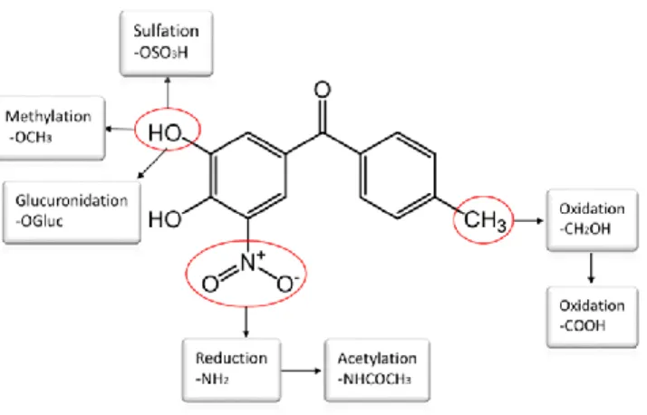
as tolcapone metabolites related ions. Figure 2 shows the possible routes of tolcapone metabolism 16
in the human body; the biotransformation of tolcapone is confined exclusively to the periphery of 17
the molecule with the benzophenone nucleus remaining unaltered.27 Next, comparison between 18
the collected metabolite-related ions with the possible tolcapone metabolite formulas was 19
conducted, and the ions within 10 ppm of theoretical m/z values were stored for MS/MS 20
confirmation. 21
22
MS/MS analysis was performed using the Agilent ESI-QTOF MS under the same LC-MS 23
parameters and with 20 V collision energy. The first Q was operated in the mass filter mode to 24
transmit only the parent ion of the putative tolcapone metabolite. This potential metabolite then 25
underwent collision induced dissociation and entered the TOF to obtain the m/z values of the 26
fragments. Data were acquired by Agilent MassHunter Data Acquisition B.05.00 (CA, USA) and 27
analyzed by Agilent MassHunter Qualitative Analysis B.06.00 (CA, USA) to generate the MS/MS 28
spectra. The ESI-QTOF was calibrated daily using the standard tuning solution from Agilent. 29
During analysis, the instrument was calibrated in real time with two different reference masses 30
(m/z 121.0509, 922.0098 for positive mode and m/z 112.9856, 1033.9881 for negative mode) with 31
8
6 μL/min constant infusion. The MS/MS spectra were interpreted manually, using the previously 1
published method,28 with the help of ChemDraw (version 18.0) Fragmentation Tools. 2
3
Metabolomics of Tolcapone-dosed Human-on-a-chip System 4
The LC-MS separation and detection conditions were the same as those in the tolcapone 5
metabolism study. Nine control samples and nine tolcapone-dosed samples obtained from separate 6
chips were used for metabolomics. Data acquired in Agilent .d format were analyzed using the 7
Molecular Feature Extraction (MFE) tool from Mass Hunter Qualitative Analysis (B.06.00) to 8
obtain the molecular features. “Small molecules (chromatographic)” extraction algorithm was 9
used. The features were characterized by retention time, chromatographic peak intensity and 10
accurate mass, with the quality score ≥ 80. To identify different ion species coming from the same 11
metabolite, H+, Na+, and K+ adducts were considered for positive ionization, while the H- and 12
HCOO− adducts were considered for negative ionization. The extracted features were then 13
analyzed with Agilent Mass Profiler Professional (MPP) software (B.12.05) for peak picking, 14
alignment and internal standard normalization. Only features with an intensity larger than 10,000 15
counts and detectable in 80% of the subjects in at least one of the treatment groups were kept for 16
further processing.29 Data were aligned with a 15 ppm mass tolerance and a retention-time window 17
tolerance of 0.15 min, and normalized to spiked N-acetylaspartic acid-2,3,3-D3 internal standard. 18
19
Next, principal component analysis (PCA) was carried out with the MPP (B.12.05) statistical 20
package. Unpaired t-test was used to find endogenous metabolites differing between the control 21
group and the tolcapone-dosed group; metabolites with fold change (FC) larger than 1.5 and p-22
value smaller than 0.05 were considered to be statistically significant.30 The exact masses of 23
putative compounds with significant changes were searched against the Human Metabolome 24
Database (HMDB, version 4.0, http://www.hmdb.ca), METLIN (version 3.7.1, 25
http://metlin.scripps.edu), and MassBank (version 2.0, http://www.massbank.jp). The matched 26
exact masses were stored for further MS/MS identification of metabolites. 27
28
MS/MS spectra were also generated on the Agilent QTOF 6530 mass spectrometer to confirm the 29
identity of metabolites. A targeted list, which included the previously determined exact masses 30
according to database search results, was generated for MS/MS analysis. Targeted MS/MS mode 31
9
was carried out with collision energies of 20 V and 40 V. Nitrogen was the collision gas. Lastly, 1
the commercially available metabolites were also confirmed by comparison with the chemical 2
standards. The metabolites are reported according to the criteria recommended by Metabolomics 3
Standard Initiative (MSI), which defined four levels of metabolite identification. These included 4
identified metabolites with reference standards (level 1), putatively annotated compounds (level 5
2), putatively characterized compound classes (level 3), and unknown compounds (level 4).31,32 6
7
Metabolic pathway analysis was conducted by MetaboAnalyst (version 4.0, 8
www.metaboanalyst.ca) to sort the significantly changed metabolites associated with tolcapone 9
treatment into biological pathways. The pathway library of Homo sapiens (KEGG) was selected 10
for the study. We considered a pathway as significant if its p-value was less than a designated 11
cutoff (i.e., 0.05). MetaboAnalyst is a web-based platform for comprehensive analysis of 12
metabolomic data, which aids in the visualization of metabolites within the context of metabolic 13
pathways.33-36 14
15
RESULTS AND DISCUSSION
16Tolcapone Metabolism in Systemic Circulation 17
To demonstrate that our human-on-a-chip platform can reflect human drug responses in vivo, we 18
first investigated the tolcapone metabolic fate by analyzing the circulated medium. Figure 2 shows 19
the structural features and possible routes of tolcapone metabolism. Given the variety of metabolic 20
pathways for tolcapone, it is theoretically possible that some of the metabolites may be toxic either 21
directly or through the formation of reactive species.27 Identification of tolcapone metabolites has 22
therefore been a topic of both pharmaceutical and toxicological interest. 23
10 1
Figure 2. Structure features and possible routes of tolcapone metabolism in the human body. 2
3
The circulated medium samples from the mixer chamber were analyzed by LC-MS to determine 4
tolcapone metabolic processes. Both positive and negative ionization modes were used due to the 5
different ionization efficiencies of possible metabolites. The principal metabolic pathways of 6
tolcapone are illustrated in Figure 3. All 12 metabolites were identified by accurate mass and 7
confirmed by MS/MS analysis. The primary metabolic pathways undergone by tolcapone in the 8
system were oxidative hydroxylation (M2) and reduction reactions (M3), as well as the conjugative 9
reactions involving glucuronidation (M4) and sulfation (M5). M2 and its sulfate (M6) and methyl 10
(M8) conjugates were subsequently oxidized to the carboxylic acids (M7, M9 and M10). In 11
addition, M11 was derived from M3, and oxidative hydroxylation of M4 further produced M12. 12
The metabolites M1, M2, M3, M4, M7 and M11 were all detected in human biofluids after oral 13
administration of tolcapone,26 reflecting that the human-on-a-chip system can successfully mimic 14
major human metabolic features. The metabolites M5, M6, M10 have also been reported in rat 15
urine after tolcapone treatment.28 To our knowledge, metabolites M8, M9 and M12 have not been 16
previously reported. 17
11 1
Figure 3. Potential routes of tolcapone metabolism in the circulated medium of human-on-a-chip system. 2
All metabolites were identified with exact mass measurement, and MS/MS confirmation. Metabolites
3
labeled with * were newly identified in this study, metabolites labeled with § were reported in rat but new
4
in human samples.
5 6
All these tolcapone metabolites were identified with a combination of exact mass measurement 7
and MS/MS confirmation. The tandem mass spectra of three newly-found metabolites (M8, M9, 8
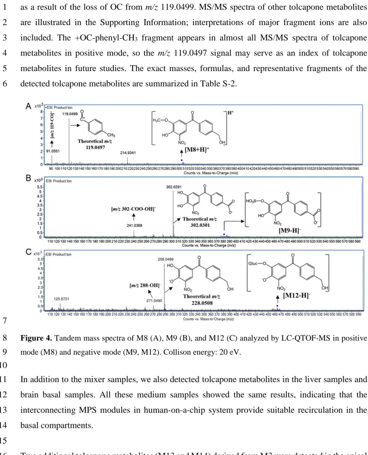
M12) are illustrated in Figure 4. Taking M8 as an example, as shown in Figure 4A, the base peak 9
at m/z 119.0499 represents the +OC-phenyl-CH3 fragment, while the ion of 91.0551 was produced 10
12
as a result of the loss of OC from m/z 119.0499. MS/MS spectra of other tolcapone metabolites 1
are illustrated in the Supporting Information; interpretations of major fragment ions are also 2
included. The +OC-phenyl-CH3 fragment appears in almost all MS/MS spectra of tolcapone 3
metabolites in positive mode, so the m/z 119.0497 signal may serve as an index of tolcapone 4
metabolites in future studies. The exact masses, formulas, and representative fragments of the 5
detected tolcapone metabolites are summarized in Table S-2. 6
7
Figure 4. Tandem mass spectra of M8 (A), M9 (B), and M12 (C) analyzed by LC-QTOF-MS in positive 8
mode (M8) and negative mode (M9, M12). Collison energy: 20 eV.
9 10
In addition to the mixer samples, we also detected tolcapone metabolites in the liver samples and 11
brain basal samples. All these medium samples showed the same results, indicating that the 12
interconnecting MPS modules in human-on-a-chip system provide suitable recirculation in the 13
basal compartments. 14
15
Two additional tolcapone metabolites (M13 and M14) derived from M3 were detected in the apical 16
brain MPS. As shown in Figure S-2A, M3 was converted to M13 by acetylation of the 5-amino 17
13
group, and M14 was produced by the reaction of M13 and glutathione (GSH). This reaction may 1
have two steps; M13 is first oxidized to the reactive quinone-imine intermediate, which is then 2
trapped with GSH to form GSH conjugate (M14) (Figure S-2B). Theoretically, M1, M2, M3, M7 3
and M13 all could carry out this reaction by conversion to their bioactive o-quinone or quinone-4
imine species, but only M14 (production of M13) was observed in the study. This GSH conjugation 5
reaction is supportive evidence for the formation of reactive o-quinone or quinone-imine species, 6
which are predicted to play a role in tolcapone-induced toxicity.37,38 7
8
Effects of Tolcapone on Apical Brain MPS Metabolomics 9
Tolcapone metabolites are expected to be detected in the post-dose samples but absent in control 10
samples. Endogenous compounds, however, would be expected in both control and dosed samples 11
but possibly with altered concentrations responding to tolcapone. Endogenous metabolites are 12
sensitive to cellular changes and these altered metabolites could serve as indicators of cell states. 13
The metabolomic study of tolcapone-dosed human-on-a-chip system is mainly focused on the 14
changes of endogenous metabolites in human brain MPS (apical medium) during tolcapone 15
treatment. 16
17
Using the LC-QTOF-MS method, 5670 putative compounds were extracted. Among them, 1115 18
putative compounds were significantly changed (FC > 1.5, p < 0.05), with the majority being down 19
regulated (Table S-3). Principal component analysis (PCA) was then performed to highlight the 20
metabolic differences between the control and tolcapone-dosed samples. As shown in Figure 5, 21
PCA revealed excellent separations of the two sample groups under both positive and negative 22
modes. 23
24
Figure 5. Separation of tolcapone-dosed human brain MPS samples (blue) and control samples without 25
tolcapone dosing (red) by principal component analysis under both positive (A) and negative (B) modes,
14
with the first three principal components account for 86.9% (A) and 91.1% (B) of the variance in analysis.
1
Each dot represents a sample and each color represents the type of the sample.
2 3
Identification of the significantly changed metabolites was carried out by database searches on 4
exact masses and MS/MS analysis. As an example, argininosuccinic acid decreased 2.43-fold in 5
the tolcapone-dosed samples. A search of the exact mass of 290.1234 (or m/z 291.1312) with ±10 6
ppm mass window generates 3-4 hits depending on different databases. MS/MS analysis was then 7
conducted in positive mode and the result is illustrated in Figure S-3. The characteristic product 8
ions of argininosuccinic acid at m/z 70.0656 and m/z 116.0703 confirmed the identity. The 9
commercially available metabolites, such as N-acetyl-L-aspartic acid, were also confirmed by 10
comparison with the chemical standards. In total, we identified 18 MSI Levels 1 and 2 metabolites 11
that were significantly changed between control and the tolcapone-dosed human brain MPS 12
samples. These metabolites were all confirmed by MS/MS analysis and are summarized in Table 13
1. 14 15
Table 1. Significantly changed metabolites in the apical brain MPS of tolcapone-dosed human-on-a-chip 16 system 17 Metabolitea HMDB Exact mass Fold
changeb p value Representative fragments (m/z)c Pathway
Indoleacetaldehyde 01190 159.0681 -1.61 2.24*10-3 103.0542, 130.0649, 142.0653* Tryptophan metabolism
Serotonin 00259 176.0947 -2.46 1.39*10-4 132.0769, 160.0733* Tryptophan metabolism
Acetyl-N-formyl-5-methoxykynurenamine 04259 264.1097 -3.47 1.03*10
-3 73.0525, 122.0611, 160.0753* Tryptophan metabolism
Anthranilic acid 01123 137.0477 3.08 2.37*10-4 65.0375, 94.0628, 120.0393* Tryptophan metabolism
Formyl-5-hydroxykynurenamine 12948 208.0844 -1.91 1.0*10 -3 150.0547, 164.0707, 192.0649* Tryptophan metabolism Hippuric acid† 00714 179.0575 -4.83 7.62*10 -7 77.0387, 105.0332* Phenylalanine metabolism -3.93 5.84*10-6 134.0603§ Phenylacetylglycine† 00821 193.0732 -2.24 1.37*10 -5 76.0373, 91.0520* Phenylalanine metabolism -2.48 9.75*10-5 74.0235§
L-dopa† 00181 197.0686 1.65 5.67*10-3 152.0706, 181.0490* Phenylalanine metabolism
Choline† 00097 103.1001 2.23 1.97*10-2 58.0658, 60.0814* Glycerophospholipid LysoPC(14:0) 10379 467.3010 4.87 5.64*10-4 78.9586, 227.2039, 452.2810§ Glycerophospholipid LysoPC(18:3) 10387 517.3140 1.72 4.10*10-2 96.9691, 242.0780§ Glycerophospholipid PE(35:3) 09351/ 08903 727.5125 -3.70 4.78*10 -3 305.2499, 502.2963§ Glycerophospholipid Dihydroxyacetone phosphate 01473 169.9981 -2.33 6.93*10-6 78.9588, 96.9692§ Glycerophospholipid Glycolysis and gluconeogenesis cis-Aconitic acid 00072 174.0167 -2.93 8.82*10-4 68.9970, 85.0303§ TCA cycle
Oxalacetic acid 00223 132.0051 -2.09 7.05*10-6 59.0130, 87.0079§ TCA cycle
Aspartate metabolism Argininosuccinic acid 00052 290.1234 -2.43 6.16*10-7 70.0656, 116.0703* Aspartate metabolism
15
a Metabolites labeled with † were verified with authentic standards (MSI Level 1). b Fold change was calculated from average values of
tolcapone-1
dosed samples and control samples. Fold change with a positive value indicates up-regulated metabolite in tolcapone-dosed samples, while a 2
negative value indicates down-regulated metabolite in tolcapone-dosed samples. c Fragments labeled with * were analyzed by MS/MS in positive
3
mode (ESI+), fragments labeled with § were analyzed by MS/MS in negative mode (ESI-). 4
5
Metabolic pathway analysis was then conducted for these 18 altered MSI Levels 1 and 2 6
metabolites. Using MetaboAnalyst 4.0 and KEGG databases, eight metabolic pathways were 7
found to be perturbed (Table S-4). The most relevant ones were glycerophospholipid metabolism, 8
tryptophan metabolism, aspartate metabolism, energy metabolism, and phenylalanine metabolism. 9
These newly detected metabolites and related pathways reveal the metabolic implications of 10
tolcapone treatment, which are important for our understanding of tolcapone and its influences on 11
the human brain. 12
13
As a COMT inhibitor, tolcapone affects central metabolism of L-dopa and would cause 14
compensatory effects on the metabolism of other neurotransmitters.13 Perturbed tryptophan 15
metabolism and phenylalanine metabolism were observed, which most likely correspond to altered 16
tetrahydrobiopterin BH4 cycle (Figure S-4). Most of the BH4 comes from the methylation cycle. 17
Methylation of BH2 into BH4 provides the co-factor necessary to turn tryptophan and tyrosine into 18
their respective neurotransmitters serotonin and dopamine.39 The involvement of the BH4 cycle 19
was further studied by analysis of the related metabolites of tryptophan, phenylalanine, tyrosine, 20
serotonin, and L-dopa. These metabolites were all detected in the brain medium, and levels of 21
serotonin and L-dopa were significantly changed (Table 1). Tryptophan, phenylalanine, and 22
tyrosine were the components of the brain culture medium with relatively high concentrations, so 23
their FC data were lower than 1.5. Previous studies from Misu et al. demonstrated that L-dopa is 24
an endogenous neurotransmitter or neuromodulator in the CNS.40,41 The increased L-dopa level 25
(+1.65 fold) indicated that the COMT inhibitor tolcapone is active in brain MPS. This also implied 26
the successful establishment of the human brain model.23 As shown in Figure S-4, the altered L-27
dopa level could affect BH4 cycle, which would further perturb tryptophan metabolism and 28
phenylalanine metabolism. 29
30
N-Acetyl aspartic acid† 00812 175.0489 -1.63 4.65*10-4 58.0301, 88.0401§ Aspartate metabolism
16
The decreased levels of hippuric acid and phenylacetylglycine observed in this study indicated 1
dysregulated phenylalanine metabolism after tolcapone dosing (Figure 6A). The pathways from 2
tryptophan to serotonin, to tryptamine and to kynurenine were all perturbed (Figure 6B). The 3
decreased level of serotonin, formyl-5-hydroxykynurenamine and acetyl-N-formyl-5-4
methoxykynurenamine indicated the inhibition of serotonin pathway, which may be ascribed to 5
the decreased tryptophan hydroxylase (TPH) activity. At the same time, the tryptamine pathway 6
was also inhibited (indoleacetaldehyde, -1.61 fold) while the kynurenine pathway was activated 7
(anthranilic acid, +3.08 fold). These observations can be explained as the result of three 8
competitive metabolism pathways for tryptophan where inhibition of one pathway also caused 9
changes to the other two pathways. 10
11
As shown in Figure 6C, glycerophospholipid metabolism was impacted by tolcapone. The level of 12
phosphatidylethanolamine was decreased, while that of lysophosphatidylcholine was increased. 13
These results indicated that phospholipase A2 (PLA2) activity in human brain was elevated after 14
tolcapone dosing.42 In neural membranes, PLA2 activity is linked to dopamine,43,44 so an 15
abnormality in dopamine metabolism induced by tolcapone may be responsible for this 16
stimulation. The decrease of phosphatidylethanolamine caused the down-regulation of 17
dihydroxyacetone phosphate, while the increase of lysophosphatidylcholine led to the up-18
regulation of choline. On the other hand, phospholipase activation has been proposed as an 19
important mechanism of cell killing because increased phospholipase activity can mediate 20
pathophysiological reactions by accelerating membrane breakdown or by generating toxic 21
metabolites.45 Therefore, the altered glycerophospholipid metabolism may suggest tolcapone-22
induced neurotoxicity. 23
24
Previous publications revealed that tolcapone could uncouple mitochondrial respiration by 25
carrying protons across the inner mitochondrial membrane and thus impair energy metabolism.46,47 26
In our study, the decreased levels of dihydroxyacetone phosphate, cis-aconitic acid and oxalacetic 27
acid also suggested disrupted energy metabolism (Figure 6D). Similar effects were observed by 28
Borroni et al., who reported that tolcapone reduced the mitochondrial membrane potential, and 29
influenced cellular respiration and mitochondrial oxygen consumption.27 Related metabolites in 30
the citrate cycle, and glycolysis were consumed and the amounts detected in cell medium were 31
17
decreased. At the same time, aspartate metabolism was perturbed by tolcapone treatment, 1
evidenced by decreased N-acetyl aspartic acid (NAA), argininosuccinic acid and oxalacetic acid. 2
Aspartate metabolism is at the upstream of TCA cycle, the altered TCA cycle may be the reason 3
of the altered aspartate metabolism. Additionally, NAA is a well-known neuron-specific marker 4
associated with neuronal cell loss,48 so the significantly decreased NAA level also indicates the 5
neurotoxic effects of tolcapone. 6
7
Figure 6. Untargeted metabolomics of the apical brain MPS revealed metabolic pathways perturbed by 8
tolcapone dose in human brain. (A) Phenylalanine metabolism. (B) Tryptophan metabolism. (C)
18
Glycerophospholipid metabolism. (D) Energy metabolism and aspartate metabolism (the up-regulated
1
metabolites are labeled in red, while the down-regulated metabolites are labeled in green).
2 3
CONCLUSIONS
4In conclusion, we have presented a comprehensive mass spectrometric study on tolcapone 5
metabolite profiling and metabolomics in a tolcapone-dosed human-on-a-chip system. Three new 6
tolcapone metabolites were identified, and the biological pathways perturbed by tolcapone 7
treatment were reported in the brain MPS. The integration of tolcapone metabolism with 8
metabolomics not only provides a powerful approach to identify tolcapone metabolites and 9
endogenous biomarkers, but also widens our insights into the metabolic pathways of tolcapone 10
itself and other pathways perturbed by tolcapone treatment, which are important for the 11
investigation of tolcapone efficacy and toxicity in human brain. Direct studies focusing on the 12
human brain, however, are difficult to plan and conduct for ethical and practical reasons. The 13
human-on-a-chip platform for combined drug metabolism and brain biochemistry is therefore an 14
attractive surrogate for such investigations. This strategy of combining drug metabolism, 15
metabolomics and cell engineering opens a new window for applying analytical chemical methods 16
to evaluate human responses to xenobiotics and other insults. 17 18
ASSOCIATED CONTENT
19 Supporting Information 20The Supporting Information is available free of charge via the Internet at http://pubs.acs.org.
21
MS/MS results of tolcapone metabolites in systemic circulation with the interpretations of major
22
fragment ions, additional figures as described in the text (Figure S-1 to Figure S-4), summary of sampling
23
location, molecular features comparison and pathway analysis.
24 25
AUTHOR INFORMATION
26 Corresponding Author 27*MIT 56-731A, 32 Vassar St, Cambridge, MA, 02139. Tel: (617) 253-3729. Fax: (617) 252-1787. E-mail:
28
srt@mit.edu; xinw@mit.edu.
19 Notes
1
The authors declare no competing financial interest.
2 3
ACKNOWLEDGMENTS
4This work was supported by the U.S. Army Research Office and the Defense Advanced Research Project
5
Agency under Cooperative Agreement Number W911NF-12-2-0039.
6 7
REFERENCES
8(1) Long, T. J.; Cosgrove, P. A.; Dunn, R. T.; Stolz, D. B.; Hamadeh, H.; Afshari, C.; McBride, H.; Griffith, L.
9
G. Drug Metab. Dispos. 2016, 44, 1940-1948.
10
(2) Pirnay, J. P.; Baudoux, E.; Cornu, O.; Delforge, A.; Delloye, C.; Guns, J.; Heinen, E.; Van den Abbeel, E.;
11
Vanderkelen, A.; Van Geyt, C.; van Riet, I.; Verbeken, G.; De Sutter, P.; Verlinden, M.; Huys, I.; Cockbain, J.;
12
Chabannon, C.; Dierickx, K.; Schotsmans, P.; De Vos, D.; Rose, T.; Jennes, S.; Sterckx, S. Embo Rep. 2015, 16,
13
557-562.
14
(3) Li, Z. Y.; Guo, Y. Q.; Yu, Y.; Xu, C.; Xu, H.; Qin, J. H. Integr. Biol. 2016, 8, 1022-1029.
15
(4) Maass, C.; Stokes, C. L.; Griffith, L. G.; Cirit, M. Integr. Biol. 2017, 9, 290-302.
16
(5) Abaci, H. E.; Shuler, M. L. Integr. Biol. 2015, 7, 383-391.
17
(6) Esch, E. W.; Bahinski, A.; Huh, D. Nat. Rev. Drug Discov. 2015, 14, 248-260.
18
(7) Tsamandouras, N.; Chen, W. L. K.; Edington, C. D.; Stokes, C. L.; Griffith, L. G.; Cirit, M. Aaps J. 2017,
19
19, 1499-1512. 20
(8) Edington, C. D.; Chen, W. L. K.; Geishecker, E.; Kassis, T.; Soenksen, L. R.; Bhushan, B. M.; Freake, D.;
21
Kirschner, J.; Maass, C.; Tsamandouras, N.; Valdez, J.; Cook, C. D.; Parent, T.; Snyder, S.; Yu, J. J.; Suter, E.;
22
Shockley, M.; Velazquez, J.; Velazquez, J. J.; Stockdale, L.; Papps, J. P.; Lee, I.; Vann, N.; Gamboa, M.;
23
LaBarge, M. E.; Zhong, Z.; Wang, X.; Boyer, L. A.; Lauffenburger, D. A.; Carrier, R. L.; Communal, C.;
24
Tannenbaum, S. R.; Stokes, C. L.; Hughes, D. J.; Rohatgi, G.; Trumper, D. L.; Cirit, M.; Griffith, L. G. Sci. Rep.
25
2018, 8, 4530-4547.
26
(9) Alvarez, F. J. Expert Rev. Neurother. 2016, 16, 1023-1032.
27
(10) Leegwater-Kim, J.; Waters, C. Expert Rev. Neurother. 2007, 7, 1649-1657.
28
(11) Poewe, W.; Antonini, A.; Zijlmans, J. C. M.; Burkhard, P. R.; Vingerhoets, F. Clin. Interv. Aging 2010, 5,
29
229-238.
30
(12) Stocchi, F.; De Pandis, M. F. Clin. Interv. Aging 2006, 1, 317-325.
31
(13) Gordin, A.; Brooks, D. J. J. Neurol. 2007, 254, 37-48.
32
(14) Borges, N. Expert Opin. Drug Saf. 2005, 4, 69-73.
20
(15) Detrait, E. R.; Carr, G. V.; Weinberger, D. R.; Lamberty, Y. Behav. Pharmacol. 2016, 27, 415-421.
1
(16) Sant'Anna, R.; Gallego, P.; Robinson, L. Z.; Pereira-Henriques, A.; Ferreira, N.; Pinheiro, F.; Esperante, S.;
2
Pallares, I.; Huertas, O.; Almeida, M. R.; Reixach, N.; Insa, R.; Velazquez-Campoy, A.; Reverter, D.; Reig, N.;
3
Ventura, S. Nat. Commun. 2016, 7, 10787-10799.
4
(17) Roussos, P.; Giakoumaki, S. G.; Bitsios, P. Biol. Psychiatry 2009, 66, 997-1004.
5
(18) Farrell, S. M.; Tunbridge, E. M.; Braeutigam, S.; Harrison, P. J. Biol. Psychiatry 2012, 71, 538-544.
6
(19) Truong, D. D. Clin. Interv. Aging 2009, 4, 109-113.
7
(20) Johnson, C. H.; Gonzalez, F. J. J. Cell. Physiol. 2012, 227, 2975-2981.
8
(21) Khoo, S. H. G.; Al-Rubeai, M. Biotechnol. Appl. Biochem. 2007, 47, 71-84.
9
(22) Fairbanks, B. D.; Schwartz, M. P.; Halevi, A. E.; Nuttelman, C. R.; Bowman, C. N.; Anseth, K. S. Adv.
10
Mater. 2009, 21, 5005-5010. 11
(23) Schwartz, M. P.; Hou, Z. G.; Propson, N. E.; Zhang, J.; Engstrom, C. J.; Costa, V. S.; Jiang, P.; Nguyen, B.
12
K.; Bolin, J. M.; Daly, W.; Wang, Y.; Stewart, R.; Page, C. D.; Murphy, W. L.; Thomson, J. A. Proc. Natl. Acad.
13
Sci. USA 2015, 112, 12516-12521. 14
(24) Powers, M. J.; Domansky, K.; Kaazempur-Mofrad, M. R.; Kalezi, A.; Capitano, A.; Upadhyaya, A.;
15
Kurzawski, P.; Wack, K. E.; Stolz, D. B.; Kamm, R.; Griffith, L. G. Biotechnol. Bioeng. 2002, 78, 257-269.
16
(25) Sarkar, U.; Ravindra, K. C.; Large, E.; Young, C. L.; Rivera-Burgos, D.; Yu, J. J.; Cirit, M.; Hughes, D. J.;
17
Wishnok, J. S.; Lauffenburger, D. A.; Griffith, L. G.; Tannenbaum, S. R. Drug Metab. Dispos. 2017, 45,
855-18
866.
19
(26) Jorga, K.; Fotteler, B.; Heizmann, P.; Gasser, R. Br. J. Clin. Pharmacol. 1999, 48, 513-520.
20
(27) Borroni, E.; Cesura, A. M.; Gatti, S.; Gasser, R. Funct. Neurol. 2001, 16, 125-134.
21
(28) Sun, J. C.; Von Tungeln, L. S.; Hines, W.; Beger, R. D. J. Chromatogr. B 2009, 877, 2557-2565.
22
(29) Bijlsma, S.; Bobeldijk, L.; Verheij, E. R.; Ramaker, R.; Kochhar, S.; Macdonald, I. A.; van Ommen, B.;
23
Smilde, A. K. Anal. Chem. 2006, 78, 567-574.
24
(30) Xi, B.; Gu, H.; Baniasadi, H.; Raftery, D. Mass spectrometry in metabolomics: methods and protocols;
25
Humana Press, 2014.
26
(31) Salek, R. M.; Steinbeck, C.; Viant, M. R.; Goodacre, R.; Dunn, W. B. Gigascience 2013, 2, 13-15.
27
(32) Naz, S.; Gallart-Ayala, H.; Reinke, S. N.; Mathon, C.; Blankley, R.; Chaleckis, R.; Wheelock, C. E. Anal.
28
Chem. 2017, 89, 7933-7942. 29
(33) Xia, J.; Wishart, D. S. Curr. Protoc. Bioinformatics 2016, 55, 14.10. 1-14.10. 91.
30
(34) Xia, J.; Sinelnikov, I. V.; Han, B.; Wishart, D. S. Nucleic Acids Res. 2015, 43, W251-W257.
31
(35) Xia, J.; Wishart, D. S. Nat. Protoc. 2011, 6, 743-760.
32
(36) Weng, R.; Shen, S. S.; Tian, Y. L.; Burton, C.; Xu, X. Y.; Liu, Y.; Chang, C. L.; Bai, Y.; Liu, H. W. Sci.
33
Rep. 2015, 5,11864-11876.
34
(37) Hughes, T. B.; Swamidass, S. J. Chem. Res. Toxicol. 2017, 30, 642-656.
21
(38) Smith, K. S.; Smith, P. L.; Heady, T. N.; Trugman, J. M.; Harman, W. D.; Macdonald, T. L. Chem. Res.
1
Toxicol. 2003, 16, 123-128. 2
(39) Werner, E. R.; Blau, N.; Thoeny, B. Biochem. J. 2011, 438, 397-414.
3
(40) Misu, Y.; Goshima, Y. Trends Pharmacol. Sci. 1993, 14, 119-123.
4
(41) Misu, Y.; Goshima, Y. Neurobiology of DOPA as a Neurotransmitter; CRC Press, 2005.
5
(42) Piomelli, D.; Astarita, G.; Rapaka, R. Nat. Rev. Neurosci. 2007, 8, 743-754.
6
(43) Kudo, I.; Matsuzawa, A.; Imai, K.; Murakami, M.; Inoue, K. J. Lipid Mediators Cell Signalling 1996, 14,
7
25-31.
8
(44) Farooqui, A. A.; Yang, H. C.; Rosenberger, T. A.; Horrocks, L. A. J. Neurochem. 1997, 69, 889-901.
9
(45) Klein, J. J. Neural. Transm. 2000, 107, 1027-1063.
10
(46) Korlipara, L. V. P.; Cooper, J. M.; Schapira, A. H. V. Neuropharmacology 2004, 46, 562-569.
11
(47) Eakins, J.; Bauch, C.; Woodhouse, H.; Park, B.; Bevan, S.; Dilworth, C.; Walker, P. Toxicol. In Vitro 2016,
12
34, 161-170. 13
(48) Jasperse, B.; Jakobs, C.; Eikelenboom, M. J.; Dijkstra, C. D.; Uitdehaag, B. M. J.; Barkhof, F.; Polman, C.
14 H.; Teunissen, C. E. J. Neurol. 2007, 254, 631-637. 15 16 17 18
22 For TOC only
1