Publisher’s version / Version de l'éditeur:
Vous avez des questions? Nous pouvons vous aider. Pour communiquer directement avec un auteur, consultez la
première page de la revue dans laquelle son article a été publié afin de trouver ses coordonnées. Si vous n’arrivez
Questions? Contact the NRC Publications Archive team at
PublicationsArchive-ArchivesPublications@nrc-cnrc.gc.ca. If you wish to email the authors directly, please see the first page of the publication for their contact information.
https://publications-cnrc.canada.ca/fra/droits
L’accès à ce site Web et l’utilisation de son contenu sont assujettis aux conditions présentées dans le site LISEZ CES CONDITIONS ATTENTIVEMENT AVANT D’UTILISER CE SITE WEB.
PLoS ONE, 15, 5, pp. 1-35, 2020-05-29
READ THESE TERMS AND CONDITIONS CAREFULLY BEFORE USING THIS WEBSITE.
https://nrc-publications.canada.ca/eng/copyright
NRC Publications Archive Record / Notice des Archives des publications du CNRC :
https://nrc-publications.canada.ca/eng/view/object/?id=07743eaf-fd48-458a-ae4d-ee0be40d4d65 https://publications-cnrc.canada.ca/fra/voir/objet/?id=07743eaf-fd48-458a-ae4d-ee0be40d4d65
Archives des publications du CNRC
This publication could be one of several versions: author’s original, accepted manuscript or the publisher’s version. / La version de cette publication peut être l’une des suivantes : la version prépublication de l’auteur, la version acceptée du manuscrit ou la version de l’éditeur.
For the publisher’s version, please access the DOI link below./ Pour consulter la version de l’éditeur, utilisez le lien DOI ci-dessous.
https://doi.org/10.1371/journal.pone.0232266
Access and use of this website and the material on it are subject to the Terms and Conditions set forth at
Molecular interactions between monoclonal oligomer-specific antibody
5E3 and its amyloid beta cognates
Khorvash, Massih; Blinov, Nick; Ladner-Keay, Carol; Lu, Jie; Silverman,
Judith M.; Gibbs, Ebrima; Wang, Yu Tian; Kovalenko, Andriy; Wishart,
David; Cashman, Neil R.
RESEARCH ARTICLE
Molecular interactions between monoclonal
oligomer-specific antibody 5E3 and its
amyloid beta cognates
Massih Khorvash1,2☯, Nick Blinov3,4☯, Carol Ladner-Keay4,5, Jie Lu2, Judith M. Silverman2, Ebrima Gibbs2, Yu Tian Wang2, Andriy Kovalenko
ID3,4, David Wishart4,5,6, Neil
R. CashmanID1,2¤*
1Department of Medicine, University of British Columbia, Vancouver, British Columbia, Canada, 2 University of British Columbia, Djavad Mowafaghian Centre for Brain Health, Vancouver, British Columbia, Canada, 3Department of Mechanical Engineering, Edmonton, Alberta, Canada, 4 National Research Council of Canada, Edmonton, Alberta, Canada, 5 Department of Biological Sciences, University of Alberta, Edmonton, Alberta, Canada, 6 Department of Computing Science, University of Alberta, Edmonton, Alberta, Canada
☯ These authors contributed equally to this work.
¤ Current address: Center for Brain Health, Vancouver, BC, Canada *neil.cashman@vch.ca
Abstract
Oligomeric amyloidβ (Aβ) is currently considered the most neurotoxic form of the Aβ pep-tide implicated in Alzheimer’s disease (AD). The molecular structures of the oligomers have remained mostly unknown due to their transient nature. As a result, the molecular mecha-nisms of interactions between conformation-specific antibodies and their Aβ oligomer (AβO) cognates are not well understood. A monoclonal conformation-specific antibody, m5E3, was raised against a structural epitope of Aβ oligomers. m5E3 binds to AβOs with high affin-ity, but not to Aβ monomers or fibrils. In this study, a computational model of the variable fragment (Fv) of the m5E3 antibody (Fv5E3) is introduced. We further employ docking and molecular dynamics simulations to determine the molecular details of the antibody-oligomer interactions, and to classify the AβOs as Fv5E3-positives and negatives, and to provide a rationale for the low affinity of Fv5E3 for fibrils. This information will help us to perform site-directed mutagenesis on the m5E3 antibody to improve its specificity and affinity toward oligomeric Aβ species. We also provide evidence for the possible capability of the m5E3 antibody to disaggregate AβOs and to fragment protofilaments.
Introduction
The most common form of dementia is associated with Alzheimer’s disease (AD), which is a fatal neurodegenerative disorder [1]. Typically, abundant presence of neurofibrillary tangles and senile amyloid plaques are displayed in individuals with AD [2]. The amyloid plaques pre-dominantly composed of densely packed Aβ fibrils [3]. Aβ is the cleavage product of the trans-membrane amyloid precursor protein byβ- and γ-secretases. The chain length of Aβ varies depending on the cleavage site ofγ-secretase [4]. The two most common forms of Aβ present
a1111111111 a1111111111 a1111111111 a1111111111 a1111111111 OPEN ACCESS
Citation: Khorvash M, Blinov N, Ladner-Keay C, Lu J, Silverman JM, Gibbs E, et al. (2020) Molecular interactions between monoclonal oligomer-specific antibody 5E3 and its amyloid beta cognates. PLoS ONE 15(5): e0232266.https://doi.org/10.1371/ journal.pone.0232266
Editor: Human Rezaei, INRA Centre de Jouy-en-Josas, FRANCE
Received: October 23, 2019 Accepted: April 12, 2020 Published: May 29, 2020
Copyright:© 2020 Khorvash et al. This is an open access article distributed under the terms of the
Creative Commons Attribution License, which permits unrestricted use, distribution, and reproduction in any medium, provided the original author and source are credited.
Data Availability Statement: The Open Source Framework links to data are included in the Supporting Information files. The permissions were obtained to publish the systems that contain Abeta aggregate models that were not deposited publicly by their authors.
Funding: DW, AK, and NRC received funding from the Alberta Prion Research Institute for this project. The funder had no role in study design, data collection and analysis, decision to publish, or preparation of the manuscript.
in AD brain are Aβ1-40 (Aβ40) and Aβ1-42 (Aβ42). Aβ40 is the most abundant isoform over-all, but Aβ42 is the dominant isoform in plaques [5].
Monomeric Aβ is amyloidogenic. A few Aβ monomers can aggregate to form what is called an oligomer. These oligomers can further nucleate the formation of higher order oligomers or fibrils. The correlation between the deposition of amyloid plaques and AD is not as strong as was initially thought [6]. Multiple immunotherapeutic efforts against Aβ fibrils has shown lim-ited efficacy [7]. The monomeric form of Aβ has been shown to have physiological roles [8,9], and thus should not be the target of a therapeutic approach against AD. A vaccination against the monomeric form of Aβ also induces an autoimmune response [10] therefore; the mono-meric form should not be targeted by an antibody [11]. Recent studies have focused on AβOs as they are linked to the age of onset of AD [12], are more toxic than fibrils [13], and lead to cognitive impairment [14]. An oligomer-specific antibody may not have the disadvantages of antibodies against the fibrils and monomers.
In an attempt to discover the toxic AβOs responsible for AD, various AβOs-dimers [15], tri-mers [16], and dodecamers [16,17] have been purified from diseased brains. Various protocols were also developed for generating synthetic AβOs, including Aβ-derived diffusible ligands [18], globulomers [19], amylospheroid [20], annular protofibrils [21], and toxic soluble Abeta assembly (TAbeta) [22]. A powerful approach for discovering the agent that causes AD is to raise oligomer-specific antibodies that can recognize only the toxic oligomeric form of Aβ and not its monomeric or fibrillar form [23,24]. A monoclonal antibody that specifically recog-nizes toxic AβOs could be useful for neutralizing the toxicity of such oligomers. An oligomer-specific antibody could also be useful as a biomarker to distinguish AD from other dementing syndromes. Its cognate mimotope can also be used to immunize a patient to harness the host immune system [25].
The amino acid sequence of Aβ is identical in monomeric, oligomeric or fibrillar forms. An Aβ oligomer-specific antibody must therefore differentiate between the conformations of olig-omers, and other forms of Aβ. The mouse monoclonal oligomer-specific antibody, m5E3, was raised against the cyclic CGSNKGC peptide (cSNK), the central five residues of which are native to the Aβ peptide, flanked by non-native cysteines to cyclize the immunogen. The resi-dues 25GSNKG29 of Aβ can adopt a sharp turn conformation in some AβOs [26]. The K28 residue was hypothesized to be solvent-exposed in some AβOs [23,26]. K28 on the contrary is known to typically form an internal salt bridge in Aβ fibrils [27–30]. The sharp turn at these residues and the solvent exposed K28 were assumed to differentiate the structure of AβOs from monomers and fibrils.
Aβ monomers need to adopt a sharp turn conformation at the epitope residues
25GSNKG29 in order to be recognized by m5E3. However, Aβ monomers are relatively disor-dered [31], and are unlikely to adopt this turn. Multiple m5E3 epitopes are usually located close to each other in fibrils preventing the individual epitopes to enter the binding pocket of m5E3.
The difficulty of isolating AβOs with a specific structure and presence of various AβOs with heterogeneous structures are among the main reasons behind the failures in developing thera-peutics for AD [32]. Atomic-level resolution of AβO structures have proven elusive, perhaps due to the transience and plasticity of these entities, theoretical and experimentally-informed structural models have been proposed ranging from dimers to large aggregates characterized by different secondary and tertiary structures. The interactions of the model of m5E3 with published AβO models may provide a ranking of how likely they are to exist in vivo. We selected representative structures of AβOs to parse the ranking of reactivity. While it is possible that m5E3 is reactive with only subclasses of AβOs, it is also possible that the activity of m5E3 with these models will help validate a particular structure for plausibility. With experimental
Competing interests: The authors have declared that no competing interests exist.
limitations to resolve tertiary structure of AβOs, oligomers have usually been reported by their sizes and secondary structures. A brief overview of the models of AβOs used in this work fol-lows. A trimer resolved experimentally by Kreutzer et al. (Panel A ofS1 Figof Supporting Information (SI)) is made ofβ-hairpins in a triangular shape [33]. A tetramer was revealed experimentally by Streltsov et al. (Panel B ofS1 Figof SI) with individual Aβ peptides forming two connected loop conformations [34]. An octadecamer developed based on experimental constraints by Gu et al. (Panel C ofS1 Figof SI) is made of stacks ofβ-sheets from individual Aβ peptides with three antiparallel β-strands [35]. A hexamer proposed theoretically by Shafrir et al. (Panel D ofS1 Figof SI) has aβ-barrel structure with individual peptides forming three antiparallelβ-strands [36]. A hexamer hypothesized theoretically by Laganowsky et al. (Panel E ofS1 Figof SI) has a nanotube-like conformation with individualβ-strands [37]. A dodeca-mer assembled theoretically by Gallion (Panel F ofS1 Figof SI) is composed of two stacked disc-shaped sub-units. The discs are built of Aβ peptides from the tetramer by Streltsov et al., and haveα-helical N-terminal residues [38]. It is worth noting that a wide range of structural features for proposed molecular models of AβOs may be indicative of a polymorphic nature for oligomers.
We hereby characterize different structural features of the above models relevant in the context of oligomer recognition by the m5E3 antibody. K28 is solvent-exposed in the trimer by Kreutzer et al., some chains of the tetramer by Streltsov et al., the last layer ofβ-sheets of the octadecamer by Gu et al., the hexamer by Shafrir et al., the hexamer by Laganowsky et al., and partly in some chains of the dodecamer by Gallion. A sharp turn at the epitope residues is formed in the trimer by Kreutzer et al., the hexamer by Shafrir et al., and the octadecamer by Gu et al. A wide-turn at the epitope residues is formed in the tetramer by Streltsov et al. and the dodecamer by Gallion. In the hexamer by Laganowsky et al. the epitope residues do not form a turn structure.
As Aβ fibrils are stable, various experimental structures are available for them. The struc-ture of a fibril with three-fold symmetry was revealed by Lu et al. (Panel A ofS2 Figof SI) with a wide-turn at the epitope residues and a salt bridge between K28 and D23 of the same chain [27]. A structure of a fibril with two-fold symmetry was resolved by Petkova et al. (Panel B of
S2 Figof SI) with a wide-turn at the epitope residues and a salt bridge between K28 and D23 of either±2 neighboring strands [28]. Schmidt et al. determined the structure of a dimer with a zipper-like two-fold symmetry in a fibril (Panel C ofS2 Figof SI) with no turn conformation at the epitope residues and a partially solvent-exposed K28 [29].
A model for a cross-β sub-unit was reported by Lu¨hrs et al. based on the observed protofila-ment of a fibril (Panel D ofS2 Figof SI) with a wide-turn at the epitope residues and a salt bridge between K28 and D23 of the adjacent chains [39]. Xiao et al. presented a model for a cross-β sub-unit (Panel E ofS2 Figof SI) with no sharp turn in the epitope region. This model’s partly solvent-exposed K28 forms a salt bridge with the carboxyl of C-terminus A42 of the same chain and not D23 [40]. The structure of the synthetic Aβ fibrils containing such Aβ cross-β sub-units was also published by both Colvin et al. [41] and Wa¨lti et al. [30] (Panel F of
S2 Figof SI). Various features of the oligomers, fibrils and cross-β sub-units are summarized inS1 Tableof SI.
An established method of classifying AβOs experimentally is based on conformation-spe-cific antibodies [42]. Here, we computationally classify the models of AβOs as Fv5E3-positives or Fv5E3-negatives using our Fv model of the m5E3 antibody. Understanding how m5E3 binds to its Aβ cognates can be used to design even better monoclonal or single chain variable fragment (ScFv) oligomer-specific antibodies. In the following sections, we first present an Fv model for the m5E3 antibody and show how it detects its cyclic mimotope. We then explore the molecular mechanisms of Fv5E3 interaction with AβOs. Finally, we show why m5E3 has a
lower affinity for Aβ fibrils. We distinguish between the fibrils and the cross-β sub-units and explain how Fv5E3 interacts with the cross-β sub-units.
Results
A variable fragment (Fv) model of the m5E3 conformation-specific
monoclonal antibody
The sequence of the Aβ42 monomer is DAEFRHDSGYEVHHQKLVFFAEDVGSNKG AIIGLMVGGVVIA. The residues 25-29 of Aβ42 (GSNKG) with a solvent-exposed lysine in a sharp turn conformation were hypothesized to be the epitope of an AβO specific antibody [23]. The residue K5 is solvent-exposed when the epitope residues are cyclized by a disulfide bond, CGSNKGC (cSNK). The disulfide bond also forces the turn structure of GSNKG to be sharp. The mouse monoclonal antibody 5E3 was raised against cSNK. It has been demon-strated that the m5E3 antibody has a much higher affinity for AβOs compared to Aβ fibrils or monomers [23].
Here, we present a model for the Fv fragment of the m5E3 antibody. We used this model to study how m5E3 binds to AβOs, and why it has a low affinity for Aβ fibrils. After translating the partial nucleotides’ sequence of m5E3 [43] to the corresponding amino acid sequence using the online ExPASy server (https://web.expasy.org/translate/) [44], we obtained the par-tial sequences for the light and heavy chains of m5E3. A search for a similar framework for m5E3 using NCBI’s BLAST tool (blastp algorithm) with default parameters (https://blast.ncbi. nlm.nih.gov/Blast.cgi) returned the Fab (fragment, antigen binding) 48G7 (pdb entry 2rcs) [45]. The BLAST score for the light and heavy chains were 147 and 151, respectively. The cor-responding E-values were 5e-43 and 2e-51. These alignments indicate that there is an 83% identity between the light chains of m5E3 and 48G7, and a 63% identity between the heavy chains of m5E3 and 48G7 (Panels A and B ofFig 1).
To build a homology Fv model for m5E3, we mutated the residues of the Fv region of 48G7 Fab fragment to the corresponding residues of the m5E3 antibody. We also used the Antibody module of Rosetta software to predict an Fv model for m5E3 [46]. The models obtained with these two approaches are very similar. Their least root mean square deviation (LRMSD) of Cα
atoms is only 3.6Å. In this paper, we use the homology model of m5E3 built from the frame-work of 48G7. The docking algorithm [47] takes advantage of the same components of the homology modeler [46] used to build a structural model of the antibody, and remodels the antibody in presence of each antigen. The CDRs of the m5E3 antibody were determined using the protocol provided in Ref. [48]. CDR1, CDR2, and CDR3 of the light chain of m5E3 include residues RASQEISGYLT, AASTQDS, and LQYGNYPRT, respectively. CDR1, CDR2, and CDR3 of the heavy chain of m5E3 include residues ASGYIFTSYY, IYPGNVNT, and ARM-DYEAHY, respectively. The resulting Fv model of m5E3 with the highlighted CDRs is shown in Panel C ofFig 1. The model is stable as assessed in a 100 ns-long MD simulation. The LRMSD of this model, with over 200 Cαatoms, only changes by about 5Å during the
simula-tion (Panel D ofFig 1). The RMSF of the Cαatoms of Fv5E3 also demonstrate that the main
fluctuations occur at the N-termini and C-termini residues of the heavy and light chains. The residues 74-76 of the heavy chain which are part of a non-CDR turn conformation, and the residues following the CDR2 of the light chain are also flexible. (Panel E ofFig 1). The second-ary structure content of the model does not vsecond-ary much during the simulation as determined by the Wordom software [49] (Panel F ofFig 1). The relaxed Fv5E3 obtained in this simulation was used in docking simulations.
Fig 1. A) Alignment of the m5E3 and 48G7 light chains. B) Alignment of the m5E3 and 48G7 heavy chains. C) Model of the m5E3 antibody built from the framework of the 48G7 Fab fragment. Complementarity determining region 1 (CDR1), CDR2, and CDR3 of the light chain are shown in blue, cyan and purple colors, respectively. CDR1, CDR2, and CDR3 of the heavy chain are shown in orange, yellow and red colors, respectively. D) LRMSD of Cαatoms of
Throughout this paper, we use the term m5E3 for the monoclonal 5E3 antibody to refer to experimental interactions. The term Fv5E3 is reserved for the computational Fv model of m5E3 if we refer to interactions in silico.
Interaction between Fv5E3 and the cyclic mimotope of m5E3
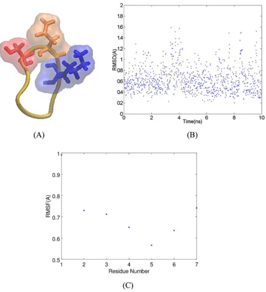
In this section, we analyze how the cSNK mimotope interacts with Fv5E3 [50]. The molecular model of cSNK was obtained in a previous study (Panel A ofFig 2). The LRMSD of this model from a 10-ns MD simulation performed in the current study confirms that the model repre-sents an average conformation of the cSNK peptide (Panel B ofFig 2). The backbone atoms of cSNK are restrained due to the presence of the disulfide bond (Panel C ofFig 2). The cSNK residues are therefore forced to stay within a certain distance from each other. The distance between the Cαatoms of G2 and G6 of cSNK is 5.55Å. There is no space within this sharp
turn for K5 to be buried; therefore, the side chain of K5 is always solvent-exposed. We use the distance between the Cαatoms of G2 and G6 as a measure of how sharp the turn conformation
in the models of Aβ aggregates is.
Fv5E3 has three solvent-exposed acidic residues in its CDRs. These residues are E28 in CDR1 of the light chain, and D100 along with E102 from CDR3 of the heavy chain. The cSNK has a net charge of +1 at a neutral pH. The negative charges of the acidic CDR residues located at the solvent-exposed surface of Fv5E3 create an affinity for the positive charge of the K5 resi-due of cSNK. The presence of a strong negative electrostatic field around the acidic resiresi-due E102 in CDR3 of the heavy chain was determined using the APBS electrostatics plugin of the VMD software [51] (S3 Figof SI). We believe that this region is the main binding pocket for cSNK, and that the initial detection of cSNK by the m5E3 antibody is driven mainly electrostatically.
To further elucidate how cSNK interacts with m5E3, we first docked cSNK to our Fv model of the m5E3 antibody. In the top hundred complexes from the docking simulation, the cSNKs are docked mainly in the superior gap (based on the orientation of the model in Panel C ofFig 1) between the light and heavy chains. The top-ranked docked structure of Fv5E3 and cSNK with the Rosetta score of -150.59 (-21.51 per residue) provides an initial complex to study how cSNK interacts with Fv5E3 (Panel A ofFig 3). The cSNK peptide does not deviate substantially from its initial docked binding site during a 30-ns simulation (Panels A and B ofFig 3). There is a small 3Å increase in LRMSD of the complex in the first 15 ns, which could be because of the adjustments by the antibody for the presence of cSNK (Panel C ofFig 3). The simulation converges towards the last 10 ns. The small 2-3Å fluctuations in the last 15 ns are due to the changes by the antibody to adjust for the small fluctuation of the cyclic peptide in the binding pocket (Panel C ofFig 3in black).
The backbone of G6 from the cSNK peptide forms high occupancy hydrogen bonds with D100 of the heavy chain, and G92 of the light chain of Fv5E3. G6 also forms low occupancy hydrogen bonds with the residues Y33 of the heavy chain, Y94 and R96 of the light chain of Fv5E3 (S2 Tableof SI). Hydrogen bonds formed by G6 make it an anchor to keep cSNK in the binding pocket. A salt bridge (a hydrogen bond accompanied by an ionic interaction) is formed between the E102 of CDR3’s heavy chain, and K5 of cSNK with high occupancy (Panel B ofFig 3,S2andS3Tables of SI). The K5 of cSNK also forms hydrogen bonds with the
the 100 ns-long MD simulation. The heavy chain is from residue 1 to 115 and the light chain is from 116 to 223. F) The secondary structure content of Fv5E3 from the first and last frames of the 100 ns-long MD simulation. The letters B and E stand for isolatedβ-bridge and extended β-sheet, respectively. The letter G stands for 310helix. The letters T, S,
and L stand for hydrogen bonded turn, bend, and unstructured loop, respectively.
residues M99, and D100 from CDR3 of the heavy chain (S2 Tableof SI). K5 acts as an addi-tional anchor to stabilize the complex. There is also a cation-π interaction between K5 of cSNK and Y32 of CDR1 of the heavy chain of Fv5E3 (S3 Tableof SI). The other (low occupancy) hydrogen bonds are formed between the G2 and S3 of cSNK, and the G92 and Y32 residues of Fv5E3, respectively. No hydrogen bonds are formed between N4 of cSNK, and the antibody. No hydrophobic interaction was identified between the final conformation of Fv5E3 and cSNK from the 30-ns simulation.
While in the binding pocket, cSNK does not dissociate from Fv5E3 even after its lysine (K5) is mutated to a glycine. This is supported by an MD simulation. Thus, the stability of the cSNK-Fv5E3 complex is due to the many hydrogen bond interactions formed between cSNK and the antibody. The average binding free energy for the association of cSNK and Fv5E3 in pure water during the simulation is -41.56 kcal/mol (standard deviation (std. dev.) of 4.97), which indicates that the interaction between cSNK and Fv5E3 is a favorable one. The K5 of cSNK as expected has a favorable pairwise contributions to the binding free energy from
Fig 2. A) Cyclic CGSNKGC mimotope. The SNK residues are shown in stick and solvent-exposed surface representations in red, orange and blue, respectively. B) The LRMSD of heavy atoms of cSNK in a 10-ns MD simulation. C) The RMSF of Cαatoms of cSNK in the 10-ns MD simulation.
interactions with E102 (-10 kcal/mol), D100 (-6.15 kcal/mol), and with M99 (-4.96 kcal/mol), all from the heavy chain of the antibody. Contribution to the binding free energy from the interaction of G6 of cSNK with D100 of the heavy chain is -4.20 kcal/mol.
Previously, a similar computational approach was used to compare the stability of another cyclic peptide and its linear form for a different antibody [52]. As a negative control, we carried out docking simulation of cSNK, and the B10 fibril specific antibody fragment [53]. In the top hundred docked complexes, cSNK interacts predominantly to the framework of B10, and only rarely close to the CDRs of B10.
Interaction between Fv5E3 and A
β oligomers
In the following sections, we go through the models of AβOs proposed in the literature and classify them into Fv5E3-positives and possibly Fv5E3-negatives. This classification was per-formed based on the combination of docking and MD simulation results. We believe Fv5E3-positive Aβ aggregates should have structural characteristics similar to cSNK that is a sharp turn at the epitope residues G25-G29, a solvent exposed K28, and available space around a few
Fig 3. A) Top-ranked docked structure of Fv5E3 and cSNK. K5 residue of cSNK and the E102 residue of Fv5E3 are shown in solvent-exposed surface and stick representations in yellow and orange, respectively. B) Complex after 30 ns of MD simulation. C) The LRMSD of Cαatoms of the complex (red), Fv5E3 (black), and cSNK (blue) during the 30-ns
MD simulation.
of the turns to enter the binding pocket of Fv5E3. In the following sections, we also reveal the molecular details of the interactions between Fv5E3 and the cognate AβOs for which structures are either resolved experimentally or predicted computationally.
Experimental models of Aβ oligomers. AβOs are transient entities; this has made it
diffi-cult to determine their molecular structures. To overcome the transient nature of AβOs, vari-ous modifications have been performed on the sequence of Aβ to generate stabilized
oligomers. It is hard to judge whether these modified constructs represent well the structure of the physiologically relevant AβOs. Below, we analyze how these proposed experimental models of AβOs interact with Fv5E3.
The trimer model of Aβ17-36 oligomers by Kreutzer et al. The crystal structure of a
tri-mer from a cyclized Aβ17-36 was determined by Kreutzer et al. [33] (pdb entry 5hoy, Panel A ofS1 Figof SI). Higher order oligomers were observed to form from these trimers, as each tri-mer has two large hydrophobic surfaces [33]. Since the trimers are the building block of these higher order oligomers, we focus on the interaction between Fv5E3 and an individual trimer. To force the formation of aβ-hairpin by the Aβ17-36 peptide, an extra ornithine residue was introduced at position 16 of each individual peptide. The amino group of the side chain of this ornithine residue is connected to V36. The residues V24 and G29 were also mutated to cys-tines. The disulfide bond between the cystines stabilizes theβ-hairpin conformation. The resi-due G33 was also N-methylated (sarcosine) to avoid uncontrolled aggregation in vivo. We used a disulfide bond instead of the ornithine bond for the docking and MD simulations. We also used a glycine instead of the sarcosine at position 33 in silico. The G25-G29 residues form a sharp turn in the trimer model by Kreutzer et al. with a distance of 6.6Å from G25 to G29, and there is plenty of space between the turns to allow a turn to enter the binding pocket of Fv5E3. The K28 residues are also solvent exposed in this model.
To show how Fv5E3 interacts with the trimer by Kreutzer et al., we performed a docking simulation. Fv5E3 interacts with the turn conformation at the epitope residues, the edge of the
β-strands, or rarely with the two hydrophobic surfaces within the three β-hairpins, in the top
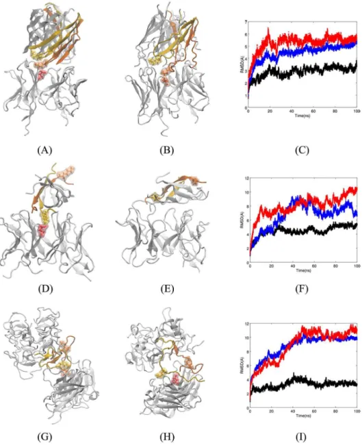
hundred docked complexes. Fv5E3 interacts with the turn conformation at the epitope resi-dues of the trimer by Kreutzer et al. in the top-ranked docked structure with the Rosetta score of -206.45 (-3.27 per residue, Panel A ofFig 4). The three-dimensional structures of the indi-vidual Aβ peptides are fairly preserved during the 100-ns MD simulation (Panel B ofFig 4). The individual Cαatoms fluctuate substantially as it is apparent from the broadened LRMSD
line (Panel C ofFig 4in blue). The quaternary triangular structure is also lost. Despite the loss of the triangular shape, the trimer stays as a trimer (Panel B ofFig 4) and does not disaggregate as the overall LRMSD is plateaued during the simulation (Panel C ofFig 4in blue). It would be interesting to see if two or three Fv5E3 simultaneously bound to the trimer can disaggregate it. The simulation converges in the last 50 ns (Panel C ofFig 4in red). The change in the LRMSD of the complex in the first 50 ns of the simulation is mostly because of the minor adjustments by the antibody to account for the presence of the oligomer (Panel C ofFig 4in black).
While some of the hydrogen bonds between Fv5E3 and the trimer are formed with the framework residues of Fv5E3 (S4 Tableof SI), a salt bridge with high occupancy is formed between the K28 of the oligomer and the E102 of the heavy chain of Fv5E3 (S4andS5Tables of SI). The hydrophobic and ionic interactions may also partially stabilize the interaction between Fv5E3 and the trimer (S5 Tableof SI). There are no aromatic-aromatic, aromatic-sul-phur or cation-π interactions formed between them. The average binding free energy for the association of the oligomer and Fv5E3 during the simulation is -31 kcal/mol (std. dev. of 7.43). This negative average binding free energy is indicative of a favorable interaction between the oligomer and the antibody. The most favorable interactions contributing to the binding free energy are between E22 of chain A of the trimer and K60 (-13.45 kcal/mol), S56 (-4.8 kcal/
mol) of the light chain, K28 of chain B of the trimer and E102 (-4.23 kcal/mol) of the heavy chain, and G33 of chain B of the trimer and Q54 of the light chain (-2.94 kcal/mol). The sum of pairwise contributions to the binding free energy involving the hydrophobic residues that form the two hydrophobic surfaces of the trimer is not a large positive number (-0.31 kcal/ mol) which may indicate that the trimer will not dissociate from the antibody.
Fig 4. A) Top-ranked docked structure of Fv5E3 and the trimer by Kreutzer et al. The stick and solvent-exposed surface representations of the K28 residue of chain B of the trimer and the E102 of the heavy chain of Fv5E3 are shown in yellow and orange colors, respectively. B) Complex after 100 ns of MD simulation. C) The LRMSD of Cαatoms of
the complex (red), Fv5E3 (black), and the oligomer (blue) during the 100-ns MD simulation. D) Top-ranked docked structure of Fv5E3 and the tetramer model of AβOs by Streltsov et al. The stick and solvent-exposed surface
representations of the K28 residue of the chain D of the tetramer, and the E102 residue of the heavy chain of Fv5E3 are shown in yellow and red, respectively. Chain E is shown in orange. E) Complex after 100 ns of MD simulation. F) The LRMSD of Cαatoms of the complex (red), Fv5E3 (black), and the tetramer (blue) during the 100 ns MD simulation. G)
Top-ranked docked structure of Fv5E3 and the octadecamer by Gu et al. The chains that are close to Fv5E3 are shown in various colors. H) Complex after 100 ns of MD simulation. I) The LRMSD of Cαatoms of the complex (red), Fv5E3
(black), and the prefibrillar oligomer (blue) during the 100 ns MD simulation.
The tetramer model of Aβ18-41 oligomers by Streltsov et al. A crystal structure for a
tetrameric Aβ18-41 oligomer was resolved by Streltsov et al. [34] (pdb entry 3moq, Panel B of
S1 Figof SI). Each Aβ molecule was stabilized by a chimerical fusion with a shark immuno-globulin new antigen receptor. The residues G25-I31 form a solvent-exposed wide-turn struc-ture with a distance of 11.66Å between G25 and G29. The side chains of K28s in two of the chains (A/D) are solvent-exposed, and do not form any hydrogen bond. The antibody m5E3 may be able to detect an individual tetramer as the K28 residues are part of turns, and do not always form salt bridges. There is also enough space around the epitope turn to allow its entrance into the binding pocket of m5E3. The top and bottom of the tetramers are covered by hydrophobic surfaces. The hydrophobic residues are also running alongside the tetramer. The tetramer is therefore hypothesized as the building block of higher order oligomers. It seems unlikely that m5E3 would be able to detect multimer oligomers made from these tetramers, as their close packing may not allow an individual epitope to enter the binding pocket of m5E3.
The docking simulation also predicts a possible interaction between the tetramer and Fv5E3. The top hundred docked complexes show a similar mode of interaction between Fv5E3 and the tetramer as in the top-ranked complex with a negative score of -247.37 (-2.51 per resi-due, Panel D ofFig 4). The tetramer after 100 ns of MD simulation stays bound to Fv5E3 (Panel E ofFig 4). One of the two chains of the tetramer (chain E) that is not interacting with Fv5E3 starts to unfold (Panels D and E ofFig 4in orange). The reason for the 1.5Å increase of the oligomer’s LRMSD during the last 20 ns of the simulation is also the unfolding of chain E of the oligomer (Panel F ofFig 4in blue). A longer simulation may demonstrate if the oligomer disaggregates at some point. The Aβ18-41 within the fusion complex and the Aβ17-42 variant of the tetramer by Streltsov et al. were shown to be stable in simulations [54]. The complex and the antibody are fairly stable during the simulation (Panel F ofFig 4in red, and black, respec-tively), and the simulation converges in the last 20 ns. The small 2-3Å fluctuation in the LRMSD of the complex and the antibody about 40 ns in the simulation is due to the fluctuation of the N-termini residues of the Fv fragment.
The two epitope residues that form high occupancy hydrogen bonds with Fv5E3 are N27 and K28 (S4 Tableof SI). Specifically, the N27 residue of chain B of the oligomer forms hydro-gen bonds with D100, E102 and Y32 of the heavy chain of Fv5E3 with high occupancies. The K28 residue of chain D of the oligomer forms a salt bridge with E102 of the heavy chain of Fv5E3 (S4andS5Tables of SI). Hydrophobic interactions also play an important role in stabi-lizing the binding of Fv5E3 to the tetramer (S5 Tableof SI). The residue V24, which precedes the epitope residue G25, participates in a hydrophobic interaction with A50 of CDR2 of Fv5E3’s light chain with a fairly high occupancy. There are also aromatic-aromatic interactions between Fv5E3 and the oligomer (S5 Tableof SI). No aromatic-sulphur or cation-π interac-tions exist at the end of the simulation between Fv5E3 and the tetramer.
The interaction between the tetramer of Streltsov et al. and Fv5E3 is favorable as its average binding free energy is negative (-17.10 kcal/mol with std. dev. of 3.26). We note however that residues 1-17 are not present in this model, and their presence may block Fv5E3’s access to the epitope residues G25-G29. The interaction between K28 of chain D of the tetramer and E102 of the heavy chain of the antibody contribute most to the binding free energy (-16.04 kcal/ mol). The interactions between N27 of chain B of the tetramer and D100 (-5.29 kcal/mol), E102 (-3.88 kcal/mol) and Y32 (-3.15 kcal/mol) of the heavy chain of Fv5E3 are the next best contributors to the binding free energy.
The octadecamer model of Aβ oligomers by Gu et al. An octadecamer model of AβOs
based on restraints from the site-directed spin labeling and electron paramagnetic resonance studies was presented by Gu et al. [35]. Gu et al. stabilized AβOs by fusing the sequence of Aβ42 with the sequence of the chaperone GroES followed by the sequence of the ubiquitin
protein. With the latter fusion, this aggregate is trapped in the oligomeric state with fibril-like
β-sheets (Panel C ofS1 Figof SI).
The residues G25-G29 form a sharp turn in this model with a distance of 7.8Å between G25 and G29. As the epitope turn is sharp, the side chain of K28 residue is not buried inside the turn, and does not form a salt bridge with D23 of its own strand. It is however trapped between adjacentβ-sheets, and can form a salt bridge with D23 of the adjacent β-strand in the nextβ-sheet. The distance between the amine of K28 and carboxyl of D23 in this model is 1.88-5.57Å. When the K28 residue does not form a salt bridge with D23, it participates in an ionic interaction with the mainchain carbonyl oxygen of V24 of mostly the same chain. It can also participate in an ionic interaction with the mainchain carbonyl oxygen of K28 in an adja-centβ-sheet. In the last layer of β-sheets, the K28 residues are solvent-exposed (Panel C ofS1 Figof SI in red), where they may form an intrachain salt bridge with E22. Since the side chains of K28 residues are fully engaged in hydrogen bonding, salt bridges and ionic interactions within the octadecamer model of AβOs, and there is little space around each epitope turn to allow the entrance of a small number of epitopes to the binding pocket of the m5E3 antibody, we anticipate that this prefibrillar oligomer cannot be detected by the m5E3 antibody specifi-cally through the epitope residues.
Contrary to our prediction, the result of the docking simulation shows a possible interac-tion between this model of AβOs and the Fv model of the antibody close to the epitope resi-dues. In the top hundred docked complexes, Fv5E3 binds along the epitope turns of the octadecamer. This interaction for the best docked complex with the Rosetta score of -596.59 (-0.88 per residue) is shown in Panel G ofFig 4. Despite a favorable Rosetta score, the complex is not stable in a 100-ns simulation (Panel I ofFig 4in red). The layer ofβ-sheets in the octade-camer model close to Fv5E3 is disrupted during the simulation (Panel H ofFig 4). The tertiary structure of the two layers ofβ-sheets in the middle are fairly preserved. The C-termini β-hair-pin of one of the Aβ peptides in the layer of β-sheets furthest from Fv5E3 also moves away from the rest of the octadecamer. As the octadecamer goes through a lot of changes, Fv5E3 makes adjustments to try to detect it (Panel I ofFig 4). With many changes in the layer close to Fv5E3, it seems plausible to hypothesize that Fv5E3 is responsible for this disaggregation. A simulation of the octadecamer model by itself should be performed in a follow-up study to confirm this hypothesis.
In the course of the MD simulation, the occupancies of the hydrogen bonds between the prefibrillar oligomer of Gu et al. and Fv5E3 are however low (S4 Tableof SI). The occupancies of hydrophobic interactions are low as well (S5 Tableof SI). There are also no salt bridges, aro-matic-aromatic, aromatic-sulphur or cation-π interactions present between the final complex of Fv5E3 and the oligomer. As the LRMSDs do not converge, it is not possible to properly esti-mate the binding affinity between the oligomer and Fv5E3.
Computational and theoretical models of Aβ oligomers. In addition to the experimental
models considered in the previous sections, some of the structural models of the oligomers have been built computationally based on available experimental constraints, and the rest of the proposed models are purely theoretical and do not rely on any experimental data. The computational and theoretical models may not be as accurate as the experimentally resolved models presented in the previous section. However, the analyses of their interactions with Fv5E3 allow us to propose experiments with oligomers that have similar structures. In the fol-lowing sections, we will assess Fv5E3’s ability to bind to these computational and theoretical models of the oligomers.
The hexamer model of Aβ42 oligomers by Shafrir et al. Shafrir et al. developed many
computational barrel-based models for soluble and membrane-bound AβOs [36]. The forma-tion ofβ-barrels is a natural mechanism that keeps β-sheets from growing into larger
aggregates [55]. Eleven of these models are for soluble hexameric AβOs. These models differ mainly in the secondary structures (β-sheets vs α-helices) of the three regions defined by Sha-frir et al. for Aβ (D1-H14, Q15-K28, and G29-A42), and in their orientations with respect to each other (parallel vs antiparallel). It is not feasible to work on every singleβ-barrel model proposed by Shafrir et al., and such an inspection does not seem to provide us much more information regarding howβ-barrel models might interact with Fv5E3 than the study of a sin-gle one. Here, we choose one of these models as a representative of theβ-barrel models.
In this hexamericβ-barrel model, each Aβ monomer is made of three antiparallel β-strands. Parallelβ-strands of adjacent Aβ peptides align with each other around the model (Panel D of
S1 Figof SI). K28s are solvent-exposed in this model, and in six otherβ-barrel models pro-posed by Shafrir et al. In three of the models, the K28s’ side chains are located on the surface, but they do not stick out to the solvent, and are stabilized by interactions with other residues. In one of the elevenβ-barrel models; however, K28s of three chains are completely buried inside the turns.
For the selectedβ-barrel model, sharp turns at the epitope residues are formed with a dis-tance of 7.30Å between G25 and G29 of each chain. In six β-barrel models, sharp turns are present at the epitope residues. The uncrowded space around the epitope residues of the selectedβ-barrel model allows these residues to enter the binding pocket of Fv5E3. In nine otherβ-barrel models by Shafrir et al., enough space around the epitope residues are also avail-able to enter the binding pocket of Fv5E3.
In the top hundred docked complexes, Fv5E3 binds to the edge around the top (based on the view of the model in Panel D ofS1 Figof SI) of the hexamer by Shafrir et al., where K28s’ side chains are sticking out to the solvent. Only in one of the top hundred complexes Fv5E3 interacts with the side of the barrel. Fv5E3 interacts with a GSNKG turn of the hexamer by Shafrir et al. in the best docked complex with the Rosetta score of -416.88 (-1.65 per residue, Panel A ofFig 5). The interaction between Fv5E3 and the hexamer is stable during the last 80 ns of the trajectory (Panel C ofFig 5in red). The antibody is also fairly stable during the sim-ulation with minor adjustments following the changes in the oligomer (Panel C ofFig 5in black). The LRMSD of the oligomer however does not reach a plateau during the course of the simulation (Panel C ofFig 5in blue). The hexamer by Shafrir et al. seems to start to disag-gregate in the presence of Fv5E3 after 100 ns of MD simulation (Panel B ofFig 5). Involve-ment of the chains of the AβO in many interactions with Fv5E3 possibly will lead to the separation of those chains from the AβO. A longer simulation of the complex is needed to confirm the disaggregation of the AβO by Fv5E3. The hexamer by itself was shown to be sta-ble [36].
In the context of the oligomer recognition by the antibody, it is important to note that K28 of the chain B of the oligomer forms a high occupancy salt bridge with the heavy chain’s E102 of Fv5E3 (S6andS7Tables of SI). We think that this interaction is the driving force that brings the antibody and the oligomer together. This interaction was also present between Fv5E3 and the cyclic mimotope. Another hydrogen bond with high occupancy is between D1 of chain B of the hexamer, and R46 of Fv5E3 light chain’s framework (S6 Tableof SI). The other low occupancy hydrogen bonds may contribute to the stability of the complex as well (S6 Tableof SI).
The two high occupancy hydrophobic interactions during the course of the simulation are between A30 of chain E and I31 of chain G of the hexamer, and Y52 of the heavy chain and Y94 of the light chain of Fv5E3, respectively (S7 Tableof SI). The other low occupancy hydro-phobic, ionic and cation-π interactions may also contribute to the overall stability of the com-plex. No aromatic-aromatic or aromatic-sulphur interaction exists between Fv5E3 and the hexamer in the final conformation after 100 ns. The MM-GBSA average binding free energy of
the complex is -30.46 kcal/mol (std. dev. of 7.98). This negative binding free energy confirms the favorable interaction between the hexamer by Shafrir et al. and Fv5E3. The interaction that contributes most to the binding free energy is between K28 of chain B of the oligomer and E102 of the heavy chain of Fv5E3 (-13.85 kcal/mol).
Fig 5. A) Top-ranked docked structure of Fv5E3 and the hexamer by Shafrir et al. Two K28 residues of the oligomer and the E102 residue of Fv5E3 are shown in solvent-exposed surface and stick representations in yellow, orange, and red, respectively. B) Complex after 100 ns of MD simulation. C) The LRMSD of Cαatoms of the complex (red), Fv5E3
(black), and the oligomer (blue) during the 100-ns MD simulation. D) Top-ranked docked structure of Fv5E3 and the hexamer by Laganowsky et al. The K28 residues of two chains of the oligomer, and the E102 residue of the heavy chain of Fv5E3 are shown in yellow, orange and red colors, respectively. E) Complex after 100 ns of MD simulation. F) The LRMSD of Cαatoms of the complex (red), Fv5E3 (black), and the oligomer (blue) during the 100-ns MD simulation.
G) Top-ranked docked structure of Fv5E3 and the disc-shaped oligomers. The stick and surface representations of K28 residues of two of the chains of the oligomer and the E102 residue of Fv5E3 are shown in yellow, orange and red, respectively. H) Complex after 100 ns of MD simulation. I) The LRMSD of Cαatoms of the complex (red), Fv5E3
(black), the oligomer (blue) during the 100 ns MD simulation.
Otherβ-barrel models of AβOs have been proposed by Pan et al. [26], Lendel et al. [56], and Nguyen et al. [57]. Pan et al. developed a protocol for the in vitro generation of the small stable Aβ40 oligomers. The schematic representation of their model based on the mass spec-trometry data represents a tetramericbarrel structure. Each Aβ40 is expected to form a β-hairpin conformation with a solvent-exposed K28 [26]. Theseβ-barrel tetrameric oligomers of Pan et al. can be used for experimental verification of m5E3’s ability to bind to theβ-barrel oligomers. These oligomers can also be used to examine whether m5E3 can disaggregate the β-barrel oligomers in vitro. The other interesting hexamericβ-barrel model was developed by Lendel et al. as a building block for a modified Aβ42 protofibril [56]. The constituting modi-fied Aβ42 peptides have two mutations at positions 21 and 30 to cystines. The undeposited coordinate file of this model prevents computational analysis of the interaction between this β-barrel model and Fv5E3. Nevertheless, some conclusions regarding Fv5E3’s ability to bind to this hexamer can be obtained based on structural features discussed in Ref. [56]. The residues A24-N27 of this model form a sharp turn. Since this turn is sharp, the side chain of K28 cannot be buried inside the turn. The K28 side chain is solvent-exposed. The K28 residue however makes a salt bridge with D23 by folding back over the turn rather than within the turn. Since individual sharp turns at A24-N27 from various Aβ peptides are not packed too close to each other, there is an opportunity for the turns to enter the binding pocket of the Fv5E3 antibody. Nguyen et al. proposedβ-barrel models for Aβ40 and Aβ42 in aqueous solution. The K28 resi-due rarely forms a salt bridge in the proposedβ-barrel models by Nguyen et al. [57]. This model might also be potentially Fv5E3-positive.
The hexamer model of Aβ26-40 oligomers by Laganowsky et al. A model of the
Aβ26-40 oligomer was built by Laganowsky et al. using the molecular structure of theα-crystalline oligomer as a template [37]. No experimental or computational work was performed to vali-date the direction of side chains or position of residues along the surface of model. This theo-retical model has aβ-barrel/nanotube-like structure. A nanotube is a tube-like structure with a diameter in the nanometer range. The interior diameter of the hexamer model by Laganowsky et al. is 1.1 nm. In this model, the residues S26 to G29 are close to its top/bottom and direct away from the interior space of the structure with a solvent-exposed K28 (Panel E ofS1 Figof SI), which creates enough space for the epitope to possibly interact with Fv5E3. However, the residues S26-G29 do not form a sharp turn in this model (a distance of 9.47Å between the S26 and G29 Cαatoms).
The Fv model of m5E3 antibody interacts with the side, top, or bottom of the hexamer model by Laganowsky et al. in the top hundred docked complexes. In the best docked struc-ture, Fv5E3 binds to the side of the hexamer by Laganowsky et al. with the Rosetta score of -212.33 (-2.35 per residue), which suggests a possible favorable interaction between them (Panel D ofFig 5). The formation of a salt bridge between K28 of the hexamer’s chain B, and the E102 of Fv5E3’s heavy chain may initially bring them close to each other. The complex is however not stable in the course of a 100-ns MD simulation (Panel F ofFig 5in red), and the hexamer by Laganowsky et al. moves away from its initial docked position (Panel E ofFig 5). The occupancies of hydrogen bonds formed during the simulation are low (S6 Tableof SI). No ionic, aromatic-aromatic, aromatic-sulphur, or cation-π interactions are also present between the final conformation of Fv5E3 and the hexamer by Laganowsky et al. after 100 ns. The occupancies of hydrophobic interactions are also low (S7 Tableof SI). The hexamer model is stable in the last 60 ns (Panel F ofFig 5in blue). As the LRMSD of the complex does not converge in the course of the simulation (Panel F ofFig 5in red), no MM-GBSA binding free energy is reported for this system.
Two other similar models of Aβ nanotubes have been proposed. Nicoll et al. provided a model of Aβ nanotubes that was reconstructed from electron microscopy images [58]. The
model by Nicoll et al. did not provide enough molecular details to judge whether it could be detected by the m5E3 antibody. Yong et al. developed a computational nanotube model of Aβ40 oligomers in which the side chains of amino acid residues oriented alternately on either side of each Aβ peptide [59]. The K28 residue is solvent-exposed in this model. The curvature around the nanotube is however not sharp, so the turn at the epitope residues cannot be sharp. Individual Aβ peptides along the surface of the nanotube are also very close to each other; therefore, individual epitopes cannot enter the binding pocket of Fv5E3. Although, the coordi-nate file of this nanotube model was not deposited, based on its structural characteristics dis-cussed above, it is unlikely that Fv5E3 can bind to these nanotubes specifically through its assumed epitopes.
The dodecamer model of Aβ42 oligomers by Gallion. A disc-shaped dodecamer model
of Aβ42 oligomers was assembled by Gallion [38] (Panel F ofS1 Figof SI). As with many other computational models, the model of Gallion is not based on a single type of oligomers but rather based on different experimental data. AFM result by Ahmed et al. showed that a type of stabilized Aβ42 oligomers have a disc-shape [60]. A disc-shaped computational model based on the proposed structure by Ahmed et al. disaggregated in a 60-ns MD simulation [61], while the Gallion model did not [38]. Gallion’s dodecamer is composed of two hexameric disc-shaped sub-units stacked on top of each other. Each Aβ peptide was taken from the crystal structure of the tetramer model of AβOs by Streltsov et al. [34], with a wide solvent-accessible turn at G25-G29. The distance between G25 and G29 of individual Aβ peptides in this model is 11.51Å. The dodecamer model by Gallion assumes that the K28 residue is buried inside, as it was shown to be buried inside the globulomers [19]. In the Gallion’s model, the K28 side chain interacts with the mainchain carbonyls of A21 and D23, and not the side chain of D23. The K28 residue is still partly solvent-exposed in some chains of the Gallion’s model.
The N-termini residues (1-16) of the model obstruct the access of Fv5E3 to the G25-G29 turn. If the N-termini residues were rigid and had little flexibility, then Fv5E3 would not be able to detect these types of oligomers specifically through its epitope. The N-termini residues of the dodecamer model were shown to be very flexible, and start to become random coil dur-ing a simulation [38]. The N-termini residues of the dodecamer are also sticking out to the sol-vent, and do not seem to participate in any interaction with the rest of the model as shown in Fig 1 of Ref. [38]. This suggests that their removal shall not affect the overall stability of the oligomer. In fact, the LRMSD of the dodecamer model was shown to reach a plateau status over the course of a simulation in the absence of the N-terminiα-helices [38]. We therefore removed the N-termini residues to open up a space for Fv5E3 to bind to a turn epitope.
The top hundred docked complexes reveal that Fv5E3 binds around the “discs”, where the upper and lower discs meet (based on the view of the dodecamer in Panel F ofS1 Figof SI). Fv5E3 interacts in a similar way to the dodecamer in the top-ranked docked complex with the Rosetta score of -396.42 (-1.43 per residue, Panel G ofFig 5). The K28 residue is not solvent-exposed in the chain that docked to Fv5E3. The LRMSD of Cαatoms of the complex converges
towards the last 50 ns of the simulation (Panel I ofFig 5in red). The Fv5E3 is fairly stable dur-ing the whole simulation with minor adjustments with respect to the changes in the oligomer. (Panel I ofFig 5in black). The oligomer is deviated 10Å from its initial structure, and seems to be stable in the last 50 ns of the simulation (Panel I ofFig 5in blue). Only the two interacting chains of the oligomer with Fv5E3 start to dissociate from the rest of the oligomer during the course of the 100-ns simulation (Panel H ofFig 5). Multiple Fv5E3 can bind around the dode-camer, and may destabilize the other parts of the oligomer. A follow-up study with a simula-tion with multiple Fv5E3s can confirm this predicsimula-tion.
A broad range of residues in the chain D of the oligomer (residues 18 to 33) form hydrogen bonds with Fv5E3, as the chain D unfolds upon interaction with Fv5E3. The residues of chain
U form hydrogen bonds only with the framework residues of Fv5E3. The assumed epitope res-idues in chain U participate in low occupancy hydrogen bonds. The hydrogen bonds with the highest occupancies are between E22 of chain D and D23 of chain U of the oligomer, and R96 and R46 of the light chain of Fv5E3, respectively (S6 Tableof SI). There are also many low occupancy hydrophobic and aromatic-aromatic interactions between the oligomer and Fv5E3 (S7 Tableof SI). These interactions further stabilize the interaction between the oligomer and Fv5E3. There is no aromatic-sulphur or cation-π interaction between the oligomer and Fv5E3. The average MM-GBSA binding free energy of the oligomer and Fv5E3 is -25.18 kcal/mol (std. dev. of 5.61), which also suggests the existence of a favorable interaction. The most favorable interactions contributing to the binding free energy are between R96 and R46 of the light chain of Fv5E3, and E22 of chain D (-16.49 kcal/mol) and D23 of chain U (-11.3 kcal/mol) of the oligomer, respectively.
Interaction between Fv5E3 and models of A
β fibrils
Various structural models have been proposed for Aβ fibrils [28,40,62–64]. A common struc-tural signature of the Aβ fibrils is the cross-β structural motif. It is characterized by β-sheets running parallel along a fibril axis withβ-strands oriented perpendicular to the axis. Each fibril may be formed from two or more cross-β sub-units [39,64]. Theβ-sheets of the cross-β sub-units are formed of repetitive individual Aβ peptides, which create a general crowding around the m5E3 epitope. This crowding around the m5E3 epitope by itself may impede the binding of m5E3 to fibrils. The m5E3-specific epitope can also be sequestered among cross-β sub-units. This provides a second barrier for m5E3 binding to fibrils specifically through its epi-tope. To verify these claims computationally, we perform detailed analyses of the interactions between available structural models of the Aβ fibrils and the Fv model of the m5E3 antibody.
The model of Aβ40 fibrils by Lu et al. Lu et al. resolved with nuclear magnetic resonance
(NMR) experiments a quaternary molecular structure for the Aβ40 fibrils seeded with the brain-derived Aβ fibrils [27] (pdb entry 2m4j, Panel A ofS2 Figof SI). The deposited structure of the protofilament has three layers (4.8Å apart from each other) perpendicular to the axis of the fibril. Each layer has a three-fold symmetry of the Aβ40 peptide.
In our analyses, to better represent a fibrillar surface, we aligned three of the protofilaments along the fibrillar axis to make a longer structure with nine layers. This longer model has now meaningful ends, and a fibrillar surface between the two ends. The use of a much longer model of the fibril may not be feasible in silico, and does not seem to provide us with more insights compared to the model with nine layers. It is also not possible to perform our simulations with an infinite layer fibril using periodic images [65], as Fv5E3 can be docked to the end of the fibril. Unlike many other models of Aβ fibrils, the N-terminal residues of Aβ in this model were shown to be a part of the quaternary structure. In this structure, the distance between G25 and G29 is 10-12Å, and thus the residues G25-G29 do not form a sharp turn. The wide turn by the epitope residues removes the conformational restriction on the side chain of K28 to reorient towards D23 for the formation of a salt bridge from within the turn, and thus not to be solvent exposed. In this model of the Aβ fibril, many adjacent epitopes are aligned closely along the axis of the fibril, and thus preventing Fv5E3 from accessing an individual epitope. The N-termini residues additionally block Fv5E3’s access to the epitope residues.
To validate our prediction for the low affinity of Fv5E3 to this model of the Aβ fibril, we performed docking of this fibrillar model and Fv5E3. In the top-ranked docked structure with the Rosetta score of -828.69 (-0.76 per residue), Fv5E3 is bound to the end of the fibril and not to the surface between the two ends (Panel A ofFig 6). In the top hundred docked complexes, Fv5E3 shows a similar mode of binding only to the end of the fibril.
We further examined the stability of the top-ranked complex in a 100-ns MD simulation. The LRMSD of the complex does not converge, while Fv5E3 is fairly stable (Panel C ofFig 6in red and black, respectively), and stays bound to the end of the fibril in the course of the simula-tion (Panel B ofFig 6). The model of the fibril however shows significant deviation of its N-ter-minal residues from their initial conformations, and starts to lose the Aβ peptides from its two ends (Panel B ofFig 6, and Panel C ofFig 6in blue). The loss of the peptides at the ends of the
Fig 6. A) Top-ranked docked structure of Fv5E3 and the model of Aβ40 fibrils by Lu et al. The leading chains of the
fibril are shown in yellow, orange and red. B) Complex after 100 ns of MD simulation. C) The LRMSD of Cαatoms of
the complex (red), Fv5E3 (black), and the fibril (blue) during the 100 ns MD simulation. D) Top-ranked docked structure of Fv5E3 and the two-fold symmetry model of Aβ40 fibrils. The leading chains of the fibril are shown in
yellow and orange. E) Complex after 100 ns of MD simulation. F) The LRMSD of Cαatoms of the complex (red),
Fv5E3 (black), the fibril (blue) during the 100-ns MD simulation. G) Top-ranked docked structure of Fv5E3 and the zipper-like model of Aβ42 fibrils. The leading chains of the fibril are shown in yellow and orange. H) Complex after 100 ns of MD simulation. I) The LRMSD of Cαatoms of the complex (red), Fv5E3 (black), and the fibril (blue) during
the 100-ns MD simulation.
fibril is not due to the presence of the antibody, as it happens at both ends and has been shown to happen in simulation of the fibril by itself especially if the number of layers in the model of the fibril is small [65].
Among many hydrogen bonds observed during the simulation, none are involved the epi-tope residues G25-G29 (S8 Tableof SI). There are also many hydrophobic interactions, salt bridges and aromatic-aromatic interactions between Fv5E3 and the model of the fibril by Lu et al. Again, no epitope residues are present in any of these interactions (S9 Tableof SI). R50 and K59 of Fv5E3’s heavy chain form salt bridges simultaneously with both E22 and D23 of chain V of the fibril (S8andS9Tables of SI). R50 and K59 are the immediate residues before and after the CDR2 of m5E3. The residues close to CDRs may participate in recognition of a cognate. R66 of the light chain of Fv5E3 forms a salt bridge with E11 of chain V of the fibrillar model. R50, K59 and R66 do not form salt bridges with AβOs, while participate in salt bridge formation with the fibril. Mutating them to acidic residues or glycine residues possibly can prevent their interaction with the model of the fibril by Lu et al.
As the LRMSD of the complex does not converge, it is not possible to properly measure the MM-GBSA binding affinity between Fv5E3 and this fibrillar model. As Fv5E3 interacts only with the end of the fibril, and considering the very long size of the fibrils, built of many Aβ pep-tides, m5E3 should have a low affinity to this type of fibrils in vitro.
A similar model with three-fold symmetry was also provided by Paravastu et al. for syn-thetic Aβ40 fibrils (pdb entries 2lmp and 2lmq) [64]. These fibrils were generated under the quiescent growth protocol with intermittent sonication during growth. The NMR data sug-gested a disordered N-terminal segment up to residue Y10. The residues 11-22 and 30-39 formedβ-strands, and residues D23-G29 formed a turn between the two β-strands. The dipole-dipole couplings between K28 Nz and D23 Cγ of these fibrils were shown to indicate a 5Å distance between them [64]. A solvent-separated salt bridge interaction between K28 and D23 were proposed to occur [64]. The twenty deposited models (pdb entries 2lmp and 2lmq) rarely show the presence of salt bridges between D23 and K28. It is possible that the intermit-tent sonications during the growth of the fibrils break these salt bridges. K28 is anyway buried between theβ-sheets in these models. As the structure of the fibrillar model by Paravastu et al. is very similar to the one resolved by Lu et al. discussed above, Fv5E3 may again only bind to the end of the fibrils by Paravastu et al.
The model of Aβ40 fibrils by Petkova et al. A quaternary structure of synthetic Aβ40
fibrils was resolved by Petkova et al. using NMR data [28] (pdb entries 2lmn and 2lmo, Panel B ofS2 Figof SI). The preparation of these Aβ fibrils was performed with gentle agitation. Pet-kova et al. proposed a two-fold symmetric model for the Aβ40 fibrils. The first eight structur-ally disordered residues of each Aβ peptide are not present in the model. The side chains of K28s almost always form salt bridges with D23s of±2 neighboring strands. From residues E22 to A30, a wide-turn is usually present. The distance between G25 and G29 of this model is 9.76-13.28Å, which indicates the presence of a wide turn at the epitope residues. It is interest-ing that D23 and K28 in this model do not always form salt bridges with each other. In fact, some of the Aβ peptides have conformations at the epitope residues very similar to the confor-mation of cSNK (chain H in model 6 of 2lmn, and chain D in model 7 of pdb 2lmo with dis-tances of 7.87Å and 6.65 Å between G25 and G29, respectively). The epitope is also not always very crowded by the presence of adjacent epitopes. This may indicate a possible interaction/ pathway between these fibrils and m5E3-positive AβOs.
The interaction between the fibril and Fv5E3 in the top-ranked docked complex occurs at the end of the fibril with the Rosetta score of -375.60 (-0.97 per residue, Panel D ofFig 6). In the top hundred docked complexes, Fv5E3 is also associated with the end of the fibril. The antibody stays bound to the end of the fibril after a 100 ns simulation of the best-ranked
docked complex (Panel E ofFig 6). The complex is fairly stable in the course of the simulation, and its LRMSD converges in the last 10 ns (Panel F ofFig 6in red). The antibody goes through many minor changes to allow its continuous binding to the fibril. The small 2Å fluctuation in LRMSD of Fv5E3 in the last 20 ns of the simulation occurs because of the interactions of the N-terminal residues of Fv fragment with the fibril. The model of the fibril by Petkova et al. twists around the axis of the fibril during the simulation. It however maintains its overall shape (Panel E ofFig 6, and Panel F ofFig 6in blue). The same twisting behavior is observed when a sixβ-hairpin layer model of the fibril was simulated [65]. Due to the high stability of the fibril, Fv5E3 is not able to dissociate any of the Aβ peptides from the rest of the fibril.
Many hydrogen bond interactions are present between Fv5E3 and the fibrillar model. Some of the interactions occur with the framework residues. The framework residues N77 and D61 of the heavy chain are not part of the m5E3’s original sequence, and hence the interactions with these two residues may not be present in vitro. From the epitope residues, only S26 and N27 form hydrogen bonds (with low occupancies) (S8 Tableof SI). The residue E11 of chain E of the fibril, and K59 of the heavy chain form a high occupancy ionic interaction. R66 also forms a salt bridge with E22 of the fibril (S9 Tableof SI). The R50 of Fv5E3’s heavy chain and Y91 of the light chain participate in high occupancy cation-π interactions with Y10 and K16 of the fibril, respectively (S9 Tableof SI). R50, K59 and R66 interact with the fibril, but do not interact with the AβOs. Site-directed mutagenesis of these residues to acidic residues or glycine residues may provide an opportunity to prevent m5E3 from interacting with this type of fibrils in vitro.
The interaction between Fv5E3 and the fibrillar model is stabilized by a combination of hydrogen bonds, hydrophobic interactions, salt bridges, aromatic-aromatic and cation-π inter-actions (S8andS9Tables of SI). The MM-GBSA binding free energy between the fibril and Fv5E3 is favorable (-8.59 kcal/mol with std. dev. of 6.45). No epitope residues however partici-pate in the hydrophobic, ionic, aromatic-aromatic or cation-π interactions (S9 Tableof SI). As the fibril is very long, consisting of many Aβ peptides, the likelihood of the m5E3 antibody to bind to only the ends of the long fibril is low. Therefore, m5E3 should have a low affinity for this type of fibrils in vitro. If this interaction however occurs, the m5E3 antibody may block further growth of the fibril.
The model of Aβ42 fibrils by Schmidt et al. A zipper-like model for the structure of an
Aβ42 dimer in fibrils was predicted by Schmidt et al. using electron cryo-microscopy data [29] (pdb entry 5aef, Panel C ofS2 Figof SI). The distance between residues G25 and G29 in this model is 15.04Å, which means the assumed epitope residues G25-G29 do not form a sharp turn. The K28 residues are solvent-exposed in this dimer. Schmidt et al. also provided a mini-mal fibrillar model with six of such dimers in which the distance between adjacent dimers is 4.7Å. We extended that fibrillar model to one with nine adjacent dimers. This longer model is a better representative of the fibril with proper ends, and a surface between the ends.
In the top hundred docked complexes, Fv5E3 binds to the end of the fibril. The top-ranked docked complex of Fv5E3 and the fibril with the Rosetta score of -460.63 (-0.6 per residue) is shown in Panel G ofFig 6. This complex after 100 ns of MD simulation maintains its overall structure (Panel H ofFig 6). The N-termini residues of the fibril move closer to theβ-sheet formed by its C-termini residues and lose their secondary structure. The leading chains of the fibril start to dissociate from both ends, which could be because the model of the fibril is formed of a limited number of peptides [65] (Panel H ofFig 6). The LRMSDs of Cαatoms of
the complex, the antibody and the fibril are fairly stable during the simulation (Panels I ofFig 6in red, black and blue, respectively). The simulation is converged in the last 80 ns of the simulation.
Many hydrogen bond interactions occur between Fv5E3 and the zipper-like model, but none of them are formed with the epitope residues. The hydrogen bonds with highest occu-pancies are between D7 of chain M, V20 of chain A, I16 of chain A of the fibril, and R96 of the light chain, S31 of the heavy chain, I28 of the heavy chain of Fv5E3, respectively (S8 Tableof SI). There is one ionic interaction between D7 of chain M of the fibril, and R96 of the light chain of Fv5E3 (with high occupancy). There are a few hydrophobic interactions between Fv5E3 and the zipper-like model (not with high occupancies) (S9 Tableof SI). There is also an aromatic-aromatic interaction between the F3 of chain K of the zipper-like model, and Y32 of the light chain of Fv5E3 with a low occupancy (S9 Tableof SI). None of the hydrophobic, ionic, aromatic-aromatic interactions are with the assumed epitope residues (S9 Tableof SI). There are no aromatic-sulphur or cation-π interactions between Fv5E3, and the zipper-like model after 100 ns of simulation.
The average MM-GBSA binding free energy between Fv5E3 and the zipper-like model of the fibril is -48.95 kcal/mol (std. dev. of 7.18). This is the most favorable binding free energy among the different Aβ aggregates considered in this work. Although, the interaction is favor-able, it occurs at the end of the fibril. As the fibrils are very long, consisting of many Aβ pep-tides, initial binding of m5E3 to the end of the fibril is a very rare event. However, if m5E3 binds to the end of the fibril, it blocks its further growth.
Interaction between Fv5E3 and cross-
β sub-units of Aβ fibrils
The construction of the spine of a fibril from a single cross-β sub-unit may not be trivial [64,
66]. As the fibrils may grow from these minimal structures, which may be present in vivo, we included them in our analyses. We are borrowing the cross-β unit terminology from Ref. [64]. The cross-β sub-units have also been used as models for AβOs, and the interaction of a single-domain antibody with the end or the surface between the two ends of these cross-β sub-units have been examined [67].
The model of Aβ42 cross-β sub-units by Lu¨hrs et al. The protofilament of the Aβ42
fibrils resolved in an NMR experiment by Lu¨hrs et al. consists of fiveβ-hairpins [39] (pdb entry 2beg, Panel D ofS2 Figof SI). The protofilament by Lu¨hrs et al. is the cross-β sub-unit of the fibril. The residues 1-16 in the Aβ fibril were disordered, and hence their coordinates were not resolved. The residues G25-G29 of this model are part of a wide-turn from S26 to I31 with a distance of 12.56Å between G25 and G29. The K28s residues almost always form salt bridges with D23s of the adjacent chains, while facing the inner part of the turns. K28 can become sol-vent exposed in the leading chain (Panel D ofS2 Figof SI). As the epitopes are also packed close to each other, it may not be possible for Fv5E3 to detect these protofilaments by specifi-cally binding to a small number of epitopes. Lu¨hrs et al. also proposed a model of the fibrils consisting of four strands of such cross-β sub-units [39]. The coordinate file of this fibrillar model is not deposited.
Contrary to our prediction, in the top hundred docked complexes, Fv5E3 binds to the ends, the turn between the twoβ-sheets, or the surface of β-sheets between the two ends of Lu¨hrs et al. model. In the best docked complex, Fv5E3 interacts with the surface of theβ-sheet formed by residues 17-24 with the Rosetta score of -237.08 (-2.1 per residue, Panel A ofFig 7). The antibody stays bound to the model after 100 ns of MD simulation (Panel B ofFig 7). This cross-β sub-unit goes through a lot of minor fluctuations especially between 40-80 ns (Panel C ofFig 7in blue). Its LRMSD also only reaches a plateau in the last 20 ns. The final conforma-tion of the cross-β sub-unit of the Lu¨hrs et al. (Panel B ofFig 7) after the 100-ns simulation is very similar to what was reported from a simulation of the model by itself [68]. The antibody is fairly stable in the presence of the protofilament. The minor fluctuations of Fv5E3 are the