HAL Id: hal-02272556
https://hal.archives-ouvertes.fr/hal-02272556
Submitted on 27 Aug 2019HAL is a multi-disciplinary open access archive for the deposit and dissemination of sci-entific research documents, whether they are pub-lished or not. The documents may come from teaching and research institutions in France or abroad, or from public or private research centers.
L’archive ouverte pluridisciplinaire HAL, est destinée au dépôt et à la diffusion de documents scientifiques de niveau recherche, publiés ou non, émanant des établissements d’enseignement et de recherche français ou étrangers, des laboratoires publics ou privés.
genes in Physarum polycephalum
Gérard Pierron, Marianne Bénard, Edmond Puvion, Ronald Flanagan,
Helmut Sauer, Dominick Pallotta
To cite this version:
Gérard Pierron, Marianne Bénard, Edmond Puvion, Ronald Flanagan, Helmut Sauer, et al.. Replica-tion timing of 10 developmentally regulated genes in Physarum polycephalum. Nucleic Acids Research, Oxford University Press, 1989, 17 (2), pp.553-566. �10.1093/nar/17.2.553�. �hal-02272556�
Replication timing of 10developmentally regulated genes in Physarum polycephalum
GerardPierron*, MarianneBenard, Edmond Puvion, RonaldFlanagan', HelmutW.Sauerl and DominickPallotta2
Institut deRecherches Scientifiques sur le Cancer, Villejuif, 94802 France, 'Department of Biology, Texas A& M University, College Station, TX 77843, USA and2Ddpartementde Biologie, Universitd Laval, Quebec GIK7P4, Canada
ReceivedOctober 25, 1988; Revised andAcceptedDecember 9, 1988
~ID
I
ABSTRATWe havetested the hypothesis which stipulates that only early-replicating genes are capable of expression. Within onecell type ofPhysarum - theplasmodium - wedefinedthe temporal order of replicationof 10genes which were known to bevariably expressed in 4 different developmental stagesofthe Physarum lifecycle.Southern analysis of density-labeled, bromodesoxyuridine-substituted DNA reveals that 4 genes presumably inactive within the
plasmodium, were notrestricted to any temporal compartment of S-phase: 1 is replicated in
early S-phase, 2in midS-phase and 1 in lateS-phase. On theotherhand,4outof 6 active genes analysed areduplicated early, with the first30% of thegenome. Surprisingly, the two othersactive genes are replicated late inS-phase. Bygene-dosageanalysis,based on
quantitation ofhybridization signals from early and late replicating genes throughout S-phase,
we couldpinpoint the replication of one of these two genes at a stage where 80-85% of the genome hasduplicated.Ourresultsdemonstratethat latereplication during S-phase doesnot preclude gene activity.
INTRODUJCTION
Theduplication of eukaryotic chromosomes is achievedthroughacomplexprocess involving thousands ofreplication unitswhich areactivated in a non-random temporal order(1,2). Ahigh number of active genes have nowbeen showntobe
replicated early
during S-phase(3-1 1). On the contrary inactive transcription units(6,7), heterochromaticsegments ofchromosomes(1 2)or eveninactivated X chromosome in female mammals
appeared tobe preferentially replicated late in
S-phase(13).
Basedon theseobservations,
models linking thespecific expression ofgenes tothetemporalorder ofreplication
have been proposed(6,14). In short, early replication would be a prerequisite forgeneexpression
whereas late replication would precludegene
activity.
Consequently,
thechronology
ofreplication would be the highestlevel ofgene control and would necessarlybestrictlydefined within onecell lineage and variable in the time course of the differentiation
process(5,6,14).
We areusingtheslime mold Physarumpolycephalum as amodelsystemforstudying
thechronology ofreplication within the
eukaryotic
genome.ln onestageof its lifecycle, Physarum
developsasalargemultinucleatedcell,calleda
plasmodium,
which containsas manyas 108 nuclei(15). Being ina commoncytoplasm, these nuclei behave synchronously and divideevery10 hours .Theythenimmediatelyembarkon a3hour synchronous S-phase as
previously demonstrated by flow-cytometry(16). This provides a unique situation where DNA replication studies do not require artificial synchronization orcumbersome sorting of a random population of cells. Interestingly, the relation between the timing ofreplication of genes and theirexpression is also apparent in the naturally synchronous plasmodium of Physarum. Previously, we havevisualized onplasmodialchromatin spreads asetof active geneswhich are replicated early in S-phasein the form of transcription units locatedon
nascent replicons(17). Next, as a test-case, wedetermined the timing ofreplication and the
transcriptional activityof the 4 members of the Physarum actin multigene family .We found that 3ofthe 4 loci are replicated earlywhereas the fourth locus is late(18). Subsequent analysisof 233 actincDNA clonesrevealed thatall these cloned mRNAoriginatedfromtwoof the earlyreplicated loci(19). Moreover,a probe specific for anactin sequence from the late replicating ardA locus did nothybridize to RNAonNorthern blots, suggesting an inactivityof this actinsequence(19), which, as revealedby sequencing(20), isa bonafide isocodingactin gene .It had also been shown that thetwohistoneH4genesofPhysarumareduplicatedinearly S-phase(21).
In this study, we determined thetiming of replication within the plasmodium of 10
developmentally regulated genes of Physarum inorderto further compare thechronologyof replicationof active and inactive genes.Much to our surprise,we foundthat 2 genes expressed
within the plasmodium arereplicated late in S-phase. These results are notcompatible with
thecurrentconcept which stipulates that geneexpression is restricted to the early replicating
compartment ofthe genome (5,6).
MATERIAL ANDMETHODS
Cultures
Two Physarum strains (Tu291 and M3CIV) were used in this investigation. Both are
diploid derivativesof the Wisl natural isolate and therefore haveacommon genetic
background. The Tu291 strain has been previously utilised for most DNA replication studies in Physarum (18,21). The DNA content of the nuclei is slightly reduced in the M3CIV strain. A flow-cytometric analysis had indicated that this was due to a reduction oflate-replicating DNA (16). Sofar, however, all the genes we have analysed were present and had equivalent replication timing in both strains.
The synchronous cultures (macroplasmodia;5-6 cm in diameter) were grown on Whatman filter paper according to published procedures (22). The second and third synchronous mitosis generally took place about 15 and 24 hours after feeding as judged by phase contrast microscopy.
Isolation ofBrdUrd-Substituted DNA
Cultures were treated in mitosis (about 10 min before the onset of DNA replication) until harvested in S-phase with a mixture of BrdUrd (100 jg/ml), fluorodeoxyuridine (5
jg/mI)
and uridine (100 jig/ml) (2,18) . After a BrdUrd pulse, about 3x10⁸nuclei were isolated from a single macroplasmodium essentially as in (23).The DNA was then extracted andpurified asdescribed in (24).To separate the newly replicated DNAfrom the unreplicated
fraction of the genome, 150
jig
ofisolated DNA were digested witheither HindlIlorEcoRI and thendiluted to 9 ml in 10mM Tris-HCL,pH 8.0/1mM EDTA. After addition of 11 g of solidCsCI and 20jg
ofethidium bromide,the restricted DNA wascentrifuged 60h at40,000 rpm in a Beckman 50 Ti rotor. The light-light (LL) and heavy-light (HL) DNA bandswere then visualised under UV-light and withdrawn separately with a syringe and a 21 gauge needle. The twofractions were desalted by dialysis andfinally ethanolprecipitated.Hybridization probes:
The 10 cDNAs used in this study had all been cloned in the Pstl site ofpBR322. Bulk
preparationsof the different plasmids were digested with Pstl and the inserts purified by standard methods (25)afteragarose gelelectrophoresis .The length of the cDNA inserts is as follow: LAV1-1: 860 nt ; LAV1-2: 800 nt ; LAV1-3: 370 nt ; LAV1-4: 480 nt ; LAV1-5: 560
nt; LAV3-1: 600 nt ; LAV3-2: 550 nt; LAV3-3: 850 nt (26); LAV2-1: 1040 nt (27); LAV5-1: 650 nt (28).
Hybridization analysis:
The LL and HL restrictedfractionsof the genome wereelectrophoresedon0.7% agarose
gel.The relativeconcentration ofthe twofractions wasadjustedas afunction of their
complexity. Sinceonethird ofthe genome isreplicated during the first 40 min ofS-phase
(16),we loaded twice as much LL DNA as HL DNA on thegel aftera40 min BrdUrdpulse.
Equal amounts of LL and HL DNA were loaded after a 60 min BrdUrd pulse, atime
corresponding to50% ofgenome replication.As a 90 min pulsecorresponds to 75% of genome
replication,weloadedthe LLand HL DNA in a 1 to 3 ratio.
After blotting onto nitrocellulose the LL and HL DNA were hybridizedwith a
32P-labelled
cDNA insert for 20 hours under standard conditions (0.45 M NaCVO.045M sodium citratepH7, 680C, 10% dextran sulfate). Probes were labelled by either nick-translation(29) or random -labelling (30) to a specific activityof 1 to
8x10⁸
cpm/ug with32P-dCTP.
The presence ofGC tailsin thecDNA inserts which hybridizeto repetitivesequences in thePhysarum genome rendered highlystringentwashes of the filtersmandatory.The two final washes werefor 5 min at700C , in 15 mMNaCI, 1.5 mMSodiumcitrate pH7 and 0.1%
Sodium
dodecyl
sulfate.Northern-blothybridizations
wereperformed
aspreviously
describedGene dosage analysis
Following DNA extraction at various time-points in S-phase, equivalent amountsof S-andG2-phase DNA samples digested with EcoRI or HindlIl were electrophoresed on 0.7% agarose gels. Afterblotting onto nitrocellulose filter, hybridizations were performed with two
32P-labelled
cDNAs known todetect genes replicated in different comparments in S-phase.The twocDNAswere labelled inasinglerandom-priming reaction in such awaythat the resulting hybridization signals were of similar intensities. This was required for comparingthe copynumber of both genes within the linear order of response of the X-rayfilms. The
autoradiographswerescanned withaChromoscan3Joyce-Loebldensitometer attachedtoa
computerequipped with theJoyce-Loebl data system software. The timing ofreplication of
tworestriction fragmentswasdeduced from the deviation in S-phaseof the ratio of their hybridization signals obtained in G2-phase (18).
RESULTS
Southern analysis of the LAV genes of Physarum
The
Physarum
life cycle offers agreatvariety of different cell types which are easilypropagated in the laboratory.
Physarum
canbe cultured as uninucleatedhaploid amoeba which under certain circumstances fuse inpairs toform adiploid zygote. These diploid cells differentiate into large multinucleated plasmodia by intranuclear mitosis in the absence of cell division. Under unfavorable conditions, aplasmodiumcan either form resistant cysts(spherules) or initiate another differentiation program (sporulation) which culminates
with the formation ofhaploid spores (31). Differential screening of cDNA libraries established from poly(A+) RNA from these 4 distinct differentiation stages of Physarum
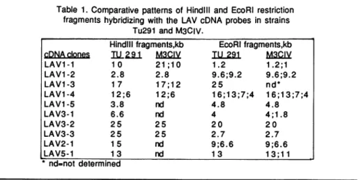
Table 1. Comparative patterns of HindlIl and EcoRI restriction fragments hybridizing with the LAV cDNA probes in strains
Tu291 and M3CIV.
HindlIlfragments,kb EcoRI fragments,kb
cDNAdclones
I291
MV3CV TU 291 M LAV1-1 10 21 ;10 1.2 1.2;1 LAV1-2 2.8 2.8 9.6;9.2 9.6;9.2 LAV1-3 17 17;1 2 25 nd* LAV1-4 12;6 12;6 16;13;7;4 16;13;7;4 LAV1-5 3.8 nd 4.8 4.8 LAV3-1 6.6 nd 4 4;1.8 LAV3-2 25 25 20 20 LAV3-3 25 25 2.7 2.7 LAV2-1 15 nd 9;6.6 9;6.6 LAV5-1 13 nd 13 13;11 nd=not determinedallowed forthe isolation of cDNA probes specific fordevelopmentally regulatedgenes (26,27,28).
Inthis communication , we established the timing ofreplication, within the plasmodium,
of genes which are either expressed in the plasmodium (26) (LAV1-1; 1-2; 1-3; 1-4; 1-5;
3-2) , or apparently not expressed in the plasmodium butreadily active in the amoeba (26)
(LAV 3-1; 3-3) , during spherulation (27) (LAV 2-1) or during sporulation (28) (LAV 5-1).Onegene(LAV3-2) isexpressed both in the amoeba and theplasmodiumbut theamount
and the size of the mRNAs differ inthesetwodevelopmental stages(seefig.3a).
InTablel, the EcoRI andHindill restriction patterns of these genes in the diploidstrains Tu291 andM3CIVaresummarized. It can be noted that many of these genes show a simple restriction pattern in the Tu291 strain, suggestive of single copy genes. However, there exists frequent restriction fragment length polymorphism in the M3CIVstrain. HindlIl and Eco RI
restriction fragment length polymorphism was previously shown to affect the 4 actin loci in the M3CIVstrain but only one actin locus of the Tu291 strain(32,18). As predicted from the
commongeneticbackgroundofthetwostrains,the allelicactin sequences found in Tu291 represented a subset of the M3CIV alleles(32,18). This isalso the case for the genes analysed in this communication(Table 1).
Chronologyofreplication of the LAV genes of Physarum
Wesubdivided the3 hour S-phaseof Physarum intofourcompartments by extracting
DNA after on vivo BrdUrd incorporation for either40, 60 or 90 minutes . About 1/3 of the genomeisduplicated during the first 40 minutes of
S-phase,
aboutone half after 60 min and 3/4 after 90 min (16). By analysing the segregation of hybridisationsignals
from thecell -type specific cDNA clonesbetween the LL and HLfractionsofthesepreparations
wededucedT LL. H L T I.L1 L. T LL IlL T LLL [{L
U.v
a b c d
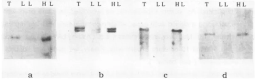
Figure 1: Early replication in S-phaseof four genes which areactive within theplasmodium
ofPhysarumr this is deduced from the preferential hybridizationof
32P-labelled
cDNAs to the HL DNA after in vivobromodeoxyuridine
incorporation for the first 40min in S-phase. T=EcoR1 digestedTotalDNA;LL-EcoR1 digested non-replicatedDNA;HL= EcoRidigestedDNA replicated during 0-40min in S-phase . a) LAVI-1 cDNA b) LAV1-2 cDNA c) LAV1-3 cDNAFigure
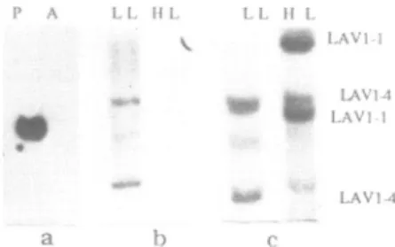
2:Expression
andreplication
timing
of the LAV1-4gene in theplasmodium
of a) Hybridization ofthe 32P-labelled LAV1-4 cDNAto plasmodial (P) and amoebal (A)poly(A+)RNA. Note the presenceofa1500 nucleotideslong mRNA detected exclusivelyin the plasmodial RNA.
b) Non-early replication of the LAV1-4 gene as demonstrated by preferential
hybridization of the LAV1-4 cDNAtothe unreplicated (LL) fraction of HindlIl digested Tu 291 DNA after BrdUrd incorporation for 0-40min in S-phase.
c) Late replication of the LAV1-4 geneas evidenced by double-hybridization experiment.
The cDNAof the LAV1-4 gene and of the early-replicating LAV1-1 genewere p32 -labelled and hybridized tothe LL and HL DNA fractions obtained after BrdUrd treatment for the first 90min ofS-phase inthe M3CIV strain. Thetwo HindlIl
fragments
of the LAV1-4 gene(12
and 6kb) are enriched in the LL fraction whereas, as internal controls, the two HindlIl LAV1-1fragments (21
and 10kb)
arefound in the HLfraction.thetiming ofreplication of the genes. Genesnot replicated by90minutes in S-phasewere
assumedtoreplicatevery late in S-phase.(See below)
First, we analysed the timing of replication of 6 genes selectively expressed within the plasmodium. Not surprisingly, most of these active genes (4 out of 6) are replicated early in
S-phase. In Figure1,wedisplayed thehybridization patterns of these 4genes. It is obvious that all the respective cDNA hybridizedpreferentially tothe HL fraction following a 40 min
pulseof BrdUrd. It is therefore concluded that these 4genes are replicated by the time 1/3 of the genome isduplicated. As control experiments, we hybridized the same filters with differentprobes, including acloned actin gene, which recognize early and late replicating restriction fragments (18) .Moreover, some genes ( e.g. LAV1-1, LAV1-2 ) were found to be replicating early in at least 10 different experiments. Finally, we performed double-hybridization experiments ( see below ).
On the other hand,2active genes, LAV1-4 and LAV3-2, were found to be replicated late inS-phase .These unexpected results are presented in Figures 2 and 3.
That the LAV1-4 gene is really expressed in the plasmodium is ascertain by the detection
on a Northern blot of a relatively abundant mRNA of 1500 nucleotides (fig.2a, lane P).ln
P A LIL Hi, LL HL *''''
~~~~LAV-1-LAVI-2
a b c
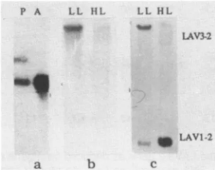
Figure 3: Expressionand timingof replicationof the LAV3-2gene of
Physarum.
a) Detection of specific transcripts within plasmodial (P) and amoebal (A) RNApreparations by hybridization with the LAV3-2 cDNA.Of the two plasmodial mRNAs of 870 and 1.000 nucleotides (lane P), note that the 870 nucleotides long mRNA is the only transcript found inthe amoebal RNA (lane A).
b) Non-early replication of the active LAV3-2 gene as evidenced by preferential hybridization of the32p - labelled LAV3-2 cDNA to the LL DNA fraction after 60min of BrdUrd treatment (strain Tu 291, Hindlil digest).
c) Late replication of the LAV3-2 gene as shown by double- hybridization experiment. The LL and HL DNA fractions obtained after 90min of BrdUrd treatment were hybridized with thecDNAs of the LAV3-2 gene and of the early replicating LAV1-2 gene as internal control. The 25kb LAV3-2 HindlIl fragments is found in the unreplicated fraction whereas the 2.8kb LAV1-2 HindlIl fragment is clearly enriched in the HL fraction (strain Tu291).
(Fig.2a,lane A). This confirms that the steady state level of the LAV1-4 mRNAvaries notably
during the development in Physarum (26).
BrdUrd incorporation studies consistently indicated a late replication of this active gene. As an example, we show a preferential hybridization of the LAV1-4cDNA to the LL DNA fraction after 40 min of BrdUrd substitution (fig.2b). We obtained a similar result for a 60 min BrdUrd treatment (data not shown) .Moreover, this holds true after 90min in S-phase (75% genome replication).ln this case, in order to rule out eventual artefacts, we performed
a double-hybridization by mixing the cDNA of the early replicating LAVI-1 genewith the LAV1-4 probe (Fig.2c). As expected, the21 and 10 kb Hindlll restriction fragments of the LAV1-1 gene (M3CIV strain) were found exclusively within the HL fraction whereas the 12
and 6 kb LAV1-4 fragments were comparatively enriched in the unreplicated fraction (Fig.2c). Therefore, the LAV1-4 gene is shown tobe active andlate
replicating
within the plasmodium of Physarum.Inaddition,wefound that the LAV3-2 gene alsobelongstothis class ofeukaryoticgenes. This is demonstrated in fig.3. Again Northern-blot analysis clearly indicates expression of the LAV3-2 gene in theplasmodium.Two mRNAs of1 000and 870 nucleotidesaredetectedby
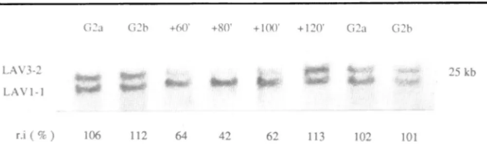
Figure4: Latereplication of theLAV3-2geneasdeduced fromgenedosageanalysis.
Samplesofsynchronous DNApreparations from the M3CIVstrain, extracted in S and G2
-phase weredigested withHindlil,electrophoresed on0.7% agarosegel andblotted.Theywere
hybridized with a mixtureof p32 -labelled cDNAs of the early replicating LAV1-1 gene and of the LAV3-2 gene. The 25kb LAV3-2 and the 21kb LAV1-1 Hindlilfragments are presented.
The hybridization signals were
quantified
bydensitometry
and the relative intensities (r.i ) of the LAV3-2 to LAV1-1 signals calculated. Asan internal control, duplicated samples from 2 differentG2-phase DNA preparations (G2aand G2b, isolated respectively 7 and 8 hours after mitosis)wereloadedoneach side of theS-phase samples. We assumed that thereare2copiesofeach gene in the
G2-phase
samples and in the 120 min extract (r.i of 106, 112, 102, 101 and 113%respectively) and 1 copy of the LAV3-2 gene for 2copiesof the LAV1-1 gene in the DNAextracted at60,80 and 100 min after the onsetofS-phase
(r.i of64, 42and 62%respectively).This experiment indicate replication of LAV1-1 between 0-60 min and
replication of LAV3-2 between 100 and 120min in S-phase.
levelof both mRNAs differs in the amoebal poly(A+) RNA where the1 000nucleotides RNA is undetectable and the 870 nucleotides message is very abundant(Fig.3a,lane A).
BrdUrd incorporation studies indicated that the 20 kb
EcoRi
fragmentcontaining the LAV3-2 gene was notreplicated by 40 min in S-phase (not shown). This is also true for the 25 kb Hind IlIl LAV3-2 fragment which is found in the unreplicated LL fraction after 60min inS (Fig.3b). Testing for the replicationof the LAV3-2 gene by 90 min in S-phase, we performed another double-hybridization. As an internal control, the cDNA of the early replicating LAV1-2 gene (Fig.1) was mixed with the LAV3-2 probe. In this experiment, where the BrdUrd was incorporated in 75% of the genome, the hybridization clearly
distinguished the 25 kbHindlIl LAV3-2fragment, which is found in the unreplicated LL fraction from the 2.8kb LAV1-2 fragment which is enriched in the replicated HL fraction ( Fig.3c ). We therefore conclude that both the LAV1-4 and LAV3-2 genes are not replicated by the time 75% of the genome has duplicated. We next wanted to determine how late in
S-phasethese genes were replicated . This was studied by gene dosage analysis. Gene
dosage
analysis
Highly synchronous DNA preparations that can be obtained in Physarum permitted us
todetectthe replication of genes by measuring the relative copy number of two restriction fragments throughout S-phase . Originally, we demonstrated the effectiveness of this method
BrdUrd
0 4*0 0 -. n60
e1 'l
d
Eigure
5: Timing ofreplication of 4genes inactive within the plasmodium of Physarum.After BrdUrd treatment for 0-40min (left) or 0-60min in S-phase (right), DNA was
extracted, restricted and the LL and HL fractions wereseparatedon CsCI gradients.
a) Hybridization of the spherulation specific LAV2-1 cDNA toEcoRI digested LLand HL fractions; strain Tu291. Note thatthe two LAV2-1 EcoRI fragment are not replicated by 60min in S.
b) Hybridization ofthe amoebalspecificLAV3-1 cDNAtoEcoRI digestedTu291 DNA.Note thatthe 4kb LAV3-1 fragment is enriched in the LL DNA for a 0-40min treatmentand in the HL DNAfor a 0- 60min treatment.
c) Hybridization of the amoebal specific LAV3-3 cDNAto Hindlil
(left)
and EcoRI(right)
Tu291 DNA. Preferential hybridization to the LL DNA (left,0-40min) and tothe HL DNA (right, 0-60min) demonstrates the replication ofthe LAV3-3 gene between 40 and 60min in S- phase.
d) Early replication of the sporulation specific LAV5-1 gene as shown by a double-hybridization. The LAV5-1 and LAV2-1
32P-labelled
cDNAswere hybridized to EcoRI digested Tu291 DNA extracted after 40 min of BrdUrd treatment. The early replication of the LAV5-1 gene is deduced from the presence of the 13 kb LAV5-1 fragment inthe HL fraction whereas the 9 and 6.6kb LAV2-1 fragments,as internal control, are found in the LL fraction .by comparing the relative intensities of the hybridization signals from restriction
fragments
containing the early and late replicating
physarum
actin genes(18).
Inthe presentwork,wechoseto compare the relative intensities of the LAV3-2 and LAV1-1 genes sinceour BrdUrd incorporation studies indicated an
early
replication
ofLAV1-1 and a late replication of LAV3-2. Therefore, we displayed HindlIl digestedS- andG2 -phaseDNA samples of the M3CIVstrainon a0.7% agarosegel.Inordertoestimate
experimentalvariations, duplicated samplesfrom 2G2-phase DNApreparations
(G2a
and G2b isolated respectively 7and 8 hours aftermitosis) wereloaded onthesamegel.Afterelectrophoresis and blotting, thesesampleswere hybridized with a mixture of the LAV1-1 and LAV3-2 p32 -labelled cDNAs,
genera:.ng
the hybridization signals of the 25kb LAV3-2 Hindlil fragment and of the 21 and 10kb LAV1-1 fragments. We restricted the quantitative analysisby densitometry to the 25kb LAV3-2 and 21kb LAV1-1fragments
(Figure 4).
In G2 phase, we found that the relative intensities of the LAV3-2to the LAV1-1 hybridization signalswerealmost identical in theduplicated samplesfrom the 2 DNApreparations
(106 and 102% for theG2a samplesand 112 and 101% for theG2b samples, Figure
4). This defines the ratio of the hybridisation signals at acell-cycle stagewhere there are 2copiesof each gene. In contrast, inthe DNA extracted 60min after theonsetofS-phase,at
astagewhereweknew that LAV1-1 is replicated and LAV3-2 is not, the relative intensitydecreasedto
64%. Moreover, this low ratiowas also observed at80and 100 min in
S-phase,
42and 62%respectively, suggesting that this unequal gene copy number ismaintained. However, the relative intensityofthe two hybridization signals changed again between 100 and 120 min in
S-phaseand returnedto aG2 like value (113%).We conclude that the LAV3-2 genereplicates during this intervaloflateS-phase,at a stage where 80-85% of the genome hasalready
replicated (1 6).
These results are in perfect agreement with the data obtained with our BrdUrd incorporation studies which had shown that by 90 min in S-phasethe LAV1-1 gene was
replicated whereas the LAV3-2 gene was not(Fig.2c).
Timing
ofreplication
of fourinactive LAVgenesAsmentioned above,4of the cDNAs used in this study are specific of genes highly expressed atdistinct stages of the Physarum life cycle but apparently notactive in the plasmodium because they do not hybridize to plasmodial RNA on Northern Blot (26,27,28).
By BrdUrd incorporation studies, we found that these 4 inactive genes could not be assignedto asingle compartment inS-phase (Fig.5). One gene, LAV2-1, which is highly expressed during spherulation, was not replicated by 90min in S-phase. This is shown in
Fig.5awhere after40 or 60 min of BrdUrd incorporation the two LAV2-1 Eco Ri fragments wereexclusively found in the LL fraction. We obtained a similar result for 90 min of BrdUrd
treatment (data not shown)
In contrast,the two cDNAs LAV3-1 and LAV3-3, specific to the amoebal stage, recognized genes replicated in mid S-phase (Fig. 5). Both were found in the LL fraction after40min of
BrdUrd incorporation but were density-shifted in the DNA preparations obtained after 60 minof BrdUrd incorporation.This is shown in
Fig.5b
for the LAV3-1 gene and in Fig.5c for the LAV3-3 gene. We conclude that they replicate between 40 and 60min in S-phase, in the interval of 33-50% of genome replication.The lastpresumably inactive gene that we tested was found to replicate early in S-phase. This gene, LAV5-1, is highly expressed during sporulation (28). Weanalysed its timing of replication by a double-hybridization experiment. The LAV5-1 probe was mixed with the LAV2-1 cDNA and hybridized to the LL and HL fractions of a 40 min BrdUrd incorporation DNA preparation (Fig.5d). The 13 kb EcoRi fragment of the LAV5-1 gene was present in the HL fraction whereas the 9 and 6.6kb fragments of the late replicating LAV2-1 gene were, as expected, found in the LL lane. This demonstrate the replication of LAV5-1 by 40min in S-phase.
DISCUSION
Theinactive X chromosome in mammals has long been known to be replicated later in S-phase than its active counterpart (33). More recent resultsobtained at the gene level have strengthened this apparent relation between early replication and gene activitysince the active genesanalysed sofar have been foundtoreplicate in the first half ofS-phase
(3-11).Moreover, some genes - either rearranged or not - are replicated early in cell-lines in which they are expressed and at later times in cell-lines in which they are not expressed (7,11,34).This demonstrates that the fixed temporal order of replication within one cell-type can varyduring differentiation. Hypotheses have been proposed in which latereplication
in S-phase would preclude gene activity (5,6,14). Therefore, a late replicating and consequently inactive gene would have first to be recruited in anearly replicating
compartmentof the genomebefore it could beactivated andtranscribed. This isclearly not alwaysthecase in Physarum whereweshowthat2active genes are replicatedverylate in S-phase, atastagewhereover80%of the genome hasduplicated.
This timing of replication has been obtained through twoindependent experimental approaches. One,the classicalBrdUrd-dependent densitylabelling ofthe
newly
replicated
DNA, indicatedthat theLAVI-4and LAV3-2 genesare notreplicatedat atime where 75% of the genomehasduplicated. Thiscontrasts withthe earlyreplication offourother active genes(LAV1-1;
1-2; 1-3; 1-5),that we eventually used as internal controls indouble-hybridization experiments(See Fig.2c and3c). These resultswereconfirmed and extended
by
genedosageanalysis.ln
this case,thereplication
ofageneisdeduced from thecomparison
of the hybridization signals throughout S-phase oftwo restrictionfragments
which are not replicated in the same interval ofS-phase
(18).1n
totalagreement
with thedensity-shift
experiments, the genedosageanalysispinpointsthe replicationof the LAV3-2 gene between 100 and 120 min in S-phase i.e about 80 to 85% of genome replication (Fig.4). Moreover, this experiment confirmed the early replication of the LAV1-1 gene in the absence of any treatment ofthe cells, since they were neithersynchronized nor treated with athymidine
analog. Basedontheseevidence,wefirmlyconclude that in
EPysarum
at least twoactivelytranscribed genesare duplicated late inS-phase.
Atfirst sight,thechronologyof genereplication appearstobe different in Physarum and in themammaliancell-lines wherea high number of active genes werefoundto bereplicated
early (3,11). However, there are recent indications that both the dihydrofolate reductase and
anactive H2A gene mightbe replicatedlate in theS-phase ofone outof 9 different murine cell lines (11). Furthermore, in Physarum too , most of the active genes are
duplicated
in the first 40 minof a3 hours S-phase.This is true for2 actin genes (18) , 2 histones H4 genes (21) and 4 LAV genes studied inthis com- munication (Fig.1). Finally, of the 5 genes potentially inactive within the plasmodium which have been identified sofar, only one ,thesporulation specific LAV5-1 gene is replicated during the first third of
S-phase (Fig.5d).
Two others genes (LAV3-1 and3-3), whichare highly expressed only in amoeba,werefound toreplicate in mid S-phase (33-50% genome replication; Figs 5b and 5c). Lastly, the encystmentspecific LAV2-1 gene is duplicated late inS-phase (Fig.5a) ,asis the inactive actin
ard&gene
(18). Therefore, the relation between early replication and geneactivity is often seenin Physarum too, yet there are exceptions asdemonstrated in this communication. From the late replication of the two active LAV3-2 andLAV1-4 genes ofPhysarum, we conclude that latereplication does notprevent gene activity.In turn, our results do not supportamodel inwhich the activity of a gene would
determine its timing of replication. Indeed, one could have hypothesized a greater accessibility ofthe replication factorsto theactive genes known to be in an "open" chromatin structure (35). This would result in a precocious replication of the genes transcribed within one cell. Yet, the LAV1-4and LAV3-2 genes which are expressed and consequently are in an active chromatin conformation within the plasmodium were invariably found to replicate late in S-phase (Fig.2and3). Clearly, what dictates the chronology of gene replication is unknown.
Ithas been postulated that the timing of replication could provide a mechanism of coarse control forthe expression of the genes in the complex genome of eukaryotic organisms. In a particularly attractive hypothesis, early replication would provide an advantage for gaining
access to limiting amount of transcriptional factors (36). A possible example for such a mechanism is the selective expression of the early replicating somatic 5S rRNA genes in
Xenopus
at low level of thetranscription TFIIIA during embryogenesis (8), and the expression of some oocyte specific 5S rRNA in a cell line where some of the respective geneshave been rearranged into an early replicatingchromosomal region (9). This model, based on
competition for trans-acting factors, can accomodateour results if it is assumed that the concentration of the trans-acting factors fluctuates during development (as is the case for TFIIIA which is abundant in the oocyte). In limiting theconcentration of a specific factor, only thecorresponding early replicating genes would have accesstothe factor whereas under
non-limiting conditions, the factor would bind to,and activate both early and late replicating genes. This would provide a general advantage to theearlyreplicated genes and would notpreclude, in
some instances, the expression of late replicating genes.
Obviously, the understanding of the coordination of the replication and transcription
activities in S-phase orduring the development ofan organism will require a better knowledge of specificchromosomal replicons.The characterization ofchromosomal originsof replication and their position relative to thetranscription units on the genome would be of particular interest (37-40). In this context, we are studying more closely some of the active and early replicating genes characterized in thisstudy.For 3 of them,we have established that they are replicated in the first 10 min in S-phase,suggesting thatthey are flanked by
functional origins of replication (M.B andG.P; manuscript in preparation).They might correspond ,therefore,tosomeof the genes we had observed onchromatinspreads asactive transcription units located at the center of nascent replicons (17).Whether the origins of replication of these 3replicons are contained within these genes, as tentatively concluded from our electron microscope observations, is currently under investigation.
ACKNOWLEDGMENTS
We thank Jacqueline Pedron for expert technical assistance. M.B. is a fellow of the "Institut de Formation Sup6rieure Bio-M6dicale" (Villejuif). G.P. is supported by grant
n01301 from the "Association pour la Recherche sur le Cancer" (Villejuif). H.W.S. is supported by grant DCB-860817 from NSF.
*Towhomcorrespondence should be addressed
REERNCES
1. Taylor, J.H. (1960) J. Biophys. Biochem. Cytol. 7, 455-464.
2. Braun, R., Mittermayer, C. & Rush, H.P. (1965) Proc. NatI. Acad. Sci. USA 53, 924-931.
3. Furst, A., Brown, E.N., Braunstein, J.D. & Schildkraut, C.L. (1981) Proc. Natl. Acad.
Sci. USA 78, 1023-1027.
4. Epner, E., Rifkind, R.A. & Marks, P.A. (1981) Proc. Natl. Acad. Sci. USA 78, 3058-3062.
5. Goldman, M.A., Holmquist, G.P., Gray, M.C., Caston, L. & Nag, A. (1984) Science 224, 686-692.
6. Goldman, M.A. (1988) BioEssays 9, 50-55.
7. Calsan, R.E., Eckardt, L.A., DelGiudice, T. & Schildkraut, C.L. (1984) Cell 36, 689-696.
8. Gilbert, D.M. (1986) Proc. Natl. Acad. Sci. USA 83, 2924-2928.
9. Guinta, D.R., Yun Tso, J., Narayanswani, S., Hamkalo, B.A. & Korn, L.J. (1986) Proc.
Natl. Acad. Sci. USA (1986) 83, 5150-5154.
10. lqbal, M.A., Chinsky, J., Didamo, V. & Schildkraut, C.L. (1987) Nucleic Acids Res. 15,
87-103.
11. Hatton, K.S., Dhar, V., Brown, E.H., lqbal, M.A.,Stuart,S., Didamo, V.T. & Schildkraut,
C.L. (1988) Mol. Cell. Biol. 6, 2536-2542.
12. Holmquist, G., Gray, M., Porter, T. & Jordan, J. (1982) Cell 31, 121-129.
13. Hand, R. (1978) Cell 15, 317-325.
14. Taylor, J.H. (1984) Mol. Cell. Biochem. 61, 99-109.
15. Rusch, H.P. (1980) in "Growth and Differentiation in Physarum polycephalum", eds. W.D. Dove and H.P. Rusch (Princeton university press) p. 1-13.
16. Kubbies, M. & Pierron, G. (1983) Exp Cell Res. 149, 57-67.
17. Pierron, G., Sauer, H.W., Toublan, B. & Jalouzot, R. (1982) Eur. J. Cell Biol. 29, 104-113.
18. Pierron, G., Durica, D.S. & Sauer, H.W. (1984) Proc. Natl. Acad. Sci. USA 81, 6393-6397.
19. Hamelin, M., Adam, L., Lemieux, G. & Pallotta, D. (1988)- DNA 7, 317-328. 20. Nader, W.F., Isenberg, G. & Sauer, H.W. (1987) Gene 48, 133-144.
21. Jalouzot, R., Toublan, B., Wilhelm, M.L. & Wilhelm, F.X. (1985) Proc. Natl. Acad. Sci.
USA 82, 6475-6479.
22. Daniel, J.W. & Baldwin, H.M. (1964) in "Methodsof Cell Physiology", ed. Prescott, D.M. (Academic, New York) 1, 9-41.
23. Christensen, M.E., Moloo, J.,Swischuck, J.L. & Schelling, M.E. (1986) Exp. Cell Res.
166, 77-93.
24. Hardman, N., Jack, P.L., Brown, A. & Mc Lachpan, A. (1979) Eur. J. Biochem. 94,
179-1 87.
25. Dretzen, G., Bellard, M., Sassone-Corsi, P. & Chambon, P. (1981) Anal. Biochem. 112, 295-298.
26. Pallotta, D., Laroche, A.,Tessier, A., Schinnick, T. & Lemieux, G. (1986) Biochem. Cell.
Biol. 64, 1294-1302.
27. Bernier, F., Lemieux, G. & Pallotta, D., (1987) Gene 59, 265-277.
28. Martel, R., Tessier, A., Pallotta, D. & Lemieux, G. (1988) J. of Bacteriol (in the press). 29. Rigby, P., Dickmann, M., Rhodes, C. & Berg, P. (1977) J. Mol. Biol. 113, 237-251.
30. Feinberg, A.P. & Vogelstein, B. (1983) Anal. Biochem. 132, 6.
31. Sauer, H.W. (1982) Developmental and Cell Biology: Developpemental Biologyof Physarum (Cambridge Univ., New York), Ser. 11, p. 237.
32. Schedl, T. & Dove, W.F. (1982) J. Mol. Biol. 160, 41-57. 33. Lyon, M.E. (1972) Biol. Rev. 47, 1-35.
34. Brown, E.H., lqbal, M.A.,Stuart, S., Hatton, K.S., Valinsky, J. &
Schildkraut,
C.L.(1987) Mol. Cell Biol. 7, 450-457.
35. Hutchinson, N. & Weintraub, H. (1985) Cell 43, 471-482. 36. Gottesfeld, J. & Bloomer, L.S. (1982) Cell 28, 781-791. 37.
Smithies,
0. (1982) J. Cell Physiol.Suppl.
1, 137-143.38. Trempe, J.P., Lindstrom, Y.l. & Leffak, M. (1988) Mol. Cell. Biol. 8, 1657-1663. 39. Brewer, B.J. (1988) Cell 53, 679-686.
40. Huberman, J.A., Zhu, J., Davis, L.R. & Newlon, C.S.