HAL Id: inserm-00526519
https://www.hal.inserm.fr/inserm-00526519
Submitted on 4 Apr 2011HAL is a multi-disciplinary open access
archive for the deposit and dissemination of sci-entific research documents, whether they are pub-lished or not. The documents may come from teaching and research institutions in France or abroad, or from public or private research centers.
L’archive ouverte pluridisciplinaire HAL, est destinée au dépôt et à la diffusion de documents scientifiques de niveau recherche, publiés ou non, émanant des établissements d’enseignement et de recherche français ou étrangers, des laboratoires publics ou privés.
Microtubule-binding agents: a dynamic field of cancer
therapeutics.
Charles Dumontet, Mary Ann Jordan
To cite this version:
Charles Dumontet, Mary Ann Jordan. Microtubule-binding agents: a dynamic field of cancer thera-peutics.. dressNature Reviews Drug Discovery, 2010, 9 (10), pp.790-803. �10.1038/nrd3253�. �inserm-00526519�
Microtubule binding agents: a dynamic target for cancer therapeutics Charles Dumontet
Inserm, U590, Lyon, F-69008, France ; Université Lyon 1, ISPB, Lyon, F-69003, France ; Mary Ann Jordan
Dept. Mol., Cell., Devel. Biology/ Neuroscience Res. Inst. University of California Santa Barbara
Santa Barbara, California, 93106-9610, U.S.A.
Acknowledgements:
This work was supported in part by the Association pour la Recherche contre le Cancer and the Ligue contre le Cancer
Conflicts of interest:
CD has received research funding from Pierre Fabre, Sanofi-Aventis and has worked as a consultant for Sanofi-Aventis and Bristol Myers Squibb
MAJ has received research support from Bristol Myers Squibb, Eisai Pharmaceuticals, and Immunogen
Correspondence should be addressed to: Charles Dumontet INSERM 590 Faculté Rockefeller 8 avenue Rockefeller 69008 Lyon France Tel 33 4 78 77 72 36 Fax 33 4 72 11 95 05 E-mail: charles.dumontet@chu-lyon.fr
Preface
2
Microtubules are dynamic filamentous cytoskeletal proteins that are an important therapeutic target in tumor cells. Microtubule binding agents have been part of the pharmacopoeia of cancer for
4
decades, and until the advent of targeted therapy microtubules were the only alternative to DNA as a therapeutic target in cancer. The screening of a variety of botanical species and marine organisms
6
has yielded promising new antitubulin agents with novel properties. Enhanced tumor specificity, reduced neurotoxicity, and insensitivity to chemoresistance mechanisms are the three main
8
objectives in the current search for novel microtubule binding agents.
10
Introduction
12
Microtubules play several key roles that are important in cell proliferation, trafficking, signalling, and migration in eukaryotic cells. For this reason several microtubule binding agents have been
14
developed with different aims, including as pesticides, antiparasitics and anticancer agents. In mammalian cells microtubules are present both in interphase cells and in dividing cells. In the latter,
16
microtubules constituting the mitotic spindle are highly dynamic and exquisitely sensitive to
therapeutic inhibitors. This explains why compounds altering microtubule function have proven to be
18
highly active in patients with cancer. The vinca alkaloids, identified over 50 years ago 1 and the taxanes, first isolated almost 40 years ago 2,3 are currently administered in a large variety of
20
indications including solid tumors and haematological malignancies 4-6. They are most often
integrated in combination chemotherapy regimens, including in some curative regimens, for example
22
in patients with non-Hodgkin’s lymphoma. Taxanes have become an essential component in the adjuvant and advanced setting of patients with breast cancer and are also extensively used in
24
patients with ovarian cancer, non small cell lung cancer (NSCLC), and Kaposi’s sarcoma7,8.
26
A peculiarity of microtubule binding agents is their extreme structural diversity and, in many cases, structural complexity (Figure 1). It should be stressed that many agents were isolated from marine
28
organisms or botanicals which are not cultivated, and in which they are present in minute amounts 9. Many of the most active agents such as taxanes were difficult to develop in the clinic due to scarcity
30
of their natural sources (Pacific yew bark in the case of taxol), a problem which was in some cases later solved by partial or total synthesis of the compounds of interest, although total synthesis has
32
not proven to be the best option for some compounds such as taxanes 10. This problem is still prevalent today for many of the novel microtubule binding agents, explaining, at least in part, the
34
slow clinical development of many of the newer agents 11-13.
36
In the age of small molecule targeted therapies and therapeutic monoclonal antibodies it is noteworthy that extensive resources and scores of clinical trials are still being devoted to the
38
identification and evaluation of microtubule-targeted agents including taxanes, epothilones, vinca alkaloids, halichondrins, maytansinoids, colchicine-site binding agents, and others. This is partly due
40
to the extremely large untapped reservoir of potential therapeutic natural compounds which
influence microtubule dynamics and also to our growing understanding of the role of the microtubule
42
cytoskeleton in cancer cells. After briefly reviewing mechanisms of action of and resistance to anticancer microtubule binding agents, we will focus on novel agents, in particular those that have
44
recently been approved or reached the stage of clinical trials. An increasingly important issue is that of toxicity, since many of these agents cause significant neurological toxicity.
46
Mechanisms of action
48
A large number of chemically diverse substances generally originating from natural sources bind to
50
tubulin and/or microtubules (Table 1), altering microtubule polymerization and dynamics in diverse ways. A reasonable hypothesis is that plants and animals evolved this vast number of compounds
that mimic endogenous regulators of microtubule behavior in order to avoid predation.All of these compounds are antimitotic agents that inhibit cell proliferation by binding to microtubules and
54
suppressing microtubule dynamics during the particularly vulnerable mitotic stage of the cell cycle (Figure 2). To document the suppressive effects of these agents on microtubule dynamics, most
56
studies have used time-lapse microscopy to analyse interphase microtubules in live cells 14. Spindle microtubule dynamics are more difficult to analyse because of microtubule density but may be
58
indirectly evaluated by the study of centromere dynamics. 15,16 These studies have confirmed that inhibition of spindle and interphase microtubule dynamics occurred at the same concentrations as
60
those inducing mitotic arrest (Box 1).
62
Depolymerizing vs. stabilizing agents
The microtubule-targeted antimitotic drugs are often classified into two major groups, the
64
microtubule-destabilizing agents and the microtubule-stabilizing agents, according to their effects at high concentrations on microtubule polymer mass. The so-called “destabilizing” agents inhibit
66
microtubule polymerization when present at high concentrations. Most of these agents bind in one of two domains on tubulin, the “vinca” domain and the “colchicine” domain (Table 1). Vinca site
68
binders include the vinca alkaloids (vinblastine, vincristine, vinorelbine, vindesine, and vinflunine), the cryptophycins, the dolastatins, eribulin, spongistatin, rhizoxin, maytansinoids, and tasidotin.
70
Colchicine-site binders include colchicine and its analogs, podophyllotoxin, combretastatins, CI-980, 2-methoxyestradiol, phenylahistins (diketopiperazine), steganacins, and curacins 17,18 . Some of the
72
destabilizing agents, including the hemiasterlins, estramustine, noscapine, herbicides such as carbendazim, psychoactive drugs such as phenytoin, and food components such as sulforaphane
74
found in cruciferous vegetables 19,20, bind to novel sites on tubulin. The “microtubule-stabilizing” agents enhance microtubule polymerization at high drug concentrations and include taxol (paclitaxel,
76
Taxol™), docetaxel (Taxotere™), the epothilones, ixabepilone (Ixempra™) and patupilone, discodermolide, eleutherobins, sarcodictyins, cyclostreptin, dictyostatin, laulimalide, rhazinilam,
78
peloruside A, certain steroids and polyisoprenyl benzophenones. Most of the stabilizing agents bind to the same, or an overlapping, taxoid binding site on beta tubulin which is located on the inside
80
surface of the microtubule 21. However, two of the agents, laulimalide and peloruside A, are not displaced by paclitaxel and for this reason are believed to bind to a novel site on tubulin 22,23. Overall
82
several hundred compounds have been reported to arrest mitosis by their effects on microtubules. In all cases where it has been investigated, they do so most potently by suppressing microtubule
84
dynamics 24,25 .
86
Suppression of microtubule dynamics
Both classes of drugs, those that increase and those that decrease microtubule polymerization at
88
high concentrations, potently suppress microtubule dynamics at 10 to 100-fold lower concentrations. The sensitivity of microtubule dynamics to regulation means that both kinds of
microtubule-90
regulating drugs can kinetically stabilize the microtubules without changing the microtubule polymer mass. At a very basic mechanistic level, these two classes of drugs act similarly to block mitosis.
92
Supporting this common mechanism of action is the finding that taxanes and vincas or estramustine can be combined clinically in chemotherapy regimens with no apparent antagonism 26-28. In addition,
94
combinations of taxanes with vincas, estramustine or colchicine analogs have shown synergism in vitro 29,30. At high concentrations, there are clear differences in their cellular effects on microtubule
96
mass 31. However, to target cells as they enter mitosis in order to gain maximum therapeutic efficacy it may be important it may be more important to maintain a low drug concentration in the tumor
98
cells or in their adjacent endothelial cells for a reasonably long duration than to achieve a brief pulse of high intracellular drug concentration 32.
100
Antiangiogenic and vascular-disrupting effects
102
The tumor vasculature is a superb therapeutic target as it is easily accessible to blood-borne drugs, and tumor cells generally die unless continually supplied with oxygen and nutrients from the blood.
4
The two approaches to inhibit vascular function are to inhibit angiogenesis (the formation of new blood vessels), and to destroy the integrity of existing tumor vasculature using vascular-disrupting
106
agents 33. Formation of new blood vessels involves both proliferation and migration of endothelial cells, and both of these processes appear to be extraordinarily sensitive to microtubule-targeted
108
drugs 25,34. It has been suggested that prolonged exposure times and frequent dosing of low
concentrations of microtubule-targeted drugs, the so-called “metronomic” schedules, may favor the
110
antiangiogenic properties of these agents but clinical confirmation of such an effect will require both randomized trials and the demonstration of an antiangiogenic effect in patients 32,35.
112
Since the late 1990’s, the combretastatins and N-acetylcolchicinol-O-phosphate, compounds that
114
resemble colchicine and bind in the colchicine domain on tubulin, have undergone extensive development as vascular-disrupting agents 36. When combretastatin-A-4 phosphate (CA-4-P) is
116
added to cultures of endothelial cells, microtubules rapidly depolymerize, cells become round within minutes, bleb and detach 37. When administered to rodents, the bloodflow may drop by >95% in less
118
than an hour, vascular permeability increases and haemorrhaging from peripheral tumor vessels occurs 38-40. These vascular-disrupting agents appear to be fairly specific for tumor vasculature
120
although the reasons for this specificity are not known. Since the targeted endothelial cells are non-tumor cells, a potential advantage of this approach is that the cells may be less susceptible to the
122
development of resistance to these drugs than genetically unstable tumor cells. The development of these agents has also prompted novel methods aiming to evaluate changes in tumor perfusion, such
124
as dynamic MRI measurements of gadolinium diethylenetriaminepentaacetate uptake and washout, and positron emission tomography of 15O-labeled water or dynamic contrast enhanced magnetic
126
resonance imaging. 40-42
128
Several currently-used microtubule-targeted agents, such as the vinca alkaloids, damage tumor vasculature in animal models. It is our belief that the difference between these classical anti-mitotic
130
anti-proliferative microtubule-targeted agents and the novel agents that are undergoing clinical testing as vascular-disrupting agents may rely on the fact that the effects of novel vascular-disrupting
132
agents are more rapidly reversible, either because of the reversibility of their binding to tubulin, or their lack of long-term retention in cells. Those agents which exert depolymerizing effects over a
134
short period of time may act best as anti-vascular agents while those that are retained and induce a long-term mitotic arrest may work best as antiproliferative agents.
136 138
Mechanisms of resistance
Understanding mechanisms of resistance to microtubule-binding agents is a key element in the
140
development of novel, more potent targeted compounds. Resistance to microtubule-binding agents can occur at several levels in the pharmacodynamics of these agents, including
142
primarily cellular efflux of the anticancer agents, ineffective interaction with the target, and deficient induction of apoptosis. In addition, resistant tumors and cell lines show a multitude of changes in
144
protein and microRNA expression whose relationship to the actions of microtubules is not always easy to discern.
146 148
ABC proteins and drug efflux
150
Membrane efflux pumps of the ATP binding cassette (ABC) family represent the primary resistance mechanism developed by tumor cells when these are exposed to microtubule binding agents in vitro
152
43
. While Pgp, the product of the mdr1 gene is responsible for the “classical multidrug resistant phenotype” (MDR) and actively effluxes both vincas and taxanes, thereby reducing their intracellular
154
concentrations and cytotoxic activity, other transporters transport only some types of antitubulin agents. Vincas are actively transported by the MRP1 protein, taxanes are substrates for MRP2 and
MRP7, and epothilone B is transported by MRP7 44-46. Given the potential importance of these efflux pumps as mechanisms of resistance to chemotherapy, newer agents which are insensitive to active
158
efflux have been identified and further developed (Table 1). The clinical relevance of ABC pumps in patients with cancer remains controversial, with limited data to support the routine study of these
160
proteins in patients 47. While the expression of these pumps in primary tumors often correlates with a lower response rate to therapy with microtubule-targeted agents, the presence and/or function of
162
ABC proteins in clinical samples is not generally used to tailor therapy in individual patients due to difficulties in standardizing assays 48,49. Attempts to reverse drug resistance by combining
164
microtubule agents with inhibitors of drug efflux proteins have been disappointing 50. Conversely, the fact that microtubule-binding agents constitute substrates for ABC efflux pumps significantly limits
166
their diffusion inside the central nervous system, and constitutes an obstacle to their oral administration, suggesting that novel compounds which are less susceptible to transport by ABC
168
proteins could possess original pharmacokinetic profiles 51.
170
Alterations in microtubules
172
A second level of resistance to antitubulin agents consists in alterations in the target of these agents, the tubulin/microtubule complex. Qualitative or quantitative modifications of microtubules which
174
can influence drug binding or the effects of drug binding on tubulin conformation and/or GTPase activity are likely to influence sensitivity to microtubule binding agents. These microtubule-based
176
mechanisms of resistance to microtubule binding agents are extremely varied, and concern either individual components of the microtubule array itself or regulatory proteins. A variety of proteins
178
participate in tubulin protein folding, tubulin dimer sequestration, microtubule dynamics or interact with microtubules and tubulin and participate in their regulatory pathways. These include the
180
proteins FHit, survivin, MAP2, MAP4, stathmin, STOP and survivin 24,52-57. Alterations in the levels, intracellular localizations (nuclear or cytoplasmic), post-translational modifications and function of
182
these proteins are likely to influence sensitivity to microtubule binding agents.
184
Microtubules are composed of at least 13 isotypes of α- and β-tubulin. The quantitative tubulin isotype composition of microtubules has been reported to influence sensitivity to microtubule
186
binding agents. Most notably, increased levels of beta tubulin III is associated with reduced response rates to taxanes in several tumors including lung, breast and ovarian cancers 54,58. In contrast,
188
epothilones may be indifferent to beta III tubulin content 59. In addition to beta III tubulin, increased levels of beta V and beta II tubulins have also been associated with taxane resistance 60-62. In
190
contrast, decreased expression of class III beta-tubulin and increased levels of MAP4 protein have been detected in vinca resistant cell lines along with increased microtubule stability in these resistant
192
cells as identified by the high levels of polymerized tubulin 63. However, in contrast, small interfering RNA-mediated knockdown of either betaII- or betaIVb-tubulin hypersensitized lung cancer cell lines
194
to Vinca alkaloids 64. It is worth noting that the role of beta III tubulin expression in cancer may extend beyond its role in drug resistance. Recent studies have found that beta III tubulin appears to
196
be a “survival factor” that can increase the incidence and progression of cancer irrespective of drug treatments65. These preclinical data have been confirmed in the clinic since high levels of beta III
198
tubulin have been found to be associated with worse prognosis and lower response rates in a variety of tumor types 58,66.
200
There are several reports of mutations in tubulin genes in cell lines resistant to microtubule binding
202
agents 67-69. However, confirmation of these observations in the clinic is currently lacking. In spite of early suggestions that mutations in the taxol binding site were found in patients with NSCLC 70,
204
subsequent studies have found no evidence that polymorphisms in beta tubulin genes are frequent events in clinical samples 71,72.
206 208 210
6
Resistance due to deficient apoptotic signaling
A third mechanism of resistance to microtubule binding agents involves apoptotic signalling
212
downstream of the microtubule insults to which tumor cells are exposed. Microtubules physically interact with a variety of cell organelles and various regulatory proteins (Box 2). An interesting case is
214
that of P53 protein and sensitivity to taxol. High hopes were raised by the observation that inactivation of P53 — a common mechanism of resistance to anticancer agents — induced
216
preferential sensitivity to taxol in normal human or murine fibroblasts 73. However, later observations suggested that P53 status had little or no impact on sensitivity to taxanes74,75. Several studies have
218
failed to establish P53 as a predictive factor of response to taxanes in the clinic76,77. p53 may influence sensitivity to microtubule binding agents by regulating microtubule composition and
220
dynamics thereby suggesting that p53 is not only a guardian of the genome but also of the microtubule cytoskeleton as well 57. Apoptotic regulators or effectors also influence sensitivity to
222
taxanes, for example a small molecule inhibitor of BclXL sensitized tumor cells to paclitaxel 78.
224
It is also becoming clear that the balance of expression of proteins that have no currently recognized direct interactions with microtubules or tubulin can also play a role in resistance or sensitivity to
226
microtubule-targeted drugs, possibly through a complex web of interactions with other proteins that are part of the recognized microtubule functions in transport, cell cycle, signalling, and apoptosis.
228
Examples of these include prohibitin, glutathione-S-transferase π, α-defensins, inflammation, GTSE-1 (G(2) and S phase-expressed-1)-protein modulation of p21, and hypoxia and hypoxia-inducible factor
230
1 α {Bublik #249;Patel #250;Huang #251;Bauer #252;Townsend, 2003 #253}. Micro RNAs have also been found to contribute to resistance to microtubule-targeted drugs. For example miR-125b
232
conferred resistance to paclitaxel by suppressing the pro-apoptotic BAK1 and miR-148a increased sensitivity to paclitaxel by decreasing expression of mitogen and stress-activated protein kinase
234
MSK1 {Zhou #254;Fujita #255}.
236 238
Novel microtubule targeted agents and/or formulations
Microtubule-binding agents are unique among anticancer agents not only because of their original
240
mechanisms of action but also because of their extreme structural diversity. In most cases natural agents with potent antitumor activity have led the way for original synthetic analogues. Surprisingly
242
this remains true even for the vinca and taxane families, the first members of which have been in clinical use for decades (Table 2).
244
Vinca domain binding agents
246
Vinca alkaloids (vincristine, vinblastine, vindesine and vinorelbine), originally isolated from the periwinkle plant Catharanthus rosea, represent the oldest and to this day most diversified family (in
248
terms of number of approved compounds within a given family) of microtubule targeted agents. Vinflunine (Javlor™), a novel fluorinated compound which was obtained by superacid transformation
250
of vinorelbine in the presence of fluorhydric acid, has recently been approved for the second-line treatment of bladder cancer 86. Also a liposomal formulation of the off-patent agent vincristine,
252
which allows a prolonged and regular delivery of this active compound, is currently the object of clinical trials.
254
The dolastatin family, originally identified by isolation of marine peptides from the ocean shell-less
256
mollusk Dolabella auricularia, includes dolastatin 10, cemadotin, tasidotin (ILX651), soblidotin, and malevamide E 87. While dolastatin 10 itself was not active in patients with various tumors including
258
advanced breast cancer or pancreaticobiliary cancers, its analog soblidotin jnduced minor responses in patients with NSCLC and a partial response in a patient with advanced esophageal cancer in a
260
which also possesses activity as an HDAC inhibitor, was recently found to be active in cutaneous T cell
262
lymphoma, with a 34% objective response rate.90
Eribulin mesylate, a synthetic halichondrin derivative, was found to be active in patients with
264
metastatic breast cancer relapsing after anthracyclines and taxanes. In a randomized phase III trial patients receiving single agent eribulin mesylate benefited from significant improvement in overall
266
survival when compared to patients treated according to physician’s choice 91.
268
Taxol domain binding agents
Besides paclitaxel (Taxol™) and docetaxel (Taxotere™), cabazitaxel (Jevtana™, XRP 6258, RPR116258,
270
Sanofi-Aventis) has displayed promising results in patients with breast and prostate cancer and has recently been approved by the FDA for the treatment of hormone-resistant metastatic prostate
272
cancer after failure of docetaxel 92,93. Issues with currently available taxanes include their mode of administration, currently limited to the intravenous route, their poor water solubility, requiring the
274
use of surfactants such as Cremophor and ethanol for intravenous administration, with an associated risk of hypersensitivity reactions 94,95, and the nearly universal recurrence of disease when patients
276
are treated in the advanced setting. Some of the novel taxanes are poor substrates for ABC transport pumps and may in some cases be administered orally or pass through the blood-brain barrier, a
278
particularly important property for the treatment of CNS metastases.
280
Conversely a phase II trial evaluating BMS 275183 given orally twice weekly in patients with relapsing NSCLC was terminated because of highly variable pharmacokinetics. Unpredictable individual
282
pharmacokinetics is a major limitation in the development and use of orally administered anticancer agents.
284
Novel taxane formulations are being developed with the intent of reducing issues associated with
286
poor solubility or hypersensitivity. In a phase III trial comparing nanoparticle albumin-bound paclitaxel (Abraxane™, nab-paclitaxel) and conventional docetaxel for the therapy of patients with
288
metastatic breast cancer, nab-paclitaxel was associated with better outcome as well as with a lower rate of severe neutropenia and a similar rate of reversible sensory neuropathy 96. Nab-paclitaxel has
290
also demonstrated activity in other settings including melanoma, gynaecological tumors and prostate cancer 97-99. Several novel generic formulations of paclitaxel and docetaxel aim to eliminate
292
surfactants from current formulations, which may eventually lead to reduced hypersensitivity reactions 100.
294
Epothilones were originally isolated from the myxobacterium Sorangium cellulosum. They represent
296
a promising novel family of agents for cancer treatment as they may retain activity against taxane-resistant tumors 101,102. Epothilones are easier to produce than taxanes, display good water solubility
298
and do not appear to be substrates for the Pgp efflux pump 103, allowing passage through the blood brain barrier 104. Besides ixabepilone (Ixempra™), a semisynthetic analog of epothilone B, which is
300
currently approved for the treatment of advanced taxane-resistant breast cancer in the United States, several other epothilones are currently being studied in clinical trials. These include
302
patupilone 105, sagopilone 106-108 and KOS-862 (epothilone D) 109,110 which are being evaluated in various solid tumor types.
304 306
Colchicine domain binding agents
308
Combretastatins represent an exciting family of microtubule targeted agents as they are lead
310
compounds of the vascular targeting or vascular disrupting agents, compounds which produce rapid disruption of tumor blood flow, probably by their effects on the microtubule cytoskeleton of
312
endothelial cells. In phase I trials combretastatin A4 (CA4), isolated from the Combretum caffrum tree, induced unusual toxicities including tumor pain, ataxia and cardiovascular modifications,
314
including prolonged QTc interval and ECG modifications consistent with acute coronary syndrome 111-113
. Fosbretabulin (CA4 phosphate) is currently being evaluated in combination trials in patients with
8
anaplastic thyroid cancer and with chemotherapy naïve lung cancer 114. Other antivascular agents that have undergone clinical evaluation include ZD6126 115, OXI4503 116, ombrabulin (AVE8062A)117,
318
crinobulin (EPC2407)118as well as auristatin PE (TZT-1027, a dolastatin derivative) 119 which binds in the Vinca domain. A key issue for the approval of this family of agents will be the lack of significant
320
toxicity on normal vasculature, as well as the mode of administration in combination with other agents.
322
Additional agents binding at or near the colchicine binding site of tubulin such as CI-980 and 1069C85
324
have been discontinued while ABT-751, and indibulin are currently in phase I 120. 2-methoxyestradiol (ME2), displayed limited activity in patients with hormone-refractory prostate cancer 121, breast
326
cancer 122 and multiple myeloma 123 leading to improved formulations consisting of nanocrystal colloidal solutions 124. The lack of myelosuppression by ME2 has been attributed to the resistance of
328
the hematopoietic-specific beta tubulin to this agent 125.
330 332
Other agents
Several other agents with original properties have undergone clinical evaluation. Cevipabulin
(TTI-334
237) is an unusual agent which appears to bind the vinca site but promotes microtubule
polymerization 126 Noscapine, which has the ability to cross the blood-brain barrier 127 is currently
336
being evaluated in a phase I/II trial in patients with multiple myeloma (NCT00912899). A number of analogs with increased potency are under investigation.
338 340
Toxicity of microtubule targeted agents
342
The evaluation of some microtubule binding agents has been discontinued because of significant toxicity. This is exemplified by the discodermolides which are highly potent natural polyketide
344
products isolated from the Caribbean sponge Discodermia dissolute, which appear to be synergistic with taxol 128,129. A phase I trial of this compound (Novartis) initiated in 2004 was interrupted because
346
of significant pulmonary toxicity. Dictyostatin is a structurally related compound for which the total synthesis has recently been obtained 130. Cryptophycins were obtained from cyanobacteria or were
348
prepared by total synthesis. While some disease stabilisation was observed in patients receiving cryptophycin 52 (LY355703), there were no responses in patients treated for advanced NSCLC in spite
350
of significant neurological toxicity 131,132.
352
Neurological toxicity
A major limitation in the use of microtubule-targeted agents is the high rate of neuropathy induced
354
by these compounds 133. This potentially severe and limiting side effect, which is dose-cumulative and more frequent in patients with preexisting neuropathy, be it due to chronic
356
alcoholism or diabetes mellitus, usually manifests itself as a painful and debilitating peripheral axonal neuropathy for which there is currently no effective symptomatic treatment 134. This has prompted
358
the search for predictive factors such as neurologic function tests or biological markers such as myelin basic protein and gliofibrillar acid protein 135-137 Other manifestations include constipation or
360
intestinal paralysis due to neurological toxicity against the autonomic nervous system. While symptoms tend to disappear a few months after the end of treatment, some patients retain
362
significant sequelae several years after therapy. The preferential toxicity of these agents for the nervous system is not understood at a mechanistic level but can be partially explained both by the
364
relative abundance of tubulin in neurons, and the importance of an intact, functional microtubule cytoskeleton for adequate nerve conduction.
366
Peripheral neuropathy has been a limiting factor in the development of several agents, leading, as in
368
the case of cryptophycins, to termination of their development. In contrast, there have been few reports of central nervous system (CNS) toxicity with the currently administered agents, partly due to
370
development of newer agents which are not substrates of Pgp might be associated with CNS toxicity,
372
or with activity against tumors within the CNS 104.The question of neuropathy is particularly important when considering the combination of these agents with other potentially neurotoxic
374
agents. Among the classical agents, the platinum compounds, which induce peripheral neurotoxicity to various degrees, are commonly used in combination with taxanes and vincas, in particular in
376
patients with NSCLC or with germ cell tumors, in the latter case with a large proportion of long term survivors 138. Among the more recently approved agents, several compounds, such as bortezomib or
378
thalidomide, can also induce high grade peripheral neuropathy in a significant proportion of patients. The mechanisms of neurotoxicity have not been precisely determined for all of these compounds and
380
may or may not be related to microtubules 139,140. The combination of these agents with microtubule-targeted agents may therefore prove to be difficult and assays, quite likely based on genetic
382
polymorphisms, predicting high grade sensory neuropathy in individual patients would be of great use.
384
A major difficulty in the screening of novel agents is the lack of adequate preclinical models of
drug-386
induced peripheral neuropathy. Glial cell cultures are extensively used to analyse this type of toxicity in vitro, but animal models that reliably correlate with or predict neurotoxicity in patients remain
388
imperfect 141-145. The development of reliable predictive models would be of great use for the future development of novel agents and of neuroprotective compounds. Alternatively the identification of
390
differences between the microtubule cytoskeleton in peripheral nerves and tumor cells could serve as a basis to design or select novel agents with reduced neuropathy. Eribulin induced no significant
392
reduction in nerve conduction velocity or amplitude in caudal and digital nerves when administered to mice at the maximal tolerated dose 146. Phase I and II clinical trials of eribulin demonstrated
394
significant activity with only a low incidence of neuropathy and no grade 4 neuropathy 147. Indibulin (ZIO-301/D-24851) has been reported to distinguish between mature neuronal tubulin and
non-396
neuronal tubulin and has entered clinical evaluation as an oral formulation 148,149. In a phase I study, ispinesib (SB-715992), a kinesin inhibitor was found to induce myelosuppression but no neurotoxicity
398
150
. Phase II trials evaluating ispinesib as a single agent have not yet demonstrated significant activity 151,152
.
400
Other toxicities
402
Myeloid toxicity is frequently observed with microtubule-targeted agents, with subtle differences
404
between compounds within the same family 153. Neutropenia is often the most frequent and/or severe side-effect observed in combination regimens including these agents 70,86,154. In several recent
406
phase II studies neutropenia was one of the dose-limiting toxicities 88,155-158. This toxicity, which is often added to similar toxicities of other agents used in combination regimens, is usually
408
manageable. In contrast, some toxicities are relatively compound specific, such as fluid retention observed in patients receiving docetaxel or diarrhoea after patupilone therapy 159-161.
410
An intriguing issue concerns the possible mutagenic properties of microtubule binding agents and
412
henceforth the risk that they may increase the risk of secondary tumors. Given the fact that cells exposed to these compounds can develop aneuploidy due to missegregation, there is a theoretical
414
risk that these agents might increase the risk of iatrogenic leukemias and/or solid tumors.
Chromosomal instability and an aneuploid-prone phenotype have been described to be correlated
416
with response to taxanes 162,163. Administration of paclitaxel to nude mice and to rhesus monkeys has caused prolonged aneugenicity and abnormal mitoses, respectively, but clinical confirmation of such
418
an effect has yet to be demonstrated 163,164. As these agents have been widely used in combination with alkylating agents, and the initial indications mostly concerned patients whose life expectancy
420
was short, it has been difficult to establish whether these agents are potentially carcinogenic per se. As a result of the widespread use of these agents in the adjuvant setting, in patients whose prognosis
422
may be globally favorable, the question of whether microtubule-targeted agents increase the risk of secondary neoplasms has become clinically relevant.
10
Improving therapy with microtubule-targeted agents
426
Microtubules represent a highly-validated target in cancer therapy, explaining the abundance of
428
efforts to develop novel agents directed against this target. All of the currently approved compounds bind directly to tubulin, either to soluble tubulin or to tubulin that is polymerized into microtubules,
430
although the binding occurs at different sites on the tubulin molecule or to different regions of the microtubule. Novel approaches aim to improve upon existing compounds either by selecting agents
432
that are insensitive to resistance mechanisms, that increase tumor selectivity, that reduce side effects such as peripheral neuropathy or by targeting the numerous other components of the
434
tubulin/microtubule complex.
436
Several promising agents have been reported in preclinical models. These include eleutherobin165, laulimalide166,167, hemiasterlins168, peloruside A22,169, taccalonolide170, coumarins171 and
438
cyclostreptin172 . Most of the novel agents have been selected because of their activity in models that show resistance to taxanes. Several of these novel agents are not substrates of efflux pumps such as
440
Pgp or other ATP-Binding Cassette proteins. In some cases these agents are also insensitive to the presence of mutations in beta tubulin and/or to overexpression of specific tubulin isotypes, in
442
particular tubulin βIII. This has led some investigators to identify either βIII-indifferent agents, or β III-targeted agents 170,173. The demonstration that tumor aggressivity and in some cases of sensitivity to
444
chemotherapy is influenced by the content of βIII tubulin isotype suggests that the development of agents targeting this isotype would be of particular interest in patients with high risk disease due to
446
high expression of this isotype. Such a strategy is corroborated by the reports that inhibition of tubulin III by oligonucleotides and by silencing RNA induced sensitization of tumor cells to various
448
anticancer agents 62,64. In this regard, secotaxoids, which are predicted to bind well to beta III tubulin isotype and retain activity in paclitaxel resistant preclinical models appeared to be particularly
450
promising but have not been further evaluated in the scope of recent clinical trials174. Another attractive approach involves vectorisation of microtubule binding agents to the tumor cell using a
452
monoclonal antibody. Maytansine conjugates are being studied in various indications, in particular in haematological diseases and breast cancer 175-177. A recent trial of trastuzumab-DM1, a
454
maytansinoid conjugated to the anti-HER2 therapeutic antibody trastuzumab, showed good efficacy in metastatic breast cancer and the CD-56 targeting antibody-maytansine conjugate,
lorvotuzumab-456
mertansine, has shown promising results in solid and liquid tumors that express CD56 178,179.
458
It is now clear that alterations in microtubule dynamics are the main mechanism of action of microtubule binding agents 24,180. Given the multiple roles of microtubules, several proteins other
460
than tubulin itself are likely to constitute therapeutic targets in cancer cells. These potential targets include proteins involved in the lifecycle of tubulin peptides and dimers as well as proteins involved
462
in microtubule nucleation, dynamics, and interaction with chromosomes or cellular organelles. Of particular interest are the motor proteins such as kinesin Eg5 (for which the first inhibitors such as
464
AZD4877are currently being evaluated 181) and tau protein182, a key microtubule-associated protein which has been correlated with outcome in patients with breast cancer. Another potential target is
466
survivin183, a protein that is intimately involved in spindle microtubule behaviour as well as apoptosis. Other potential targets include MCAK, a mitotic centromere-associated protein that regulates
468
microtubule dynamics184, and stathmin185, an important regulator of the soluble tubulin dimer pool as well as dynamics.
470
Another important avenue for the optimization of microtubule binding agents is the identification of
472
patient subsets most susceptible to respond to therapy or to develop significant toxicity, using tumor-related parameters or patient characteristics 186. This approach is of particular interest in
474
diseases such as lung cancer, in which there are several therapeutic alternatives, none of which has clearly proven to be superior 187. A randomized trial is currently analyzing the potential benefit of
476
ixabepilone in patients with βIII tubulin-positive lung cancer (NCT00723957). Analyses of targeted polymorphisms in patients receiving microtubule-binding agents has not yet allowed the
identification of patients with the highest chance of response or the highest risk of developing dose-limiting side effects of chemotherapy 188. High throughput analyses of large patient cohorts and
480
validation series will help establish personalized therapy with microtubule-binding agents.
482
Concluding thoughts
In light of the development of microtubule-targeted agents over the past decades, the recent
484
approvals of a novel vinca alkaloid, a novel taxane and the first epothilone, and the recent advances in the understanding of the role of the microtubule cytoskeleton in cancer cells, the stakes are high
486
that this family of anticancer compounds not only will still be in use years from now, but will also will be considerably enriched with less toxic and highly active molecules. The tremendous diversity of
488
naturally occurring compounds interacting with mammalian microtubules represents a largely untapped source for future anticancer agents. A major aim in this very dynamic field will be to purify,
490
Legends to Figures
Figure 1. Chemical structures of microtubule binding agents according to binding domains
This figure shows the extreme chemical diversity as well as the complexity of many of these agents. The complex structure of certain natural compounds explains the difficulty encountered by chemists to perform total synthesis of these molecules.
Figure 2. Microtubule formation and binding sites of microtubule inhibitors
Fig 2A. Soluble tubulin dimers containing one alpha tubulin peptide and one beta tubulin peptide polymerize to form a “nucleus”. Additional dimers are added head-to-tail and the resulting microtubules are highly dynamic structures containing a (+) end characterized by an exposed β tubulin peptide and a (-) end characterized by an exposed α tubulin peptide.
Fig 2B. Binding sites of microtubule inhibitors. While vinca alkaloids bind to microtubule ends, colchicine binds to soluble dimers which can be incorporated within the microtubules. Taxanes bind along the interior surface of the microtubules.
Box 1. Microtubule dynamics
Microtubules are dynamic structures composed of αβ-tubulin molecules that are constantly
integrated or shed into the cytoplasm as the microtubules dramatically grow and shorten. Dynamics can be measured in live cells using fluorescently labelled tubulin (either labelled ex vitro and
microinjected or using an expressed GFP-tag) and video-microscopy. Several parameters of dynamics can be assessed to determine the effects of microtubule targeted drugs on dynamics. These include the rates and durations of growing and shortening events and the mean frequency of rescue or catastrophe. Although these parameters are generally analysed on interphase cytoplasmic microtubules and not on spindle microtubules, systems using markers of the ends of spindle microtubules such as GFP-CENP-B have found that the suppressive effects of drugs on dynamics of interphase microtubules are very similar to their suppressive effects on mitotic microtubules.
Box 1A:Time-lapse sequence analysis of microtubules, using fluorescent-labelled tubulin microinjected into human mammamy adenocarcinoma MCF7 cells
Box 1B: Reduced length changes of individual microtubules in the presence of taxol show suppression of microtubule dynamic instability by taxol
Control
7.5 nM
paclitaxel
Control
7.5 nM
paclitaxel
0 5 10 15 20 0 20 40 60 80 100 120 140 Length ( m) 0 5 10 15 20 0 20 40 60 80 100 120 140 Time (seconds) Length (µµµµ m)14
Box 2. Interactions of microtubules with other proteins and cellular organelles
Microtubules interact with a variety of intracellular components including mitochondria, the Golgi apparatus, the endoplasmic reticulum, and lysosomes. In the mitotic spindle microtubules allow the proper alignment of chromosomes during metaphase, followed by the equal distribution of
chromatids to the two daughter cells during anaphase. This phenomenon relies on the physical interaction between microtubule (+) ends of the microtubules and the kinetochores.
A number of key proteins involved in cell cycle and/or apoptosis have also been shown to physically interact with microtubules. P53 is physically associated with dynein, a microtubule motor protein. Bcl2, survivin and several other proteins that play a role in cell survival also colocalize with
microtubules although it is not clear whether microtubules serve as molecular scaffolds for these proteins to exert their activity or whether the proteins are sequestered by microtubules and therefore functionally inactive.
Glossary
Adjuvant therapy: a treatment which is administered to patients with minimal or no detectable sign of disease, in order to prevent disease recurrence
Catastrophe: the switch of a growing or stable microtubule end to rapid shortening
Dynamics: the nonequilibrium dynamic behaviors of microtubules in cells which are crucial to their functions. The two kinds consist of “dynamic instability" in which the ends of individual microtubules randomly switch between phases of growth and shortening and “treadmilling” which consists of net growth at one microtubule end and a balanced net shortening at the opposite end resulting in a flow of tubulin subunits through the microtubules. Microtubule dynamics are much faster during mitosis than in interphase and are crucial to cell division, making mitotic cells highly susceptible to
microtubule-targeted drugs. They are also important in the trafficking of elements within the cell and for cell migration; their suppression is thought to impair cell metastasis.
Kinetochore: the complex assemblage of proteins at the chromosome centromere to which dynamic mitotic spindle microtubules attach, ultimately producing equal segregation of chromosomes to the daughter cells.
Microtubule-associated proteins (MAPs): a number of proteins bind very tightly to microtubules and can be purified along with the microtubules. The most famous of these are tau and Microtubule-associated proteins 2 and 4. In addition, many proteins can bind less tightly to microtubules in cells and regulate their behaviour.
Microtubule binding agents: drugs and endogenous regulators of microtubule dynamics can bind selectively to several sites on a microtubule. They can bind preferentially to one or both microtubule ends (vincas, eribulin, cryptophycins, maytansinoids and others) or to the sides of the microtubule (taxanes, epothilones). They may also copolymerize into the microtubule with the tubulin
(colchicines).
Tubulin dimer: the heterodimeric protein subunit that polymerizes into microtubules. Each subunit is composed of one α-tubulin and one β-tubulin molecule.
Tubulin isotype: there are at least 13 different isotypes of α- and β-tubulin. The tubulin isotype composition of cells varies between cell types within the same tissue and between tissues. For example, brain cells contain high amounts of βIII-tubulin, but non-neuronal cells generally contain only low amounts of this isotype. Isotype content also differs between tumor cells and the non-tumor cells of the same tissue. The complement of tubulin isotypes can be induced to change in response to treatment by many drugs.
Rescue: the switch of a shortening microtubule end to growth or to a state of stable microtubule length
16 .
Agent Sensitivity to ABC efflux pumps Sensitivity to β-tubulin content references Vincas MDR sensitive MRP sensitive Sensitive to βIII-tubulin content 44,189-191
Cryptophycins MDR insensitive n.a. 192,193
Dolastatins MDR sensitive n.a. 194
Taxanes MDR sensitive MRP2 and MRP7 sensitive Sensitive to βIII-tubulin content 45,46 Epothilones MDR sensitive No 103,195 Discodermolides MDR sensitive MRP1 sensitive Sensitive to βIII-tubulin content 196,197
Cyclostreptin MDR insensitive n.a. 172
Laulimalides MDR insensitive n.a. 198
Taccalonolide MDR insensitive More active if high beta III
content
170
Peloruside MDR insensitive n.a. 169
Hemiasterlin MDR insensitive n.a. 168
Combretastatins MDR insensitive Yes 199-201
2 methoxyestradiol MDR insensitive Inactive against beta I 125,202 Table 1. Characteristics of microtubule binding agents
Binding domain
Family Agent Approved
Indications *
Clinical trials Comments
Vincristine ALL, lymphomas Various solid tumors Various tumor types Natural compound Generic Parenteral administration Vinblastine Lymphomas Various solid tumors Various tumor types Natural compound Generic Parenteral administration Vinorelbine Breast, NSCLC Various tumor
types
Semi-synthetic Generic
Oral and parenteral administration
Vindesine ALL, lymphoma
Lung cancer Various tumor types Semi-synthetic Generic Parenteral administration Vinflunine (Javlor®, Pierre Fabre) Bladder Breast in combination with Herceptin Semi-synthetic Parenteral administration Vincas
Liposomal vincristine - leukemia
melanoma, myeloma, sarcoma
Prolonged and regular delivery soblidotin (TZT-1027) - Phase I in advanced solid tumors No ongoing trials Responses in NSCLC and esophageal cancer romidepsin Istodax® Gloucester Pharmaceuticals Cutaneous T cell lymphoma Myeloma, lymphoma, solid tumors Dolastatin 15 analog Dolastatins brentuximab vedotin (SGN 35)
- Phase III trial
recruiting in Hodgkin’s disease
Antibody-vectorized agent directed against CD30 positive malignancies Cryptophycins Cryptophycin 52 LY355703 - Phase II NSCLC Terminated Caused significant neurological toxicity Halichondrin Eribulin (E7389, NSC 707389) - Phase III in advanced breast cancer
Improved OS when compared to treatment of physician’s choice
Hemiasterlin E-7974 - Phase I Hematological MTD
Maytansinoids Mertansine immunoconjugates (BT-062, IMGN388, BIIB015)
- Head and neck,
oesophagus, advance HER2 positive breast cancer, myeloma
Phase II and III underway Vinca Folate vectorized vinca alkaloid EC-145 Ovarian, endometrial, lung cancer
Folate-targeted vinca alkaloid conjugate Paclitaxel Taxol® Ovarian, breast, NSCLC Various solid tumor types
May induce hypersensitivity reactions Docetaxel Taxotere® Breast, NSCLC, prostate, stomach, head and neck Various solid tumor types
May induce hypersensitivity reactions cabazitaxel (XRP6258) Jevtana® Metastatic hormone-resistant prostate Approved June 2010 Milataxel (MAC-321, TL-139) - Phase II mesothelioma
Active in preclinical models of resistance to taxanes 203,204
Larotaxel (XRP9881) - Phase III
pancreatic
Active in preclinical models of resistance to taxanes, poor MDR substrate 205-207. Ortataxel IDN-5109 BAY 59-8862 - Phase II taxane-resistant tumors Active in Pgp-expressing models 208 Taxane Taxanes
18 DJ-927 Phase II colorectal Phase II melanoma Is not transported by Pgp 209,210 BMS 275183 - Phase II NSCLC Terminated Oral administration Unpredictable pharmacokinetics TPI 287 (ARC-100) - Phase II prostate cancer Phase I pediatric CNS cancers Investigated in neurological tumors in combination with temozolomide Nab-paclitaxel (ABI-007) Abraxane® Abraxis Bioscience Nab-docetaxel (ABI-008)
Breast cancer Various solid tumors
Prostate cancer
Shorter infusion times than paclitaxel
Does not require premedication
NKTR-105 Phase I PEGylated formulation of
docetaxel ; pre-treatment with corticosteroids not required
Ixabepilone Ixempra®
Bristol Myers Squibb
Breast cancer Solid tumors Several ongoing trials in solid tumors
Is not a substrate for Pgp Patupilone (epothilone B) - Brain metastases in breast cancer, ovarian, melanoma, other solid tumors Penetrates in the CNS Is not a substrate for Pgp Possesses radiosensitizing properties
Sagopilone - Glioblastoma,
prostate, lung cancers
First fully synthetic epothilone Penetrates in the CNS Epothilones KOS 1584 (epothilone D) - NSCLC Phase II
Investigated in breast and prostate cancer
Discodermolide - - Phase I
Terminated
Pulmonary toxicity
CI-980 - - Phase II trials
Terminated
No responses observed in sarcoma or colorectal cancer 211,212 2 methoxy-estradiol (ME2) Panzem® EntreMed - - Phase II in prostate, myeloma, glioblastoma Endogenous metabolite of estradiol with no affinity for estrogen receptor Side effects : DVT and increased transaminases 1069C85 - - Phase I Terminated Oral administration 213 ABT 751 E7010 - - Phase II in various solid tumors No ongoing trials Orally bioavailable sulfonamide Neurotoxicity 214
Indibulin - - Phase I/II in
metastatic breast cancer
Discriminates between neuronal and non-neuronal tubulin 148,215 Fosbretabulin (CA4 phosphate) - Phase II in lung and thyroid cancer, glioma
Vascular disrupting agent
Verubulin Phase II
glioblastoma
Vascular disrupting agent
Crinobulin - Phase I Vascular disrupting agent
Plinabulin Phase I Vascular disrupting agent
Colchicin e
Combretastatins
Ombrabulin - Phase III in
sarcoma
Vascular disrupting agent
Noscapinoids Noscapine - Phase II multiple
myeloma
Oral opium alkaloid used as antitussive
Other
Estramustine - Prostate cancer Combination with
taxanes, vincas, ixabepilone in prostate cancer Generic Binds to microtubule associated protein
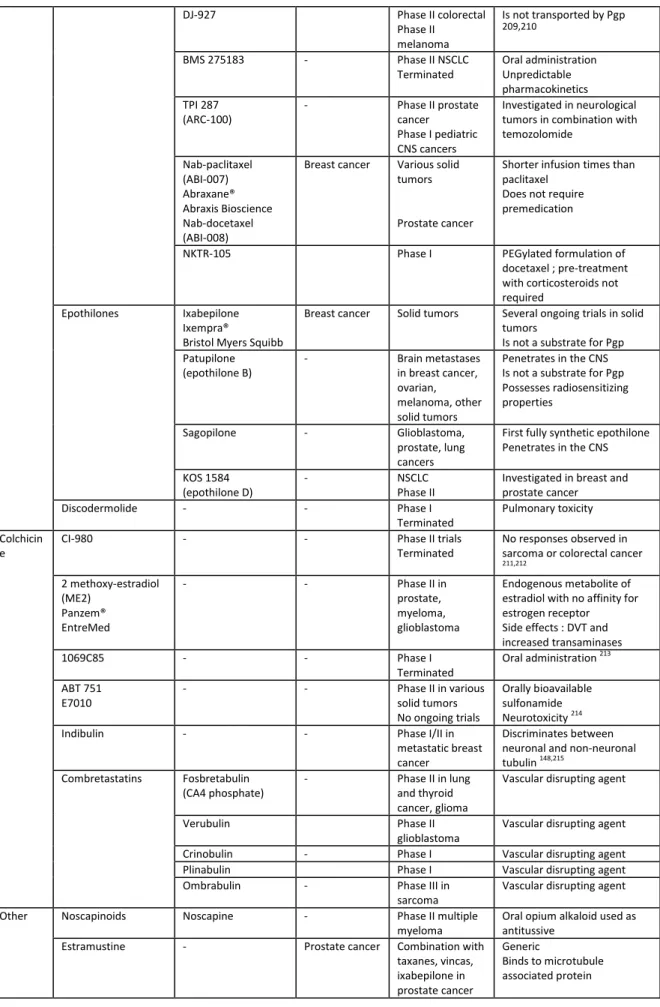
Table 2. Selected microtubule-binding agents which have been approved or have undergone clinical evaluation
ALL: acute lymphoblastic leukemia; CNS: central nervous system; DVT: deep vein thrombosis; MTD: maximal tolerated dose; NSCLC: non small cell lung cancer; OS: overall survival; Pgp: P glycoprotein
Data in this table have been obtained from clinicaltrials.gov, Pubmed, ASCO, company sites and the Thomson Pharma Partnering database.
20
References
1. Noble, R. L., Beer, C. T. & Cutts, J. H. Role of chance observations in chemotherapy: Vinca rosea. Ann N Y Acad Sci 76, 882-94 (1958).
2. Schiff, P. B., Fant, J. & Horwitz, S. B. Promotion of microtubule assembly in vitro by taxol. Nature 277, 665-7 (1979).
Initial description that paclitaxel induces tubulin polymerization.
3. Wani, M. C., Taylor, H. L., Wall, M. E., Coggon, P. & McPhail, A. T. Plant antitumor agents. VI. The isolation and structure of taxol, a novel antileukemic and antitumor agent from Taxus brevifolia. J Am Chem Soc 93, 2325-7 (1971).
4. Pajk, B. et al. Anti-tumor activity of capecitabine and vinorelbine in patients with anthracycline- and taxane-pretreated metastatic breast cancer: findings from the EORTC 10001 randomized phase II trial. Breast 17, 180-5 (2008).
5. Norris, B. et al. Phase III comparative study of vinorelbine combined with doxorubicin versus doxorubicin alone in disseminated metastatic/recurrent breast cancer: National Cancer Institute of Canada Clinical Trials Group Study MA8. J Clin Oncol 18, 2385-94 (2000).
6. Dimitroulis, J. & Stathopoulos, G. P. Evolution of non-small cell lung cancer chemotherapy (Review). Oncol Rep 13, 923-30 (2005).
7. Gridelli, C. et al. Treatment of advanced non-small-cell lung cancer in the elderly: results of an international expert panel. J Clin Oncol 23, 3125-37 (2005).
8. Markman, M. Antineoplastic agents in the management of ovarian cancer: current status and emerging therapeutic strategies. Trends Pharmacol Sci 29, 515-9 (2008). 9. Amador, M. L., Jimeno, J., Paz-Ares, L., Cortes-Funes, H. & Hidalgo, M. Progress in
the development and acquisition of anticancer agents from marine sources. Ann Oncol
14, 1607-15 (2003).
10. Nicolaou, K. C. et al. Total synthesis of taxol. Nature 367, 630-4 (1994).
11. de Lemos, E. et al. Total synthesis of discodermolide: optimization of the effective synthetic route. Chemistry 14, 11092-112 (2008).
12. Busch, T. & Kirschning, A. Recent advances in the total synthesis of pharmaceutically relevant diterpenes. Nat Prod Rep 25, 318-41 (2008).
13. Wender, P. A., Hegde, S. G., Hubbard, R. D. & Zhang, L. Total synthesis of (-)-laulimalide. J Am Chem Soc 124, 4956-7 (2002).
14. Sammak, P. J. & Borisy, G. G. Direct observation of microtubule dynamics in living cells. Nature 332, 724-6 (1988).
15. Kelling, J., Sullivan, K., Wilson, L. & Jordan, M. A. Suppression of centromere dynamics by Taxol in living osteosarcoma cells. Cancer Res 63, 2794-801 (2003). 16. Okouneva, T., Azarenko, O., Wilson, L., Littlefield, B. A. & Jordan, M. A. Inhibition
of centromere dynamics by eribulin (E7389) during mitotic metaphase. Mol Cancer Ther 7, 2003-11 (2008).
17. Hamel, E. & Covell, D. G. Antimitotic peptides and depsipeptides. Curr Med Chem Anticancer Agents 2, 19-53 (2002).
18. Lacey, E. & Gill, J. H. Biochemistry of benzimidazole resistance. Acta Trop 56, 245-62 (1994).
19. Azarenko, O., Okouneva, T., Singletary, K. W., Jordan, M. A. & Wilson, L.
Suppression of microtubule dynamic instability and turnover in MCF7 breast cancer cells by sulforaphane. Carcinogenesis 29, 2360-8 (2008).
20. Lobert, S., Ingram, J. W. & Correia, J. J. Additivity of dilantin and vinblastine inhibitory effects on microtubule assembly. Cancer Res 59, 4816-22 (1999).
21. Buey, R. M. et al. Microtubule interactions with chemically diverse stabilizing agents: thermodynamics of binding to the paclitaxel site predicts cytotoxicity. Chem Biol 12, 1269-79 (2005).
22. Hamel, E. et al. Synergistic effects of peloruside A and laulimalide with taxoid site drugs, but not with each other, on tubulin assembly. Mol Pharmacol 70, 1555-64 (2006).
23. Huzil, J. T. et al. A unique mode of microtubule stabilization induced by peloruside A. J Mol Biol 378, 1016-30 (2008).
24. Jordan, M. A. & Kamath, K. How do microtubule-targeted drugs work? An overview. Curr Cancer Drug Targets 7, 730-42 (2007).
25. Zhou, J. & Giannakakou, P. Targeting microtubules for cancer chemotherapy. Curr Med Chem Anticancer Agents 5, 65-71 (2005).
26. Infante, J. R. et al. Phase II trial of weekly docetaxel, vinorelbine, and trastuzumab in the first-line treatment of patients with HER2-positive metastatic breast cancer. Clin Breast Cancer 9, 23-8 (2009).
27. William, W. N., Jr. et al. Phase II Study of Vinorelbine and Docetaxel in the
Treatment of Advanced Non-Small-Cell Lung Cancer as Frontline and Second-Line Therapy. Am J Clin Oncol (2009).
28. Hudes, G. R. et al. Phase II study of estramustine and vinblastine, two microtubule inhibitors, in hormone-refractory prostate cancer. J Clin Oncol 10, 1754-61 (1992). 29. Giannakakou, P., Villalba, L., Li, H., Poruchynsky, M. & Fojo, T. Combinations of
paclitaxel and vinblastine and their effects on tubulin polymerization and cellular cytotoxicity: characterization of a synergistic schedule. Int J Cancer 75, 57-63 (1998).
Preclinical study analysing cytotoxicity on cell lines showing that under certain conditions a vinca alkaloid and a taxane can be synergistic.
30. Photiou, A., Shah, P., Leong, L. K., Moss, J. & Retsas, S. In vitro synergy of
paclitaxel (Taxol) and vinorelbine (navelbine) against human melanoma cell lines. Eur J Cancer 33, 463-70 (1997).
31. Jordan, M. A., Toso, R. J., Thrower, D. & Wilson, L. Mechanism of mitotic block and inhibition of cell proliferation by taxol at low concentrations. Proc Natl Acad Sci U S A 90, 9552-6 (1993).
Demonstration that taxol modifies microtubule dynamics at concentrations that do not affect microtubule mass and shares a common antiproliferative
mechanism with vinblastine.
32. Ng, S. S. et al. Influence of formulation vehicle on metronomic taxane chemotherapy: albumin-bound versus cremophor EL-based paclitaxel. Clin Cancer Res 12, 4331-8 (2006).
33. Tozer, G. M., Kanthou, C. & Baguley, B. C. Disrupting tumour blood vessels. Nat Rev Cancer 5, 423-35 (2005).
34. Lippert, J. W., 3rd. Vascular disrupting agents. Bioorg Med Chem 15, 605-15 (2007). 35. Dark, G. G. et al. Combretastatin A-4, an agent that displays potent and selective
toxicity toward tumor vasculature. Cancer Res 57, 1829-34 (1997).
36. Griggs, J., Metcalfe, J. C. & Hesketh, R. Targeting tumour vasculature: the development of combretastatin A4. Lancet Oncol 2, 82-7 (2001).
37. Kanthou, C. & Tozer, G. M. The tumor vascular targeting agent combretastatin A-4-phosphate induces reorganization of the actin cytoskeleton and early membrane blebbing in human endothelial cells. Blood 99, 2060-9 (2002).
38. Tozer, G. M. et al. Mechanisms associated with tumor vascular shut-down induced by combretastatin A-4 phosphate: intravital microscopy and measurement of vascular permeability. Cancer Res 61, 6413-22 (2001).
22
These authors describe the rapid and reversible in vivo effect of the vascular disrupting agent combretastatin in a tumor implanted in a rat model.
39. Hori, K., Saito, S. & Kubota, K. A novel combretastatin A-4 derivative, AC7700, strongly stanches tumour blood flow and inhibits growth of tumours developing in various tissues and organs. Br J Cancer 86, 1604-14 (2002).
40. Anderson, H. L. et al. Assessment of pharmacodynamic vascular response in a phase I trial of combretastatin A4 phosphate. J Clin Oncol 21, 2823-30 (2003).
41. Beauregard, D. A. et al. Magnetic resonance imaging and spectroscopy of
combretastatin A4 prodrug-induced disruption of tumour perfusion and energetic status. Br J Cancer 77, 1761-7 (1998).
42. Galbraith, S. M. et al. Combretastatin A4 phosphate has tumor antivascular activity in rat and man as demonstrated by dynamic magnetic resonance imaging. J Clin Oncol
21, 2831-42 (2003).
43. Fojo, A. T. & Menefee, M. Microtubule targeting agents: basic mechanisms of multidrug resistance (MDR). Semin Oncol 32, S3-8 (2005).
44. Breuninger, L. M. et al. Expression of multidrug resistance-associated protein in NIH/3T3 cells confers multidrug resistance associated with increased drug efflux and altered intracellular drug distribution. Cancer Res 55, 5342-7 (1995).
45. Huisman, M. T., Chhatta, A. A., van Tellingen, O., Beijnen, J. H. & Schinkel, A. H. MRP2 (ABCC2) transports taxanes and confers paclitaxel resistance and both processes are stimulated by probenecid. Int J Cancer 116, 824-9 (2005).
46. Hopper-Borge, E., Chen, Z. S., Shchaveleva, I., Belinsky, M. G. & Kruh, G. D. Analysis of the drug resistance profile of multidrug resistance protein 7 (ABCC10): resistance to docetaxel. Cancer Res 64, 4927-30 (2004).
47. Kuttesch, J. F. et al. P-glycoprotein expression at diagnosis may not be a primary mechanism of therapeutic failure in childhood rhabdomyosarcoma. J Clin Oncol 14, 886-900 (1996).
48. Beck, W. T. et al. Methods to detect P-glycoprotein-associated multidrug resistance in patients' tumors: consensus recommendations. Cancer Res 56, 3010-20 (1996).
49. Meisel, C., Roots, I., Cascorbi, I., Brinkmann, U. & Brockmoller, J. How to manage individualized drug therapy: application of pharmacogenetic knowledge of drug metabolism and transport. Clin Chem Lab Med 38, 869-76 (2000).
50. Lhomme, C. et al. Phase III study of valspodar (PSC 833) combined with paclitaxel and carboplatin compared with paclitaxel and carboplatin alone in patients with stage IV or suboptimally debulked stage III epithelial ovarian cancer or primary peritoneal cancer. J Clin Oncol 26, 2674-82 (2008).
51. Fromm, M. F. P-glycoprotein: a defense mechanism limiting oral bioavailability and CNS accumulation of drugs. Int J Clin Pharmacol Ther 38, 69-74 (2000).
52. Chaudhuri, A. R. et al. The tumor suppressor protein Fhit. A novel interaction with tubulin. J Biol Chem 274, 24378-82 (1999).
53. Cheung, C. H. et al. Survivin counteracts the therapeutic effect of microtubule de-stabilizers by stabilizing tubulin polymers. Mol Cancer 8, 43 (2009).
54. Don, S. et al. Neuronal-associated microtubule proteins class III beta-tubulin and MAP2c in neuroblastoma: role in resistance to microtubule-targeted drugs. Mol Cancer Ther 3, 1137-46 (2004).
55. Tian, G. et al. Pathway leading to correctly folded beta-tubulin. Cell 86, 287-96 (1996).
These authors report the nature and interactions between the tubulin binding
56. Alli, E., Bash-Babula, J., Yang, J. M. & Hait, W. N. Effect of stathmin on the sensitivity to antimicrotubule drugs in human breast cancer. Cancer Res 62, 6864-9 (2002).
57. Galmarini, C. M. et al. Drug resistance associated with loss of p53 involves extensive alterations in microtubule composition and dynamics. Br J Cancer 88, 1793-9 (2003). 58. Seve, P. & Dumontet, C. Is class III beta-tubulin a predictive factor in patients
receiving tubulin-binding agents? Lancet Oncol 9, 168-75 (2008).
59. Dumontet, C., Jordan, M. A. & Lee, F. F. Ixabepilone: targeting betaIII-tubulin expression in taxane-resistant malignancies. Mol Cancer Ther 8, 17-25 (2009). 60. Bhattacharya, R. & Cabral, F. Molecular basis for class V beta-tubulin effects on
microtubule assembly and paclitaxel resistance. J Biol Chem 284, 13023-32 (2009). 61. Haber, M. et al. Altered expression of M beta 2, the class II beta-tubulin isotype, in a
murine J774.2 cell line with a high level of taxol resistance. J Biol Chem 270, 31269-75 (1995).
62. Kavallaris, M., Burkhart, C. A. & Horwitz, S. B. Antisense oligonucleotides to class III beta-tubulin sensitize drug-resistant cells to Taxol. Br J Cancer 80, 1020-5 (1999). 63. Kavallaris, M. et al. Multiple microtubule alterations are associated with Vinca
alkaloid resistance in human leukemia cells. Cancer Res 61, 5803-9 (2001).
64. Gan, P. P., Pasquier, E. & Kavallaris, M. Class III beta-tubulin mediates sensitivity to chemotherapeutic drugs in non small cell lung cancer. Cancer Res 67, 9356-63 (2007). 65. McCarroll, J. A., Gan, P. P., Liu, M. & Kavallaris, M. Beta III-Tubulin Is a
Multifunctional Protein Involved in Drug Sensitivity and Tumorigenesis in Non-Small Cell Lung Cancer. Cancer Res 70, 4995-5003 (2010).
Study showing that tubulin III peptide is involved not only in resistance to therapy, an observation confirmed in clinical trials by several authors, but in tumorigenesis as well.
66. Ferrandina, G. et al. Class III beta-tubulin overexpression is a marker of poor clinical outcome in advanced ovarian cancer patients. Clin Cancer Res 12, 2774-9 (2006). 67. Giannakakou, P. et al. Paclitaxel-resistant human ovarian cancer cells have mutant
beta-tubulins that exhibit impaired paclitaxel-driven polymerization. J Biol Chem 272, 17118-25 (1997).
68. Gokmen-Polar, Y. et al. beta-Tubulin mutations are associated with resistance to 2-methoxyestradiol in MDA-MB-435 cancer cells. Cancer Res 65, 9406-14 (2005). 69. Hari, M. et al. Paclitaxel-resistant cells have a mutation in the paclitaxel-binding
region of beta-tubulin (Asp26Glu) and less stable microtubules. Mol Cancer Ther 5, 270-8 (2006).
70. Monzo, M. et al. Paclitaxel resistance in non-small-cell lung cancer associated with beta-tubulin gene mutations. J Clin Oncol 17, 1786-93 (1999).
This study linking mutations of tubulin genes to taxane resistance was later found to be mistaken due to sequencing of tubulin pseudogenes.
71. Sale, S. et al. Conservation of the class I beta-tubulin gene in human populations and lack of mutations in lung cancers and paclitaxel-resistant ovarian cancers. Mol Cancer Ther 1, 215-25 (2002).
72. Sale, S., Oefner, P. J. & Sikic, B. I. Genetic analysis of the beta-tubulin gene, TUBB, in non-small-cell lung cancer. J Natl Cancer Inst 94, 776-7 (2002).
73. Wahl, A. F. et al. Loss of normal p53 function confers sensitization to Taxol by increasing G2/M arrest and apoptosis. Nat Med 2, 72-9 (1996).
These data suggested that paclitaxel could be more active in cells which had lost normal P53 function, a common occurrence in tumor cells.