HAL Id: inserm-00743408
https://www.hal.inserm.fr/inserm-00743408
Submitted on 18 Oct 2012
HAL is a multi-disciplinary open access
archive for the deposit and dissemination of
sci-entific research documents, whether they are
pub-lished or not. The documents may come from
teaching and research institutions in France or
abroad, or from public or private research centers.
L’archive ouverte pluridisciplinaire HAL, est
destinée au dépôt et à la diffusion de documents
scientifiques de niveau recherche, publiés ou non,
émanant des établissements d’enseignement et de
recherche français ou étrangers, des laboratoires
publics ou privés.
cancer cells.
Anne-Marie Gaben, Michèle Sabbah, Gérard Redeuilh, Monique Bedin, Jan
Mester
To cite this version:
Anne-Marie Gaben, Michèle Sabbah, Gérard Redeuilh, Monique Bedin, Jan Mester. Ligand-free
estrogen receptor activity complements IGF1R to induce the proliferation of the MCF-7 breast cancer
cells.. BMC Cancer, BioMed Central, 2012, 12 (1), pp.291. �10.1186/1471-2407-12-291�.
�inserm-00743408�
R E S E A R C H A R T I C L E
Open Access
Ligand-free estrogen receptor activity complements
IGF1R to induce the proliferation of the MCF-7
breast cancer cells
Anne-Marie Gaben
1,2*, Michèle Sabbah
1,2, Gérard Redeuilh
1,2, Monique Bedin
1,2and Jan Mester
1,2Abstract
Background: Ligand-dependent activation of the estrogen receptor (ER) as well as of the insulin-like growth
factor type 1 (IGF1R) induces the proliferation of luminal breast cancer cells. These two pathways cooperate and
are interdependent. We addressed the question of the mechanisms of crosstalk between the ER and IGF1R.
Methods: We evaluated the mitogenic effects of estradiol (E2; agonist ligand of ER) and of insulin (a ligand
of IGF1R) in the MCF-7 cells by flow cytometry and by analyzing the cell levels of cell cycle-related proteins
(immunoblotting) and mRNA (RT-QPCR). To verify the requirement for the kinase activity of Akt (a downstream
target of IGF1R) in the mitogenic action of estradiol, we used shRNA strategy and shRNA-resistant expression
vectors.
Results: The activation of the ER by E2 is unable to induce the cell cycle progression when the phosphatidyl
inositol-3 kinase (PI3K)/Akt signaling is blocked by a chemical inhibitor (LY 294002) or by shRNA targeting Akt1
and Akt2. shRNA-resistant Akt wild-type constructs efficiently complemented the mitogenic signaling activity of
E2 whereas constructs with inactivated kinase function did not. In growth factor-starved cells, the residual PI3K/Akt
activity is sufficient to complement the mitogenic action of E2. Conversely, when ER function is blocked by the
antiestrogen ICI 182780, IGF1R signaling is intact but does not lead to efficient reinitiation of the cell cycle in
quiescent, growth factor-starved MCF-7 cells. The basal transcription-promoting activity of ligand-free ER in
growth factor-starved cells is sufficient to complement the mitogenic action of the IGF1R-dependent signaling.
Conclusions: The basal ER activity in the absence of ligand is sufficient to allow efficient mitogenic action of IGF1R
agonists and needs to be blocked to prevent the cell cycle progression.
Background
Therapies based on hormonal manipulations are
rou-tinely applied in breast cancer patients whose tumors
express estrogen receptor α (ER) (“luminal breast
cancer”, some 75–80% of all breast cancers); of these,
some 50% benefit from objective responses. The current
methods use the inhibition of action of endogenous
estro-gens by selective estrogen receptor modulators (SERM)
such as tamoxifen, or by the suppression of endogenous
estrogen production by aromatase inhibitors [1,2].
The primary lack of sensitivity to these therapies of
a subset of luminal tumors, as well as the secondary
resistance which sets in after an initial response, prevent
the cure of patients from their cancer by hormonal
ther-apy alone. There has been extensive speculation
con-cerning the mechanisms of resistance. Activating ER
mutations or cyclic AMP-dependent phosphorylation [3]
account only for a small fraction of relapses. The
major-ity of relapses of breast cancer under hormone therapy
probably results from alternative mitogenic pathways
triggered by polypeptide growth factors (HER family and
IGF) whose actions are transmitted by membrane
recep-tors [4-6]. These pathways have their own impact on cell
survival and proliferation but can also phosphorylate the
ER (and/or the appropriate transcriptional co-activators)
and reinforce its activity. Laboratory research using
breast cancer-derived cell lines produced abundant
information concerning mitogenic signaling pathways
* Correspondence:anne-marie.gaben@inserm.fr
1Inserm U938, Centre de Recherche Saint-Antoine, Hôpital Saint-Antoine, Bâtiment Kourilsky, 34 rue Crozatier, 75571, Paris cedex 12, France 2
Université Pierre-et-Marie-Curie Paris 6, 75005, Paris, France
© 2012 Gaben et al.; licensee BioMed Central Ltd. This is an Open Access article distributed under the terms of the Creative Commons Attribution License (http://creativecommons.org/licenses/by/2.0), which permits unrestricted use, distribution, and reproduction in any medium, provided the original work is properly cited.
dependent on estrogens as well as on polypeptide growth
factors. However, the data presented by different research
groups are sometimes contradictory. In particular, the
action of estrogens has been reported to be mediated by
direct transcription-promoting activity of the ER [7] or by
activation of kinase cascades identical to those triggered
by cell surface receptors of polypeptide growth factors [8].
Data obtained in our laboratory [9] argue in favor of the
direct transcriptional mechanism, but nonetheless
con-firm the fact that inhibition of the PI3K/Akt cascade by
chemical inhibitors or by shRNA prevents the mitogenic
activity of estradiol in the MCF-7 cells. The importance
of PI3K activity in the IGF-I-induced mitogenic signaling
in the MCF-7 cells has been reported by Dufourny et al.
[10]. Similarly, although to a lesser extent, the inhibition
of the MEK/ERK pathway reduces the mitogenic activity
of estradiol (E2). Conversely, it has been reported that the
mitogenic activity of IGF1R is blocked by ICI 182780
[11,12]; this anti-estrogen belongs to the category of
selective estrogen receptor down-regulators (SERD) since
its presence in the cell culture medium leads to a
sub-stantial decrease in the content of ER [13]. These data
suggest the importance of crosstalk between the signaling
by ER and by growth factor receptors.
In this work we have addressed two questions: first,
the requirement of the PI3K activity and in particular of
the kinase function of its downstream mediator Akt in
the estrogen-induced cell cycle progression, and second,
the interplay between the ER- and IGF1R-dependent
mitogenic signaling pathways.
Methods
Cell culture
Breast cancer-derived cell lines (MCF-7, MELN) were
propagated in DMEM supplemented with 10% fetal
bo-vine serum (FBS).
For experiments, the cells were seeded at approximately
20.10
3/cm
2, allowed to attach overnight, washed twice and
placed in phenol red-free, serum- free DMEM containing
or not 10 nM ICI 182780 for various times as indicated.
Mitogenic stimulation was carried out by pipetting the
reagents directly into the culture medium in the dish to
produce final concentrations: 1 μM estradiol (100-fold
excess over the antiestrogen) or 1 μM insulin (sufficient to
activate the IGF1R), or 10 nM IGF-I. The final
concentra-tions of other drugs used in some experiments were
20 μM for LY 294002 and 10 μg/mL for cycloheximide.
The distribution of cells among the phases of the cell
cycle was evaluated by staining with propidium iodide
and flow cytometry.
Expression vectors and shRNA
The shRNA Akt (1 + 2) vector was a gift of Dr. F.
Czauderna. It contains a sequence (cloned under pol III
promoter in a U6 vector) common to isoforms of Akt1
and Akt2 [14].
The effective and specific suppression of Akt
expres-sion by this sequence in the HeLa cells has been verified
by these authors and we have confirmed this
suppres-sion in the MCF-7 cells (Additional file 1: Figure S1).
To create wild-type Akt1 (Akt1R) and Akt2 (Akt2R)
vectors, resistant to shRNA Akt (1 + 2), we used the
HA-Akt1 and HA-Akt2 expression vectors (obtained from
Xiao GH, Altomare DA and Testa JR, Fox Chase Cancer,
Philadelphia, USA) [15]. We introduced silent mutations
of 3 codons within the shRNA target common sequence.
The following sequences were used: Akt1, forward 5′
CCAACACCTTCATCATCCggTgTCTCCAgTggACCAC
TgTCATCg-3′; reverse 5′-CgATgACAgTggTCCACTggA
gACACCggATgATgAAggTgTTgg-3′ and Akt2, forward
5′-CCAACACCTTTgTCATACggTgTCTCCAgTggACC
ACAgTCATCG-3′; reverse 5′-CgATgACTgTgTggTCCA
CTggAgACACCgTATgACAAAggTgTTgg-3′.
To replace the endogenous Akt1 or Akt2 by
kinase-dead, sh-RNA-resistant variants, we introduced additional
mutation substituting alanine for lysine at position 179 or
181 for Akt1 and Akt2 respectively in the catalytic
domains of Akt1R and Akt2R kinases [16]. Point
muta-tion was accomplished by PCR primer mutagens using
the QuikChange II Site-Directed Mutagenesis Kit
(Strata-gene). The following sequences were used: Akt1R/KD
(Kinase Dead), forward 5′-CgCTACTACgCCATggCgAT
CCTCAAgAAgg-3′;
reverse
5′-CCTTCTTgAggA
TCgCCATggCgTAgTAgCg-3′ and Akt2R/KD (Kinase
dead), forward 5′-CgCTAC TACgCCATggCgATCCTgCg
AAAgg-3′; reverse 5′-CCTTTCgCAggATCgCCATggCgT
Ag TAgCg-3′.
Control cells were transfected with the empty pcDNA3
vector. For each transfection, the total quantity of
trans-fected plasmid DNA was completed to 2 μg by the
addition of pcDNA3 plasmid (Invitrogen, Life
Technolo-gies, Carlsbad, CA). The indicator plasmid used was
pCA-Luc (luciferase cDNA cloned downstream of the
cyclin A promoter) [17].
Transfection experiments
Cells were transfected with expression vectors
con-taining: shRNA sequence complementary to Akt1 and
Akt2 mRNA (shAkt1 + 2); shRNA-resistant Akt1 or Akt2
(Akt1R and Akt2R); shRNA-kinase dead Akt1 and Akt2
(Akt1R/KD and Akt2R/KD); cyclin A-luciferase
(indica-tor of late G1 phase; β-galactosidase (indica(indica-tor of
trans-fection efficiency). Transtrans-fections were carried out by the
Lipofectamine Plus method (Invitrogen) according to
the manufacturer’s protocol. After 3 h incubation with
the DNA-containing liposomes, the cells were rinsed
and incubated 40 h in serum-free, phenol red-free
DMEM with 10 nM ICI 182780 prior to stimulation
with E2 for additional 24 h. Cells were then lysed in
Reporter Lysis Buffer (Promega) and the luciferase and
β-galactosidase (Galacto-Star-Applied Biosystems)
activ-ities were determined.
Western blotting
Cells were harvested on ice in a Tris (50 mM, pH 7.4)
buffer containing EDTA (20 mM) Nonidet P-40 (0.5%),
NaCl (150 mM), dithiothreitol (1 mM), aprotinin (1 mg/
mL), leupeptine (1 mg/mL), phenylmethylsulfonyl
fluor-ide (0.3 mM), NaF (1 mM), and sodium orthovanadate
(1 mM). The lysates were clarified by centrifugation
(10,000 × g for 5 min). The total protein concentration
was determined by Bio-Rad assay (Bio-Rad, Hercules,
CA). 100 μg of total protein were denaturated by boiling
in Læmmli buffer containing sodium dodecyl sulfate (1%
final concentration) and 2-mercaptoethanol (100 mM)
before fractionation by electrophoresis in a
polyacryl-amide gel (8% or 10% as needed). The proteins were
then electrotransferred onto a Hybond membrane and
incubated with the appropriate antibodies followed by
the peroxidase-tagged secondary antibody. The primary
antibodies used were: from Cell Signaling Technology
(Beverly, MA) for Akt, phospho-Ser473-Akt, IGF1R
(β chain), phospho-GSK3α/β, p21
WAF1/CIP1, cyclin A;
from Santa Cruz Biotechnology (Santa Cruz, CA, USA)
for p27 (C-19); from Thermo Fisher Scientific Fremont,
(CA, USA) for cyclin D1 (cyclin D1/bcl-1 Ab-1, clone
DCS-6); from Millipore Corporation (Temecula, CA,
USA) for phospho-ER; from BD Pharmingen (Le Pont
de Claix, France) for Rb.
The detection of the signal was carried out with the
enhanced chemoluminescence kit (Amersham
Bios-ciences, Saclay, France.).
mRNA quantification
RNA was isolated by using Trizol (Euromedex). One
microgram of total RNA was reverse transcribed with
200 ng random primers (Invitrogen) and ImProm-II
reverse transcriptase (Promega) for 60 min at 42°C, in
20 μl final volume.
The cDNA (equivalent of 0.2 μL of the RT reaction
mix) was subjected to Q-PCR using Sybr green (Applied
Biosciences, Foster City, CA) and appropriate primers.
The mRNA contents were evaluated based on the
com-parative ΔCT method and normalized to the
housekeep-ing gene 36B4 as described previously [18].
Results
To reduce the risk that experimental results may be
influenced by cell heterogeneity, we subcloned MCF-7
cells by limiting dilution. All clones analyzed (18 in
total) ceased to proliferate in serum- and estrogen-free
medium, and responded to mitogenic stimulation by E2
and insulin. Four clones were further analyzed and
found to express the ER and PR (inducible by E2). One
of these clones was used in all subsequent experiments.
1. The kinase function of Akt is required for the
E2-dependent cell cycle progression.
In our previous work we showed that depletion
of Akt1 and 2 prevented the mitogenic signaling
by E2 in the MCF-7 cells. At the same time,
E2 stimulation failed to induce the activating
phosphorylation of Akt on Ser 473. This opened
the possibility that Akt may have a function
unrelated to its kinase activity, as has been suggested
in a different context [
16
,
19
]. We therefore
produced Akt1 and Akt2 expression vectors carrying
silent mutations in the sequence targeted by shRNA,
as well as in the kinase domain. As reported by
Nakatani et al. [
20
] and Zinda et al. [
21
], Akt3 is
not expressed in the MCF-7 cells. We tested these
constructs for their capacity to “rescue” the
mitogenic action of E2 in cells exposed to shRNA
targeting Akt1 and 2. The end-point was the
activation of the promoter of the cyclin A gene
cloned upstream of a luciferase coding sequence,
as an indicator of late G1 phase.
When cells were transfected with the
shRNA-expression vector Akt (1 + 2) directed against a
sequence shared by Akt1 and 2 mRNAs, the
activation of the cyclin A promoter by E2 was
blocked and co-transfection of expression vectors
coding for shRNA-resistant, wild type kinase
variants of the Akt isoforms (Akt1R, Akt2R)
restored the cyclin A promoter activation as
revealed by the induction of luciferase. Akt2
appeared to be more efficient to restore the full
mitogenic effect of E2 than Akt1 (Figure
1A
).
Next we compared the wild-type, shRNA-resistant
Akt constructs with their kinase-dead counterparts
Akt1R/KD and Akt2R/KD. In these experiments,
the inclusion of the KD variants resulted in a
reduced transfection efficiency documented by the
diminished activity of the indicator β-galactosidase.
Therefore, we treated groups of dishes with E2 and
kept other groups of dishes as controls (in the
serum-free medium supplemented with ICI 182780),
to calculate the induction factor for the luciferase/
β-galactosidase ratios. The results showed that with
the kinase-dead mutants, there was only a partial
restoration of luciferase induction (Figure
1B
) as
compared with the wild-type Akt2R used as a
positive control. The results of these experiments
demonstrate that the kinase function of exogenous
Akt is required for efficient rescue of E2-inducible
cell cycle progression when endogenous Akt is
knocked down.
2. Cells deprived of serum in the absence of ICI
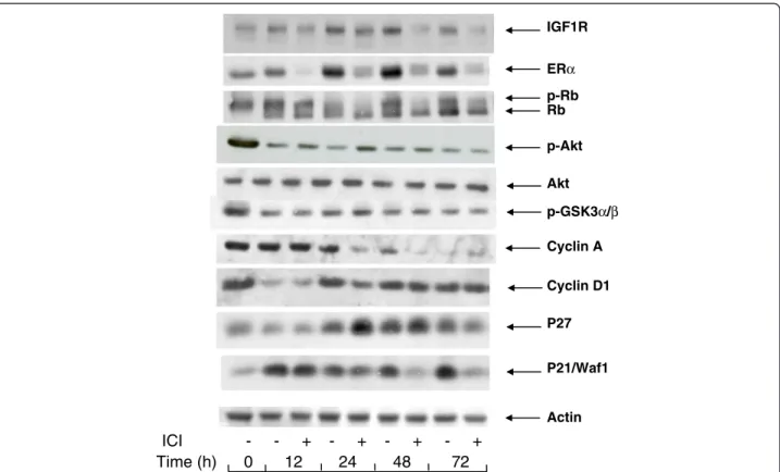
182780 continue to express cell cycle markers.
The arrest of proliferation by depriving the MCF-7
cells of exogenous mitogens was characterized by
changes in the cell contents of certain markers of
mitogenic signaling of the cell cycle (Figure
2
).
Interruption of the mitogenic signaling is illustrated
by the changes in the phosphorylation status of the
Rb protein, a substrate of cyclin-dependent kinases
and a modulator of late G1-phase gene expression.
After incubation for 24 h or longer in serum and
phenol red-free medium containing ICI 182780, Rb
was dephosphorylated, whereas a significant fraction
of Rb remained phosphorylated when ICI 182780
was omitted. This indicates that the suppression of
ER by the antiestrogen is required for an efficient
block of the induction of cyclin-dependent kinases.
This conclusion is also supported by the presence of
a residual cyclin A in cells deprived of serum in the
absence of the antiestrogen whereas in the presence
of the antiestrogen, the cyclin A signal is nearly
eliminated (Figure
2
).
The cdk inhibitory proteins p21
WAF1/CIP1and p27
accumulated in cells deprived of serum. Whereas
the addition of ICI 182780 in the starvation medium
made no difference for p27, it led to a strongly
sh Akt 1+2 (µg) Akt 2 R (µg) Akt 1 R/KD (µg) Akt 2 R/KD (µg) 0 1 2 3 4 5 Fold Induction cyclinA-Luc/ β -Gal 0 0.6 1.2 1.8 sh Akt 1+2 (µg) Akt 1R (µg) Akt 2R (µg)
A
B
0.4 0.8 -0.8 -- -- -0.5 0.5 0.5 0.5 0.5 0.5 0.5 0.4 - - - -- - - - -- -0.5 0.5 0.5 0.5 0.5 0.5 0.5 0.5 0.25 0.25 0.25 0.25Figure 1 Akt kinase activity is required for the E2 -induced reinitiation of the cell cycle progression. A: MCF-7 cells seeded in 35 mm dishes were transfected with shRNA Akt (1 + 2) (0.5 μg per dish) together with expression vectors of shRNA-resistant mutants of Akt1 (Akt1R) or Akt2 (Akt2R) as indicated, and an indicator plasmid encoding luciferase cloned downstream of cyclin A promoter (0.5 μg). A β-galactosidase vector (100 ng) was included to allow the correction for transfection efficiency. For more details see Materials and Methods. The cells were lysed and the activity of luciferase and β-galactosidase were determined. The results shown are means and s.e.m. of triplicates. B: The cells were transfected as in A with shAkt (1 + 2), Akt2R, or kinase-dead shRNA-resistant mutants of Akt1 (Akt1R/KD) or Akt2 (Akt2R/KD) as indicated. After transfection, the cells were made quiescent as in A. To a set of dishes of each transfection mix, E2 was added to a final concentration of 1 μM, the remaining dishes were left in the medium containing ICI 182780 (control). The data presented show the induction factor (E2 vs. control) calculated for the luciferase/β-galactosidase activities. The results shown are means +/− s.e.m. of triplicates.
reduced cell content of p21
WAF1/CIP1after a
transient increase seen at 12 h (Figure
2
). The
expression of IGF1R also showed a slightly higher
level in cells deprived of serum in a medium without
the antiestrogen. As the suppression of ER by ICI
182780 leads to a reduced expression of certain
genes (see below, section 6), it is likely that the
levels of their protein products result from the basal
transcription-regulating activity of ligand-free ER.
As expected, in the cells serum-starved in medium
with ICI 182780, ER was rapidly eliminated, the
signal being near absent at 12 h. In spite of the
continued presence of ICI 182780, ER became again
detectable at later times. Starvation of serum and
E2 in the absence of the antiestrogen led to a
progressive accumulation of ER, as seen between
24 and 72 h.
It is to be noted that the cell contents of cyclin D1,
a marker of early G1 phase, showed an early
decrease at 12 h but then regained about the initial
level and remained approximately constant
throughout the 72 h incubation in serum-free
medium. The presence of ICI 182780 did not reduce
the level of cyclin D1 in mitogen-deprived cells
(Figure
2
).
3. Serum and estrogen deprivation does not eliminate
phospho-Akt.
Since the presence of the wild-type form of Akt is
a prerequisite for the mitogenic signaling by E2
and since E2 does not induce the activating
phosphorylation of Akt, we set out to verify by
Western blotting the presence of
phospho-Ser473-Akt (p-phospho-Ser473-Akt) in the MCF-7 cells incubated in serum
and estrogen-free medium. In these experiments the
intensity of the p-Akt signal became weaker during
serum deprivation but remained detectable, whether
the cells had been incubated in a medium deprived
of serum and exogenous estrogens, or in the same
medium supplemented with ICI 182780. GSK3α/β a
substrate of Akt kinase, showed a similar profile of
phosphorylation (Figure
2
).
In order to verify that the signal detected with
the anti-P-Ser473-Akt antibody represented the
phosphorylated Akt rather than a non-specific
antigen co-migrating incidentally with Akt, we
ICI
-
-
+ -
+
-
+
-
+
Time (h)
0
12
24
48
72
p-Rb Rb p-Akt p-GSK3α/β P21/Waf1 IGF1R P27 Cyclin D1 ERα Akt Cyclin A ActinFigure 2 Changes in the levels of cell cycle-related and signaling proteins during serum- and E2 -starvation. MCF-7 cells were seeded in 60 mm dishes and allowed to attach overnight. They were then placed in serum- and phenol red-free medium containing or not 10 nM ICI 182780 for periods of time as indicated. After lysis, the different proteins were revealed by Western blotting with the appropriate antibodies.
treated the cell lysates with phosphatase. This
treatment abolished the p-Akt signal both in cell
lysates prepared from the quiescent MCF-7 cells and
in cells treated for 1 h with insulin, a powerful
inducer of the PI3K/Akt signaling (see Additional
file
2
: Figure
S2
).
The phosphorylation of Akt in the quiescent MCF-7
cells could be a consequence of signaling by an
autocrine factor. To test this possibility, we
harvested conditioned medium from cells after 48 h
of incubation in the absence of serum and we
compared the phosphorylation of Akt in quiescent
cells placed in fresh DMEM with that detected in
cells incubated with the conditioned medium.
No difference was seen, suggesting that the Akt
phosphorylation resulted from endogenous
mechanisms and was not mediated by a secreted
autocrine factor (see Additional file
3
: Figure
S3
).
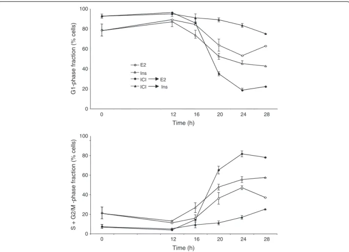
4. IGF1R signal transduction is not sufficient to drive
the G1 phase progression.
Stimulation of the IGF1R signaling pathway induces
a rapid and lasting phosphorylation of Akt. IGF-I
and -II, as well as insulin at supra-physiological
concentrations, are efficient mitogens in
estrogen-deprived MCF-7 cells. Also, simultaneous
stimulation of this pathway and of the ER acts in
synergy to induce the MCF-7 cells’ proliferation. It
has been reported by the laboratory of R. Sutherland
that suppression of ER-dependent signaling by ICI
182780 prevents the mitogenic activity of insulin
in these cells whereas antiestrogens of the type
“SERM” do not show this effect [
22
]. Varma and
Conrad [
12
] showed that the direct effects of IGF,
phosphorylation of IGF1R and of Akt, are unaffected
by ICI 182780, in contrast with the inhibition of
the mitogenic action. We have addressed the
mechanisms underlying the cooperation of the ER
and IGF1R pathways. We analyzed the effects of E2
and insulin on the distribution of cells among the
phases of the cell division cycle (Figure
3
; Additional
file
4
: Figure
S4
). Remarkably, even after 48 h
incubation in serum-free medium, the MCF-7 cells
did not become fully quiescent, with approximately
20% of the total population in S+G2/M-phase
(Figure
3
, time 0, lower panel). If the serum-free
culture medium contained ICI 182780, after 48 h
there remained practically no S+G2/M-phase cells.
Stimulation with E2 or with insulin triggered the
re-entry of G0/G1 arrested cells into the cell division
cycle (Figure
3
). The most marked mitogenic effect
was seen when the cells were fully synchronized by
serum-starvation in the presence of ICI 182780 and
subsequently stimulated by the addition of E2
(100-fold excess over ICI 182780). In these conditions,
insulin produced only a weak and delayed effect. In
contrast, insulin was an efficient mitogen when ICI
182780 was omitted from the culture medium
(Figure
3
, lower panel).
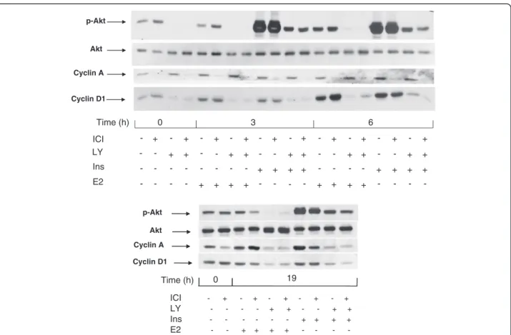
These data confirm that pretreatment of the MCF-7
cells with ICI 182780 strongly reduces their
sensitivity to the mitogenic action of insulin
(Figure
3
) while the signal transduction by IGF1R is
intact as documented by the strong induction of
Akt phosphorylation by insulin in such cells, similar
to that seen in cells deprived of serum in the
absence of the antiestrogen (Figure
4
, upper panel).
We also observed an induction of cyclin D1 in cells
starved of serum with and without ICI 182780,
confirming that this process reflects direct IGFR1
signaling and is not sufficient for the cell cycle
progression. There was though a correlation
between the induction of cyclin D1 accumulation
and the mitogenic action as shown by the FACS
data: stronger induction by E2, weaker by insulin in
antiestrogen-exposed cells.
The fact that chemical inhibitors of PI3K block the
mitogenic signaling in breast cancer cells has been
reported earlier [
9
,
23
]. This is also illustrated by the
effect of LY 294002 on the expression of cyclin A
(Figure
4
, upper panel). In cells starved of mitogens
in a medium without antiestrogen, cyclin A
remained detectable, and its content did not
diminish during a short (3 to 6 h) incubation with
LY 294002 (Figure
4
, upper panel). The expression
of cyclin A in these conditions is probably the
consequence of the incomplete quiescence
(Figure
3
). When the cells were stimulated with E2
or with insulin for 19 h (late G1 phase), cyclin A
was strongly induced and this induction was
abolished by LY 294002 (Figure
4
, lower panel).
As expected, the effect of IGF-I (10 nM) was the
same as that of insulin (1 μM) (Additional file
5
:
Figure
S5
).
As ICI 182780 is a SERD-type antiestrogen (inducer
of ER degradation), the lack of ER after pretreatment
with this compound could be a reason for the
diminished sensitivity of the cells to insulin. This is
however unlikely to be the case as the reinitiation
of the cell cycle progression by E2 in ICI
182780-pretreated cells is actually stronger than that of cells
not pretreated with the antiestrogen, in spite of the
strong reduction of the cell contents in ER
(Figure
2
). The recent report of Wardell et al. [
24
]
demonstrates that the efficacy of ICI 182780 as an
antiestrogen does not rely on its ability to induce
ERα degradation.
5. Effect of the suppression of the PI3K pathway on
the expression of cyclin D1 and c-myc protein
and mRNA.
We were intrigued by the continuous presence of
cyclin D1 in serum- and estrogen-deprived cells,
non-suppressible by long-term treatment with ICI
182780. Signaling by the PI3K/Akt pathway favors
the accumulation of the cyclin D1 protein by
post-transcriptional mechanisms: accelerated translation
[
25
,
26
] as well as inhibition of degradation of the
cyclin D1 protein due to the inhibition of GSK3 α/β
through phosphorylation by Akt [
27
].
In order to verify the role of the basal level of
phosphorylated Akt in the expression of cyclin D1,
we examined the effect of the PI3K inhibitor LY
294002. A 3 h incubation of serum-deprived cells
with this drug strongly reduced the p-Akt signal,
indicating that the basal phosphorylation of Akt seen
in mitogen-deprived cells depended on PI3K activity.
Further, our experiments showed a strong inhibition
of the basal cyclin D1 expression by a 3 h exposure
of the cells to LY 294002 (Figure
4
, upper panel,
t = 0). The presence of LY294002 led to a reduction
of the contents in cyclin D1 also when the cells
were stimulated with either insulin or E2 (Figure
4
,
upper panel). Next we examined the transcriptional
regulation of the CCND1 gene (Figure
5
). The
presence of ICI 182780 during serum deprivation
did not modify the level of cyclin D1 mRNA. After
48 h in serum-free medium, an incubation for 3 h
with 20 μM LY294002 led to a 2 to 3-fold decrease
E2Ins ICI E2 ICI Ins
S + G2/M -phase fraction (% cells)
Time (h) Time (h) 0 20 40 60 80 100 28 24 20 16 12 28 24 20 16 12 0
G1-phase fraction (% cells)
0 20 40 60 80 100 0
Figure 3 Reinitiation of the cell cycle progression in quiescent cells. Blocking the ER function inhibits the insulin-induced reinitiation of the cell cycle progression. MCF-7 cells were placed in serum- and phenol red-free medium containing or not 10 nM ICI 182780 for 48 h. Subsequently the cells were stimulated by addition of insulin (Ins; 1 μM) or E2 (1 μM). The cells were harvested for analysis of their DNA contents by flow cytometry at t = 0, 12, 16, 20, 24 and 28 h as shown. The data (means of 2 to 4 experiments) are plotted as percent of cells in the G1 phase and S phase. S.e.m. are shown (unless smaller than the size of the symbol).
of cyclin D1 mRNA contents, indicating that the
basal activity of PI3K was required to maintain the
expression of the CCND1 gene (Figure
5
, bars 3,
4 vs. 1, 2). Stimulation of the quiescent cells with
either E2 or insulin induced the accumulation
of cyclin D1 mRNA (about 3-fold at 4 h). The
amplitude of this induction paralleled the pattern
of reinitiation of the cell cycle progression
(Figure
3
): insulin was more efficient when serum
deprivation had been carried out without ICI
182780 (Figure
5
, bar 5 vs. 6), whereas the effect of
E2 was more marked in cells rendered quiescent in
the presence of ICI 182780 (bar 9 vs. 10). The
induction of cyclin D1 mRNA by E2 was not
prevented by LY 294002 (about 3-fold at 4 h
compared with the level in control, LY
294002-exposed cells; Figure
5
); although the absolute level
was lower than that reached without LY 294002,
the induction of CCND1 transcription by estradiol
apparently proceeded unhindered (compare bars
3 vs. 11 and 4 vs. 12; differences significant at
respectively p ≤0.05 and p ≤ 0.01). On the other
hand, the induction of the expression of the
CCND1
gene by insulin was efficiently inhibited
by LY294002.
In contrast, in cells cultured in serum-free medium,
a 3 h exposure to LY 294002 did not affect the
level of the c-myc mRNA (Figure
5
, bars 1 vs. 3 and
2 vs. 4). The same result was noted when the cells
were stimulated with insulin (absence of effect of
LY 294002; bars 5 vs. 7 and 6 vs. 8). The induction
of c-myc mRNA accumulation by E2 was actually
increased by LY294002 (difference significant for
cells maintained without ICI 182780; bars 9 vs. 11,
p ≤ 0.01). It is to be noted that ICI 182780 prevented
the induction of c-myc mRNA accumulation by
insulin (Figure
5
, compare bars 5 vs. 6 and 7 vs. 8).
6. Transcriptional activity of unliganded ER in
serum-deprived MCF-7 cells.
The essential consequence of the presence of ICI
182780 is the suppression of the basal level of
Cyclin A Cyclin D1 p-Akt Akt Time (h) 0 19 ICI LY Ins E2 - + - + - + - + - + - - - - + + - - + + - - - + + + + - - + + + + - - - -- - + + + + - - - + + + + - - - -ICI LY Ins E2 Time (h) 0 3 6 p-Akt Akt Cyclin A Cyclin D1 -- -- -+ + + + + + + - - -+ + + + + + + + + + + + + + + + + + + + +
Figure 4 Effect of the inhibition of PI3K by LY 294002 on the induction of cyclins D1 and A. MCF-7 cells were starved of serum and E2 during 48 h in the presence or absence of ICI 182780 (10 nM). Last 3 hours of serum deprivation (time −3 h to 0 h), one series of dishes was treated with LY 294002 (LY; 20 μM). Subsequently, at t = 0, all cells were stimulated by the addition of insulin (ins; 1 μM) or E2 (1 μM) for 3 h or 6 h (upper panel) or 19 h (lower panel). After lysis, the different proteins were revealed by after Western blotting using specific antibodies.
ER-dependent gene expression. This was
documented by monitoring the levels of two
transcripts encoded by genes with estrogen response
elements in their promoters, pS2 and PR
(progesterone receptor). ICI 182780 caused a strong
decrease in the expression of these genes (by
approximately 90% after 48 h) whereas in the
absence of the antiestrogen their mRNA levels
decreased respectively by approximately 50% as
compared to those observed in the exponential cells
(Figure
6
).
In order to obtain a more direct information about
the ER-dependent transcription in the absence of
ligand, we evaluated the expression of luciferase in
the MELN cell line derived from the MCF-7 cells by
stable transfection with ERE-TK-LUC [
28
]. When
placed in serum- and phenol red-free medium, the
cell content in luciferase varied little, whereas the
addition of ICI 182780 led to a rapid extinction of
the indicator enzyme, at a rate similar to that caused
by the protein synthesis inhibitor cycloheximide,
after a delay of about 3 h (Figure
7A
). This delay is
understandable: cycloheximide blocks all de novo
synthesis of luciferase protein whereas ICI 182780
prevents the synthesis of mRNA coding for
luciferase and not the translation of pre-existing
mRNA. To ascertain that the continued expression
of luciferase was not due to a possible residual
estrogen, we cultured the MELN cells for more than
a month (a minimum of 4 passages) in estrogen-free
medium supplemented with charcoal-stripped serum
plus 100 nM Insulin. The cells were then placed in
serum-free medium, without insulin, with or
without ICI 182780. Similar results were obtained:
ICI 182780 rapidly extinguished the expression of
luciferase whereas in the absence of the antiestrogen
the level of luciferase increased with time
(Figure
7B
)
A possible explanation of these results is the
existence of pathways that lead to the
phosphorylation of the ER and of co-activators that
participate at the regulation of its transcriptional
activity. This possibility is sustained by the fact that
phospho-Ser118-ER is detected in the
serum-deprived MCF-7 cells (data not shown). The
0 1 2 3 4 5Cyclin D1
c-Myc
ICI Ins E2 LY Time (h) Fold induction Fold induction -- -- - -- - - -- -+ + + + ++ ++ + + + + + + + + + + + + -0 0 0 0 4 4 4 4 4 4 4 4 -0 1 2 3 4 5 6 1 2 3 4 5 6 7 8 9 10 11 12 1 2 3 4 5 6 7 8 9 10 11 12 * * ** ** ** ** ** ** ** * *Figure 5 Effect of LY 294002 on the induction of cyclin D1 and c-myc mRNA. MCF-7 cells were starved of serum and E2, in the presence or absence of ICI 182780, for 48 h. In one series of dishes, LY294002 was added for the last 3 h of serum deprivation (time −3 h to 0 h). The cells were then stimulated with E2 (1 μM) or insulin (1 μM) for 4 h. The cells were then harvested for the isolation of RNA and RT-QPCR analysis. Fold induction of ΔΔCT (means of three independent experiments performed in triplicate) related to the values at t = 0 in cells starved of serum in the absence of ICI 182780, are presented. The differences between bars 3 vs 11, 4 vs 12, 6 vs 8 and 9 vs 11 are significant at p ≤ 0.05 (*) or p ≤ 0.01 **).
mechanism responsible for ER phosphorylation
remains unknown at this moment. As in the case of
the basal, constitutive phosphorylation of Akt, it is
probably the result of an endogenous process, not
requiring added or secreted factors.
Discussion
“Hormone-dependent” breast cancer cells, by definition,
require estrogens for their proliferation. Many
experi-mental models used in the literature employ culture
conditions where cells (usually MCF-7, a line mimicking
luminal breast cancer) are placed in a medium without
phenol red (a weak estrogen) and supplemented with
FBS treated with active charcoal to remove serum
estro-gens. However, the dependence of the MCF-7 cells on
estrogens is not absolute and, in such estrogen-free
media, these cells continue to proliferate, albeit at a slow
rate. Charcoal-stripped FBS contains residual
polypep-tide growth factors (e.g. IGFs), which can stimulate
the proliferation of the MCF-7 cells, but even after 48 h
incubation in serum-free medium, the MCF-7 cells
do not become fully quiescent (Figure 3, time 0). To
obtain quiescence, the serum starvation medium needs
to be supplemented by a “complete” antiestrogen ICI
182780. Even at quiescence, the cellular
phospho-Ser473-Akt (enzymatically active form, dependent on
PI3K signaling) is not completely suppressed. We have
verified that serum-deprived MCF-7 cells do not secrete
autocrine growth factors capable to activate the PI3K/
Akt pathway.
We analyzed the mechanisms that may drive the
re-sidual cell division cycle in estrogen-deprived cells. We
also addressed the question of the role of the PI3K/Akt
signal in the crosstalk between ER and IGF1R in the
G1-phase progression.
We observed that unliganded ER continues to act as a
transcriptional activator in mitogen-deprived cells, and
that this action is blocked by ICI 182780. This is
docu-mented by our data obtained using the MELN cell line
derived from the MCF-7 cells by stable transfection with
an ERE-TK-Luc construct [28]. The basal expression of
the indicator gene in these cells stabilizes at
approxi-mately 50% of the initial level by 48 h and is not
elimi-nated by long-term estrogen deprivation, but is abruptly
blocked by the addition of ICI 182780.
The activity of the unliganded ER results also in a
higher expression of certain cellular genes as compared
with that observed when ER activity is cancelled by ICI
182780. This is the case of the PS2 gene, which contains
an ERE sequence at its promoter, as well as PR
(regu-lated by atypical half-ERE sequences). A higher
expres-sion in serum-starved cells without ICI 182780 is also
seen for certain cellular proteins not known as
ER-targets. For example, p21
WAF1/CIP1increases with the
time of incubation in serum-free medium when ICI
182780 is omitted. This increase may be an indirect
con-sequence of either the unliganded ER activity during
in-cubation in serum-free medium or of the arrest of the
cell cycle (or both). Our laboratory reported earlier that
p21
WAF1/CIP1cooperates with the ER in the regulation of
the expression of genes, apparently with a preference
for those genes that are characteristic of differentiation
of the mammary gland cells [18].
The cell content of ER is enhanced when the cells are
starved of serum and E2 (Figure 2). The expression of
ER-target genes in the absence of agonist ligand may be
reinforced by this increase during serum starvation [29].
In contrast, the levels of cyclin D1 protein or mRNA
were similar irrespective of the presence or not of ICI
182780 during serum deprivation. The CCND1 gene
0 0.5 1 1.5 ICI
PR
pS2
Time (h) - - + - - + 0 48 0 48Standardized mRNA level
Figure 6 ICI 182780 is required for the efficient suppression of ER-dependent endogenous transcripts during serum starvation. Exponentially growing MCF-7 cells (shaded bars) and cells incubated for 48 h in serum- and phenol red-free medium, with or without ICI 182780 (10 nM), were analyzed by RT-QPCR for the ER-inducible pS2 and progesterone receptor (PR) mRNAs. The values of ΔΔCT (means of two independent experiments performed in duplicate) in relation to the exponentially growing cells are presented.
does not contain ERE, and its induction by E2 relies on
the action of ER as a transcriptional co-activator [30].
The sustained expression of CCND1 in serum and
estrogen-deprived MCF-7 cells results apparently from
the activity of other transcription factors [31].
Besides its “canonical” role as a Cdki and its
cooper-ation with ER, p21
WAF1/CIP1protein appears also to be
involved in the activation of Cdk4 [32]. The elevated
expression of p21
WAF1/CIP1could therefore reinforce
the mitogenic signaling resulting from the activation of
IGF1R in cells not exposed to ICI 182780.
As we reported earlier, E2 did not rapidly induce Akt
phosphorylation [9] (Figure 4) similar observations have
been published by others, e.g. [33]. However, the
experi-ments in which we knocked down Akt1 and Akt2 by
targeting their shared nucleotide sequence demonstrated
that the Akt protein is necessary for the full mitogenic
activity of the E2/ER pathway [9]; the present work
moreover indicates that the kinase function of Akt is
required. Akt2 was more efficient than Akt1, in
agree-ment with the report of Morelli et al. [34]. At the same
time, the induction of the PI3K/Akt pathway alone is at
best only weakly mitogenic, as illustrated by the weak/
delayed effect of insulin on the cell cycle progression in
cells where ER activity is suppressed by ICI 182780.
Note that overexpression of IGF1R may restore the
mitogenic activity of IGF [35]. This is in contrast with
the fact that stimulation of the cells with insulin was
sufficient not only to ensure the direct actions of IGF1R
including the phosphorylating activation of Akt (and of
its substrate GSK3β resulting in the post-translational
actions such as the stabilisation of the cyclin D1
pro-tein), but also the transcriptional activation of CCND1.
Our data point to cyclin D1 as the critical element for
the estrogen-induced, PI3K/Akt-dependent cell cycle
progression. However, cyclin D1 alone is not sufficient
to reinitiate the cell cycle progression: cyclin D1 is
present in quiescent cells, and, although its level is
increased by insulin stimulation (Figures 3 and 5), this
is not sufficient for a mitogenic effect [36]. Additional
events driven by ER-dependent transcription are
neces-sary. The nature of these additional events is not clear.
They do take place in mitogen-deprived cells, albeit at
a low rate, due to the transcriptional activity of
ligand-free ER and are efficiently blocked by ICI 182780.
Activation of IGF1R has been reported to augment the
transcription-promoting activity of the ER [37], at least
in part via activation of Akt [34]. ER regulates the
tran-scription of numerous genes involved in cellular
func-tions including cell cycle progression, as well as genes
coding for other transcriptional regulators, autocrine/
paracrine factors, and cell survival [38,39]. It is plausible
that the basal expression of such genes is required for
triggering the G1-phase progression, in coordination
with an enhanced cellular level of cyclin D1. C-Myc is a
candidate for this complementary function of ligand-free
ER-dependent transcription as it is induced by insulin in
cells starved of serum in the absence but not in the
pres-ence of ICI 182780 (Figure 5).
Blocking the PI3K/Akt signaling by LY 294002 led to a
strong reduction of the CCND1 transcript, both at
qui-escence and in mitogen-treated cells. The promoter of
the CCND1 gene contains several regulatory elements
on which the PI3K/Akt signal can participate. For
instance, transcription of CCND1 is inhibited by FOXO
0 5000 10000 15000 0 3 6 9 12 24 48 RLU Time (h) DMEM Change ICI CH DMEMA
B
RLU 0 10000 20000 30000 40000 - ICI +ICI 0 12 24 48 Time (h)Figure 7 ICI 182780 is required for the efficient suppression of ER-dependent luciferase during serum starvation of MELN cells. A. Exponentially growing MELN cells were placed in serum- and phenol red-free medium, with or without ICI 182780 (10 nM) or cycloheximide (CH; 10 μg/mL). In a series of dishes, the medium was refreshed at 3, 6, 9, 12 and 24 h. At the times shown, the cells were harvested for the determination of luciferase activity. The results presented are means of duplicates. B. MELN cells were cultivated for more than one month in phenol red-free DMEM supplemented with 5% charcoal-stripped fetal bovine serum and 100 nM insulin. They were then incubated in phenol red-free DMEM without serum and without insulin, with or without ICI 182780 as indicated. At different time intervals, the cells were harvested for the determination of luciferase activity.
family transcription factors, which are inactivated by
phosphorylation by Akt [40-42] suggesting a mechanism
to account for this observation. The effect was selective
as, for instance, the expression of the c-Myc gene was
not reduced.
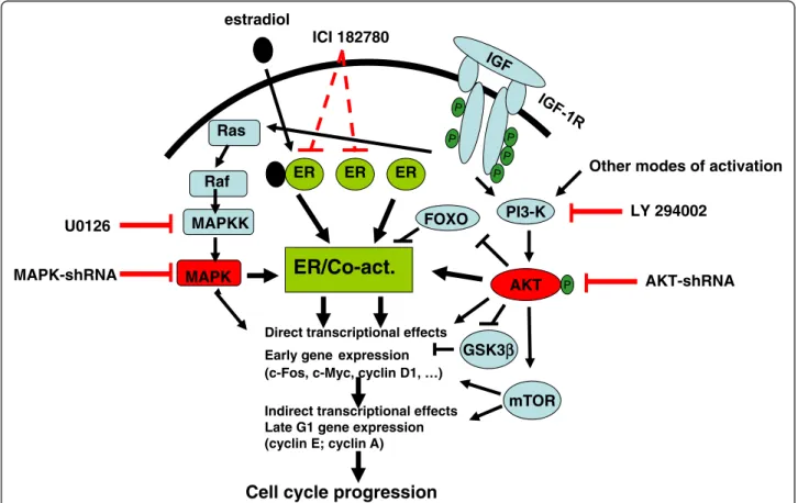
We propose that, in order to induce the cell cycle
pro-gression in the MCF-7 cells, both the presence of
func-tional Akt kinase and the transcripfunc-tional activation by
the ER are required (Figure 8). The basal,
ligand-independent transcriptional activation of ER is sufficient
to complement the mitogenic signaling via IGF1R/PI3K/
Akt; the expression of the c-Myc gene may be part of
this mechanism. Conversely, the basal level of
phospho-Akt present in the serum- and estrogen-deprived cells,
with or without ICI 182780, is sufficient to supply the
indispensable activity of the Akt kinase needed for the
full mitogenic activity of the E2/ER complex. The basal
level of phospho-Akt is a consequence of intracellular
processes, not requiring added or secreted (autocrine)
factors. The precise mechanism which leads to the basal
PI3K/Akt activity is not known. The function of the Akt
kinase in the mitogenic signaling may be to maintain a
sufficient level of phosphorylation of FOXO
transcrip-tion factors and of GSK3β in order to ensure the
tran-scription of the CCND1 gene and to stabilize the cyclin
D1 protein, necessary for the activation of Cdk4/6 and
the primary phosphorylation of Rb. A critical role of
cyclin D1 in the breast cancer cell proliferation has
been proposed by several laboratories and recently
docu-mented in the signaling by anterior gradient-2 [43]. In
practical terms, we believe that the development of
hor-monal therapies based on “full” antiestrogens (lacking
agonist action) could improve the outcome of both early
and advanced breast cancer. Suppression of estrogen
synthesis by the use of aromatase inhibitors is clearly
not sufficient to abolish the participation of
ligand-free ER in the mitogenic signaling by other growth
factors. An additional and substantial improvement
Cell cycle progression
Direct transcriptional effects Early gene expression (c-Fos, c-Myc, cyclin D1, …)
estradiol
Indirect transcriptional effects Late G1 gene expression (cyclin E; cyclin A)
ICI 182780
Ras
MAPK
Raf
ER/Co-act.
U0126
MAPK-shRNA
MAPKK
FOXO
ER
ER
ER
PI3-K
AKT
PLY 294002
AKT-shRNA
mTOR
Other modes of activation
GSK3
β
β
Figure 8 Mitogenic signaling in MCF-7 cells. The resumption of cell cycle progression in quiescent MCF-7 cells requires cooperation between two signaling pathways: ER-dependent transcription and PI3K/Akt activity. In the absence of ligand, the ER-dependent transcription proceeds at baseline level. This is sufficient to complement the growth factor-stimulated kinase cascades (Ras/MAPK and PI3K/Akt) to achieve sufficient level of expression of early- and late-G1 genes. The cell concentration of the encoded proteins such as cyclin D1 is also regulated by
post-transcriptional processes (e.g. translational regulation by mTOR). The G1/S transition requires in addition the cascade of cyclin-dependent kinases whose activation requires the expression of the appropriate cyclins (D1, E, A) as well as specific phosphorylation and dephosphorylation of the kinases themselves. In the absence of growth factor stimulation, the basal activity of PI3K/Akt is sufficient to complement the full (estrogen-stimulated) transcriptional activation of ER to induce G1-phase progression.
would require simultaneous targeting the PI3K/Akt
pathway but, until now, no clinically applicable methods
have been reported. Also, while most research
addres-sing the need to complement targeted therapies of breast
cancer concentrates on the HER family [44], an
alterna-tive approach directed at the IGF1R-dependent signaling
deserves attention. The interest of the IGF1R pathway is
well understood for the development of targeted
therap-ies in other solid tumors including the basal-like, triple
negative breast cancer [45]; there is now ample evidence
that this pathway is important also in luminal-type breast
cancer and may play a role in the recurrence after
endo-crine therapy.
Conclusion
We show that transcriptional activity of the ligand-free
estrogen receptor is sufficient to complement the
mito-genic action of the IGF1R-induced kinase cascade.
Reciprocally, PI3K/Akt activity is required to complement
the mitogenic effect of the agonist-activated ER. The basal
level of PI3K/Akt present in cells in the absence of
exo-genous growth factors is sufficient for the full mitogenic
effect of estradiol. Thus, both ER and PI3K/Akt need to
be targeted for an effective inhibition of the proliferation
of hormone-dependent breast cancer cells.
Additional files
Additional file 1: Figure S1. Knock-down of the Akt signal by shAkt. Transfections were carried out by the Icafectin method (Eurogentec) according to the manufacturer’ protocol with the shRNA as indicated. The cells were serum-starved during 48h and then harvested and analyzed by Western blotting with the Akt antibody. Actin was used as control.
Additional file 2: Figure S2. p- Akt signal is abolished by phosphatase. The cells were starved as in Figure 2 and then stimulated by addition of insulin (1 mM) for 1 h. The cells were lysed in a buffer without EDTA and proteases inhibitors. Portions of lysates (200 μg of total protein) were incubated with calf intestinal alkaline phosphatase (0.05 U/mg protein) for 1 h at 37°C. The lysates were analyzed by Western blotting for Phospho Ser473-Akt.
Additional file 3: Figure S3. Serum-deprived MCF-7 cells do not secrete autocrine factors. The cells were made quiescent in medium with ICI 182780 during 48 h. They were then placed for 6 h in fresh medium (serum- and phenol red-free) with ICI 182780; this was used as conditioned medium (CM). Another series of dishes were stimulated for 1 h or 3 h with CM or with insulin as a positive control, lysed and analyzed for phospho-Ser473 Akt.
Additional file 4: Figure S4. Cell cycle progression of cells stimulated by insulin or E2. MCF-7 cells were deprived of serum in phenol red-free medium with or without ICI 182780 during 48 h, and then stimulated with insulin or with E2 as described in the text. Cells harvested at the different time points were labeled with propidium iodide and analyzed by flow cytometry. The data were evaluated using the ModFit LT software.
Additional file 5: Figure S5. Akt phosphorylation is equally induced by IGF-I and insulin in cells exposed to ICI 182780. Serum- and
E2-starved cells exposed or not to ICI 182780 during 48 h were stimulated with IGF-I (10 nM) or insulin (1 mM) for 1 h. The lysates were analyzed for phospho- Ser473 Akt.
Abbreviations
cdk: Cyclin dependent kinase; cdki: Cyclin dependent kinase inhibitory protein; E2: Estradiol; ER: Estrogen receptor; ERE: Estrogen response element; GSK: Glycogen synthase kinase; HER: Human epidermal growth factor receptor; IGF: Insulin-like growth factor; IGF1R: Insulin-like growth factor type 1 receptor; PI3K: Phosphatidyl inositol-3 kinase; SERD: Selective estrogen receptor degrader; SERM: Selective estrogen receptor modulator; shRNA: Short hairpin RNA.
Competing interests
The authors declare that they have no conflicts of interests concerning this work.
Authors’ contributions
AMG participated at the design, execution and interpretation of the experiments, as well as writing up of the manuscript. MS participated at the RTQPCR experiments and the presentation of the manuscript. MB participated at the cell culture experiments and the presentation of the manuscript. GR participated at the interpretation of the data and the presentation of the manuscript. JM participated at the design, execution and interpretation of the experiments, as well as writing up of the manuscript. All authors read and approved the final manuscript.
Acknowledgements
We are grateful to Dr. F Czauderna for the shAkt (1 + 2) vector and to Dr. Xiao G H for the Akt1 and Akt2 expression vectors. We thank Dr. A.M Faussat for analyzing the cells by flow cytometry.
Received: 25 November 2011 Accepted: 26 June 2012 Published: 16 July 2012
References
1. Burstein HJ, Griggs JJ: Adjuvant hormonal therapy for early-stage breast cancer. Surg Oncol Clin N Am 2010, 19(3):639–647.
2. Janni W, Hepp P: Adjuvant aromatase inhibitor therapy: outcomes and safety. Cancer Treat Rev 2010, 36(3):249–261.
3. Michalides R, Griekspoor A, Balkenende A, Verwoerd D, Janssen L, Jalink K, Floore A, Velds A, van’t Veer L, Neefjes J: Tamoxifen resistance by a conformational arrest of the estrogen receptor alpha after PKA activation in breast cancer. Cancer Cell 2004, 5(6):597–605.
4. Di Cosimo S, Baselga J: Targeted therapies in breast cancer: where are we now? Eur J Cancer 2008, 44(18):2781–2790.
5. Massarweh S, Osborne CK, Creighton CJ, Qin L, Tsimelzon A, Huang S, Weiss H, Rimawi M, Schiff R: Tamoxifen resistance in breast tumors is driven by growth factor receptor signaling with repression of classic estrogen receptor genomic function. Cancer Res 2008, 68(3):826–833. 6. Saxena R, Dwivedi A: ErbB family receptor inhibitors as therapeutic
agents in breast cancer: current status and future clinical perspective. Med Res Rev 2012, 32(1):166–215.
7. Prall OW, Rogan EM, Musgrove EA, Watts CK, Sutherland RL: c-Myc or cyclin D1 mimics estrogen effects on cyclin E-Cdk2 activation and cell cycle reentry. Mol Cell Biol 1998, 18(8):4499–4508.
8. Castoria G, Migliaccio A, Bilancio A, Di Domenico M, de Falco A, Lombardi M, Fiorentino R, Varricchio L, Barone MV, Auricchio F: PI3-kinase in concert with Src promotes the S-phase entry of oestradiol-stimulated MCF-7 cells. EMBO J 2001, 20(21):6050–6059.
9. Gaben AM, Saucier C, Bedin M, Redeuilh G, Mester J: Mitogenic activity of estrogens in human breast cancer cells does not rely on direct induction of mitogen-activated protein kinase/extracellularly regulated kinase or phosphatidylinositol 3-kinase. Mol Endocrinol 2004, 18(11):2700–2713. 10. Dufourny B, Alblas J, van Teeffelen HA, van Schaik FM, van der Burg B,
Steenbergh PH, Sussenbach JS: Mitogenic signaling of insulin-like growth factor I in MCF-7 human breast cancer cells requires
phosphatidylinositol 3-kinase and is independent of mitogen-activated protein kinase. J Biol Chem 1997, 272(49):31163–31171.
11. Lai A, Sarcevic B, Prall OW, Sutherland RL: Insulin/insulin-like growth factor-I and estrogen cooperate to stimulate cyclin E-Cdk2 activation and cell Cycle progression in MCF-7 breast cancer cells through differential regulation of cyclin E and p21(WAF1/Cip1). J Biol Chem 2001, 276(28):25823–25833.
12. Varma H, Conrad SE: Antiestrogen ICI 182,780 decreases proliferation of insulin-like growth factor I (IGF-I)-treated MCF-7 cells without inhibiting IGF-I signaling. Cancer Res 2002, 62(14):3985–3991.
13. Wittmann BM, Sherk A, McDonnell DP: Definition of functionally important mechanistic differences among selective estrogen receptor down-regulators. Cancer Res 2007, 67(19):9549–9560.
14. Czauderna F, Fechtner M, Aygun H, Arnold W, Klippel A, Giese K, Kaufmann J: Functional studies of the PI(3)-kinase signalling pathway employing synthetic and expressed siRNA. Nucleic Acids Res 2003, 31(2):670–682. 15. Xiao GH, Jeffers M, Bellacosa A, Mitsuuchi Y, Vande Woude GF, Testa JR:
Anti-apoptotic signaling by hepatocyte growth factor/Met via the phosphatidylinositol 3-kinase/Akt and mitogen-activated protein kinase pathways. Proc Natl Acad Sci U S A 2001, 98(1):247–252.
16. Remy I, Montmarquette A, Michnick SW: PKB/Akt modulates TGF-beta signalling through a direct interaction with Smad3. Nat Cell Biol 2004, 6(4):358–365.
17. Henglein B, Chenivesse X, Wang J, Eick D, Brechot C: Structure and cell cycle-regulated transcription of the human cyclin A gene. Proc Natl Acad Sci U S A 1994, 91(12):5490–5494.
18. Fritah A, Saucier C, Mester J, Redeuilh G, Sabbah M: p21WAF1/CIP1 selectively controls the transcriptional activity of estrogen receptor alpha. Mol Cell Biol 2005, 25(6):2419–2430.
19. Conery AR, Cao Y, Thompson EA, Townsend CM Jr, Ko TC, Luo K: Akt interacts directly with Smad3 to regulate the sensitivity to TGF-beta induced apoptosis. Nat Cell Biol 2004, 6(4):366–372.
20. Nakatani K, Thompson DA, Barthel A, Sakaue H, Liu W, Weigel RJ, Roth RA: Up-regulation of Akt3 in estrogen receptor-deficient breast cancers and androgen-independent prostate cancer lines. J Biol Chem 1999, 274 (31):21528–21532.
21. Zinda MJ, Johnson MA, Paul JD, Horn C, Konicek BW, Lu ZH, Sandusky G, Thomas JE, Neubauer BL, Lai MT, et al: AKT-1, -2, and -3 are expressed in both normal and tumor tissues of the lung, breast, prostate, and colon. Clin Cancer Res 2001, 7(8):2475–2479.
22. Carroll JS, Lynch DK, Swarbrick A, Renoir JM, Sarcevic B, Daly RJ, Musgrove EA, Sutherland RL: p27(Kip1) induces quiescence and growth factor insensitivity in tamoxifen-treated breast cancer cells. Cancer Res 2003, 63(15):4322–4326.
23. Polo ML, Arnoni MV, Riggio M, Wargon V, Lanari C, Novaro V:
Responsiveness to PI3K and MEK inhibitors in breast cancer. Use of a 3D culture system to study pathways related to hormone independence in mice. PLoS One 2010, 5(5):e10786.
24. Wardell SE, Marks JR, McDonnell DP: The turnover of estrogen receptor alpha by the selective estrogen receptor degrader (SERD) fulvestrant is a saturable process that is not required for antagonist efficacy. Biochem Pharmacol 2011, 82(2):122–130.
25. Rosenwald IB, Setkov NA, Kazakov VN, Chen JJ, Ryazanov AG, London IM, Epifanova OI: Transient inhibition of protein synthesis induces expression of proto-oncogenes and stimulates resting cells to enter the cell cycle. Cell Prolif 1995, 28(12):631–644.
26. Vinals F, Chambard JC, Pouyssegur J: p70 S6 kinase-mediated protein synthesis is a critical step for vascular endothelial cell proliferation. J Biol Chem 1999, 274(38):26776–26782.
27. Diehl JA, Cheng M, Roussel MF, Sherr CJ: Glycogen synthase kinase-3beta regulates cyclin D1 proteolysis and subcellular localization. Genes Dev 1998, 12(22):3499–3511.
28. Gagne D, Balaguer P, Demirpence E, Chabret C, Trousse F, Nicolas JC, Pons M: Stable luciferase transfected cells for studying steroid receptor biological activity. J Biolumin Chemilumin 1994, 9(3):201–209. 29. Fowler AM, Solodin NM, Valley CC, Alarid ET: Altered target gene
regulation controlled by estrogen receptor-alpha concentration. Mol Endocrinol 2006, 20(2):291–301.
30. Sabbah M, Courilleau D, Mester J, Redeuilh G: Estrogen induction of the cyclin D1 promoter: involvement of a cAMP response-like element. Proc Natl Acad Sci U S A 1999, 96(20):11217–11222.
31. Klein EA, Assoian RK: Transcriptional regulation of the cyclin D1 gene at a glance. J Cell Sci 2008, 121(Pt 23):3853–3857.
32. Dupont J, Karas M, LeRoith D: The cyclin-dependent kinase inhibitor p21CIP/WAF is a positive regulator of insulin-like growth factor I-induced cell proliferation in MCF-7 human breast cancer cells. J Biol Chem 2003, 278(39):37256–37264.
33. Becker MA, Ibrahim YH, Cui X, Lee AV, Yee D: The IGF pathway regulates ERalpha through a S6K1-dependent mechanism in breast cancer cells. Mol Endocrinol 2011, 25(3):516–528.
34. Morelli C, Lanzino M, Garofalo C, Maris P, Brunelli E, Casaburi I, Catalano S, Bruno R, Sisci D, Ando S: Akt2 inhibition enables the forkhead transcription factor FoxO3a to have a repressive role in estrogen receptor alpha transcriptional activity in breast cancer cells. Mol Cell Biol 2010, 30(3):857–870.
35. Zhang Y, Moerkens M, Ramaiahgari S, de Bont H, Price L, Meerman J, van de Water B: Elevated insulin-like growth factor 1 receptor signaling induces antiestrogen resistance through the MAPK/ERK and PI3K/Akt signaling routes. Breast Cancer Res BCR 2011, 13(3):R52.
36. Mawson A, Lai A, Carroll JS, Sergio CM, Mitchell CJ, Sarcevic B: Estrogen and insulin/IGF-1 cooperatively stimulate cell cycle progression in MCF-7 breast cancer cells through differential regulation of c-Myc and cyclin D1. Mol Cell Endocrinol 2005, 229(1–2):161–173.
37. Lee AV, Weng CN, Jackson JG, Yee D: Activation of estrogen receptor-mediated gene transcription by IGF-I in human breast cancer cells. J Endocrinol 1997, 152(1):39–47.
38. Charpentier AH, Bednarek AK, Daniel RL, Hawkins KA, Laflin KJ, Gaddis S, MacLeod MC, Aldaz CM: Effects of estrogen on global gene expression: identification of novel targets of estrogen action. Cancer Res 2000, 60(21):5977–5983.
39. Frasor J, Danes JM, Komm B, Chang KC, Lyttle CR, Katzenellenbogen BS: Profiling of estrogen up- and down-regulated gene expression in human breast cancer cells: insights into gene networks and pathways underlying estrogenic control of proliferation and cell phenotype. Endocrinology 2003, 144(10):4562–4574.
40. Reagan-Shaw S, Ahmad N: RNA interference-mediated depletion of phosphoinositide 3-kinase activates forkhead box class O transcription factors and induces cell cycle arrest and apoptosis in breast carcinoma cells. Cancer Res 2006, 66(2):1062–1069.
41. Burgering BM: A brief introduction to FOXOlogy. Oncogene 2008, 27 (16):2258–2262.
42. Zou Y, Tsai WB, Cheng CJ, Hsu C, Chung YM, Li PC, Lin SH, Hu MC: Forkhead box transcription factor FOXO3a suppresses estrogen-dependent breast cancer cell proliferation and tumorigenesis. Breast Cancer Res 2008, 10(1):R21.
43. Vanderlaag KE, Hudak S, Bald L, Fayadat-Dilman L, Sathe M, Grein J, Janatpour MJ: Anterior gradient-2 plays a critical role in breast cancer cell growth and survival by modulating cyclin D1, estrogen receptor-alpha and survivin. Breast Cancer Res BCR 2010, 12(3):R32.
44. Johnston SR: New strategies in estrogen receptor-positive breast cancer. Clin Cancer Res 2010, 16(7):1979–1987.
45. Klinakis A, Szabolcs M, Chen G, Xuan S, Hibshoosh H, Efstratiadis A: Igf1r as a therapeutic target in a mouse model of basal-like breast cancer. Proc Natl Acad Sci U S A 2009, 106(7):2359–2364.
doi:10.1186/1471-2407-12-291
Cite this article as: Gaben et al.: Ligand-free estrogen receptor activity complements IGF1R to induce the proliferation of the MCF-7 breast cancer cells. BMC Cancer 2012 12:291.
Submit your next manuscript to BioMed Central
and take full advantage of:
• Convenient online submission • Thorough peer review
• No space constraints or color figure charges • Immediate publication on acceptance
• Inclusion in PubMed, CAS, Scopus and Google Scholar • Research which is freely available for redistribution
Submit your manuscript at www.biomedcentral.com/submit