HAL Id: hal-00143841
https://hal.archives-ouvertes.fr/hal-00143841
Submitted on 27 Apr 2007
HAL is a multi-disciplinary open access
archive for the deposit and dissemination of sci-entific research documents, whether they are pub-lished or not. The documents may come from teaching and research institutions in France or abroad, or from public or private research centers.
L’archive ouverte pluridisciplinaire HAL, est destinée au dépôt et à la diffusion de documents scientifiques de niveau recherche, publiés ou non, émanant des établissements d’enseignement et de recherche français ou étrangers, des laboratoires publics ou privés.
alpha7B integrin changes in mdx mouse muscles after
L-arginine administration.
Delphine Chazalette, Karim Hnia, François Rivier, Gérald Hugon, Dominique
Mornet
To cite this version:
Delphine Chazalette, Karim Hnia, François Rivier, Gérald Hugon, Dominique Mornet. alpha7B inte-grin changes in mdx mouse muscles after L-arginine administration.. FEBS Letters, Wiley, 2005, 579 (5), pp.1079-84. �10.1016/j.febslet.2004.12.081�. �hal-00143841�
(Article publié dans FEBS Letters, (2005); 579: 1079-1084.)
Alpha7 Beta1 Integrin changes in mdx mouse muscles after L-arginine
administration
Delphine Chazalettea, Karim Hniaa,b, François Riviera,Gérald Hugona, Dominique Morneta,*
a
EA 701, Muscles et Pathologies Chroniques, Institut de Biologie, Boulevard Henri IV, 34060 Montpellier, France
b
Institut Supérieur de Biotechnologie & U.R. 08/39 Faculté de Médecine, Monastir, Tunisia
Abstract: Muscle .fibers attach to laminin in the basal lamina using two mechanisms, i.e., dystrophin with its associated proteins and α7β1 integrin. In humans, gene-mutation defects in one member of these complexes result in muscular dystrophies. This study revealed changes after L-arginine treatment of utrophin-associated proteins and the α7β1 integrin subunit in mdx mouse, a dystrophin-deficient animal model. In the two studied muscles (cardiac muscle and diaphragm), the α7β1 integrin subunit was increased in 5-week-old treated mice. Interestingly, the diaphragm histopathological appearance was significantly
improved by L-arginine administration. These results highlight a possible way to compensate for dystrophin deficiency via α7β1 integrin.
.
Keywords: α7β1 Integrin; Utrophin; mdx; Cardiac muscle; Diaphragm; L-Arginine
*Corresponding author. Fax: +33 467 606 904. E-mail address: [email protected] (D. Mornet).
Abbreviations: NO, nitric oxide; PAGE, polyacrylamide gel electrophoresis; Ss, saline solution; SDS, sodium dodecyl sulfate
HAL author manuscript hal-00143841, version 1
HAL author manuscript
FEBS Lett 579, 5 (14/02/2005) 1079-84
HAL author manuscript hal-00143841, version 1
HAL author manuscript
1. Introduction
The actin network located under the muscle membrane is linked to the extracellular laminin by two distinct mechanisms. The first one involves dystrophin at the inner cytoplasmic membrane. The linkage is mediated through dystrophin associations with transmembrane proteins, i.e., dystroglycan and sarcoglycan–sarcospan complexes [1–3]. There are also important associations with the subsarcolemmal complex, i.e., dystrobrevins, syntrophins and neuronal-type nitric oxide synthase (nNOS) [4]. The second mechanism involves muscle-specific α7β1 integrin. This transmembrane laminin receptor also links the extracellular matrix with the cell cytoskeleton of skeletal and cardiac muscles [5,6]. The dystrophin-associated protein complex (DAPC) and the α7β1 integrin both participate in the molecular continuity between the extracellular matrix and myofibers, which is essential for muscle membrane structural and functional integrity.
Defects in the dystrophin gene result in a lack of dystrophin, which weakens muscle .fiber association with the surrounding basal lamina and underlies Duchenne and Becker muscular dystrophies [7]. The absence of dystrophin is followed by persistence of an homologous protein called utrophin [8]. Utrophin is present all around the muscle membrane during foetal development and then only maintained at the neuromuscular junction in normal muscle [9]. In dystrophin-deficient muscles, utrophin remains associated along the muscle membrane and links to different dystrophin-associated partners such as syntrophin and nNOS [10].
An absence of dystrophin partially disrupts membrane-bound nNOS [11] followed by defects in microvascular adaptation [12]. In contrast, a nNOS transgene improves the muscular dystrophy phenotype [13]. Contraction of dystrophin-deficient muscles therefore may not properly stimulate sufficient NO production to release vasoconstriction, resulting in local muscle ischemia [14].
The NO precursor is provided by L-arginine (L-Arg), which is involved in the urea synthesis cycle and is the nNOS substrate. If muscle fibers produce sufficient level of nNOS, elevated L-Arg concentrations could influence NO synthesis and perhaps improve muscle membrane integrity. This prompted us to treat a murine model of dystrophin deficiency, i.e., mdx mouse [15], with L-Arg. Utrophin, different members of the DAPC and α7β1 integrin were analyzed in cardiac muscle and diaphragm by Western blot and immunofluorescent detection. Here, we describe specific protein changes and potential associated improvements in muscle morphology induced by L-Arg treatment in dystrophin-deficient muscles.
2. Materials and methods 2.1. Antibodies
Polyclonal antibodies directed against C-terminal sequences of α-dystrobrevin (D124: GVSYVPYCRS), β-dystroglycan (LG5: PPPYVPPP), dystrophin (H4: SRGNIPGKPMREDTM); sarcospan (C525: CSLTASEGPQQKI); utrophin (K7: CPNVPSRPQAMC) and α7β1-integrin (K6: DWHPELGPDGHPVPATA) were obtained by injecting the keyhole limpet hemocyanin-linked peptide as antigen according to a previously described protocol [16]. K6 specifically recognize the α7Β1 isoform of the α7 subunit in the α7β1 integrin complex. Commercial antibodies directed against caveolin-3 and nNOS were purchased from Santa Cruz Biotechnologies and BD Transduction Laboratory, respectively.
2.2. Laboratory animals and L-arginine administration protocol
Wild-type (C57BL/10) and dystrophin-deficient (mdx) mice were purchased from Jackson Laboratories. Intraperitoneal injections (250 μl vol.) were performed daily for 3 weeks. Control mdx and C57BL/10 mice were injected with saline solution (L-Arg-diluent) while treated mdx animals were administered with L-Arg solution (250 mg/ kg). Experiments were carried out on 5-and 13-week-old animals in duplicate. Thirty-six mice were used: Ss-treated C57BL/10 (n =2 · 6), Ss-treated mdx (n =2 · 6) and L-Arg-treated mdx (n =2 · 6). Animals were killed on the 22nd day post-treatment. Diaphragm and cardiac muscles were dissected, rapidly frozen in 2-methylbutane, cooled in liquid nitrogen and stored at .80 C until use.
2.3. Preparation of tissue lysates
0.01 g from cardiac muscle or diaphragm were homogenized in 150 ll of SDS buffer (50 mM Tris/HCl, pH 8.0, 10% SDS, 5% b-mercaptoethanol, 10% glycerol, 10 mM EDTA). Samples were centrifuged at 13000g for 10 min. Each supernatant was mixed with 50 μl of SDS buffer containing 0.01% bromophenol blue. Each tissue lysate was denatured for 3 min at 100 C and submitted in duplicate to SDS–PAGE. One resulting gel was Coomassie blue stained and the other was transferred onto nitrocellulose.
2.4. Scanning densitometric measurement standardization
Using the NIH Image software package, the relative optical density of the myosin heavy chain (MHC) present in each tissue lysate was estimated from the Coomassie blue stained gel. Independently, a standard SDS–PAGE, corresponding to known myosin amounts, was stained
with Coomassie blue. This was converted into a standard curve associating each scanning densitometric measurement of the MHC band with the corresponding amount of myosin. In reference to this curve, the protein concentration was equilibrated in each tissue lysate.
2.5. Western blot analysis
Tissue lysates were electroblotted onto nitrocellulose (0.2 lm). Each blot was blocked in Tris-buffered saline with 0.1% Tween 20 (TBST) containing 3% bovine serum albumin (w/v). All membranes were incubated with primary antibodies for 1 h at room temperature. After labeling, the membranes were washed in TBST and then incubated with the secondary antibody (phosphatase-conjugated goat anti-rabbit IgG, Chemicon International, 1/5000). Antibody-bound proteins were detected with p-nitroblue tetrazolium and 5-bromo-4-chloro-3-indoylphosphate substrate, as previously reported [17].
2.6. Measurement of relative protein concentrations
Protein band intensities were assessed using the NIH Image software package. Means of arbitrary values obtained for the two assays were calculated. To test for statistically significant differences, a one-way analysis of variance (ANOVA) was used; in the case of significant differences, the Scheffe test was applied. Statistical significance was set at P < 0.05. Means of arbitrary values were translated into mean percentages by considering the C57BL/10 mean of the arbitrary value as 100%. Data were expressed as means ± SEM (in %).
2.7. Immunofluorescence studies
Unfixed cryostat sections (10 ll thick) of frozen diaphragm and cardiac muscles were incubated with anti-utrophin or anti-a7B integrin antibody for 1 h at room temperature on slides. After washing with phosphate-buffered saline (PBS), sections were incubated for 1 h at room temperature with the secondary antibody (Cy3-conjugated goat anti-rabbit IgG, Chemicon International, 1/4000). Finally, the sections were washed with PBS, the slides were then mounted with Mowiol (CalBiochem) and observed under a Nikon optiphot-2 microscope.
2.8. Histological analysis
Unfixed cryostat sections (10 μm thick) of frozen cardiac muscle and diaphragm from 5-week-old mice were incubated with haematoxylin (Sigma). After washing with distilled
water, sections were incubated with eosin (Sigma). Finally, the sections were washed, the slides were then mounted with Mowiol and observed under a Nikon optiphot-2 microscope. A subsequent image analysis was performed using the ImageTool IT3 software package. Muscle .fiber cross-sections were analyzed in three steps: (a) determination of the muscle .fiber size, (b) determination of the percentage of muscle .fibers containing centralized nuclei and (c) assessment of the lymphocytic invasion. The geometrical parameter tested for the determination of the muscle .fiber size was the minimal ‘‘Feret.s diameter’’ (the minimum distance of parallel tangents at opposing borders of the muscle .fiber) according to recent published data [18]. The variance coefficient (VC) of the ‘‘Feret.s diameter’’ is defined as follows: VC = (standard deviation of the ‘‘Feret.s diameter’’/ mean muscle .fiber size) · 1000. The lymphocytic invasion was evaluated by measuring the inflammatory area as a percentage of the total area muscle cross-section.
3. Results
3.1. L-Arg treatment does not significantly alter the utrophin level in mdx mouse cardiac muscle and diaphragm
We .first attempted to determine whether L-Arg treatment would modify protein levels in muscles. Tissue lysates from cardiac muscle and diaphragm were subjected to Western blot analysis with antibodies directed against utrophin, a7B-integrin and dystrophin-associated proteins (i.e., caveolin-3, a-dystrobrevin, b-dystroglycan, nNOS and sarcospan).
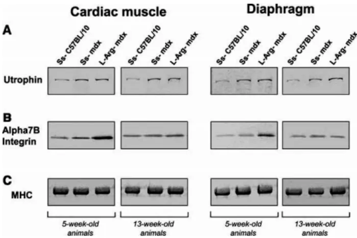
As expected, saline solution (L-Arg-diluent) did not modify the protein levels in Ss-treated C57BL/10 and Ss-treated mdx mice, as compared with protein levels in untreated mice (data not shown). In addition, L-Arg treatment had no significant effect on caveolin-3, a-dystrobrevin, b-dystroglycan, nNOS and sarcospan levels in both 5-and 13-week-old mdx mice (data not shown). Fig. 1A shows the utrophin band obtained with the K7 polyclonal antibody and Fig. 2A shows the relative utrophin concentrations (percentage) in each treated animal-type. In each studied muscle, we observed clear overexpression of utrophin in Ss-treated mdx mice relative to Ss-Ss-treated C57BL/10 mice as previously showed [19]. Histograms revealed that L-Arg treatment had no effect on cardiac utrophin level in 5-week-old mdx mice. Surprisingly, L-Arg treatment reduced the cardiac utrophin level in 13-week-old mdx mice. Finally, diaphragm utrophin levels appeared increased in 5-and 13-week-13-week-old L-Arg-treated mdx mice but it was not significant according to our statistical tests (Fig. 2A).
Figure. 1. Western blot detection of utrophin and a7B integrin in treated C57BL/10,
Ss-treated mdx and L-Arg-Ss-treated mdx mice muscles. Western blots showing relative detection of (A) utrophin and (B) the a7B integrin subunit in tissue lysates from 5-and 13-week-old mice. The six left lanes contained samples from cardiac muscles, while the six right lanes contained samples from diaphragms. (C) Coomassie blue detection of MHC after SDS–PAGE of tissue lysates.
3.2. L-Arg treatment increases the α7Β integrin level in mdx mouse cardiac muscle and
diaphragm
Interestingly, α7Β integrin levels were significantly increased after L-Arg treatment in cardiac muscle and diaphragm from 5-week-old mdx mice compared to 5-week-old Ss-treated mdx mice (Figs. 1B and 2B).
To independently confirm these observations, frozen cardiac muscle and diaphragm tissue sections from 5-week-old mdx mice were prepared and examined by immunofluorescence. Fig. 3A shows utrophin and α7Β integrin staining of cardiac cross-sections. Utrophin immunostaining of the Ss-treated C57BL/10 cardiac muscle essentially revealed microvessels (Fig. 3A, panel 1). In contrast, utrophin was present all along the sarcolemma in Ss-treated mdx mice, as expected. Finally, utrophin immunostaining was quite similar in Ss-treated and in L-Arg-treated mdx cardiac muscle. In cardiac muscles, a7B integrin immunofluorescence did not markedly increase in stained plasma membrane when comparing Ss-treated mdx and L-Arg-treated mdx muscles (Fig. 3A, panel 2).
Fig. 3B shows utrophin and α7Β integrin staining of diaphragm cross-sections. Utrophin immunostaining of the Ss treated C57BL/10 diaphragm essentially revealed microvessels, as observed in cardiac muscle. In contrast, utrophin was present along the sarcolemma, and was
quite similar in Ss-treated and L-Arg-treated mdx diaphragms (Fig. 3B, panel 3). α7Β integrin immunostaining of Ss-treated C57BL/10 diaphragm revealed all the fiber membrane, as expected (Fig. 3B, panel 4). In Ss-treated mdx diaphragm, α7Β integrin immunostaining of the .fiber membrane was less marked than in Ss-treated C57BL/10. In addition, one revertant .fiber showed intensive α7Β integrin immunostaining (indicated with an arrow and in insert). As expected, in diaphragm, the amount of fluorescence detected in L-Arg-treated mdx diaphragm exceeded that of the Ss-treated mdx sample. In addition, in L-Arg-treated mdx diaphragm, immunofluorescence staining of α7Β integrin was increased in comparison to what we observed in Ss-treated C57BL/10 mice (Fig. 3B, panel 4).
Figure. 2. Histogram analysis after utrophin and α7Β integrin detection in Ss-treated
C57BL/10, Ss-treated mdx and L-Arg-treated mdx mouse muscles. Histograms showing relative concentrations of (A) utrophin and (B) the a7B integrin subunit in tissue lysates from 5-and 13-weekold mice. The symbol * indicates that concentrations of protein differed significantly at P < 0.05.
3.3. L-Arg treatment improves muscle morphology in mdx diaphragm
We .finally attempted to evaluate whether L-Arg treatment, would improve the overall muscle pathology of these muscles. Thus, we performed H&E staining of frozen cardiac muscle and
hragm sections from 5-week-old mdx mice. diap
A
B
Figure. 3. Immunohistochemistry of the utrophin and a7B integrin distribution in 5-week-old
mouse muscles. Cryostat sections of (A) cardiac muscle and (B) diaphragm from Ss-treated C57BL/10, Ss-treated mdx and L-Arg-treated mdx mice were analyzed. Ss-treated C57BL/10, Ss-treated and L-Arg-treated mdx mice were studied comparatively using polyclonal antibodies against utrophin and the a7B integrin subunit. In the insert, a revertant .fiber in Ss-treated mdx diaphragm revealed by H4 antibody.
Cardiac muscle sections from Ss-treated and L-Arg-treated mdx mice were relatively similar to cardiac muscle sections from Ss-treated C57BL/10 mice (Fig. 4A). One possible explanation could be that cardiac muscle showed no regenerative capacity compared to skeletal muscle.
In contrast, diaphragm sections from Ss-treated mdx mice showed clear signs of dystrophic changes, including muscle .fiber size variations (VC = 393.7 ± 15.1), central nuclei (21.5 ± 0.9), lymphocytic invasion (16.0 ± 1.4) (Fig. 4B and C). Interestingly, an analysis of diaphragm muscle sections from L-Arg-treated mdx mice (n = 12) revealed few centrally
nucleated muscle fibers (14.0 ± 1.3), more uniform .fiber diameters (247.0 ± 9.9), and low signs of lymphocytic invasion (6.4 ± 0.8), thus suggesting that the dystrophic process was reduced (Fig. 4B and C).
Figure. 4. Histology of 5-week-old mouse muscles. Cryostat sections of (A) cardiac muscle
and (B) diaphragm from Ss-treated C57BL/10, Ss-treated mdx and L-Arg-treated mdx mice were subjected to H&E staining and to (C) quantitative analysis by histomorphometry. (a) Variance coefficient of the minimal ‘‘Feret.s diameter’’, (b) percentage of muscle .fibers with centralized nuclei and (c) percentage of lymphocytic invasion of multiple mouse cross-sections were compared in diaphragm. To test for statistically significant differences, an
ANOVA was used; in the case of significant differences, the Scheffe test was applied. The symbol *** indicates that data differed significantly at P < 0.0001. The summary values are reported in the table.
4. Discussion
Our results revealed that 3 weeks of intraperitoneal injections of L-Arg had no significant effect on utrophin level in 5-week-old mdx cardiac muscle. On the contrary, utrophin was down regulated in L-Arg-treated mdx 13-week-old cardiac muscle (compared to Ss-treated mdx cardiac muscle). Finally, utrophin showed an increase in 5-and 13-week-old L-Arg-treated mdx diaphragm (compared to Ss-L-Arg-treated mdx diaphragm) but the standard error was too high to conclude to a significant positive effect. These observations in cardiac muscle and diaphragm supplement but do not confirm previous results [20], which reported that L-Arg treatment of three adult mdx mice increased utrophin levels in skeletal muscles different than diaphragm, i.e., biceps femoris and semitendinous muscles.
Differential effects of the L-Arg treatment could be explained by (i) the L-Arg dose applied (250 mg/kg/day in our experiments vs 200 mg/kg/day in previous experiments), (ii) the age-related physiopathology of the mdx muscle when L-Arg treatment is begun (5-and 13-week-old animals vs unspecified-aged animals) and (iii) the different muscle types studied (cardiac muscle and diaphragm vs biceps femoris and semitendinous muscles). Further experiments will be necessary to clarify theses points. Note, however, that the mdx mouse strain, which lacks dystrophin in all of its muscles, exhibits high between-muscle variability in myofiber necrosis and contractile properties. Their hindlimb muscles undergo extensive myofiber degeneration and regeneration from 3 to 5 weeks of age [21], which continues on a more limited scale for the duration of their life cycle. Although hindlimb muscles of mdx mice undergo successful regeneration, mdx mouse diaphragm exhibits muscle pathology very similar to DMD. The underlying mechanisms by which dystrophin deficiency mediates the observed pathophysiological changes in mdx mice are unclear. However, a recent paper indicates that distinct signaling pathways are differentially activated in diaphragm and tibialis anterior muscle of dystrophic mice [22]. The results notably demonstrate that p38 activity is decreased in diaphragm, the muscle that displays the more severe phenotype in mdx mouse strain. This stress activated protein kinase p38 is the downstream target of multiple growth factors, NO and integrin mediated signaling [23].
Our analysis of cardiac muscle and diaphragm from L-Arg mdx treated mice also showed that 3 week intraperitoneal injections of L-Arg had a significant effect on the α7Β integrin level in
5-week-old mdx muscles. The exact reasons for this are unclear but may be related to the α7Β integrin localisation and function in muscle .fibers. In skeletal and cardiac muscle membranes, the DAPC is concentrated in costameres [24]. The major integrin complex of cardiac and skeletal muscle (α7β1) is also concentrated in costameres [25]. Costameres are thought to transmit mechanical force from sarcomeres to the sarcolemma, the extracellular matrix and even surrounding .fibers and require both outside-in and inside-out signaling [26]. Direct interaction between integrins and the DAPC has not been shown, but immunoprecipitation studies suggest bidirectional signaling between these complexes. In cultured L6 myocytes, the anti-β1 integrin subunit co-precipited components of the DAPC [27]. In addition, a7-integrin and dystrophin represent associated adhesion protein complexes during muscle regeneration [28].
Finally, in our study, no increase in nNOS level was observed after stimulation of the nNOS pathway by L-Arg injection (data not shown), even if, contrary to DMD, the mdx mouse strain exhibits a low but detectable nNOS level and activity [11]. However, the α7Β integrin level increase that we observed after L-Arg treatment in 5-week-old mdx mice was accompanied by a significant improvement in the histopathological appearance in the diaphragm. This improvement could be related to intracellular Ca2+ levels which are in part regulated by signaling through the α7β1 integrin [29]. This integrin may contribute to the maintenance of calcium levels in myofibers. In dystrophic muscle, the intracellular Ca2+ increase may activate Ca2+-dependent proteolysis and increase muscle degeneration [30]. If so, the a7B integrin level increase that we observed after L-Arg treatment in 5-week-old mdx mice may regulate the activity of calcium channels, stabilizing intracellular Ca2+ levels in mdx .fibers and reducing muscle degeneration.
The increased α7Β integrin level was not observed after L-Arg treatment in 13-week-old mdx muscles. This could be attributed to the age-related physiopathology of the mdx muscle when L-Arg treatment is begun (5-week-old animals vs 13-week-old animals). Then, based on histomorphometric assessment, the increased α7Β integrin-associated-improvement in the histopathological appearance was particularly evident in 5-week-old mdx diaphragms as stated in Fig. 4. This indicates that only within the short period at around 5-weekold when a large number of muscle .fibers undergoes degeneration, drug candidates like L-Arg may have a therapeutic effect on dystrophic process.
Acknowledgments: We thank Mrs. Chantal Jacquet for her technical assistance in
maintaining our wild-type (C57BL/10) and dystrophin deficient (mdx) mice housed in the IGMM (Montpellier, France) and in delivering L-Arg to animals. This work was supported by the ‘‘Association Francaise contre les Myopathies’’ Fellowship No. 10699.
References
[1] Ibraghimov-Beskrovnaya, O., Ervasti, J., Leveille, C., Slaughter, C., Sernett, S. and Campbell, K. (1992) Primary structure of dystrophin-associated glycoproteins linking dystrophin to the extracellular matrix. Nature 355, 696–702.
[2] Monaco, A.P., Neve, R.L., Colletti-Feener, C., Bertelson, C.J., Kurnit, D.M. and Kunkel, L.M. (1986) Isolation of candidate cDNAs for portions of the Duchenne muscular dystrophy gene. Nature 323, 646–650.
[3] Suzuki, A., Yoshida, M., Hayashi, K., Mizumo, Y., Hagiwara, Y. and Ozawa, E. (1994) Molecular organisation at the glycoprotein-complex-binding site of dystrophin; Three dystrophin-associated proteins binds directly to the carboxy-terminal portion of dystrophin. Eur. J. Biochem. 220, 283–292.
[4] Adams, M.E., Mueller, H.A. and Froehner, S.C. (2001) In vivo requirement of the alpha-syntrophin PDZ domain for the sarcolemmal localization of nNOS and aquaporin-4. J. Cell Biol. 155, 113–122.
[5] Song, W.K., Wang, W., Sato, H., Bielser, D.A. and Kaufman,
S.J. (1993) Expression of alpha 7 integrin cytoplasmic domains during skeletal muscle development: alternate forms, conformational change, and homologies with serine/threonine kinases and tyrosine phosphatases. J. Cell Sci. 106 (Pt 4), 1139–1152.
[6] Hodges, B.L., Hayashi, Y.K., Nonaka, I., Wang, W., Arahata, K. and Kaufman, S.J. (1997) Altered expression of the alpha7beta1 integrin in human and murine muscular dystrophies. J. Cell Sci. 110 (Pt 22), 2873–2881.
[7] Koenig, M., Beggs, A.H., Moyer, M., Scherpf, S., Heindrich, K., Bettecken, T., Meng, G., Muller, C.R., Lindlof, M. and Kaariainen, H., et al. (1989) The molecular basis for Duchenne versus Becker muscular dystrophy: correlation of severity with type of deletion. Am. J. Hum. Genet. 45, 498–506.
[8] Love, D.R., Hill, D.F., Dickson, G., Spurr, N.K., Byth, B.C., Marsden, R.F., Walsh, F.S., Edwards, Y.H. and Davies, K.E. (1989) An autosomal transcript in skeletal muscle with homology to dystrophin. Nature 339, 55–58.
[9] Karpati, G., Carpenter, S., Morris, G.E., Davies, K.E., Guerin,
C. and Holland, P. (1993) Localization and quantitation of the chromosome 6-encoded dystrophin-related protein in normal and pathological human muscle. J. Neuropathol. Exp. Neurol. 52, 119–128.
[10] Matsumura, K., Ervasti, J.M., Ohlendieck, K., Kahl, S.D. and Campbell, K.P. (1992) Association of dystrophin-related protein with dystrophin-associated proteins in mdx mouse muscle. Nature 360, 588–591.
[11] Chang, W.J., Iannaccone, S.T., Lau, K.S., Masters, B.S., McCabe, T.J., McMillan, K., Padre, R.C., Spencer, M.J., Tidball, J.G.
D. Chazalette et al. / FEBS Letters 579 (2005) 1079–1084
and Stull, J.T. (1994) Neuronal nitric oxide synthase and dystrophin-de.cient muscular dystrophy. Proc. Natl. Acad. Sci. USA 93, 9142–9147.
[12] Loufrani, L., Levy, B.I. and Henrion, D. (2002) Defect in microvascular adaptation to chronic changes in blood .ow in mice lacking the gene encoding for dystrophin. Circ. Res. 91, 1183–1189.
[13] Wehling, M., Spencer, M.J. and Tidball, J.G. (2001) A nitric oxide synthase transgene ameliorates muscular dystrophy in mdx mice. J. Cell Biol. 155, 123–131.
[14] Thomas, G.D., Sander, M., Lau, K.S., Huang, P.L., Stull, J.T. and Victor, R.G. (1998) Impaired metabolic modulation of alpha-adrenergic vasoconstriction in dystrophin-de.cient skeletal muscle. Proc. Natl. Acad. Sci. USA 95, 15090–15095.
[15] Sicinski, P., Geng, Y., Ryder-Cook, A.S., Barnard, E.A., Darlison, M.G. and Barnard, P.J. (1989) The molecular basis of muscular dystrophy in the mdx mouse: a point mutation. Science 244, 1578–1580.
[16] Pons, F., Robert, A., Fabbrizio, E., Hugon, G., Califano, J.C., Fehrentz, J.A., Martinez, J. and Mornet, D. (1994) Utrophin localization in normal and dystrophin-de.cient heart. Circulation 90, 369–374.
[17] Rivier, F., Robert, A., Royuela, M., Hugon, G., Bonet-Kerrache,
A. and Mornet, D. (1999) Utrophin and dystrophin-associated glycoproteins in normal and dystrophin de.cient cardiac muscle.
J. Muscle Res. Cell Motil. 20, 305–314.
[18] Briguet, A., Courdier-Fruh, I., Foster, M., Meier, T. and Magyar,
J.P. (2004) Histological parameters for the quantitative assessment of muscular dystrophy in the mdx-mouse. Neuromuscul. Disord. 14, 675–682.
[19] Tanaka, H., Ishiguro, T., Eguchi, C., Saito, K. and Ozawa, E. (1991) Expression of a dystrophin-related protein associated with the skeletal muscle cell membrane. Histochemistry 96, 1–5.
[20] Chaubourt, E., Fossier, P., Baux, G., Leprince, C., Israel, M. and De La Porte, S. (1999) Nitric oxide and L-arginine cause an accumulation of utrophin at the sarcolemma: a possible compensation for dystrophin loss in Duchenne muscular dystrophy. Neurobiol. Dis. 6, 499– 507.
[21] Dangain, J. and Vrbova, G. (1984) Muscle development in mdx mutant mice. Muscle Nerve 7, 700–704.
[22] Lang, J.M., Esser, K.A. and Dupont-Versteegden, E.E. (2004) Altered activity of signaling pathways in diaphragm and tibialis anterior muscle of dystrophic mice. Exp. Biol. Med. (Maywood) 229, 503–511.
[23] Ono, K. and Han, J. (2000) The p38 signal transduction pathway: activation and function. Cell Signal 12, 1–13.
[24] Minetti, C., Beltrame, F., Marcenaro, G. and Bonilla, E. (1992) Dystrophin at the plasma membrane of human muscle .bers shows a costameric localization. Neuromuscul. Disord. 2, 99–109.
[25] Belkin, A.M., Zhidkova, N.I., Balzac, F., Altruda, F., Tomatis, D., Maier, A., Tarone, G., Koteliansky, V.E. and Burridge, K. (1996) Beta 1D integrin displaces the beta 1A isoform in striated muscles: localization at junctional structures and signaling potential in nonmuscle cells. J. Cell Biol. 132, 211–226.
[26] Danowski, B.A., Imanaka-Yoshida, K., Sanger, J.M. and Sanger,
J.W. (1992) Costameres are sites of force transmission to the substratum in adult rat cardiomyocytes. J. Cell Biol. 118, 1411– 1420.
[27] Yoshida, T., Pan, Y., Hanada, H., Iwata, Y. and Shigekawa, M. (1998) Bidirectional signaling between sarcoglycans and the integrin adhesion system in cultured L6 myocytes. J. Biol. Chem. 273, 1583–1590.
[28] Kaariainen,, M., Kaariainen, J., Jarvinen, T.L., Nissinen, L., Heino, J., Jarvinen, M. and Kalimo, H. (2000) Integrin and dystrophin associated adhesion protein complexes during regeneration of shearing-type muscle injury. Neuromuscul. Disord. 10, 121–132.
[29] Kwon, M.S., Park, C.S., Choi, K., Ahnn, J., Kim, J.I., Eom, S.H., Kaufman, S.J. and Song, W.K. (2000) Calreticulin couples calcium release and calcium in.ux in integrin-mediated calcium signaling. Mol. Biol. Cell 11, 1433–1443.
[30] Denetclaw Jr., W.F., Hopf, F.W., Cox, G.A., Chamberlain, J.S. and Steinhardt, R.A. (1994) Myotubes from transgenic mdx mice expressing full-length dystrophin show normal calcium regulation. Mol. Biol. Cell 5, 1159–1167.