HAL Id: inserm-00657076
https://www.hal.inserm.fr/inserm-00657076
Submitted on 5 Jan 2012
HAL is a multi-disciplinary open access
archive for the deposit and dissemination of
sci-entific research documents, whether they are
pub-lished or not. The documents may come from
teaching and research institutions in France or
abroad, or from public or private research centers.
L’archive ouverte pluridisciplinaire HAL, est
destinée au dépôt et à la diffusion de documents
scientifiques de niveau recherche, publiés ou non,
émanant des établissements d’enseignement et de
recherche français ou étrangers, des laboratoires
publics ou privés.
GnRH neurona lfunction?
Jerome Clasadonte, Ariane Sharif, Marc Baroncini, Vincent Prevot
To cite this version:
Jerome Clasadonte, Ariane Sharif, Marc Baroncini, Vincent Prevot. Gliotransmission byprostaglandin
E2: a prerequisite for GnRH neurona lfunction?: Gliotransmitters and GnRH neuronal function.
Frontiers in Endocrinology, Frontiers, 2011, 2, pp.1-12. �10.3389/fendo.2011.00091�. �inserm-00657076�
Gliotransmission by prostaglandin E
2
: a prerequisite for
GnRH neuronal function?
Jerome Clasadonte
1,2,3, Ariane Sharif
1,2,3, Marc Baroncini
1,2,3,4and Vincent Prevot
1,2,3*
1Jean-Pierre Aubert Research Center, Inserm, U837, F-59000, Lille, France2Laboratory of Anatomy, Université Lille Nord de France, Lille, France 3School of Medicine, UDSL, Lille, France
4Department of Neurosurgery, CHULille, Lille, France
Edited by:
Carol F. Elias, University of Texas Southwestern Medical Center, USA
Reviewed by:
Sue Moenter, University of Michigan, USA
Rebecca E. Campbell, University of Otago, New Zealand
*Correspondence:
Vincent Prevot, INSERM U837, Bâtiment Biserte, Place de Verdun, 59045 Lille Cedex, France. e-mail: vincent.prevot@inserm.fr
Over the past four decades it has become clear that prostaglandin E
2(PGE
2), a
phospholipid-derived signaling molecule, plays a fundamental role in modulating the
gonadotropin-releasing hormone (GnRH) neuroendocrine system and in shaping the
hypo-thalamus. In this review, after a brief historical overview, we highlight studies revealing that
PGE
2released by glial cells such as astrocytes and tanycytes is intimately involved in the
active control of GnRH neuronal activity and neurosecretion. Recent evidence suggests
that hypothalamic astrocytes surrounding GnRH neuronal cell bodies may respond to
neu-ronal activity with an activation of the erbB receptor tyrosine kinase signaling, triggering the
release of PGE
2as a chemical transmitter from the glia themselves, and, in turn, leading
to the feedback regulation of GnRH neuronal activity. At the GnRH neurohemal junction,
in the median eminence of the hypothalamus, PGE
2is released by tanycytes in response
to cell–cell signaling initiated by glial cells and vascular endothelial cells. Upon its release,
PGE
2causes the retraction of the tanycyte end-feet enwrapping the GnRH nerve
termi-nals, enabling them to approach the adjacent pericapillary space and thus likely facilitating
neurohormone diffusion from these nerve terminals into the pituitary portal blood. In view
of these new insights, we suggest that synaptically associated astrocytes and
perijunc-tional tanycytes are integral modulatory elements of GnRH neuronal function at the cell
soma/dendrite and nerve terminal levels, respectively.
Keywords: gliotransmitter, cyclooxygenase, nitric oxide, hypothalamus, reproduction
INTRODUCTION
Sexual development, puberty, and adult fertility are achieved by
events that are initiated within the central nervous system and
require the maturation and function of a neural network that
transmits both homeostatic and external cues to the discrete
hypothalamic neuronal population that releases
gonadotropin-releasing hormone (GnRH) from neuroendocrine terminals
within the median eminence into the pituitary portal vessels to
control gonadotropins (luteinizing hormone, LH and follicle
stim-ulating hormone, FSH) secretion (
Terasawa and Fernandez, 2001
;
Herbison and Neill, 2006
;
Malpaux, 2006
;
Ojeda and Skinner, 2006
;
Plant, 2006
;
Donato et al., 2011
). In turn, these gonadotropins act
on the ovaries and testis to regulate the secretion of sex steroids
and the production of eggs and sperm.
In addition to neurons, accumulating evidence over the past
two decades indicates that glial cells, and in particular astrocytes
and tanycytes, also contribute to the neural network that
con-verges onto GnRH neurons to control reproduction. Both the
neuronal and glial elements of this GnRH neural network are
subject to the direct modulatory influence of gonadal steroids
(
Garcia-Segura and McCarthy, 2004
;
Ronnekleiv and Kelly, 2005
;
Mong and Blutstein, 2006
;
Wintermantel et al., 2006
;
Christ-ian and Moenter, 2010
;
Prevot et al., 2010a
;
Bellefontaine et al.,
2011
). Although neuronal elements regulate the activity of GnRH
neurons through a complex array of excitatory and inhibitory
synaptic inputs, glial cells communicate with GnRH neurons via
the activation of specific growth-factor-dependent signaling
path-ways (reviewed in
Melcangi et al., 2002
;
Herbison and Neill, 2006
;
Mahesh et al., 2006
;
Ojeda and Skinner, 2006
;
Sharif and Prevot,
2010
).
The main glial population in the brain consists of astrocytes
that ensheathe synapses and are in contact with blood vessels.
They regulate blood flow, provide much-needed energy to
neu-rons, and supply the building blocks for neurotransmitters at the
synapses, in addition to dynamically contributing to
informa-tion processing within the central nervous system (
Haydon and
Carmignoto, 2006
;
Martineau et al., 2006
;
Iadecola and
Neder-gaard, 2007
;
Eroglu and Barres, 2010
;
Halassa and Haydon, 2010
;
Pfrieger, 2010
;
Di Castro et al., 2011
;
Panatier et al., 2011
),
includ-ing the hypothalamus (
Hatton and Wang, 2008
;
Theodosis et al.,
2008
;
Gordon et al., 2009
;
Panatier, 2009
;
Oliet and Bonfardin,
2010
). Tanycytes are elongated radial glial cells that have many
fea-tures in common with astrocytes and are closely associated with
neuroendocrine terminals in the median eminence of the
hypo-thalamus (
Theodosis et al., 2008
;
Prevot et al., 2010a,b
;
Dale, 2011
;
Sild and Ruthazer, 2011
). As integrative hubs, astrocytes and
tany-cytes likely play a fundamental role in shaping and regulating the
GnRH system.
Here, we will review recent findings that illustrate the
remark-able interplay between glia and neurons within the
hypothalamo-hypophyseal–gonadal axis, and show that different glial cell types
regulate different aspects of the architecture, function, and
plas-ticity of the GnRH system through dynamic and often
multidi-rectional interactions with specialized neuronal junctions, such as
synapses and neurohemal junctions. We will mainly restrict our
focus to the roles of hypothalamic astrocytes and tanycytes
sub-served by the release of prostaglandin E
2(PGE
2), a molecule that
has long been known to regulate GnRH neuronal function and has
recently been identified as a gliotransmitter.
PROSTAGLANDIN E2AND THE CENTRAL CONTROL OF REPRODUCTION
Prostaglandin E
2is one of a number of prostanoids synthesized
from arachidonic acid, which is produced from membrane
phos-pholipids by a phospholipase A
2. Arachidonic acid is converted to
bioactive prostanoids by the cyclooxygenases (1 and
COX-2) and a class of terminal synthases (see for review
Bosetti, 2007
;
Figure 1). Several studies suggest that PGE
2is mainly derived
from the COX-2 pathway (
Brock et al., 1999
;
Vidensky et al., 2003
;
Sang et al., 2005
). PGE
2signaling is propagated by four
G-protein-coupled receptors, EP1–EP4 (see for review
Coleman et al., 1994
;
Figure 1).
Prostaglandin E
2has been known to play a role in the central
control of reproduction for more than 35 years. The first
indica-tion that PGE
2was involved in the process of GnRH secretion
was provided by experiments showing that PGE
2injected into
the third ventricle of the rat brain induced the release of LH
into the general circulation (
Harms et al., 1973
) and of GnRH
into the pituitary portal blood vessels (
Eskay et al., 1975
;
Ojeda
et al., 1975b
). A similar stimulatory effect of PGE
2on GnRH
release has also been documented in monkeys using push–pull
perfusion in conscious animals (
Gearing and Terasawa, 1991
).
To bring about the activation of the GnRH axis, PGE
2acts at
two main hypothalamic sites: the preoptic-anterior hypothalamic
region in which GnRH cell bodies reside, and the tuberal region
of the hypothalamus, which contains the median eminence and
GnRH-releasing neuroendocrine terminals (
Ojeda et al., 1977
).
The use of COX inhibitors such as indomethacin has provided
further support for a physiological role of the prostaglandins in
the control of GnRH release. Indomethacin administration
sup-presses the LH surge induced by estradiol during anestrus in ewes
(
Carlson et al., 1974
) and during the early follicular phase in
rhe-sus monkeys (
Carlson et al., 1977
). In rats, the intraventricular or
intrahypothalamic administration of indomethacin inhibits both
pulsatile LH release and the LH discharge induced by ovarian
steroids (
Ojeda et al., 1975a
). Other studies have demonstrated
that the microinjection of either aspirin, a non-steroidal COX
inhibitor, or N-0164, a prostaglandin and thromboxane
antago-nist, into the tuberal region of the rat hypothalamus results in the
suppression of ovulation (
Labhsetwar and Zolovick, 1973
;
Botting
et al., 1977
). Finally, experiments conducted using hypothalamic
explants in vitro have revealed that PGE
2is an effective stimulator
of GnRH release from median eminence nerve terminals (
Gallardo
and Ramirez, 1977
;
Ojeda et al., 1979, 1986b
).
A sizable body of evidence also implicates PGE
2as a
phys-iological component of the GnRH system during postnatal
FIGURE 1 | Prostaglandin E2(PGE2) biosynthesis and signaling. Upon
its release from plasma membrane phospholipids by phospholipase A2,
arachidonic acid is converted to the unstable endoperoxide intermediates, prostaglandin G2(PGG2), and prostaglandin H2(PGH2) by the
cyclooxygenases (COX-1 and COX-2, encoded by separate genes). Both COX isoforms catalyze the same reactions, but while COX-1 is constitutively expressed, COX-2 is rapidly and transiently upregulated by cytokines and growth factors. Terminal synthases convert both PGG2and
PGH2into prostaglandins [PGE2, PGD2, PGF2α, prostacyclin (PGI2)], and
thromboxane (TxA2). Once synthesized, PGE2immediately diffuses away
and activates its specific E-prostanoid receptors (EP1–4), which belong to the family of seven-transmembrane G-protein-coupled receptors. EP2 and EP4 are coupled to Gs and stimulate the adenylyl cyclase (AC)–cyclic adenosine monophosphate (cAMP)–protein kinase A (PKA) pathway. In contrast, EP3 is coupled to Gi and inhibits AC activation, resulting in decreased cAMP concentrations. EP1 is thought to be coupled to the Gq-phospholipase C (PLC) pathway, leading to an elevation of free cytosolic calcium concentrations (Milatovic et al., 2011). Notably, an examination of the capacity of the hypothalamus to metabolize arachidonic acid through the COX pathway has revealed a pubertal increase in the formation of PGE2, particularly during the first proestrus (Ojeda and Campbell, 1982).
Intriguingly, the increase in PGE2synthesis is not associated with changes
in the formation of PGF2α, PGI2, PGD2, or thromboxane A2from exogenous
arachidonic acid, suggesting that it is a specific event directly associated with the peripubertal activation of the reproductive hypothalamus (Ojeda and Campbell, 1982). Such a selective synthesis of PGE2has also been
shown to be triggered by estrogens during early postnatal development
(Amateau and McCarthy, 2002).
development. For instance, PGE
2can induce the release of GnRH
long before puberty in both mice and rats (
Ojeda et al., 1986a
;
Pre-vot et al., 2003b
). As puberty approaches, the increasing output of
estradiol from the developing ovaries induces a preovulatory surge
of GnRH/LH. Biochemical analyses at this last phase of sexual
mat-uration have demonstrated that the capacity of the reproductive
hypothalamus to metabolize arachidonic acid through the COX
pathway leads to a specific increase in PGE
2synthesis (Figure 1),
particularly during the first proestrus (
Ojeda and Campbell, 1982
).
This effect appears to be estrogen-dependent since it is mimicked
by the treatment of juvenile animals (early post-weaning period)
with estradiol at doses capable of inducing a preovulatory surge
of LH (
Ojeda and Campbell, 1982
). More recent studies have
shown that an estradiol-induced increase in hypothalamic PGE
2levels can be seen even in newborn rats (
Amateau and McCarthy,
2002
). Intriguingly, experiments showing that estradiol treatment
upregulates both COX-2 mRNA and protein synthesis in the
hypo-thalamus of female rats during postnatal development (
Amateau
and McCarthy, 2004
) raise the possibility that estrogens may act
on COX-2 expression to promote PGE
2synthesis at puberty.
Since the levels of arachidonic acid in tissue phospholipids
are to some degree influenced by the dietary intake of different
polyunsaturated fatty acids (e.g., linoleic acid), malnutrition
dur-ing early life could cause deficits in arachidonic acid synthesis (
de
Souza et al., 2011
;
Lauritzen and Carlson, 2011
), which in turn
could lead to changes in the production of prostaglandins and
thus interfere with the maturation of the GnRH system. Indeed, a
diet-related deficiency of essential fatty acids initiated before
fer-tilization in female rats has been shown to significantly delay the
onset of reproductive capacity in female offspring without
affect-ing the progression of gestation or the delivery of healthy litters by
the dams (
Smith et al., 1989
). In this study, delayed puberty was
associated with reduced PGE
2synthesis within the hypothalamus.
The deficit did not appear to be due to the impairment of COX
activity, but to diminished arachidonic acid bioavailability (
Smith
et al., 1989
).
GLIA, THE MAIN SOURCE OF PROSTAGLANDIN E2WITHIN THE GnRH NEUROSECRETORY SYSTEM
Although PGE
2was initially postulated to be an intracellular
mes-senger produced by the binding of neurotransmitters to receptors
located on GnRH neurons and acting within these neurons (
Ojeda
et al., 1982
;
Gearing and Terasawa, 1991
;
Rettori et al., 1992
),
this concept has been revisited following studies showing that
the actions of PGE
2on GnRH release are initiated by its binding
to specific membrane receptors (
Coleman et al., 1994
) expressed
by GnRH neurons (
Rage et al., 1997
) and the recognition that
astrocytes represent a major source of PGE
2in the brain (
Ma
et al., 1997
;
Bezzi et al., 1998
;
Hirst et al., 1999
). Two decades ago,
seminal studies by Ojeda and colleagues revealed that the PGE
2-mediated activation of GnRH neuronal secretory activity triggered
by estrogen at the time of puberty required the activation of
growth-factor-dependent glial signaling pathways involving
recep-tor tyrosine kinases of the erbB family (
Ojeda et al., 1990
;
Junier
et al., 1991
;
Ma et al., 1992
).
Of the four known members of the erbB family (Figure 2),
three of them, erbB1, erbB3, and erbB4, bind and are activated
by cognate ligands. In contrast, erbB2 has no known ligand, and
functions primarily as a modulator of the other members of the
family (
Hynes and Lane, 2005
). While erbB receptors do not appear
to be expressed in GnRH neurons (
Ma et al., 1994b, 1999
;
Voigt
FIGURE 2 | The erbB family of receptors and their ligands. ErbB1 (or EGFR, epidermal growth factor receptor) and erbB4 are fully functional receptors that possess an extracellular ligand-binding domain and a cytoplasmic protein tyrosine kinase domain and can function as homo- or heterodimers. In contrast, erbB2 (or neu), which lacks a ligand-binding domain, and erbB3, which is defective in its intrinsic tyrosine kinase activity (dashed lines), must heterodimerize with another member of the erbB family for signal transduction. The different EGF-like growth factors exhibit different binding specificities for the erbB receptors. While TGFα, EGF, amphiregulin, epigen, neuregulin-3, and neuregulin-4 are specific for a single member of the receptor family, the five other EGF-like ligands can bind two or three receptors each. EGF, epidermal growth factor; HB-EGF, heparin binding-EGF; TGFα, transforming growth factor α.
et al., 1996
;
Prevot et al., 2003b
), erbB1, erbB2, and erbB4, but
not erbB3, are expressed in hypothalamic astrocytes, known to
morphologically and physically interact with GnRH cell bodies
(
Witkin et al., 1995
;
Cashion et al., 2003
;
Baroncini et al., 2007
;
San-dau et al., 2011a
) both in rodents and humans (Figures 3 and 4;
Ma
et al., 1999
;
Prevot et al., 2003b
;
Sharif et al., 2009
). In addition,
hypothalamic astrocytes express the erbB1 ligand, transforming
growth factor alpha (TGFα; Figure 4), and several forms of the
erbB4 ligand, neuregulin (
Ma et al., 1992, 1994a, 1999
;
Sharif et al.,
2009
). Importantly, gonadal steroids have been found to induce
dramatic increases in the expression levels of the erbB receptors
and their ligands within the hypothalamus at puberty; no such
changes are seen in the cortex or other brain regions unrelated to
reproductive control (
Ma et al., 1992, 1994a, 1999
).
The pharmacological or genetic inhibition of erbB1, erbB2,
and/or erbB4 receptors delays the onset of puberty (
Ma et al.,
1992
;
Apostolakis et al., 2000
;
Prevot et al., 2003b, 2005
) and alters
adult reproductive function in rodents (
Prevot et al., 2005
). In vitro
studies using either hypothalamic explants or primary cultures of
hypothalamic astrocytes with a GnRH-producing neuronal cell
line have shown that erbB receptor ligands can stimulate GnRH
release from the explants or neuronal cells, but do so indirectly, by
inducing astrocytes to secrete PGE
2(
Ojeda et al., 1990
;
Ma et al.,
1997, 1999
;
Prevot et al., 2003b, 2005
). In addition, ligand
activa-tion of erbB receptors has been shown to promote morphological
rearrangements in hypothalamic astrocytes (Figure 4G–I;
Sharif
et al., 2009
) thus raising the possibility that erbB signaling may
also influence the astrocytic coverage of GnRH neurons in vivo
(see for review
Prevot et al., 2010b
).
FIGURE 3 | Astrocytes morphologically interact with GnRH neurons and express erbB4 receptors in the tuberal region of the human hypothalamus. (A–C) Photomicrographs showing a GnRH neuronal cell body (green) to which the processes of glial fibrillary acidic protein (GFAP)-immunoreactive astrocytes (red, arrows) are abundantly apposed.
Cell nuclei are stained with Hoechst (blue). Adapted from (Baroncini et al., 2007) with permission. (D–F) GFAP-immunoreactive astrocytes (red) of the tuberal region of the human hypothalamus express erbB4 receptors (green; M. Baroncini and V. Prevot, unpublished data). Scale bars = 20 µm (C), 10 µm (F).
In vitro experiments suggest that erbB signaling in
hypothal-amic astrocytes is functionally connected to the neuronal
gluta-matergic system, the primary mode of excitatory transsynaptic
communication used by hypothalamic neurons (
van den Pol and
Trombley, 1993
), and one that is known to increase GnRH
secre-tion (
Donoso et al., 1990
;
Claypool et al., 2000
) and accelerate
the initiation of puberty in both rodents and primates (
Urban-ski and Ojeda, 1987, 1990
;
Plant et al., 1989
). In hypothalamic
and non-hypothalamic astrocytes alike (
Bezzi et al., 1998
;
Zonta
et al., 2003a,b
), transmitter spillover from nearby synaptic
activ-ity results in an elevation of PGE
2release (
McCarthy et al., 2008
;
Glanowska and Moenter, 2011
). For example, neuronally released
glutamate can engage biochemical signaling in astrocytes through
the co-activation of AMPA and metabotropic glutamate receptors
to cause a ligand-dependent increase in astrocytic erbB signaling
and PGE
2release (
Dziedzic et al., 2003
). This in turn signals back
to GnRH neurons (Figure 5) facilitating neuroendocrine
devel-opment and adult reproductive function (
Prevot et al., 2003b,
2005
).
PROSTAGLANDIN E2, A NEWLY UNCOVERED GLIOTRANSMITTER WITHIN THE GnRH NEUROSECRETORY SYSTEM
Even though PGE
2has been known to trigger GnRH release from
the hypothalamic neurons controlling reproduction for almost
40 years, it is only very recently that it has been identified as a
potent excitatory regulator of GnRH neuronal activity, both in
male and female mice (
Clasadonte et al., 2011
). Using
patch-clamp recordings in brain slices from transgenic mice expressing
green fluorescent protein (GFP) under the control of the GnRH
promoter, we showed that PGE
2induced a reversible membrane
depolarization of GnRH neurons leading to the initiation of
spike firing via the postsynaptic effect involving activation of a
non-selective cation current (Figure 5;
Clasadonte et al., 2011
)
reminiscent of the ones recently described in GnRH neurons by
other groups (
Zhang et al., 2008
;
Roland and Moenter, 2011
).
Although GnRH neurons are known to express both the EP1
and EP2 subtypes of prostaglandin receptors in vivo (
Rage et al.,
1997
;
Jasoni et al., 2005
), the excitatory effect of PGE
2on GnRH
neuronal activity was selectively mimicked by the EP2 receptor
agonist butaprost (
Clasadonte et al., 2011
) previously shown to
promote GnRH release in the GnRH-producing neuronal cell
line, GT1–7 (
Rage et al., 1997
). The PGE
2-mediated membrane
depolarization of GnRH neurons was also shown to require the
cAMP/protein kinase A (PKA) pathway (
Clasadonte et al., 2011
),
which is known to be coupled to the EP2 receptor (Figure 1;
Coleman et al., 1994
;
Sang et al., 2005
) and to underlie the
stim-ulatory effect of PGE
2on GnRH secretion (Figure 5;
Ojeda et al.,
1985
).
As alluded to above, the selective disruption of erbB4
signal-ing in astrocytes by the overexpression of a dominant-negative
erbB4 receptor under the control of the human GFAP promoter
leads to diminished PGE
2release in response to ligand-dependent
erbB4 activation, leading in turn to reduced GnRH release, delayed
puberty, and disrupted adult reproductive function (
Prevot et al.,
2003b, 2005
). Intriguingly, electrophysiological analyses have
shown that the spontaneous activity of GnRH neurons in these
animals is decreased and that this deficiency is mimicked by the
bath application of either fluoroacetate, an inhibitor of astrocyte
metabolism (
Fonnum et al., 1997
;
Henneberger et al., 2010
), or the
COX blocker indomethacin, to slices of the preoptic region from
wild-type animals (
Clasadonte et al., 2011
). The fact that GnRH
neuronal activity in all these conditions can be rescued by
exoge-nous PGE
2(
Clasadonte et al., 2011
) strongly suggests that glial
FIGURE 4 | Human hypothalamic astrocytes express the molecular components required for a glia-to-neuron communication through the erbB-prostaglandin signaling system. Primary cultures of human hypothalamic astrocytes have been prepared from 9 to 12-week-old human fetuses (A). The cultures are composed of 98% of cells immunopositive for the astrocytic markers GFAP [(B) green] and the glutamate–aspartate transporter GLAST [(C) red]. Note that cells that express GFAP at low to undetectable levels are nevertheless strongly immunopositive for GLAST (arrows). (D) Human astrocytes in culture express TGFα protein (red). (E) Western blot analysis of erbB receptor expression in primary cultures of human cortical and hypothalamic astrocytes. While all four erbB receptors are expressed in the fetal brain, hypothalamic astrocytes (Hyp astro) express erbB1, erbB2, and erbB4, but not erbB3, and cortical astrocytes (Ctx astro) express erbB1, erbB2, and erbB3 but not erbB4 receptors. IB, immunoblot.
(F) Human hypothalamic astrocytes in culture are immunopositive for COX (green). (G–I) EGF ligands induce profound morphological rearrangements of human hypothalamic astrocytes in vitro. Cell morphology was examined by visualization of the actin cytoskeleton using Alexa Fluor 568-conjugated phalloidin (red). Hypothalamic astrocytes exhibit heterogeneous shapes under control conditions, i.e., polygonal cells, cells with short and thick extensions (asterisk) or long and thin processes (arrow) (G). TGFα (50 ng/mL for 3 days) stimulates the extension of long and thin processes (arrows) and the apparition of bipolar cells (double arrows) (H) while treatment with neuregulin-1 NRGβ1 (50 ng/mL for 3 days) increases the number of multipolar cells with thick processes (arrowheads) (I). Nuclei are counter-stained with Hoechst [(B,C,F–I), blue]. Scale bars = 3 mm (A), 50 µm (B,C,F), 20 µm (D), 100 µm (G–I). Adapted from (Sharif et al., 2009) with permission.
controlling GnRH neuronal excitability. The role of glia in the
con-trol of GnRH neuronal activity is further supported by a recent
study demonstrating that glial prostaglandins may regulate the
efficacy of GABAergic inputs to GnRH neurons in ovariectomized
mice (
Glanowska and Moenter, 2011
). Using GnRH–GFP
trans-genic mice and patch-clamp recordings in brain slices, the authors
demonstrated that the repeated action-potential-like
depolariza-tion of a GnRH neuron caused a short-term reducdepolariza-tion in the
frequency of spontaneous GABAergic postsynaptic currents in
the same neuron, suggesting the presence of local circuit
inter-actions between GnRH neurons and their GABAergic afferents
(
Chu and Moenter, 2005
;
Glanowska and Moenter, 2011
). It is
important to note that in this local circuit, the activation of GABA
Areceptors exerts a depolarizing action that can trigger action
poten-tial firing due to the elevated chloride levels maintained in adult
GnRH neurons (
DeFazio et al., 2002
;
Han et al., 2002
;
Herbi-son and Moenter, 2011
). Consequently, this represents a negative
feedback loop in which depolarized GnRH neurons reduce the
activity of their own excitatory GABAergic afferents. In addition
to being steroid-dependent and under the influence of both
glu-tamatergic and endocannabinoid signaling mechanisms via the
activation of presynaptic metabotropic glutamate receptors and
cannabinoid CB1 receptors respectively (
Chu and Moenter, 2005
;
Glanowska and Moenter, 2011
) this local negative feedback loop
also requires the action of glial-derived prostaglandins (
Glanowska
and Moenter, 2011
). Indeed, the incubation of brain slices with
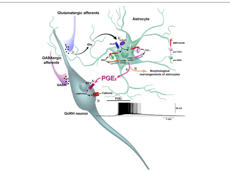
FIGURE 5 | Prostaglandin E2acts as a gliotransmitter to stimulate
GnRH neuron electrical activity. Neuronally released glutamate (Glu) (1) co-activates metabotropic glutamatergic (mGluR) and AMPA glutamatergic receptors (GluR) in astrocytes (2), stimulating the activity of
zinc-dependent matrix metalloproteinases (MMPs) of the ADAM (a disintegrin and metalloproteinase) family (3). The MMPs catalyze ectodomain shedding of the pro-EGF ligands pro-TGFα and pro-NRG (pro-neuregulin). In particular, the processing of pro-TGFα has been shown to involve the metalloproteinase ADAM17, also known as tumor necrosis factor α converting enzyme (TACE). The subsequently released mature TGFα and NRG activate erbB1/erbB2 and erbB4/erbB2 heterodimers, respectively (Dziedzic et al., 2003). The co-activation of glutamatergic receptors induces the recruitment of erbB1, erbB4, and their pro-ligands to
the cell membrane, where multiprotein complexes form, as demonstrated by the direct physical association of glutamatergic and erbB receptors (not shown). The activation of erbB receptors in hypothalamic astrocytes promotes profound morphological changes, including the retraction of cytoplasm, stellation of cells and the elongation of processes (see Figure 4G–I) (4!). The activation of erbB receptors also promotes the
release of PGE2(Ma et al., 1997, 1999;Dziedzic et al., 2003) (4), which
stimulates a cAMP/protein kinase A (PKA) pathway in GnRH neurons through the mobilization of EP2 receptors (EP2;Clasadonte et al., 2011) (5). Activation of this signaling pathway induces a reversible membrane depolarization of GnRH neurons leading to the initiation of spike firing via a postsynaptic effect involving the activation of a non-selective cation current (Clasadonte et al., 2011) (6).
indomethacin, the broad-spectrum prostaglandin receptor
antag-onist AH 6809, or fluorocitrate, which like fluoroacetate, is a
spe-cific blocker of astrocyte metabolism, prevents the
depolarization-induced suppression of GABAergic transmission in GnRH
neu-rons (
Glanowska and Moenter, 2011
). Since GABA exerts a
depo-larizing action in this local circuit, we could envisage that glial
prostaglandins, by suppressing excitatory drive, would reduce
GnRH neuronal activity. Estradiol could also differentially
influ-ence this local inhibitory feedback to exert its positive or negative
feedback effects (
Glanowska and Moenter, 2011
). Thus, in
addi-tion to exerting a direct postsynaptic excitatory acaddi-tion on the cell
body of GnRH neurons, prostaglandins released from astrocytes
can also participate in mechanisms that regulate the activity of
their GABAergic presynaptic inputs (Figure 5). In the GnRH
sys-tem, thus, PGE
2fulfills all the criteria that qualify a compound as
a “gliotransmitter” (
Parpura and Zorec, 2010
): (i) it is synthesized
by astrocytes, (ii) its regulated release is triggered by
physiolog-ical stimuli, (iii) it acutely activates the firing of GnRH neurons
and modulates the activity of their GABAergic afferents, and (iv)
it plays a role in an important physiological function, i.e., the
neuroendocrine control of reproduction, which is vital to species’
survival.
A ROLE FOR ASTROGLIAL PROSTAGLANDIN E2IN DENDRITIC SPINE PLASTICITY IN GnRH NEURONS?
Gonadotropin-releasing hormone neurons exhibit a simple
bipo-lar morphology (Figure 3A) with one or two very long dendritic
processes that can extend up to 1 mm (
Campbell et al., 2005,
2009
). Intriguingly, recent studies have demonstrated that the
den-sity of spines along these dendrites is subject to robust increases
not only during sexual development in immature animals (
Cot-trell et al., 2006
), but also at the onset of the GnRH/LH surge
induced by gonadal steroids in ovariectomized adult mice (
Chan
et al., 2011
). Although sexual maturation and the surge
mech-anism have been shown to require the neuronal expression of
sex-steroid receptors (
Wintermantel et al., 2006
;
Raskin et al., 2009
;
Mayer et al., 2010
), studies suggesting that astrocytic mechanisms
might control the stabilization of individual dendritic processes
and their subsequent maturation into spines (
Nishida and Okabe,
2007
), together with the demonstration that specific juxtacrine
signaling pathways are involved in sculpting astrocyte–dendritic
spine interactions (
Murai et al., 2003
), raise the possibility that
astrocytes play a role in the physiological changes of synaptic
structure underlying GnRH neuronal maturation and function.
PGE
2release by astrocytes could be central in this process and
PGE
2has in fact been shown to mediate the dramatic
neu-ronal spine plasticity induced by estrogens in the developing
preoptic region (
Amateau and McCarthy, 2002, 2004
;
Wright and
McCarthy, 2009
). This effect involves the activation of AMPA and
metabotropic glutamate receptors (
Amateau and McCarthy, 2002
;
Wright and McCarthy, 2009
), known to promote erbB-dependent
PGE
2release in hypothalamic astrocytes (
Dziedzic et al., 2003
), as
well as the EP2/PKA signaling pathway (
Amateau and McCarthy,
2002
), recently found to be functional in native GnRH neurons
(
Clasadonte et al., 2011
; Figure 5). Importantly, estrogens, which
have long been known to regulate neuronal spine plasticity in
the adult hippocampus (
Woolley and McEwen, 1992, 1994
), have
also been shown to promote comparable changes in the
imma-ture hippocampus (
Amateau and McCarthy, 2002
). However, in
the hippocampus, the underlying mechanisms do not appear to
require PGE
2synthesis (
Amateau and McCarthy, 2002
), suggesting
that increases in PGE
2synthesis are selectively used by estrogens
to promote dendritic spine plasticity in the developing preoptic
region. Further studies are required to determine whether
estro-genic effects on the plasticity of hypothalamic neurons such as
those seen in newborn rodents can also occur later in postnatal
life and/or in adulthood.
PROSTAGLANDIN E2IS A KEY MEDIATOR OF GnRH RELEASE, NEURONAL–GLIAL–ENDOTHELIAL INTERACTIONS, AND CELL PLASTICITY AT THE GnRH NEUROHEMAL JUNCTION
Neuroendocrine GnRH neurons send axons to the median
emi-nence, where they release their neurohormone into the pituitary
portal blood vessels for delivery to the anterior pituitary. The
median eminence, which lies ventral to the third ventricle in the
tuberal region of the hypothalamus, constitutes one of the key sites
for the regulation of GnRH release (see for review
Hrabovszky
and Liposits, 2008
;
Ojeda et al., 2008
;
Prevot et al., 2010a
;
Yin
and Gore, 2010
). The modulation of GnRH release by PGE
2within the median eminence was suggested as soon as in vitro
systems to statically incubate median eminence nerve terminals
were developed, i.e., in the late 70s (
Negro-Vilar et al., 1979
;
Ojeda
et al., 1979
). Experiments showing that the PGE
2-induced GnRH
release from median eminence explants requires the
mobiliza-tion of intracellular calcium stores (
Ojeda and Negro-Vilar, 1985
;
Ojeda et al., 1988
) have suggested a role for the EP1 receptor in
this process (Figure 1). In line with this assumption are other
findings demonstrating that GnRH neurons express EP1 receptors
in vivo and that the EP1 agonist 17-phenyl trinor PGE
2promotes
FIGURE 6 | Prostaglandin E2promotes the retraction of tanycytic
processes in vitro and induces neuroglial plasticity causing GnRH neurosecretory terminals to advance toward the pericapillary space in isolated median eminence explants. (A) Tanycytes in culture were stained with Alexa Fluor 568-conjugated phalloidin to visualize filamentous actin (red) and with Hoechst to stain nuclei (blue). In control unstimulated tanycyte cultures, actin was localized adjacent to the cell membrane (top panel, cortical actin, arrowheads) and was also diffused throughout the cytoplasm. PGE2
treatment (280 nM, 30 min) promoted tanycyte retraction (bottom panel, long arrowhead). (B) Electron micrographs of GnRH immunoreactive axon terminals (big arrowhead, green) from female rat median eminence explants
incubated for 30 min in the presence or absence of PGE2(1 µM). Under basal
unstimulated conditions (Control), GnRH nerve endings (big arrowhead, green) were maintained at a distance from the brain basal lamina (white arrow) delineating the pericapillary space (p.s., pink) by thick enclosing tanycyte end-feet (Tan., yellow). PGE2treatment caused the advancement of
GnRH axon terminals (big arrowhead, green) toward the brain basal lamina (white arrow) and the apparent retraction of most of the astroglial sheath (black arrows, yellow) from those neurosecretory terminals that were separated from the fenestrated (small arrowhead) portal capillaries (Cap., red) by only a few nanometers. end., endothelium. Scale bars = 10 µm (A), 1 µm (B). Reproduced from (de Seranno et al., 2010) with permission.
GnRH release from GT1–1 cells in vitro (
Rage et al., 1997
) without
affecting GnRH neuronal firing in brain slices (
Clasadonte et al.,
2011
).
Within the median eminence, GnRH axon terminals are
inti-mately associated with cell processes belonging to specialized
unciliated ependymal cells named tanycytes. Tanycyte cell
bod-ies are attached together at the apex by tight junctions (
Mullier
et al., 2010
) and line the floor of the third ventricle. They send
out long slender processes that eventually contact the pial
sur-face of the brain where the fenestrated pituitary portal vessels
reside, via end-feet (
Page, 1994
;
Ciofi et al., 2009
;
Mullier et al.,
2010
). These tanycyte end-feet not only enclose the GnRH nerve
terminals, possibly providing a diffusion barrier (
Kozlowski and
Coates, 1985
;
Meister et al., 1988
;
Ugrumov et al., 1989
;
King and
Letourneau, 1994
), but also display a high degree of structural
plasticity across the ovarian cycle in rats (
Prevot et al., 1998, 1999
).
During the estrous cycle, under basal conditions, e.g., in diestrus,
GnRH nerve terminals are completely wrapped up in tanycyte
end-feet (
Prevot et al., 1998, 1999
). In proestrus, following the
activation of the reproductive axis, the end-feet are retracted,
pre-sumably due to increasing levels of gonadal steroids (
King and
Letourneau, 1994
), thus allowing GnRH neurons to directly
con-tact the pericapillary space (
Prevot et al., 1998
). By analogy with
the function-related plasticity documented in the neural lobe of
the pituitary (
Hatton, 1997
), these data argue for the importance
of tanycyte structural rearrangement in delivering peak levels of
GnRH to the pituitary during the preovulatory surge. The
intrigu-ing possibility that PGE
2could be involved in the control of these
FIGURE 7 | Schematic representation of neural–glial–endothelial interactions involved in the control of GnRH neurosecretion in the median eminence. (A) Glial–neuronal interactions in the median eminence involve the production of epidermal growth factor (EGF)-related peptides by glial cells. Activation of erbB1/erbB2 and erbB4/erbB2 heterodimers by TGFα and NRG, respectively, promotes the release of PGE2from astrocytes. The
binding of TGFα to tanycytic erbB1 receptors results in the recruitment of erbB2 co-receptors and signal transduction. The ligand-dependent activation of erbB1 receptors in tanycytes results in biphasic plastic changes characterized by an initial phase of tanycyte outgrowth (1) and a secondary phase of retraction (5). Although the initial outgrowth (1) is independent of the TGFβ1 system, the subsequent retraction requires PGE2synthesis (2), a
PGE2-dependent increase in the production of TGFβ1 (3!) and matrix
metalloproteinase (MMP) activity (4). In addition to promoting TGFβ1 synthesis by tanycytes (3!), PGE2released by tanycytes (2) and astrocytes is
able to directly stimulate GnRH release at nerve endings through the EP1 receptor (EP1)-mediated mobilization of intracellular calcium stores (3). (B) Endothelial–neuronal interactions at the level of the median eminence involve the production of nitric oxide (NO) by the endothelial cells of fenestrated capillaries of the portal blood vessels. Upon its secretion, NO diffuses from its source and stimulates the production of PGE2from tanycytes. PGE2
promotes the release of GnRH into the blood stream by the direct stimulation of nerve endings (3) and by promoting their access to the pericapillary space by inducing cytoarchitectural changes in tanycyte end-feet (1–3!). Estrogens
are likely to be the key humoral factors involved in the orchestration of the endothelia-to-glia communication that allows GnRH neurons to directly contact the pituitary portal blood vessels on the day of proestrus. Estrogen treatment upregulates COX expression in tanycytes and stimulates endothelial nitric oxide synthase (eNOS) expression in median eminence endothelial cells. Adapted from (Prevot, 2002) with permission.
plastic phenomena arises from recent studies using either median
eminence explants (
de Seranno et al., 2010
) or primary cultures of
tanycytes isolated from the median eminence (
Prevot et al., 2003a
;
De Seranno et al., 2004
;
de Seranno et al., 2010
; Figure 6). When
PGE
2is applied to median eminence explants at concentrations
known to stimulate GnRH release, structural remodeling occurs
at the neurohemal junction in a matter of minutes causing GnRH
neurosecretory terminals to advance toward the pericapillary space
(
de Seranno et al., 2010
), a phenomenon that probably results from
the retraction of tanycyte end-feet (Figure 6B), as suggested by
the PGE
2-promoted tanycyte retraction seen in vitro (Figure 6A).
As extensively reviewed elsewhere (
Prevot et al., 2010a,b
;
Belle-fontaine et al., 2011
), PGE
2synthesis in tanycytes of the median
eminence could be prompted by two independent but
complimen-tary cell-based mechanisms, one involving glial–glial interactions
set in motion by the paracrine activation of TGFα/erbB1
signal-ing pathway in tanycytes, as depicted in Figure 7A (
Prevot et al.,
2003a
), and another involving endothelial–tanycyte interactions
and the release of nitric oxide (NO) by vascular endothelial cells,
which in turn directly modulates COX activity in tanycytes, as
described in Figure 7B (
De Seranno et al., 2004
;
de Seranno et al.,
2010
). Both pathways could be subject to the modulatory
influ-ence of gonadal steroids, as estrogens are known to upregulate
both TGFα expression in astroglial cells (
Ma et al., 1992, 1994a
)
and COX expression in tanycytes (
de Seranno et al., 2010
). Finally,
the physiological importance of PGE
2in the cell–cell
communi-cation processes regulating GnRH release has been highlighted by
experiments in which the COX inhibitor indomethacin is infused
directly into the median eminence, resulting in the marked
impair-ment of the rat ovarian cycle, which requires the coordinated
delivery of GnRH into the hypothalamo-hypophyseal portal
sys-tem (
de Seranno et al., 2010
). Indeed, the local inhibition of
prostaglandin synthesis has been shown to arrest the ovarian cycle
in either diestrus or estrus when GnRH release is low (
Levine and
Ramirez, 1982
) and GnRH neuroendocrine terminals are enclosed
by tanycyte end-feet (
Prevot et al., 1998, 1999
).
CONCLUSION
Several observations made over the last two decades have
demon-strated that PGE
2known for almost 40 years to play an important
role in the regulation of the hypothalamic–pituitary–gonadal axis,
is a transmitter released by astrocytes and tanycytes, and intimately
linked with GnRH neuronal function in both the preoptic region
and the median eminence of the hypothalamus, where the cell
bodies and the neuroendocrine terminals of GnRH neurons in
rodents are respectively located. However, many mysteries
regard-ing the underlyregard-ing mechanisms remain unsolved. For example,
even though recent studies suggest that GnRH neurons can directly
communicate with neighboring astrocytes via juxtacrine
signal-ing pathways (
Sandau et al., 2011a,b
), a true understanding of
how these GnRH neurons interact with hypothalamic astrocytes
to modulate PGE
2gliotransmission is missing. Are these
com-munication processes involved in sculpting astrocyte–dendritic
spine interactions and in promoting the physiological changes in
synaptic structure that underlie GnRH neuronal maturation and
function? How is PGE
2released from hypothalamic astrocytes?
Now that a general strategy for the application of molecular
genetics to the study of neuron–glia interactions and
gliotrans-mission has been elucidated, the next several years should provide
an opportunity to begin to address these questions.
ACKNOWLEDGMENTS
This research was supported by the Agence Nationale pour la
Recherche (ANR, France) grants ANR-07-NEURO-026-03 and
ANR-09-BLAN-0267, the Fondation pour la Recherche Médicale
(Equipe FRM 2005, France). Jerome Clasadonte was supported by
a doctoral fellowship from the INSERM and the Région Nord Pas
de Calais.
REFERENCES
Amateau, S. K., and McCarthy, M. M. (2002). A novel mecha-nism of dendritic spine plasticity involving estradiol induction of prostaglandin-E2. J. Neurosci. 22,
8586–8596.
Amateau, S. K., and McCarthy, M. M. (2004). Induction of PGE2
by estradiol mediates devel-opmental masculinization of sex behavior. Nat. Neurosci. 7, 643–650.
Apostolakis, E. M., Garai, J., Lohmann, J. E., Clark, J. H., and O’Malley, B. W. (2000). Epidermal growth fac-tor activates reproductive behavior independent of ovarian steroids in female rodents. Mol. Endocrinol. 14, 1086–1098.
Baroncini, M., Allet, C., Leroy, D., Beauvillain, J. C., Francke, J. P., and Prevot, V. (2007). Morphological evidence for direct interaction between gonadotrophin-releasing hormone neurones and astroglial
cells in the human hypothalamus. J.
Neuroendocrinol. 19, 691–702.
Bellefontaine, N., Hanchate, N. K., Parkash, J., Campagne, C., de Seranno, S., Clasadonte, J., d’Anglemont de Tassigny, X., and Prevot, V. (2011). Nitric oxide as key mediator of neuron-to-neuron and endothelia-to-glia communication involved in the neuroen-docrine control of reproduction.
Neuroendocrinology 93, 74–89.
Bezzi, P., Carmignoto, G., Pasti, L., Vesce, S., Rossi, D., Rizzini, B. L., Pozzan, T., and Volterra, A. (1998). Prostaglandins stimulate calcium-dependent glutamate release in astrocytes. Nature 391, 281–285. Bosetti, F. (2007). Arachidonic acid
metabolism in brain physiology and pathology: lessons from genetically altered mouse models. J. Neurochem. 102, 577–586.
Botting, J. H., Linton, E. A., and Whitehead, S. A. (1977). Block-ade of ovulation in the rat by a
prostaglandin antogonist (N-0164).
J. Endocrinol. 75, 335–336.
Brock, T. G., McNish, R. W., and Peters-Golden, M. (1999). Arachidonic acid is preferentially metabolized by cyclooxygenase-2 to prostacyclin and prostaglandin E2. J. Biol. Chem.
274, 11660–11666.
Campbell, R. E., Gaidamaka, G., Han, S. K., and Herbison, A. E. (2009). Dendro-dendritic bundling and shared synapses between gonadotropin-releasing hormone neurons. Proc. Natl. Acad. Sci. U.S.A. 106, 10835–10840.
Campbell, R. E., Han, S. K., and Her-bison, A. E. (2005). Biocytin filling of adult gonadotropin-releasing hormone neurons in situ reveals extensive, spiny, dendritic processes.
Endocrinology 146, 1163–1169.
Carlson, J. C., Barcikowski, B., Cargill, V., and McCracken, J. A. (1974). The blockade of LH release by indomethacin. J. Clin. Endocrinol.
Metab. 39, 399–402.
Carlson, J. C., Wong, A. P., and Per-rin, D. G. (1977). Luteinizing hormone secretion in the rhesus monkey and a possible role for prostaglandins. Biol. Reprod. 16, 622–626.
Cashion, A. B., Smith, M. J., and Wise, P. M. (2003). The morphometry of astrocytes in the rostral preoptic area exhibits a diurnal rhythm on proestrus: relationship to the luteinizing hormone surge and effects of age. Endocrinology 144, 274–280.
Chan, H., Prescott, M., Ong, Z., Herde, M. K., Herbison, A. E., and Camp-bell, R. E. (2011). Dendritic spine plasticity in gonadotropin-releasing hormone (GnRH) neurons activated at the time of the preovulatory surge.
Endocrinology 152, 4906–4914.
Christian, C. A., and Moenter, S. M. (2010). The neurobiology of preovulatory and estradiol-induced gonadotropin-releasing hormone surges. Endocr. Rev. 31, 544–577.
Chu, Z., and Moenter, S. M. (2005). Endogenous activation of metabotropic glutamate receptors modulates GABAergic transmission to gonadotropin-releasing hormone neurons and alters their firing rate: a possible local feedback circuit. J.
Neurosci. 25, 5740–5749.
Ciofi, P., Garret, M., Lapirot, O., Lafon, P., Loyens, A., Prevot, V., and Levine, J. E. (2009). Brain-endocrine interactions: a microvascular route in the mediobasal hypothalamus.
Endocrinology 150, 5509–5519.
Clasadonte, J., Poulain, P., Hanchate, N. K., Corfas, G., Ojeda, S. R., and Prevot, V. (2011). Prostaglandin E2 release from astrocytes
trig-gers gonadotropin-releasing hor-mone (GnRH) neuron firing via EP2 receptor activation. Proc. Natl. Acad.
Sci. U.S.A. 108, 16104–16109.
Claypool, L. E., Kasuya, E., Saitoh, Y., Marzban, F., and Terasawa, E. (2000). N-methyl D, L-aspartate induces the release of luteinizing hormone-releasing hormone in the prepuber-tal and puberprepuber-tal female rhesus mon-key as measured by in vivo push-pull perfusion in the stalk-median emi-nence. Endocrinology 141, 219–228. Coleman, R. A., Smith, W. L., and Narumiya, S. (1994). International Union of Pharmacology classifica-tion of prostanoid receptors: prop-erties, distribution, and structure of the receptors and their subtypes.
Pharmacol. Rev. 46, 205–229.
Cottrell, E. C., Campbell, R. E., Han, S. K., and Herbison, A. E. (2006). Postnatal remodeling of dendritic structure and spine den-sity in gonadotropin-releasing hor-mone neurons. Endocrinology 147, 3652–3661.
Dale, N. (2011). Purinergic signaling in hypothalamic tanycytes: potential roles in chemosensing. Semin. Cell
Dev. Biol. 22, 237–244.
de Seranno, S., d’Anglemont de Tas-signy, X., Estrella, C., Loyens, A., Kasparov, S., Leroy, D., Ojeda, S. R., Beauvillain, J. C., and Prevot, V. (2010). Role of estradiol in the dynamic control of tanycyte plastic-ity mediated by vascular endothe-lial cells in the median eminence.
Endocrinology 151, 1760–1772.
De Seranno, S., Estrella, C., Loyens, A., Cornea, A., Ojeda, S. R., Beauvillain, J. C., and Prevot, V. (2004). Vascular endothelial cells promote acute plas-ticity in ependymoglial cells of the neuroendocrine brain. J. Neurosci. 24, 10353–10363.
de Souza, A. S., Fernandes, F. S., and do Carmo, M. G. (2011). Effects of maternal malnutrition and post-natal nutritional rehabilitation on
brain fatty acids, learning, and mem-ory. Nutr. Rev. 69, 132–144. DeFazio, R. A., Heger, S., Ojeda, S.
R., and Moenter, S. M. (2002). Activation of A-type gamma-aminobutyric acid receptors excites gonadotropin-releasing hormone neurons. Mol. Endocrinol. 16, 2872–2891.
Di Castro, M. A., Chuquet, J., Liaudet, N., Bhaukaurally, K., Santello, M., Bouvier, D., Tiret, P., and Volterra, A. (2011). Local Ca(2+) detection and modulation of synaptic release by astrocytes. Nat. Neurosci. 14, 1276–1284.
Donato, J. Jr., Cravo, R. M., Frazao, R., and Elias, C. F. (2011). Hypo-thalamic sites of leptin action link-ing metabolism and reproduction.
Neuroendocrinology 93, 9–18.
Donoso, A. O., Lopez, F. J., and Negro-Vilar, A. (1990). Glutamate receptors of the non-N-methyl-D-aspartic acid type mediate the increase in luteinizing hormone-releasing hor-mone release by excitatory amino acids in vitro. Endocrinology 126, 414–420.
Dziedzic, B., Prevot, V., Lomniczi, A., Jung, H., Cornea, A., and Ojeda, S. R. (2003). Neuron-to-glia signal-ing mediated by excitatory amino acid receptors regulates ErbB recep-tor function in astroglial cells of the neuroendocrine brain. J. Neurosci. 23, 915–926.
Eroglu, C., and Barres, B. A. (2010). Reg-ulation of synaptic connectivity by glia. Nature 468, 223–231. Eskay, R. L., Warberg, J., Mical,
R. S., and Porter, J. C. (1975). Prostaglandin E2-induced release
of LHRH into hypophysial por-tal blood(1). Endocrinology 97, 816–824.
Fonnum, F., Johnsen, A., and Hassel, B. (1997). Use of fluorocitrate and fluoroacetate in the study of brain metabolism. Glia 21, 106–113. Gallardo, E., and Ramirez, V. D. (1977).
A method for the superfusion of rat hypothalami: secretion of luteiniz-ing hormone-releasluteiniz-ing hormone.
Proc. Soc. Exp. Biol. Med. 155, 79–84.
Garcia-Segura, L. M., and McCarthy, M. M. (2004). Minireview: role of glia in neuroendocrine function.
Endocrinology 145, 1082–1086.
Gearing, M., and Terasawa, E. (1991). Prostaglandin E2 mediates the
stimulatory effect of methoxamine on in vivo luteinizing hormone-releasing hormone (LH-RH) release in the ovariectomized female rhesus monkey. Brain Res. 560, 276–281.
Glanowska, K. M., and Moenter, S. M. (2011). Endocannabinoids
and prostaglandins both contribute to gonadotropin-releasing hormone (GnRH) neuron-GABAergic affer-ent local feedback circuits. J.
Neuro-physiol. doi: 10.1152/jn.00046.2011.
[Epub ahead of print].
Gordon, G. R., Iremonger, K. J., Kantevari, S., Ellis-Davies, G. C., MacVicar, B. A., and Bains, J. S. (2009). Astrocyte-mediated dis-tributed plasticity at hypothala-mic glutamate synapses. Neuron 64, 391–403.
Halassa, M. M., and Haydon, P. G. (2010). Integrated brain circuits: astrocytic networks modulate neu-ronal activity and behavior. Annu.
Rev. Physiol. 72, 335–355.
Han, S. K., Abraham, I. M., and Herbison, A. E. (2002). Effect of GABA on GnRH neurons switches from depolarization to hyperpolarization at puberty in the female mouse. Endocrinology 143, 1459–1466.
Harms, P. G., Ojeda, S. R., and McCann, S. M. (1973). Prostaglandin involve-ment in hypothalamic control of gonadotropin and prolactin release.
Science 181, 760–761.
Hatton, G. I. (1997). Function-related plasticity in hypothalamus. Annu.
Rev. Neurosci. 20, 375–397.
Hatton, G. I., and Wang, Y. F. (2008). Neural mechanisms underlying the milk ejection burst and reflex. Prog.
Brain Res. 170, 155–166.
Haydon, P. G., and Carmignoto, G. (2006). Astrocyte control of synap-tic transmission and neurovas-cular coupling. Physiol. Rev. 86, 1009–1031.
Henneberger, C., Papouin, T., Oliet, S. H., and Rusakov, D. A. (2010). Long-term potentiation depends on release of D-serine from astrocytes.
Nature 463, 232–236.
Herbison, A. E., and Moenter, S. M. (2011). Depolarising and hyperpolarising actions of GABA(A) receptor activation on gonadotrophin-releasing hormone neurones: towards an emerging consensus. J. Neuroendocrinol. 23, 557–569.
Herbison, A. E., and Neill, J. D. (2006). “Physiology of the gonadotropin-releasing hormone neuronal net-work,” in Knobil and Neill’s
Physi-ology of Reproduction, 3rd Edn, eds
E. Knobil and J. D. Neill (New York: Elsevier), 1415–1482.
Hirst, W. D., Young, K. A., Newton, R., Allport, V. C., Marriott, D. R., and Wilkin, G. P. (1999). Expression of COX-2 by normal and reactive astro-cytes in the adult rat central ner-vous system. Mol. Cell. Neurosci. 13, 57–68.
Hrabovszky, E., and Liposits, Z. (2008). Novel aspects of glutamatergic signalling in the neuroendocrine system. J. Neuroendocrinol. 20, 743–751.
Hynes, N. E., and Lane, H. A. (2005). ERBB receptors and cancer: the complexity of targeted inhibitors.
Nat. Rev. Cancer 5, 341–354.
Iadecola, C., and Nedergaard, M. (2007). Glial regulation of the cere-bral microvasculature. Nat.
Neu-rosci. 10, 1369–1376.
Jasoni, C. L., Todman, M. G., Han, S. K., and Herbison, A. E. (2005). Expression of mRNAs encoding receptors that mediate stress sig-nals in gonadotropin-releasing hor-mone neurons of the mouse.
Neu-roendocrinology 82, 320–328.
Junier, M. P., Ma, Y. J., Costa, M. E., Hoffman, G., Hill, D. F., and Ojeda, S. R. (1991). Transform-ing growth factor alpha contributes to the mechanism by which hypo-thalamic injury induces precocious puberty. Proc. Natl. Acad. Sci. U.S.A. 88, 9743–9747.
King, J. C., and Letourneau, R. J. (1994). Luteinizing hormone-releasing hor-mone terminals in the median eminence of rats undergo dra-matic changes after gonadectomy, as revealed by electron microscopic image analysis. Endocrinology 134, 1340–1351.
Kozlowski, G. P., and Coates, P. W. (1985). Ependymoneuronal spe-cializations between LHRH fibers and cells of the cerebroventricu-lar system. Cell Tissue Res. 242, 301–311.
Labhsetwar, A. P., and Zolovick, A. (1973). Hypothalamic inter-action between prostaglandins and catecholamines in promot-ing gonadotropin secretion for ovulation. Nature New Biol. 246, 55–56.
Lauritzen, L., and Carlson, S. E. (2011). Maternal fatty acid status during pregnancy and lactation and rela-tion to newborn and infant sta-tus. Matern. Child Nutr. 7(Suppl. 2), 41–58.
Levine, J. E., and Ramirez, V. D. (1982). Luteinizing hormone-releasing hor-mone release during the rat estrous cycle and after ovariectomy, as esti-mated with push-pull cannulae.
Endocrinology 111, 1439–1448.
Ma, Y. J., Berg-von der Emde, K., Moholt-Siebert, M., Hill, D. F., and Ojeda, S. R. (1994a). Region-specific regulation of transforming growth factor alpha (TGF alpha) gene expression in astrocytes of the neuroendocrine brain. J. Neurosci. 14, 5644–5651.
Ma, Y. J., Hill, D. F., Junier, M. P., Costa, M. E., Felder, S. E., and Ojeda, S. R. (1994b). Expression of epidermal growth factor receptor changes in the hypothalamus during the onset of female puberty. Mol. Cell.
Neu-rosci. 5, 246–262.
Ma, Y. J., Berg-von der Emde, K., Rage, F., Wetsel, W. C., and Ojeda, S. R. (1997). Hypothala-mic astrocytes respond to trans-forming growth factor-alpha with the secretion of neuroactive sub-stances that stimulate the release of luteinizing hormone-releasing hor-mone. Endocrinology 138, 19–25. Ma, Y. J., Hill, D. F., Creswick, K. E.,
Costa, M. E., Cornea, A., Lioubin, M. N., Plowman, G. D., and Ojeda, S. R. (1999). Neuregulins signal-ing via a glial erbB-2-erbB-4 recep-tor complex contribute to the neu-roendocrine control of mammalian sexual development. J. Neurosci. 19, 9913–9927.
Ma, Y. J., Junier, M. P., Costa, M. E., and Ojeda, S. R. (1992). Transforming growth factor-alpha gene expression in the hypothalamus is developmen-tally regulated and linked to sexual maturation. Neuron 9, 657–670. Mahesh, V. B., Dhandapani, K. M., and
Brann, D. W. (2006). Role of astro-cytes in reproduction and neuropro-tection. Mol. Cell. Endocrinol. 246, 1–9.
Malpaux, B. (2006). “Seasonal regula-tion in mammals,” in Knobil and
Neill’s Physiology of Reproduction,
eds E. Knobil and J. D. Neill (New York: Raven Press).
Martineau, M., Baux, G., and Mothet, J. P. (2006). D-serine signalling in the brain: friend and foe. Trends
Neurosci. 29, 481–491.
Mayer, C., Acosta-Martinez, M., Dubois, S. L., Wolfe, A., Radovick, S., Boehm, U., and Levine, J. E. (2010). Timing and completion of puberty in female mice depend on estrogen receptor alpha-signaling in kisspeptin neu-rons. Proc. Natl. Acad. Sci. U.S.A. 107, 22693–22698.
McCarthy, M. M., Schwarz, J. M., Wright, C. L., and Dean, S. L. (2008). Mechanisms mediating oestradiol modulation of the developing brain.
J. Neuroendocrinol. 20, 777–783.
Meister, B., Hokfelt, T., Tsuruo, Y., Hem-mings, H., Ouimet, C., Greengard, P., and Goldstein, M. (1988). DARPP-32, a dopamine- and cyclic AMP-regulated phosphoprotein in tany-cytes of the mediobasal hypothal-amus: distribution and relation to dopamine and luteinizing hormone-releasing hormone neurons and other glial elements. Neuroscience 27, 607–622.
Melcangi, R. C., Martini, L., and Gal-biati, M. (2002). Growth factors and steroid hormones: a complex inter-play in the hypothalamic control of reproductive functions. Prog.
Neuro-biol. 67, 421–449.
Milatovic, D., Montine, T. J., and Aschner, M. (2011). Prostanoid sig-naling: dual role for prostaglandin E2in neurotoxicity. Neurotoxicology
32, 312–319.
Mong, J. A., and Blutstein, T. (2006). Estradiol modulation of astrocytic form and function: implications for hormonal control of synaptic communication. Neuroscience 138, 967–975.
Mullier, A., Bouret, S. G., Prevot, V., and Dehouck, B. (2010). Differ-ential distribution of tight junc-tion proteins suggests a role for tanycytes in blood-hypothalamus barrier regulation in the adult mouse brain. J. Comp. Neurol. 518, 943–962.
Murai, K. K., Nguyen, L. N., Irie, F.,Yam-aguchi,Y., and Pasquale, E. B. (2003). Control of hippocampal dendritic spine morphology through ephrin-A3/EphA4 signaling. Nat. Neurosci. 6, 153–160.
Negro-Vilar, A., Ojeda, S. R., and McCann, S. M. (1979). Cate-cholaminergic modulation of luteinizing hormone-releasing hor-mone release by median eminence terminals in vitro. Endocrinology 104, 1749–1757.
Nishida, H., and Okabe, S. (2007). Direct astrocytic contacts regulate local maturation of dendritic spines.
J. Neurosci. 27, 331–340.
Ojeda, S. R., and Campbell, W. B. (1982). An increase in hypothalamic capacity to synthesize prostaglandin E2 precedes the first preovulatory
surge of gonadotropins.
Endocrinol-ogy 111, 1031–1037.
Ojeda, S. R., Harms, P. G., and McCann, S. M. (1975a). Effect of inhibitors of prostaglandin synthesis on gonadotropin release in the rat.
Endocrinology 97, 843–854.
Ojeda, S. R., Wheaton, J. E., and McCann, S. M. (1975b). Prostaglandin E2-induced release
of luteinizing hormone-releasing factor (LRF). Neuroendocrinology 17, 283–287.
Ojeda, S. R., Jameson, H. E., and McCann, S. M. (1977). Hypothala-mic areas involved in prostaglandin (PG)-induced gonadotropin release. I: effects of PGE2 and PGF2alpha
implants on luteinizing hor-mone release. Endocrinology 100, 1585–1594.
Ojeda, S. R., Lomniczi, A., and Sandau, U. S. (2008). Glial-gonadotrophin
hormone (GnRH) neurone interac-tions in the median eminence and the control of GnRH secretion. J.
Neuroendocrinol. 20, 732–742.
Ojeda, S. R., and Negro-Vilar, A. (1985). Prostaglandin E2-induced
luteinizing hormone-releasing hor-mone release involves mobilization of intracellular Ca+2. Endocrinology 116, 1763–1770.
Ojeda, S. R., Negro-Vilar, A., and McCann, S. M. (1979). Release of prostaglandin Es by hypothalamic tissue: evidence for their involve-ment in catecholamine-induced luteinizing hormone-releasing hor-mone release. Endocrinology 104, 617–624.
Ojeda, S. R., Negro-Vilar, A., and McCann, S. M. (1982). Evidence for involvement of alpha-adrenergic receptors in norepinephrine-induced prostaglandin E2 and
luteinizing hormone-releasing hormone release from the median eminence. Endocrinology 110, 409–412.
Ojeda, S. R., and Skinner, M. K. (2006). “Puberty in the rat,” in Knobil and
Neill’s Physiology of Reproduction,
ed. J. D. Neill (San Diego: Academic Press), 2026–2126.
Ojeda, S. R., Urbanski, H. F., Costa, M. E., Hill, D. F., and Moholt-Siebert, M. (1990). Involvement of transforming growth factor alpha in the release of luteinizing hormone-releasing hormone from the developing female hypothala-mus. Proc. Natl. Acad. Sci. U.S.A. 87, 9698–9702.
Ojeda, S. R., Urbanski, H. F., Katz, K. H., and Costa, M. E. (1985). Stimulation of cyclic adenosine 3!,5!-monophosphate production enhances hypothalamic luteinizing hormone-releasing hormone release without increasing prostaglandin E2synthesis: studies in prepubertal
female rats. Endocrinology 117, 1175–1178.
Ojeda, S. R., Urbanski, H. F., Katz, K. H., and Costa, M. E. (1986a). Acti-vation of estradiol-positive feedback at puberty: estradiol sensitizes the LHRH-releasing system at two dif-ferent biochemical steps.
Neuroen-docrinology 43, 259–265.
Ojeda, S. R., Urbanski, H. F., Katz, K. H., Costa, M. E., and Conn, P. M. (1986b). Activation of two differ-ent but complemdiffer-entary biochemi-cal pathways stimulates release of hypothalamic luteinizing hormone-releasing hormone. Proc. Natl. Acad.
Sci. U.S.A. 83, 4932–4936.
Ojeda, S. R., Urbanski, H. F., Katz, K. H., and Costa, M. E. (1988). Prostaglandin E2 releases
luteinizing hormone-releasing hormone from the female juve-nile hypothalamus through a Ca2+-dependent, calmodulin-independent mechanism. Brain Res. 441, 339–351.
Oliet, S. H., and Bonfardin, V. D. (2010). Morphological plasticity of the rat supraoptic nucleus – cellu-lar consequences. Eur. J. Neurosci. 32, 1989–1994.
Page, R. B. (1994). “The anatomy of the hypothalamo-hypophysial com-plex,” in The Physiology of
Repro-duction, eds E. Knobil and J. D.
Neill (New York: Raven Press), 1527–1619.
Panatier, A. (2009). Glial cells: indis-pensable partners of hypothalamic magnocellular neurones. J.
Neuroen-docrinol. 21, 665–672.
Panatier, A., Vallee, J., Haber, M., Murai, K. K., Lacaille, J. C., and Robitaille, R. (2011). Astrocytes are endoge-nous regulators of basal transmis-sion at central synapses. Cell 146, 785–798.
Parpura, V., and Zorec, R. (2010). Gliotransmission: exocytotic release from astrocytes. Brain Res. Rev. 63, 83–92.
Pfrieger, F. W. (2010). Role of glial cells in the formation and maintenance of synapses. Brain Res. Rev. 63, 39–46. Plant, T. M. (2006). “Puberty in
non-human primates and non-humans,” in
Knobil and Neill’s Physiology of Reproduction, ed. J. D. Neill (San
Diego: Academic Press), 2177–2230. Plant, T. M., Gay, V. L., Marshall, G. R., and Arslan, M. (1989). Puberty in monkeys is triggered by chem-ical stimulation of the hypothala-mus. Proc. Natl. Acad. Sci. U.S.A. 86, 2506–2510.
Prevot, V. (2002). Glial-neuronal-endothelial interactions are involved in the control of GnRH secretion. J.
Neuroendocrinol. 14, 247–255.
Prevot, V., Bellefontaine, N., Baroncini, M., Sharif, A., Hanchate, N. K., Parkash, J., Campagne, C., and de Seranno, S. (2010a). GnRH nerve terminals, tanycytes and neuro-haemal junction remodeling in the adult median eminence: functional consequences for reproduction and dynamic role of vascular endothe-lial cells. J. Neuroendocrinol. 22, 639–649.
Prevot, V., Hanchate, N. K., Belle-fontaine, N., Sharif, A., Parkash, J., Estrella, C., Allet, C., de Seranno, S., Campagne, C., de Tassigny, X., and Baroncini, M. (2010b). Function-related structural plasticity of the GnRH system: a role for neuronal-glial-endothelial interactions. Front.