Defining the gut microbiota in individuals with
periodontal diseases: an exploratory study
The MIT Faculty has made this article openly available.
Please share
how this access benefits you. Your story matters.
Citation
Lourenςo, Talita Gomes Baeta et al. “Defining the Gut Microbiota
in Individuals with Periodontal Diseases: An Exploratory Study.”
Journal of Oral Microbiology 10, 1 (January 2018): 1487741 © 2018
The Author(s)
As Published
http://dx.doi.org/10.1080/20002297.2018.1487741
Publisher
Co-action Publishing
Version
Final published version
Citable link
http://hdl.handle.net/1721.1/117520
Terms of Use
Creative Commons Attribution 4.0 International License
Full Terms & Conditions of access and use can be found at
http://www.tandfonline.com/action/journalInformation?journalCode=zjom20 ISSN: (Print) 2000-2297 (Online) Journal homepage: http://www.tandfonline.com/loi/zjom20
Defining the gut microbiota in individuals with
periodontal diseases: an exploratory study
Talita Gomes Baeta Louren
ς
o, Sarah J. Spencer, Eric John Alm & Ana Paula
Vieira Colombo
To cite this article: Talita Gomes Baeta Lourenςo, Sarah J. Spencer, Eric John Alm & Ana Paula Vieira Colombo (2018) Defining the gut microbiota in individuals with periodontal diseases: an exploratory study, Journal of Oral Microbiology, 10:1, 1487741, DOI: 10.1080/20002297.2018.1487741
To link to this article: https://doi.org/10.1080/20002297.2018.1487741
© 2018 The Author(s). Published by Informa UK Limited, trading as Taylor & Francis Group.
View supplementary material
Published online: 03 Jul 2018.
Submit your article to this journal
Article views: 229
Defining the gut microbiota in individuals with periodontal diseases: an
exploratory study
Talita Gomes Baeta Lourenςo a, Sarah J. Spencer b, Eric John Alm c,dand Ana Paula Vieira Colombo a
aDepartment of Medical Microbiology, Institute of Microbiology, Federal University of Rio de Janeiro, Rio de Janeiro, Brazil; bComputational and Systems Biology, Massachusetts Institute of Technology, Cambridge, MA, USA;cDepartment of Biological
Engineering, Massachusetts Institute of Technology, Cambridge, MA, USA;dCenter for Microbiome Informatics and Therapeutics,
Massachusetts Institute of Technology, Cambridge, MA, USA
ABSTRACT
Background: This exploratory study aimed to characterize the gut microbiome of individuals with different periodontal conditions, and correlate it with periodontal inflammation and tissue destruction.
Methods: Stool samples were obtained from individuals presenting periodontal health (PH = 7), gingivitis (G = 14) and chronic periodontitis (CP = 23). The intestinal microbiome composition was determined by Illumina MiSeq sequencing.
Results: A lower alpha-diversity in the gut microbiome of individuals with CP was observed, although no significant difference among groups was found (p > 0.01). Firmicutes, Proteobacteria, Verrucomicrobia and Euryarchaeota were increased, whereas Bacteroidetes were decreased in abundance in patients with periodontitis compared to PH. Prevotella (genus), Comamonadaceae (family) and Lactobacillales (order) were detected in higher num-bers in G, while Bacteroidales (order) was predominant in PH (p < 0.01). Significant correla-tions (rho = 0.337–0.468, p < 0.01) were found between OTUs representative of periodontal pathogens and attachment loss. Mogibacteriaceae, Ruminococcaceae and Prevotella were able to discriminate individuals with periodontal diseases from PH (overall accuracy = 84%). Oral taxa were detected in high numbers in all stool samples.
Conclusions: Individuals with periodontal diseases present a less diverse gut microbiome consistent with other systemic inflammatory diseases. High numbers of oral taxa related to periodontal destruction and inflammation were detected in the gut microbiome of indivi-duals regardless of periodontal status.
ARTICLE HISTORY
Received 30 January 2018 Accepted 29 May 2018
KEYWORDS
Gut microbiome; periodontal diseases; oral microorganisms; microbial metagenome; human microbiome
Introduction
Disturbance on human microbiota colonizing the various body sites has been implicated in a wide range of microbiome-related inflammatory diseases [1–5]. Among those, periodontal diseases are complex poly-microbial inflammatory diseases associated with dysbio-sis of the dental biofilm that induces a long-lasting chronic inflammation of the periodontal supporting tissues, leading to alveolar bone destruction, and even-tual tooth loss [6]. Over the years, strong evidence has accumulated to indicate that the pathogenic microbiota and the chronic inflammation established in periodon-titis contribute to the onset and/or progression of several systemic inflammatory diseases such as cardio-vascular diseases [7,8], diabetes [9], obesity [10], metabolic syndrome [11], respiratory disease [12], can-cer [13], chronic kidney disease (CKD) [14] and rheu-matoid arthritis (RA) [15]. Most research on the periodontitis-systemic disease relationship, however, has not determined causality, and the link between these diseases are bi-directional associations [16].
There are several biologically plausible mechan-isms to support these associations. The direct or indirect effects of circulating bacteria, inflammatory mediators and/or immune complexes from infected/ inflamed periodontal tissues on other body sites are some of the main mechanisms that contribute to systemic inflammation [17]. Oral bacteria can enter the circulation and cause bacteraemia by actively crossing the periodontal epithelium [18–20], or by being inoculated through mechanical procedures, including periodontal debridement, flossing and brushing [21,22]. Periodontal pathogens, such as Aggregatibacter actinomycetemcomitans, Treponema denticola and Porphyromonas gingivalis are also cap-able of invading endothelial cells [23–27], and they have been detected in atherosclerotic plaques, heart valves, aortic aneurysms, carotid and coronary vessels [28–33]. Studies in a variety of animal models have demonstrated that recurrent bacteraemia or oral administration with P. gingivalis can enhance ather-ogenesis [34,35]. Of interest, P. gingivalis is so far the only bacterium capable of causing enzymatic
CONTACT Ana Paula Vieira Colombo apcolombo@micro.ufrj.br Department of Medical Microbiology, Institute of Microbiology, Federal University of Rio de Janeiro, Rio de Janeiro, Brazil
JOURNAL OF ORAL MICROBIOLOGY 2018, VOL. 10, 1487741
https://doi.org/10.1080/20002297.2018.1487741
© 2018 The Author(s). Published by Informa UK Limited, trading as Taylor & Francis Group.
This is an Open Access article distributed under the terms of the Creative Commons Attribution License (http://creativecommons.org/licenses/by/4.0/), which permits unrestricted use, distribution, and reproduction in any medium, provided the original work is properly cited.
citrullination of peptides with subsequent develop-ment of anti-citrullinated peptide auto-antibodies, a major etiopathologic event in RA [36]. Bacterial by-products, particularly LPS from the predominant Gram-negative periodontal biofilm may also contri-bute to systemic inflammation. Wahaidi et al. [37] showed a significant increase in the levels of systemic endotoxin and in hyperactivity of circulating neutro-phils following 21 days of dental plaque accumulation (experimental gingivitis). After treatment of gingivi-tis, a reduction of endotoxemia to baseline levels was observed.
Alternatively, data have suggested that the inflamma-tory response to periodontal bacteria at the inflamed periodontal tissues represents a source of persistent chronic systemic inflammation [38]. Pro-inflammatory mediators and biomarkers are significantly more ele-vated in serum and gingival crevicular fluid of indivi-duals with periodontitis compared to periodontally healthy individuals [38–41]. In addition, periodontal treatment generally lowers most of these mediators [41,42].
Another possible mechanism linking periodontitis and inflammatory systemic diseases would be through a disturbance of the gut microbiome by a long-term, orally ingested high dose of periodontopathic micro-organisms. Based on this hypothesis, individuals with chronic periodontal diseases would eventually establish a disturbed gut microbiome commonly seen in indivi-duals affected by systemic inflammatory diseases. In fact, the novel pathogenesis model of periodontitis (the ‘keystone-pathogen hypothesis’) proposes that periodontal pathogens can orchestrate inflammatory periodontal disease by remodelling a symbiotic period-ontal microbiota into a dysbiotic one, as demonstrated in animal model studies [43,44]. However, no clinical studies in humans have evaluated the ability of periodontal pathogens to cause a dysbiosis in the gut microbiome. So far, only two studies in mice have shown that oral administration of P. gingivalis induces increased local and systemic inflammation, and signif-icant changes in the gut microbiota composition [45,46]. Furthermore, the gut microbial profile of sys-temically healthy individuals with periodontal diseases has not been explored.
Considering that periodontitis patients, commonly colonized by higher levels of periodontal pathogens in the sub-gingival biofilm, may present a unique gut microbiota, we aimed to determine and compare the composition of the gut microbiome of individuals with gingivitis and chronic periodontitis to period-ontally healthy controls in a parallel observational case-control study, using high-throughput sequen-cing of the 16S rRNA gene, and to correlate this microbiome with parameters of periodontal inflam-mation and tissue destruction.
Materials and methods
Study population
The population of this study was recruited from the Division of Graduate Periodontics of the School of Dentistry at the Federal University of Rio de Janeiro (UFRJ), between January 2015 and March 2016. Participants were individually informed about the nat-ure of the study, its risks and benefits, and signed informed consent forms. To be enrolled, patients had to be≥ 18 years of age, have ≥ 18 teeth, and be in good general health. Exclusion criteria included history of periodontal treatment and use of topical or systemic antimicrobials in the last 6 months; and use of anti-inflammatory drugs in the last 3 months previous to the initial examination; need for chemoprophylaxis; presence of diabetes, immune-deficiencies, chronic gas-trointestinal diseases or abnormal gasgas-trointestinal symptoms; history of metabolic disease; Body Mass Index (BMI) ≥30; pregnancy and nursing. This study was conducted according to the principles outlined in the Declaration of Helsinki of 1975 on experimentation involving human subjects, revised in 2000. The study protocol was approved by the Human Research Ethics Committee of the Hospital of the Federal University of Rio de Janeiro (UFRJ), Brazil (approval #685.070).
Clinical examination
At the first visit, individuals answered anamnesis ques-tionnaires and data on age, gender, race, smoking and lifestyle (eating habits, physical activity practice and use of alcoholic beverages). Clinical examinations were per-formed by calibrated periodontists and included probing depth (PD) and clinical attachment level (CAL), presence of bleeding on probing (BOP), gingival bleeding (GI), visible supragingival plaque (PL), calculus (CA) and sup-puration (SUP). Individuals were diagnosed as having periodontal health (PH), gingivitis (G), and chronic per-iodontitis (CP) according to Silva-Boghossian et al. [47]. Briefly, periodontal health was defined as≤ 10% of sites with BOP and/or GI, no PD or CAL > 3 mm, although PD or CAL = 4 mm in up to 5% of sites without BOP was allowed. Gingivitis was defined as > 10% of sites with BOP and/or GI, no PD or CAL> 3 mm, although PD or CAL = 4 mm in up to 5% of sites without BOP was allowed. Chronic periodontitis was defined as > 10% of teeth with PD and CAL≥ 5 mm with BOP.
Collection and processing of faecal samples
Patients were instructed to return within one week after clinical examination with a fresh sample of faeces preserved into a sterile recipient previously provided. The samples were immediately processed for extraction and purification of genomic DNA
(gDNA). Briefly, 1g of faeces was diluted in 10 mL of lysis buffer (0.5M Tris-HCl, 20 mM EDTA, 10 mM NaCl, 0.1% SDS and pH 9.0), vortexed for 5 min and homogenized for 10 min. A further dilution (1:2) was made in lysis buffer. The samples were homogenized again for 5 min and centrifuged at 12,000x g for 10 min [48]. The supernatant was transferred to a new tube, centrifuged and re-suspended in 150 μl of TE buffer. The samples were then incubated with 10 μl of lysozyme (20 mg/mL) overnight at 37°C prior to initiating extraction using a commercial kit, accord-ing to the manufacturer’s instructions (MasterPure DNA Purification Kit, Epicentre, Madison, WI). Measurements and purity of DNA samples were eval-uated by spectrophotometry using a Nano Drop Lite™ (ThermoFisher Scientific, São Paulo, SP). Random samples were also evaluated by agarose gel electro-phoresis (1.5%). Samples were stored at−80°C.
Illumina library preparation for 16S rRNA gene amplicons
Samples were randomly arrayed onto multi-well plates for library preparation. Both positive and negative con-trols were included on the plates for each amplification reaction. Our library preparation protocol consisted of two main amplification steps, one to amplify and tag 16S rRNA variable regions, and a second to add final Illumina adapters. Prior to the first amplification, we completed duplicate qPCRs with 1:20 and 1:200 dilutions of gDNA to determine relative concentrations and nor-malize the input gDNA. These reactions targeted the 16S rRNA gene V4 variable region with primers PE16S_V4_U515_F and PE16S_V4_E786R (Table S1). The reactions included 0.5X SYBR Green I nucleic acid gel stain (Sigma-Aldrich, St. Louis, MO), 280 nM of each primer, and the standard Phusion High-Fidelity PCR Kit (New England BioLabs, Ipswich, MA) reagents accord-ing to the manufacturer’s instructions. Cyclaccord-ing condi-tions for qPCR then included 98°C for 30 sec; 30 cycles of 98°C for 30 sec, 52°C for 30 sec, 72°C for 30 sec; and 4°C hold. The threshold Ct values were used to quantify relative concentrations of samples. These quantifications allowed us to prepare normalized dilutions of all the samples for the first step PCR. We completed the first step PCR under the same conditions described for the qPCR earlier, minus the 0.5X SYBR Green I. We set the number of cycles based on Ct calculations and dilutions described. The reactions were run in quadruplicate for each sample and then pooled after thermal cycling. Each pooled reaction was purified using AgencourtAMPure XP Beads according to the manufacturer’s instructions (Beckman Coulter, Brea, CA), and 1/4 of the final elution volume was used as input into the final library amplifica-tion. The final library amplification was conducted to add Illumina adapter sequences and sample-specific bar-codes to either end of the constructs. We used the
Phusion High-Fidelity PCR Kit according to manufac-turer’s instructions, and added 420 nM each of indexing primers PE-III-PCR-F and PE-IV-PCR-R (Table S1). These primers were added row- and column-wise respectively to array 96 barcodes from eight forward and 12 reverse primers. Thermal cycling conditions included 98°C for 30 sec; seven cycles of 98°C for 30 sec, 83°C for 30 sec, 72°C for 30 sec; and 4°C hold. These were single reactions for each sample that proceeded immediately into AgencourtAMPure XP Bead purifica-tion according to the manufacturer’s protocol. Samples were quantified with SYBR Green I and a standard curve, then pooled in equimolar ratios for 2 × 250 bp paired-end sequencing on an Illumina MiSeq. All Illumina sequence data from this study were submitted to Sequence Read Archive (SRA) under BioProject acces-sion number SRP115612.
Sequencing data processing
Raw reads were quality filtered and clustered into operational taxonomic units (OTUs) with the QIIME pipeline version 1.9.1 [49], using default parameters unless otherwise noted. After quality filtering (split_li-braries_fastq.py – barcode_type16, – min_per_rea-d_length_fraction 0.40, -q 20, – max_barcode_errors 0,– max_bad_run_length 0, – phred_offset 33), reads were clustered at the 97% similarity level, classified against the 16S rRNA GreenGenes database, as well as the Human Oral Microbiome Database (HOMD) RefSeq version 14.5, for oral taxa analysis, and aligned in order to build phylogenetic trees. We ran the QIIME commands pick_otus.py, pick_rep_set.py (-m most_abundant), and make_otu_table.py to produce the OTU table. The uclust classifier was used to assign taxonomy with default parameters. The frequency of detection of each OTU was computed for each sample. Also, the number of reads assigned to each OTU was counted and normalized to relative abundance. A rar-efaction stage was performed, standardizing the sam-ples for a total of 10,000 sequences per sample. The alpha diversity was calculated using the Shannon indices, Faith’s phylogenetic diversity [50], OTU rich-ness, and Chao1 index, and compared between groups using nonparametric two sample t-tests, using the default Monte Carlo permutations. The beta diversity was performed using the Weighted Distance Matrix Analysis (UniFrac), calculated by the difference in probability of mass of OTUs of each community for each branch [51] and Principal Coordinates Analysis (PCoA) plots to evaluate the degree of variation among the samples. The ANOSIM test was used to compare the ranged distances of beta diversity between groups, and to calculate the correlation coef-ficient and p value by permutation. The generated OTU tables combined with the clinical data were used as input for follow-up analysis.
Statistical analyses
Statistical analyses were performed using the SPSS pro-gramme (Statistical Package for the Social Sciences 21.0, IBM Brazil, São Paulo, Brazil). For demographic data, frequency and means were computed for each patient and group. Periodontal clinical parameters were aver-aged for each patient and then across groups. Comparisons among groups were evaluated by Chi-square (for categorical data), Mann-Whitney (for pairs of groups) and Kruskal-Wallis tests. The relative abun-dance at the phylum and genera taxonomic levels were calculated for each patient and averaged within each clinical group. Comparisons among groups were evalu-ated by the Mann-Whitney and Kruskal-Wallis tests. From the raw OTU tables generated, the assigned OTUs detected at numbers≥ 50 in all 44 samples were computed as mean counts of reads within each group. Associations between gut OTUs, periodontal inflamma-tion (BOP and GI) and tissue destrucinflamma-tion (PD and CAL) were evaluated by correlation analysis of Spearman. Random forest analysis using the out-of-bag method of prediction error was carried out to classify the clinical status of individuals based on the number of reads of different OTUs. The mean decrease in accuracy was assessed for each OTU to determine the variables of importance for prediction by removing the association between that variable and the target (clinical status). This was achieved by randomly permuting the values of the variable and measuring the resulting increase in error. For all analyses, significance level was set at 1%.
Results
Clinical features of the study population
A total of 82 patients were selected according to the criteria of inclusion. Of those, 44 were included into the analysis. These patients were diagnosed as having period-ontal health (PH, n = 7), gingivitis (G, n = 14) or chronic periodontitis (CP, n = 23). Demographic, lifestyle, diet and clinical data of the study population are presented in Table S2. Regarding lifestyle and diet, gender, smoking status, and race, no significant differences among groups were found. However, patients in the CP group pre-sented significantly greater mean BMI compared to the PH and G patients (Mann-Whitney test, p < 0.01). Nevertheless, no differences among groups were seen for diet (data not shown). Individuals in the CP group were significantly older than individuals with G (Mann-Whitney test, p < 0.01). Periodontitis patients presented significantly more periodontal destruction, calculus and missing teeth than PH and G patients (Kruskal-Wallis test, p < 0.01). Regarding inflammation and supragingi-val plaque, G and CP individuals presented more sites with BOP, GI and PL than controls, and no differences were seen between both diseased groups (Mann-Whitney test, p < 0.01).
Gut microbiome samples of diseased cohorts showed low diversity
Sequencing of the 253 bp segment corresponding to the V4 region of the gene encoding the 16S rRNA from the stool samples of 49 patients generated 2,508,767 sequences. After screening and rarefaction, samples from 44 patients (PH, n = 7, G, n = 14, CP, n = 23) were included in the analyses. A total of 957,061 sequences were clustered into 1367 identifiable OTUs, and a total of 1093 sequences were classified as unas-signed. The mean number of reads per sample assigned to OTUs was 21,751, ranging from 2547 to 101,648.
Different values of phylogenetic diversity among groups can be observed, with a decrease in diversity from a healthy periodontal condition (3.48 ± 1.01) to periodontitis (2.95 ± 1.15); however, no significant differ-ences among groups were observed (Figure 1(a)). Beta-diversity analysis compared bacterial communities based on their compositional structures and resulted in a PCoA (distance matrix), showing the spatial separation of the samples, with different colours indicating the clinical groups (Figure 1(b)). Seventy-four per cent of the total variance among the individual samples were explained by the first three principal components (PCs). The PC1 axis was the one with the greatest contribution, accounting for 43.3% of the variation found in the microbiota. PC2 and PC3 explained, respectively, 18.18% and 12.72% of the inter-sample variations (Figure 1(b)). It was not possible to clearly distinguish clinical status by microbial commu-nities (R =−0.0388, p = 0.758, ANOSIM test).
Increasing firmicutes, proteobacteria,
verrucomicrobia and euryarchaeota phyla in subjects with periodontitis
Figure 2 and Figure S1 show the relative abundance of bacterial taxa at the phylum level in patients from all clinical groups. Fifteen different phyla were iden-tified, with a predominance of Firmicutes (40.9%) and Bacteroidetes (40.5%), followed by Fusobacteria (7.6%), Proteobacteria (5.8%) and Tenericutes (3.6%). Phyla detected in low abundance included Actinobacteria (0.44%), Euryarchaeota (0.37%), SR1 (0.19%), Spirochaetes (0.17%), Cyanobacteria (0.16%), Verrucomicrobia (0.13%), Lentisphaerae (0.07%), Synergistetes (0.01%), Elusimicrobia (0.005%) and GN02 (0.0006%). The phyla Firmicutes, Proteobacteria, Verrucomicrobia and Euryarchaeota showed a tendency to increase in abundance in the diseased groups compared to PH, whereas Bacteroidetes were decreased in abundance in indivi-duals with periodontitis. However, these differences in abundance between groups were not significant (Mann-Whitney, Kruskal-Wallis tests, p > 0.01). Even at this high taxonomic level, a great
inter-individual variability in the proportions of these phyla can be seen in all groups (Figure S1).
Genera related to periodontal pathogens were abundant in all clinical groups
We also tested for significant differences between patient groups at finer taxonomic resolution. In particular, a total of 127 genera were detected in at least one sample. Genera detected at mean relative abundance≥ 0.1% in all samples are presented inFigure 3. The most abundant
genera were Bacteroides (29.4%), followed by Streptococcus (7.7%), Fusobacterium (6.7%) and [Prevotellaceae] Prevotella (2.8%). Although these genera accounted for most of the sequences obtained from all samples, variability in abundance among individuals within the same clinical groups could also be observed (Figure S2). Several genera related to putative period-ontal pathogens such as Fusobacterium, Prevotella, Parvimonas, Porphyromonas, Tannerella, Dialister, Filifactor, Treponema and Eubacterium were detected in stool samples of all groups. No significant differences in
(a)
(b)
Figure 1.High diversity of the gut microbiome among individuals with different periodontal conditions. (a) Alpha diversity based on relative abundance, using Shannon indices, calculated for each clinical group (p > 0.05, Student t test). (b) Beta diversity for comparison of microbial community composition among clinical groups (Periodontal Health, Gingivitis, Chronic Periodontitis). Weighted UniFrac analysis was used to generate distances among different samples, and plots were generated by using principal coordinate analysis (PCoA). The percentage of variation explained by each PC is indicated on the axes. No statistical differences among groups were observed (R = −0.0388, p = 0.758, ANOSIM test).
abundance of these predominant genera were observed among groups (Mann-Whitney, Kruskal-Wallis tests, p > 0.01).
The mean number of reads for each OTU was also calculated and compared among clinical groups (Tables S3). Only assigned OTUs detected at 50 or more reads in all samples were included in these analyses. OTUs assigned to the same genus, family or order were grouped as representative OTUs, and their numbers averaged within each clinical group. OTU classifications present in high mean number of reads included Clostridiales (order), Bacteroides (genus), [Mollicullites] RF39 (order), Rikenellaceae (family), Fusobacterium (g), Streptococcus (g), Bacteroides uniformis and Ruminococcaceae (f). Approximately 46% of the assigned OTUs were more abundant in diseased individuals (par-ticularly in patients with gingivitis), while few classified OTUs were predominant in samples from PH indivi-duals, including known oral pathogens such as Porphyromonas endodontalis and Prevotella tannerae. The numbers of OTUs assigned to the genus Prevotella,
the family Comamonadaceae, and the order Lactobacillales were significantly more abundant in gin-givitis patients than PH and CP individuals, whereas Bacteroidales (o) was detected in higher counts in PH (Kruskal-Wallis, Chi-square tests, p < 0.01).
Prediction of periodontal disease determined by mogibacteriaceae, prevotella and
ruminococcaceae in stool samples
We constructed random forest classifiers to attempt prediction of oral disease state based on the gut microbiome composition. Discrimination of PH and periodontal disease (G and CP) groups based on the number of OTUs has shown that all 37 diseased individuals were correctly classified, as demonstrated by the confusion matrix in Figure S3 (Overall predic-tion accuracy = 0.841). OTUs which improved pre-diction of periodontal disease relative to PH were determined in decreasing order of accuracy (Figure S3b). The families Mogibacteriaceae and
Figure 2.Proportional taxonomic assignments at the phylum level in stool samples from individuals with different periodontal status. Only phyla detected at mean relative abundance≥ 0.1% are presented. Upper panel correspond to phyla detected in higher mean relative abundance, and lower panel represent the phyla in low abundance. No significant differences among groups were observed (Mann-Whitney, Kruskal-Wallis test, p > 0.01).
Ruminococcaceae, and the genus Prevotella contribu-ted most to model accuracy in classification of dis-eased patients. In contrast, none of the PH patients were predicted to be healthy, likely due to healthy variability in steady states (Figure S3a).
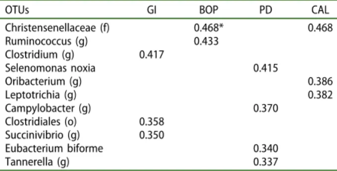
Specific gut phylotypes correlate with periodontal inflammation and attachment loss
Regardless of the periodontal status, associations between mean number of OTU reads and periodontal inflammation and periodontal attachment loss were examined (Table 1). A subset of OTUs showed sig-nificant correlations (rho = 0.337–0.468, p < 0.01) with bleeding and periodontal tissue destruction. Of interest, genera and species associated with period-ontal diseases showed significant moderate correla-tions with PD and CAL, including Selenomonas noxia, Leptotrichia, Tannerella and Campylobacter (p < 0.01).
High oral taxa counts in the gut microbiome of individuals with gingivitis
OTUs representing traditionally oral species were fre-quently detected in stool samples across the entire patient population. Over 100 species/phylotypes representative of oral microorganisms were identified. OTUs of oral organisms within species- or genus-level taxonomic groups, detected at total numbers ≥ 100 reads in all samples were selected and their numbers averaged within groups.Figure 4presents the mean number of reads for oral OTUs. The top 10 organisms detected in high mean reads were Bacteroides heparinolyticus, Alloprevotella rava, Fusobacterium spp., Streptococcus australis, Tannerella spp., Lachnospiraceae [G-2] spp., Oribacterium sp. OT102, Prevotella spp., Prevotella maculosa and Neisseria spp. Significant differences in oral taxa counts among groups were observed for S. australis, Prevotella spp., Rothia eria, Granulicatella adia-cens, Oribacterium asaccharolyticum and Porphyromonas sp. OT930 (present in high mean reads in G compared to
Figure 3.Proportional taxonomic assignments at the genus level in stool samples from individuals with different periodontal status. Genera detected at mean relative abundance ≥ 0.1% are presented. No significant differences among groups were observed (Mann-Whitney and Kruskal-Wallis tests, p > 0.01).
the other groups), and Peptostreptococcaceae [XI][G-6] which was predominant in PH individuals (Kruskal-Wallis, Mann-Whitney tests, p < 0.01). Other oral spe-cies, including several oral pathogens (Prevotella macu-losa, Prevotella veroralis, Prevotella denticola, Slackia exigua, Campylobacter curvus, Porphyromonas sp. OT930 and OT279, Dialister invisus, Peptostreptococcus stomatis, Porphyromonas endodontalis, Alloprevotella tannerae, Prevotella oulorum, Treponema maltophilum, Campylobacter rectus, Filifactor alocis and Parvimonas micra) were detected across samples regardless of period-ontal status (Figure 4).
Discussion
In addition to bacteraemia and metastatic inflammation [17], another possible pathway associating periodontal diseases to systemic inflammatory diseases may be through alterations in the gut microbiome. In individuals with periodontal diseases, the long-term swallowing of high doses of periodontal pathogenic microorganisms could induce a dysbiosis of the intestinal microbiota, favouring the establishment of an‘inflamed’ microbiome in terms of composition and/or function. In turn, this altered gut microbiota could modulate periodontal dis-eases by contributing to the progression and severity of periodontal tissue destruction. As a first approach to support this concept, we focused on determining the composition profile of the gut microbiome of individuals with periodontal diseases in comparison to periodontally healthy controls. We also searched for correlations between periodontal inflammation and attachment lost with specific microorganisms of the gut microbiota.
Along with our line of thought, some studies have addressed the possible association between periodon-tal disease/oral pathogens and the gut microbiota [45,46,52]. Arimatsu et al. [45] and Nakajima et al. [46] tested the direct impact of P. gingivalis on the gut microbiota through oral administration of high doses of this pathogen in mice. These authors reported an increase in local and systemic
inflammation, glucose blood levels, insulin resistance, systemic endotoxemia, and a decrease in gut barrier function. Although no major changes in diversity were seen, significant changes in the gut microbiota composition of P. gingivalis-administered mice were observed, with an increase in Bacteroidetes (mainly the order Bacteroidales), a decrease in Firmicutes, and an increase in Prevotella. Of interest, the proportions of Porphyromonadaceae were still very low in these animals, suggesting that the pathobiont P. gingivalis may alter the gut microbiome not by outgrowing in the gut but by indirectly inducing endotoxemia. In fact, endotoxin-induced inflammation seems to be essential for the development of many metabolic dis-orders. For instance, Fei and Zhao [53] were able to induce obesity and insulin resistance in germ free mice inoculated with an endotoxin-producing bacter-ium (Enterobacter cloacae B29) isolated from a mor-bidly obese human’s gut.
In contrast to those previous studies in animal models [45,46], our findings showed that patients with periodontal diseases tend to present lower diver-sity in the gut microbiota. Other investigations have shown that reduced alpha diversity is a reliable indi-cator of disease-associated dysbiosis [1,54–56], corro-borating our results. In relation to beta diversity, it was not possible to clearly distinguish individuals with distinct clinical status based on the composition of the gut microbiota.
Differences among clinical groups were observed in the composition of the gut microbiome at the phylum level. Considering the two major phyla commonly observed in the human gut microbiota, there was a ten-dency of Bacteroidetes to decrease and Firmicutes to increase with disease severity. In accordance with these data, a high Firmicutes/Bacteroidetes ratio in the gut microbiome has been reported in many other systemic inflammatory conditions [52,57]. Other investigations, however, have either reported a significant decrease in this ratio [45,46,58] or no changes at all [59] in systemic inflammatory conditions. Although the ratio Firmicutes/ Bacteroidetes was high in our diseased population, these phyla varied widely in abundance among patients within the same clinical group (Figure S1). Other phyla increased in abundance in G and CP patients compared PH were Proteobacteria, Verrucomicrobia and Euryarchaeota. Multiple sclerosis patients have shown to present increased relative abundance of Euryarchaeota and Verrucomicrobia compared to healthy controls [60], whereas Proteobacteria is increased in severe acute malnutrition [61]. Verrucomicrobia is also increased in abundance in dextran sodium sulphate-induced murine colitis [62]. On the other hand, evidence indicates that the species Akkermansia muciniphila of the Verrucomicrobia phylum has a protective anti-inflamma-tory effect against specific metabolic disorders and obe-sity [63].
Table 1.Correlation analysis between parameters of inflam-mation, attachment loss and OTUs detected in faeces sam-ples from 44 individuals with different periodontal status.
OTUs GI BOP PD CAL
Christensenellaceae (f) 0.468* 0.468 Ruminococcus (g) 0.433 Clostridium (g) 0.417 Selenomonas noxia 0.415 Oribacterium (g) 0.386 Leptotrichia (g) 0.382 Campylobacter (g) 0.370 Clostridiales (o) 0.358 Succinivibrio (g) 0.350 Eubacterium biforme 0.340 Tannerella (g) 0.337
GI: gingival bleeding. BOP: bleeding on probing. PD: probing depth. CAL: clinical attachment level.(o): order taxonomic level; (f): family taxo-nomic level; (g): genus taxotaxo-nomic level. *Refers to the Spearman coefficient (rho). All correlations presented were significant at p < 0.01.
Variability in abundance of different microorgan-isms in stool samples was even greater at the genus level (Figure S2). Several genera related to putative periodontal pathogens such as Fusobacterium, Prevotella, Parvimonas, Porphyromonas, Tannerella, Dialister, Filifactor, Treponema and Eubacterium were detected in stool samples of all groups.
In particular, OTUs representative of Prevotella, Comamonadaceae (f), and Lactobacillales (o) were sig-nificantly abundant in patients with gingivitis, but the order Bacteroidales was increased in stools of PH patients. We also found significant positive associations between Selenomonas noxia and some genera of key periodontal pathogens (Leptotrichia, Tannerella and Campylobacter) in the gut with periodontal attachment
loss. Finally, patients with periodontal diseases were accurately classified, and Mogibacteriaceae (f), Ruminococcaceae (f) and Prevotella (g) were the best predictors for classification of diseased patients. Contradictory results associating these gut microbial taxa with many different systemic diseases and health have been reported by others; therefore, results should be carefully interpreted. For instance, Ruminococcaceae has been related to a healthy gut [64], and Prevotella species are predominant commensal bacteria of healthy human mucosal sites [65]. Compelling data, however, have linked increased Prevotella abundance to inflammatory disorders, suggesting that some species exhibit distinct pathobiontic properties [65]. Inconsistency among stu-dies may be partially due to the high inter-individual
Figure 4.Oral taxa detected in the gut microbiota. A heat map of OTUs (mean number of reads) with > 97% identity to oral taxa, which were detected in stool samples from individuals with different periodontal status.(a) Oral taxa detected at mean numbers≥ 10; (b) Oral taxa detected at lower mean numbers (< 10). *Significant differences among groups were observed for S. australis, Prevotella spp., Rothia aeria, Granulicatella adiacens, Oribacterium asaccharolyticum, Porphyromonas sp. OT930, and Peptostreptococcaceae (Kruskal-Wallis, Mann-Whitney tests, p < 0.01). OT: oral taxon. (g) genus.
variability in gut microbiome composition at different taxonomic levels, as well as the complex multifactorial nature of periodontitis and systemic inflammatory dis-eases [66]. According to Duvallet and co-workers [54], there is not a unique dysbiotic microbiome associated with disease, and different diseases are characterized by distinct shifts in the gut microbiome. These shifts may be represented by depletion of beneficial species or enrich-ment of certain pathobionts. Of interest, many consortia of microorganisms in the gut microbiome are shared between a healthy and diseased state. This is true parti-cularly at the genus level in which different strains or species within some genera play different roles in various microbiome-associated conditions. As an example, our data showed an association between increased abun-dance of Prevotella and periodontal disease (more speci-fically gingivitis). However, different species of this genus (P. tannerae, Prevotella oulorum and Prevotella oris) were detected in higher proportions in PH or in both PH and CP individuals. These authors have also reported that the order Bacteroidales, here abundant in periodontal health, was one of these organisms non-specifically associated with health or disease [54]. Additionally, confounding factors such as diet, smoking, obesity, and stress, among others have significant impact on the composition of the gut microbiome, and therefore on possible associations between specific consortia and disease. Of the life-style
parameters here measured, only BMI was significantly higher in patients with periodontitis compared to other groups. Strong evidence has corroborated the positive association between obesity and periodontitis [67]. Therefore, the altered gut microbiome in overweight CP individuals may have contributed to the lower diver-sity and high numbers of pathogenic OTUs observed. Despite these caveats, OTUs representing periodontal pathogens were also detected in high numbers in PH controls and G who presented normal body weight. One limitation of the current study that may explain in part the lack of significant differences in microbial diversity among clinical groups or the existence of specific micro-biome associations with health or disease is the small sample population evaluated, in particular in the control group. Challenges in patient compliance resulted in some stool submissions that were not in adequate conditions during the initial clinical examination and prior to dental treatment.
The frequent detection of oral species in stools samples of our study population was reinforced by comparing sequencing data against the HOMD 16S rRNA database [68]. OTUs representative of known pathogenic oral taxa including P. endodontalis, C. rectus, D. invisus, P. micra, F. alocis, S. exigua, Treponema spp., Prevotella spp., Oribacterium spp., Tannerella spp., Leptotrichia spp., Selenomonas spp. and Fusobacterium spp. were detected
in high numbers in the gut microbiome of all patients regardless of periodontal status. Only a few species/phy-lotypes (S. australis, Prevotella spp., R. aeria, G. adiacens, O. asaccharolyticum, and Porphyromonas sp. OT930) were significantly more predominant in diseased indivi-duals compared to PH. Many of these microorganisms are associated with caries, periodontal and endodontic infections [69–72], but they have also been isolated from extra-oral infections or systemic conditions [52–54,73– 75]. In contrast, S. australis and G. adiacens have been related to periodontal health [69,70].
Koren et al. [56] brought up also the possibility of a link between oral and intestinal microbiota with inflam-matory diseases by investigating whether the oral or gut microbiota could contribute to atherosclerosis. They reported that several OTUs from oral and gut sources were also detected in atherosclerotic plaque within the same patients. In addition to the shared OTUs between atherosclerotic plaques and oral/gut microbiota, some of these OTUs were strongly correlated with disease mar-kers. For instance, Fusobacterium abundance in the oral cavity and members of the Erysipelotrichaceae and Lachnospiraceae families in the gut were positively cor-related with LDL and total cholesterol. Taken all together, our results support the idea that a relatively large variety of oral species can gain access to the intest-inal microbiota, regardless of the periodontal status. Whether these microorganisms colonize or not this body site, they may cause disturbances in this environ-ment by releasing metabolites and cell components, which induce an inflammatory state [76]. They may also interact and enhance the pathogenic effects of other species that colonize the gut [77]. The mechanisms involved in the contribution of these oral species to intestinal dysbiosis are highly complex and beyond the scope of this study, but our data indicate that microor-ganisms of known pathogenicity and associated with periodontal and/or systemic inflammation were fre-quently detected in the gut microbiota of individuals with periodontal diseases.
In summary, the present data indicate that individuals with periodontal diseases present a less diverse intestinal microbiome, characterized by an increase in the Firmicutes/Bacteroidetes ratio, as well as an enrichment of the phyla Euryarchaeota, Verrucomicrobia and Proteobacteria, consistent with microbial shifts observed in some systemic inflammatory diseases. Moreover, high numbers of oral taxa related to periodontal destruction and inflammation were detected in the gut microbiome of these individuals regardless of periodontal status. Future approaches using animal models are required to investigate the direct or indirect possible mechanisms by which specific oral consortia may affect the composition and function of the gut microbiome. Furthermore, larger longitudinal clinical investigations are encouraged to determine the impact of periodontal treatment on the restoration of the gut microbiome, as well as the
improvement of systemic health. A true causal relation-ship between periodontitis and gut dysbiosis will support the existence of an additional pathway linking period-ontal diseases to other systemic inflammatory conditions, reinforcing the role of periodontal diseases as an impor-tant risk factor for these conditions.
Acknowledgements
The sequencing efforts were supported by the National Institute of Environmental Health Sciences of the NIH, under award P30-ES002109. This study was supported in part by National Council for Scientific and Technological Development, Coordination of Improvement of Higher Education Personnel, Brasilia, Brazil; and Foundation for Research Financial Support in the State of Rio de Janeiro, Brazil. We extend our thanks to the MIT BioMicroCenter for library preparation and sequencing support. The authors also want to thank Clarissa B. Magalhães, Fatima Aparecida R. R. Hartenbach and Laís Christina P. Espíndola for their contribu-tions to this work.
Disclosure statement
No potential conflict of interest was reported by the authors.
Funding
This work was supported by the Conselho Nacional de Desenvolvimento Científico e Tecnológico [400239/2016– 7];National Institute of Environmental Health Sciences [P30-ES002109]; Fundação Carlos Chagas Filho de Amparo à Pesquisa do Estado do Rio de Janeiro [E-26/ 200.048/2016].
ORCID
Talita Gomes Baeta Lourenςo http://orcid.org/0000-0003-0966-3620
Sarah J. Spencer http://orcid.org/0000-0002-2744-8994
Eric John Alm http://orcid.org/0000-0001-8294-9364
Ana Paula Vieira Colombo http://orcid.org/0000-0002-2061-1840
References
[1] Cho I, Blaser MJ. The human microbiome: at the interface of health and disease. Nat Rev Genet.
2012;13(14):260–270.
[2] Belkaid Y, Hand TW. Role of the microbiota in immunity and inflammation. Cell.2014;157:121–141. [3] Holmes E, Li JJ, Athanasiou T, et al. Understanding
the role of gut microbiome–host metabolic signal dis-ruption in health and disease. Trends Microbiol.
2011;19:349–359.
[4] Lozupone CA, Stombaugh JI, Gordon JI, et al. Diversity, stability and resilience of the human gut microbiota. Nature.2012;489:220–230.
[5] Carding S, Verbeke K, Vipond DT, et al. Dysbiosis of the gut microbiota in disease. Microb Ecol Health Dis.
2015;26:26191.
[6] Page RC, Offenbacher S, Schroeder HE, et al. Advances in the pathogenesis of periodontitis: sum-mary of developments, clinical implications and future directions. Periodontol 2000.1997;14:216–248. [7] Tonetti MS, Van Dyke TE. Working group 1 of the joint
EFP/AAP workshop. Periodontitis and atherosclerotic cardiovascular disease: consensus report of the Joint EFP/AAP Workshop on periodontitis and systemic dis-eases. J Clin Periodontol.2013;40:S24–S29.
[8] Kholy KE, Genco RJ, Van Dyke TE. Oral infections and cardiovascular disease. Trends Endocrinol Metab.2015. 26:315–321.
[9] Borgnakke WS, Ylöstalo PV, Taylor GW, et al. of periodontal disease on diabetes: systematic review of epidemiologic observational evidence. J Periodontol.
2013;84(4 Suppl):S135–S52.
[10] Keller A, Rohde JF, Raymond K, et al. Association between periodontal disease and overweight and obesity: a systematic review. J Periodontol.2015;86:766–776.
[11] Kaye EK, Chen N, Cabral HJ, et al. Metabolic syn-drome and periodontal disease progression in men. J Dent Res.2016;95:822–828.
[12] Scannapieco FA, Bush RB, Paju S. Associations between periodontal disease and risk for nosocomial bacterial pneumonia and chronic obstructive pulmonary disease. A Systematic Review. Ann Periodontol.2003. 8:54–69. [13] Ahn J, Segers S, Hayes RB. Periodontal disease,
Porphyromonas gingivalis serum antibody levels and oro-digestive cancer mortality. Carcinog.2012;33:1055–1058.
[14] Shultis WA, Weil EJ, Looker HC, et al. Effect of period-ontitis on overt nephropathy and end-stage renal disease in type 2 diabetes. Diab Care.2007;30:306–311.
[15] De Pablo P, Chapple ILC, Buckley CD, et al. Periodontitis in systemic rheumatic diseases. Nat Rev Rheumatol.2009. 5:218–224.
[16] Winning L, Linden GJ. Periodontitis and systemic disease: association or causality. Curr Oral Health Rep.2017;4:1–7.
[17] Li X, Kolltveit KM, Tronstad L, et al. Systemic dis-eases caused by oral infection. Clin Microbiol.
2000;13:547–558.
[18] Tomas I, Diz P, Tobias A, et al. Periodontal health status and bacteremia from daily oral activities: sys-tematic review/meta-analysis. J Clin Periodontol.
2012;39:213–228.
[19] Reyes L, Herrera D, Kozarov E, et al. Periodontal bacterial invasion and infection: contribution to ather-osclerotic pathology. J Clin Periodontol. 2013;40 (Suppl. 14):S30–S50.
[20] Takeuchi H, Furuta N, Morisaki I, et al. Exit of intra-cellular Porphyromonas gingivalis from gingival epithelial cells is mediated by endocytic recycling pathway. Cell Microbiol.2011;13:677–691.
[21] Lafaurie GI, Mayorga-Fayad I, Torres MF, et al. Periodontopathic microorganisms in peripheric blood after scaling and root planning. J Clin Periodontol.2007;34:873–879.
[22] Castillo DM, Sanchez-Beltran MC, Castellanos JE, et al. Detection of specific periodontal microorgan-isms from bacteremia samples after periodontal ther-apy using molecular-based diagnostics. J Clin Periodontol.2011;38:418–427.
[23] Dorn BR, Harris LJ, Wujick CT, et al. Invasion of vascular cells in vitro by Porphyromonas endodontalis. Int Endo J.2002;35:366–371.
[24] Peters SR, Valdez M, Riviere G, et al. Adherence to and penetration through endothelial cells by oral tre-ponemes. Oral Microbiol Immunol. 1999;14:379–383.
[25] Saito A, Inagaki S, Kimizuka R, et al. Fusobacterium nucleatum enhances invasion of human gingival epithelial and aortic endothelial cells by Porphyromonas gingivalis. FEMS Immunol Med Microbiol.2008;54:349–355. [26] Schenkein HA, Barbour SE, Berry CR, et al. Invasion
of human vascular endothelial cells by Actinobacillus actinomycetemcomitans via the receptor for platelet-activating factor. Infect Immun.2000;68:5416–5419. [27] Takahashi Y, Davey M, Yumoto H, et al. Fimbria
dependent activation of pro-inflammatory molecules in Porphyromonas gingivalis infected human aortic endothelial cells. Cell Microbiol.2006;8:738–757. [28] Ohki T, Itabashi Y, Kohno T, et al. Detection of
periodontal bacteria in thrombi of patients with acute myocardial infarction by polymerase chain reac-tion. Am Heart J.2012;163:164–167.
[29] Nakano K, Nemoto H, Nomura R, et al. Detection of oral bacteria in cardiovascular specimens. Oral Microbiol Immunol.2009;24:64–68.
[30] Marques da Silva R, Caugant DA, Lingaas PS, et al. Detection of. Actinobacillus actinomycetemcomitans but not bacteria of the red complex in aortic aneur-ysms by multiplex polymerase chain reaction. J Periodontol.2005;76:590–594.
[31] Aimetti M, Romano F, Nessi F. Microbiologic analysis of periodontal pockets and carotid atheromatous pla-ques in advanced chronic periodontitis patients. J Periodontol.2007;78:1718–1723.
[32] Aquino AR, Lima KC, Paiva MS, et al. Molecular survey of atheromatous plaques for the presence of DNA from periodontal bacterial pathogens, archaea and fungi. J Period Res.2011. 46:303–309.
[33] Gaetti-Jardim E Jr, Marcelino SL, Feitosa AC, et al. Quantitative detection of periodontopathic bacteria in atherosclerotic plaques from coronary arteries. J Med Microbiol.2009;58:1568–1575.
[34] Brodala N, Merricks EP, Bellinger D, et al. Porphyromonas gingivalis bacteremia induces coron-ary and aortic atherosclerosis in normocholesterole-mic and hypercholesterolenormocholesterole-mic pigs. Arterio Thromb Vasc Biol.2005. 25:1446–1451.
[35] Jain A, Batista EL Jr, Serhan C, et al. Role for period-ontitis in the progression of lipid deposition in an animal model. Infect Immu.2003. 71:6012–6018. [36] McGraw WT, Potempa J, Farley D, et al. Purification,
characterization, and sequence analysis of a potential virulence factor from Porphyromonas gingivalis, peptidy-larginine deiminase. Infect Immun.1999;67:3248–3256. [37] Wahaidi VY, Kowolik MJ, Eckert GJ, et al. Endotoxemia
and the host systemic response during experimental gingivitis. J Clin Periodontol.2011;38:412–417.
[38] Winning L, Patterson CC, Cullen KM, et al. The association between subgingival periodontal patho-gens and systemic inflammation. J Clin Periodontol.
2015;42:799–806.
[39] Schenkein HA, Loos BG. Inflammatory mechan-isms linking periodontal diseases to cardiovascular diseases. J Clin Periodontol. 2013;40(Suppl. 14): S51–S69.
[40] Taylor JJ, Preshaw PM, Lalla E. A review of the evi-dence for pathogenic mechanisms that may link per-iodontitis and diabetes. J Clin Periodontol. 2013;40 (Suppl. 14):S113–S34.
[41] Mustapha IZ, Debrey S, Oladubu M, et al. Markers of systemic bacterial exposure in periodontal disease and cardiovascular disease risk: a systematic review and meta-analysis. J Periodontol.2007;78:2289–2302.
[42] Behle JH, Sedaghatfar MH, Demmer RT, et al. Heterogeneity of systemic inflammatory responses to periodontal therapy. J Clin Periodontol.
2009;36:287–294.
[43] Hajishengallis G, Liang S, Payne MA, et al. Low-abun-dance biofilm species orchestrates inflammatory period-ontal disease through the commensal microbiota and complement. Cell Host Micro.2011;10:450–497. [44] Hasturk H, Kantarci A, Goguet-Surmenian E, et al.
Resolvin E1 regulates inflammation at the cellular and tissue level and restores tissue homeostasis in vivo. J Immunol.2007;179:7021–7029.
[45] Arimatsu K, Yamada H, Miyazawa H, et al. Oral pathobiont induces systemic inflammation and meta-bolic changes associated with alteration of gut micro-biota. Sci Rep.2014;4:4828.
[46] Nakajima M, Arimatsu K, Kato T, et al. Oral admin-istration of P.. gingivalis induces dysbiosis of gut microbiota and impaired barrier function leading to dissemination of enterobacteria to the liver. PLoS One.2015;10:e0134234.
[47] Silva-Boghossian CM, Luiz R, Colombo AP. Risk indi-cators for increased periodontal probing depth in sub-jects attending a public dental school in Brazil. Oral Health Prev Dent.2011;9:289–299.
[48] Mohammad RA, Arash V, Alireza T, et al. Rapid DNA extraction protocol from stool, suitable for molecular genetic diagnosis of colon cancer. Iran Biom J.
2007;11(3):203–208.
[49] Caporaso JG, Kuczynski J, Stombaugh J, et al. QIIME allows analysis of high-throughput community sequencing data. Nat Methods.2010;7:335–336. [50] Shannon CE, Weaver W. The mathematical theory
of communication. Urbana: Illinois University Press;1963.
[51] Lozupone C, Lladser ME, Knights D, et al. UniFrac: an effective distance metric for microbial community comparison. ISME.2011;5:169–172.
[52] Koren O, Spor A, Felin J, et al. Human oral, gut, and plaque microbiota in patients with atherosclerosis. Proc Natl Acad Sci U S A.2011;108(Suppl 1):4592–4598.
[53] Fei N, Zhao L. An opportunistic pathogen isolated from the gut of an obese human causes obesity in germfree mice. ISME J.2013;7:880–884.
[54] Duvallet C, Gibbons SM, Gurry T, et al. Meta analysis of microbiome studies identifies disease-specific and shared responses. Nat Commun.
2017;8(1):1784.
[55] Turnbaugh PJ, Hamady M, Yatsunenko T, et al. A core gut microbiome in obese and lean twins. Nature.2008;457:480–484.
[56] Vincent C, Stephens DA, Loo VG, et al. Reductions in intestinal clostridiales precede the development of noso-comial Clostridium difficile infection. Microbiome.
2013;1:18.
[57] Ley RE, Backhed F, Turnbaugh P, et al. Obesity alters gut microbial ecology. Proc Natl Acad Sci U S A.
2005;102:11070–11075.
[58] Schwiertz A, Taras D, Schäfer K, et al. Microbiota and SCFA in lean and overweight healthy subjects. Obes.
2010;18:190–195.
[59] Duncan SH, Lobley GE, Holtrop G, et al. Human colonic microbiota associated with diet, obesity and weight loss. Int J Obes.2008;32:1720–1724.
[60] Jangi S, Gandhi R, Cox LM, et al. Alterations of the human gut microbiome in multiple sclerosis. Nat Comm. 2016;7:12015.
[61] Tidjani Alou M, Lagier JC, Raoult D. Diet influ-ence on the gut microbiota and dysbiosis related to nutritional disorders. Hum Microbiome J.
2016;1:3–11.
[62] Nagalingam NA, Phil M, Kao JY, et al. Microbial ecology of the murine gut associated with the devel-opment of DSS-colitis. Inflamm Bowel Dis. 2011;17 (4):917–926.
[63] Cani PD, De Vos WM. Next-generation beneficial microbes: the case of Akkermansia muciniphila. Front Microbiol.2017;8:1765.
[64] Wong JMW, De SR, Kendall CWC, et al. Colonic health: fermentation and short chain fatty acids. J Clin Gastroenterol.2006;40:235–243.
[65] Larsen JM. The immune response to Prevotella bac-teria in chronic inflammatory disease. Immunol.
2017;151:363–374.
[66] Eckburg PB, Bik EM, Bernstein BC, et al. Diversity of the human intestinal microbial flora. Science.
2005;308:1635–1638.
[67] Nascimento GG, Leite FR, Do LG, et al. Is weight gain associated with the incidence of periodontitis? A sys-tematic review and meta-analysis. J Clin Periodontol.
2015;42:495–505.
[68] Chen T, Yu W-H, Izard J, et al. The Human Oral Microbiome Database: a web accessible resource for investigating oral microbe taxonomic and genomic information. Database. 2010;baq013. DOI:10.1093/ database/baq013.2010.
[69] Lourenco TGB, Heller D, Silva-Boghossian CM, et al. Microbial signature profiles of periodontally healthy and diseased patients. J Clin Periodontol.
2014;41:1027–1036.
[70] Colombo AP, Boches SK, Cotton SL, et al. Comparisons of subgingival microbial profiles of refractory periodontitis, severe periodontitis, and periodontal health using the human oral microbe identification microarray. J Periodontol. 2009;80: 1421–1432.
[71] Siqueira JF, Rôças IN. Distinctive features of the microbiota associated with different forms of apical periodontitis. J Oral Microbiol.2009;1:10.
[72] Tanner AC, Kent RL, Holgerson PL, et al. Microbiota of severe early childhood caries before and after ther-apy. J Dent Res.2011;90:1298–1305.
[73] Han YW, Wang X. Mobile mcrobiome: oral bacteria in extra-oral infections and inflammation. J Dent Res.
2013;92:485–491.
[74] Väisänen M, Kiviranta M, Summanen P, et al. Porphyromonas endodontalis-like organisms from extraoral sources. Clin Infect Dis.1996;25:191–193. [75] Kim UJ, Won E, Kim JE, et al. Rothia aeria infective
endocarditis: a first case in Korea and literature review. Ann Lab Med.2014;34(4):317–320.
[76] Berezowa AB, Ernst RK, Coats SR, et al. The structurally similar, penta-acylated lipopolysacchar-ides of Porphyromonas gingivalis and Bacterolipopolysacchar-ides elicit strikingly different innate immune responses. Microb Pathog. 2009;47:68–77.
[77] Onderdonk AB. Animal models simulating anaerobic infections. Anaerobe.2005;11:189–195.