0
“ INSTITUTE OF BIOLOGY - DEPARTMENT OF CELLULAR AND MOLECULAR BIOLOGY”Rue Emile - Argand 11, 2009 Neuchâtel
“
STUDY OF MOSS VACUOLES
AND FUNCTIONAL CHARACTERIZATION OF THE PUTATIVE
VACUOLAR RECEPTORS, THE
RMR
PROTEINS
PhD thesis presented by Sanaa Ayachi PhD director Prof. Jean-Marc Neuhaus
3
Pour Toi et par Toi A ma mère et à mon père
5
Thanks
Firstly I want to thank my supervisor Prof. Jean-Marc Neuhaus for giving me the opportunity to follow this PhD project.
I want to thank sincerely Dr. Didier Schaefer, my mentor for teaching me the moss system. I want to thank all the members of the Laboratory of Molecular and Cell Biology for their support and the scientific discussions and support; Sonia Negro, Livia Autauri, Dirk Balmer, Sophie Marc-Martin, Dr. Nadia Feddermann, Dr. Guillaume Gouzerh and Noémie Fahr. I thank particularly my colleagues Dr. Egidio Stigliano and Dr. Alessandro Occhialini for being my PhD mate and for the unforgettable moment with them.
I thank Prof. Niko Geldner for the nice suggestions and the interesting discussion we had during my mid-thesis defense.
7
Table of Contents
Table of Contents ... 6 Abbreviations ... 11 Abstract ... 12 Chapter I: Introduction ... 13Part 1: The plant secretory system ... 15
1. The endomembrane system ... 16
2. The ER is the starting point of the secretory system... 18
3. The Golgi Apparatus ... 21
4. Direct traffic from ER to vacuoles ... 23
5. The Trans-Golgi-Network ... 23
6. The endosomes ... 24
7. Vesicle trafficking ... 26
8. Endocytosis process... 30
9. Vacuoles ... 31
Part 2: Vacuole biogenesis ... 33
1. Vacuole types ... 34
2. Vacuolar sorting determinants ... 36
3. Vacuolar sorting receptors ... 38
4. Vacuolar sorting pathways ... 42
5. De novo vacuole biogenesis ... 45
Part 3: P. patens as model system ... 47
1. P.patens a simple system ... 48
2. The moss vacuole ... 55
3. Moss organelles ... 59
Experimental aims ... 61
8
I. INTRODUCTION ... 64
II. Results ... 67
1. Visualization of the ER ... 67
2. Visualization of Golgi in moss cells ... 68
3. Visualization of trans-Golgi-network in moss cells... 70
4. AtTIP localization in moss cells ... 72
5. The moss vacuole structure and mobility ... 74
6. Interactions of vacuoles with other organelles ... 77
III. Discussion ... 79
1. Qualitative study of the organelles of the secretory system ... 79
2. Vacuole organization ... 79
3. Model of biogenesis of artefactual AtTIP compartments ... 80
4. TIP protein evolution ... 81
IV. Material and methods ... 83
V. Supplemental figures ... 85
Chapter 3: Vacuole biogenesis ... 87
I. INTRODUCTION ... 88
II. Results ... 91
1. Most moss cells have an acidic lytic central vacuole ... 91
2. Two vacuole types can coexist in moss cells ... 93
3. The central vacuole is formed from small vacuoles in differentiated cells ... 94
4. Vacuole regeneration ... 95
III. Discussion ... 97
1. Does moss have different vacuoles types? ... 98
2. Early steps of vacuole biogenesis ... 98
3. Evidence for vacuole enlargement ... 99
9
VI. Supplemental figures ... 103
Chapter 4: Characterization of complete RMR deletion mutants. ... 105
I. Introduction ... 106
II. Results ... 110
1. Identification of RMR genes in P. patens ... 110
2. Generation of RMR Knock-out lines ... 112
3. Phenotypic analysis ... 116
III. Discussion ... 124
1. Phenotypic analysis ... 125
2. Stress response ... 125
3. Vacuolar organization... 126
4. Secretory systems reporters ... 126
5. A rescue pathway? ... 127
IV. Material and methods ... 128
General discussion and outlook ... 131
1. Moss secretory system ... 132
1.1. The development of the moss reporters ... 133
1.2. Use of heterologous reporters ... 133
2. Vacuole biogenesis ... 134
2.1. Vacuole regenerates from tubule-like structures ... 134
2.2. One or several vacuole types? ... 134
3. Are RMRs vacuolar receptors? ... 135
3.1. Putative destinations of the RMRs cargoes in the PpRMRs ko mutant ... 135
3.2. RMRs are not a major actors of the protein targeting to the vacuole ... 136
3.3. Perspectives ... 136
Annex ... 139
10
2. Methods ... 142
3. Annex primers ... 147
4. Annex: plasmids and constructs ... 150
4.1. Cloning vectors ... 150 4.2. Knock-out vectors ... 150 4.3. Knock-in Vectors ... 151 4.4. Heterologous reporters ... 152 5. Annex: RMR sequences ... 153 Bibliography ... 155
11
Abbreviations
ABA: Abscisic AcidADP: Adenosine Diphosphate AP: Adaptin Protein
ARF: ADP Ribosylation Factor ATP: Adenosine Triphosphate
ATPase: Adenosine Triphosphate Hydrolase
Bip: Binding immunoglobulin protein
BFA: Brefeldine A
BP-80: Binding Protein of 80 kDa CCV: Clathrin-Coated Vesicle COPI: Coat Protein I
COPII: Coat Protein II Ct-VSD: C-terminal Vacuolar
SortingDeterminant
DIP: Dark-Induced Protein DNA: Deoxyribonucleic Acid DV: Dense Vesicle
ERAD: Endoplasmic Reticulum Associated
Degradation
ERES: Endoplasmic Reticulum Exportin Site ESCRT: Endosomal Sorting Complex
Required for Transport
GDP: Guanosine Diphosphate GEF: GTP Exchange Factor GFP: Green Fluorescent Protein Hsp: Heat Shock Protein Ko: Knock-out
LV: Lytic Vacuole
MS: Mass Spectrometry MVB: Multivesicular Body
PA domain: Protein Associated domain PAC: Precursor Accumulating Vesicle PM: Plasma Membrane
PSV: Protein Storage Vacuole
psVSD: Protein Structure dependent Vacuolar
Sorting Determinant
PVC: Prevacuolar Compartment Rab: Ras-related in Brain
RING: Really Interesting New Gene RFP: Red Fluorescent Protein
RMR: Receptor-like Membrane Ring-H2 RNF13: RING Finger Protein 13
SNAP: Soluble NSF Attachment protein RNA:
Ribonucleic Acid
SNARE: Soluble N-ethylmaleimide-sensitive
Protein Attachment Protein Receptor
SP: Signal Peptide
SRP: Signal Recognition Particle
ssVSD: Sequence-specific Vacuolar Sorting
Determinant
TIP: Tonoplast Intrinsic Protein UPR: Unfolded Protein Response
VAMP: Vesicle Associated Membrane Protein VSD: Vacuolar Sorting Determinant
VSR: Vacuolar Sorting Receptor YFP: Yellow Fluorescent Protein
12
Abstract
The vacuolar system of plants is a key element of plant growth and development, it fulfils many other functions. Plant cell can have more than two different vacuolar sorting systems: the lytic and the (seed) protein storage or vegetative storage vacuoles. Soluble vacuolar proteins are sorted through the secretory pathway to these vacuoles by three different routes, depending on different types of Vacuolar Sorting Determinants (VSD) and involving several types of receptors and vesicles. The AtRMR proteins has been identified in cellular structures associated with the seed storage vacuole pathway (Jiang et al. 2000). Based on its localisation and homology to a known vacuolar receptor, it has been hypothesised to be a receptor protein for the C-terminal type of VSD (ct-VSD) involved in sorting to the storage vacuole. The genome of Physcomitrella patens contains five genes coding for RMR proteins.
My work hypothesis is that the vacuolar system of higher plants has evolved from simple ancestors, which might have been preserved in lower plants. This evolution is reflected in the gene families involved in vacuole biogenesis. In a first part, we established the moss P.
patens as a model system for the study of the secretory pathways. In a second part, we
performed a comparative study of the plant-specific aspects of the vacuolar system. And finally in a third part, we tried to establish the functional role of PpRMR genes by the analysis of the complete RMR deletion mutants. Several strategies were considered to investigate a putative disorder due to RMRs loss of function. So far, no phenotype was detected in the mutants. Nevertheless the absence of the RMR family gene seems not to be necessary for moss development.
13
14
15
16 1.
The endomembrane system
The endomembrane system (secretory pathway) of plant cells has been studied by analogy with those of animals and yeasts, which have been well characterized. In plants, the secretory pathway comprises the endoplasmic reticulum (ER) as site of protein and lipid synthesis, the Golgi apparatus (GA) as maturation and sorting compartment for proteins and lipids, the Trans-Golgi-Network (TGN), the prevacuolar compartment (PVC), the vacuoles, and several types of vesicles involved in transport between these compartments and to the cell surface. In a review based mainly on observations of plant cells, Morre and Mollenhauer (Morre et al. 1974) defined the endomembrane system as the functional integration of ER, Golgi complex,secretory vesicles, plasma membrane, and hydrolytic compartments (fig. 1).
Most secretory proteins are transported by vesicles from the ER to the GA where they are matured and sorted. If they contain the necessary sorting information, they will be retained or transported to the correct target compartment (Paris et al. 1996). In the absence of such specific information, they will be packaged into vesicles that will then fuse with the plasma membrane and release their content to the apoplast (Denecke et al. 1990b).
On their way to secretion or to vacuoles, proteins have to pass several organelles such as the trans-Golgi network, early or late endosomes or prevacuoles. One final compartment of the plant secretory system is the vacuole (Marty 1999). The vacuole plays a major role in storage and recycling of various compounds (Taiz 1992); it contains the various hydrolases which degrade and recycle proteins, lipids and carbohydrates (Otegui et al. 2005). Depending on the tissue type, a single cell can harbor two vacuoles with distinct functions: the lytic vacuole (LV) and the protein storage vacuole (PSV) (Epimashko et al. 2004; Neuhaus and Rogers 1998; Paris et al. 1996; Surpin and Raikhel 2004).
17
Figure 1: Model of vacuolar protein sorting in plants
Soluble proteins with a specific sorting signal reach the vacuole via the secretory pathway: ER (Endoplasmic Reticulum), GA (Golgi Apparatus), TGN (Trans-Golgi-Network), PVC (Prevacuolar Compartment) and lytic vacuoles (LV) or protein storage vacuole (PSV). This model is based on different studies and was described in Vitale„s review (Vitale and Raikhel 1999). However this model was recently challenged.
(1) Soluble protein precursors are synthesized in the ER. The proteins lacking a specific vacuolar
sorting signal (black stars) are secreted out of the cell at the plasma membrane.
(2) Proteins with ssVSD (red stars) interact with a receptor (VSR) in the membrane of the TGN.
These ligand-receptor complexes are recruited into clathrin-coated vesicles (CCVs). The CCVs release their contents into the prevacuolar compartment (PVC). In PVC (or MVB), the cargo dissociates from the receptor. Vacuolar proteins then reach the central vacuole while the cargo receptor is recycled back to the GA for another cycle of transport.
(3) Proteins with ct-VSD (blue stars) aggregate at the rim of the cis-cisternae and at the TGN. They
are probably packed in the precursors of dense vesicles (DVs). DVs either fuse directly to PSVs or form a PVC for PSV.
(4) Storage proteins in pumpkins seeds (green stars) are transported by Precursor-Accumulating
18
2. The ER is the starting point of the secretory system
The ER can be subdivided into three domains with distinct function; the nuclear membrane, the smooth ER (SER) and the rough ER (RER). The nuclear membrane is a specialised part of the ER which harbours the nuclear pore complexes. The SER is the site of lipid biosynthesis, xenobiotic detoxification and calcium regulation (Vertel et al. 1992). The RER appears rough in the electron microscope because ribosomes are associated to its cytosolic face. It is the entry point into the endomembrane system for newly synthesized proteins (Vitale and Denecke 1999). Proteins addressed to the endomembrane system possess N-terminal signal peptide (SP) or have analogous transmembrane domains at other places within the protein. The newly synthesized SP emerges from the ribosome and is recognized by a signal recognition particle (SRP). The ribosome stops translating until the SRP-ribosome complex can attach to a receptor at the ER surface. The nascent polypeptide can then enter the RER cotranslationally through a protein pore, the translocon (Hamman et al. 1998). The translocation is a passive process which does not require additional energy, since the pressure provided by translation is enough for translocation (Vitale and Denecke 1999). Once it reaches the ER lumen, the signal peptide (SP) is cleaved by a signal peptidase and yield the maturing protein.
2.1. The role of the ER
ER-localised proteins undergo post-traductional modifications and acquire their mature conformation with the help of chaperone proteins resident in the ER (endoplasmins, calnexin, calreticulin, BiP, protein disulfide isomerases (PDI)) (Bednarek and Raikhel 1992). The best characterized ER chaperone is BiP which belongs to the Hsp 70 family (70 kDa proteins) of ATPases involved in the catalysis of protein folding and assembly (Hartl 1996). PDI which catalyses the formation and rearrangement of disulphide bonds plays also an important role in protein folding and maturation (Vitale and Denecke 1999).
The ER is also an important site for protein modification, in particular for N-glycosylation on specific asparagines (Asn) present in the consensus sequence Asn–X– Ser/Thr (X is any amino-acid except Pro). The main role of N-glycosylation in plant cells is to assure correct protein folding and to increase the protein solubility. N-glycosylation is catalyzed by the multisubunit enzyme oligosaccharyl transferase which is associated on the
19
luminal side with the translocon pore. The modification usually occurs cotranslationally but post-translational glycosylation does also occur (Vitale and Denecke 1999).
The ER can become a storage compartment in seeds. Cereal storage proteins accumulate in the ER lumen forming electron-dense structures called protein bodies (PB) (Herman and Larkins 1999). PB can be permanently stored in the ER or transferred to PSV by a specific pathway (Vitale and Ceriotti 2004). The mechanism leading to the retention of storage proteins in the ER and subsequent formation of protein bodies is still unclear. The lack of an export signal and actions of molecular chaperones such as BiP might be involved in aggregations of storage proteins in the ER. An interaction between storage proteins and the membrane bilayer has also been demonstrated (Kogan et al. 2004; Vitale and Denecke 1999). 2.2. Quality control
The ER has important check points for correct protein folding and assembly with a process called ER quality control (Hurtley and Helenius 1989). Misfolded proteins affected by physical or chemical stresses are recognized by molecular chaperones such as BiP and retained in the ER lumen. The chaperones help the proteins to refold to their native structure, recovering their normal cell functions (Hiller et al. 1996). If binding time to the chaperone is abnormally long, the malfolded proteins are degraded in a process called ER-associated degradation (ERAD) (Vitale and Boston 2008). Two ER-resident lectins (calnexin and calreticulin) are also involved in protein quality control. They recognize misfolded glycoproteins and cooperate with glucosylation and deglucosylation enzymes (Helenius et al. 1997).
2.3. ER retention signals
Soluble proteins with a C-terminal H/KDEL sequence are retained in the lumen of the ER (Pelham 1998). KDEL proteins which accidentally escaped from the ER to the GA are retrieved by membrane receptors such as ERD2 at a cis-Golgi cisterna and sent back to the ER by retrograde transport (Lewis and Pelham 1992).
20 2.4. Traffic between ER and Golgi
2.4.1. COPII vesicles
In eukaryotic cells, the ER and Golgi are connected via two types of vesicles, the COPII vesicles for the anterograde traffic from ER to Golgi and the COPI (Coatomer) vesicles for retrograde traffic from the Golgi to the ER (Barlowe et al. 1994). The anterograde traffic starts at specific ER domains called ERES (ER exporting site) (Bassham and Blatt 2008). The anterograde transport has been studied by analogy to mammalian cells and yeast, and homologues of COPII coat proteins have been identified in the A.thaliana genome (Movafeghi et al. 1999). The process of COPII vesicle formation requires a specific GTPase, a Sar1p family protein, which recruits the adaptor complex Sec23/Sec24 and cargo membrane proteins. Recruitment of Sec13/Sec31 completes the coat assembly (Movafeghi et al. 1999). Sar1p is recruited by the guanine nucleotide exchange factor (GEF) Sec12p at the membrane of ER. Once activated, Sar1p can recruit all coat proteins (Hanton et al. 2005). When Sar1p hydrolyses its bound GTP and then dissociates from the membrane, it causes the disassembly of the coat and thus allows vesicle fusion with the membrane of cis-Golgi. Proteins of the COPII coat are recycled back to ER for another cycle of vesicle formation (Hanton et al. 2005).
2.4.2. Endoplasmic reticulum export site (ERES) and signals
The anterograde ER export occurs from a specific domain named ERES where the different factors needed for COPII vesicle formation are assembled (Hawes et al. 2008). The recruitment of cargo proteins at ERES is poorly understood in plant cells. The process may be similar to mammalian cells where protein cargoes are recruited to ERES, incorporated in COPII vesicles and then transported to Golgi (Aridor et al. 2001). A study has shown an increase of the ERES site number when cargo is over-expressed (Hanton et al. 2007). In mammals and yeast, studies showed that (Sec16) a protein associated to ER exit sites was involved in ER protein export (Connerly et al. 2005). However, a protein with similar function has yet to be identified in plant.
21 2.4.3. COPI vesicles
COPI vesicle formation starts when the ARF1 GTPase interacts with the GA protein p23 (Gommel et al. 2001). Then, an ARF-GEF exchanges GTP for GDP, leading to a conformational change of ARF1 which allows its membrane binding (Helms and Rothman 1992). Activated ARF1 recruits the COPI coatomer from the cytosol causing vesicle budding (Rothman and Orci 1996). The COPI coatomer consists of the F adaptor subcomplex (four subunits: β-, γ-, δ-, δ-COP) and of the B cage subcomplex (three subunits: α-, β‟ and ε-COP) (Waters et al. 1991). ARF1 inactivation by an ARF-GAP after vesicle budding from the membrane causes coat disassembly and allows fusion with the ER membrane. ARF1 activation can be inhibited by the lactone antibiotic Brefeldin A (BFA). This prevents COPI coat formation, blocking the retrograde transport from cis-Golgi to ER and then indirectly blocking also anterograde transport. This drug is widely used to study protein transport by blocking the ER/Golgi transport (Helms and Rothman 1992; Ritzenthaler et al. 2002; Robinson et al. 2008).
3. The Golgi Apparatus
3.1. Roles of the Golgi Apparatus
After leaving their production site (ER), most of secretory proteins are transported to the Golgi apparatus (GA). It is composed of stacked cisternae. In plant cells, the GA is dispersed throughout the cytosol, and the stacks move around whereas in mammalian cells the stacks are localized near the nucleus. The stacks are interconnected by tubular elements (Andreeva et al. 1998; Mellman and Simons 1992). A Golgi stack can be composed of four to eight cisternae organized in three different regions with distinct functions: cis-Golgi, medial-Golgi and Golgi. The cis-Golgi constitutes the entrance of the apparatus while, from the trans-Golgi, vesicles transport secretory proteins to their final destination (Hawes et al. 2008; Matheson et al. 2006). The Golgi apparatus is an important traffic point between different organelles, such as ER, TGN and endosomes. This compartment is also a major site of post-translational modifications of N-glycans and O-glycosylation of glycoproteins and proteoglycan. The N-glycans modification starts already in the ER and is continued sequentially by numerous GA glycosidases and glycosyltransferases. Glycoproteins are
22
transported across the stack (Zhang and Flint 1992), each cisterna containing specific modification enzymes to form a multistage processing unit. This functional differentiation between cis-, medial and trans-Golgi was demonstrated by localizing different glycan-modifying enzymes in different cisternae of the stack (Glick 2000). The Golgi apparatus is also involved in the biosynthesis of many polysaccharides such as hemicellulose and acidic pectic polysaccharides which are very important components of the cell wall matrix (Bolwell 1988). The GA is also a biosynthesis site for lipids such as sphingolipids ubiquinone and plastoquinone, as described in spinach (Swiezewska et al. 1993).
3.2. Transport through the Golgi apparatus
Cargo from the ER passes sequentially through cis-, medial-, and trans-cisternae before arriving at the TGN. Transport through the Golgi cisternae is a matter of controversy (Pelham and Rothman 2000). Three models have been suggested to explain the vectorial transport of secretory proteins through the GA.
The first model (“old model”) postulated that the GA is a static organelle, proteins transit from cis-cisternae to trans-cisternae and are matured on their way, and anterograde protein traffic between cisternae is assured by COPI vesicles (Donaldson and Williams 2009; Rothman 1994).
Cisternal maturation was proposed as an alternative model to explain scale transport in algae (Becker and Melkonian 1996) and was studied in yeast (Glick and Malhotra 1998). In this model, the cisternae are transient structures formed by fusion of ER derived vesicles with retrograde vesicles at the GA‟s cis side. In this model, cargo proteins remain in the cisternae during maturation from cis- to trans-Golgi, while the GA enzymes modifying the cargo are relocated back to precursor cisternae by COPI vesicles. The trans cisternae are destroyed by the formation of transport vesicles for other compartments and of retrograde vesicles recycling trans-Golgi enzymes to younger cisternae. Alternatively, the trans cisternae may detach from GA stack and become a TGN. This mechanism was visualized in budding yeast, which does not have Golgi stacks but rather isolated single cisternae (Donaldson and Williams 2009; Losev et al. 2006; Matsuura-Tokita et al. 2006).
The third model proposed is a modification of the cisternal maturation model which was supported by recent evidences, especially in plants (e.g. see (Donohoe et al. 2007; Otegui et al. 2006). It is called the “rapid-partitioning model” (Patterson et al. 2008) because GA cisternae are partitioned into subdomains where enzymes perform modifications. Cargoes are
23
able to move rapidly between the cisternae via vesicles and tubular connections to undergo specific modifications (Donaldson and Williams 2009). Consequently, cargoes are able to move in a bidirectional manner by specific vesicular or tubular traffic depending on the modification location (Patterson et al. 2008). All these models are not exclusive; they may vary between different organisms and different steps of development.
4. Direct traffic from ER to vacuoles
In plant cells, a pathway from ER to protein storage vacuoles (PSV) bypassing the GA was demonstrated by electron microscopy on maturing pumpkin cotyledons. The storage protein proglobulin is sorted via precursor accumulating (PAC) vesicles (Hara-Nishimura et al. 1998), with sizes ranging from 200 to 400 nm, and containing unglycosylated precursors of storage proteins. Once in the vacuolar lumen, these precursors undergo a maturation process. PAC vesicles fuse with PSV either by autophagy or by direct membrane fusion. The autophagy uptake of PAC vesicles was observed in maturing pea seeds and in mungbean seedlings (Robinson et al. 1995). However, another study supported the fusion theory, showing small G-proteins involved in membrane fusion between PACs and PSV on the surface of PACs (Shimada et al. 1994). Most recently, a similar direct pathway was also suggested for certain membrane proteins (Rivera-Serrano et al. 2012).
5. The Trans-Golgi-Network
5.1. The TGN
The TGN is the site of cargo sorting and the organelle where proteins are targeted to their final destination (Gu et al. 2004). Its size differs, and it is mainly located near the trans-side of the Golgi (Traub and Kornfeld 1997). The plant TGN is physically and functionally distinct from the trans-Golgi (Uemura et al. 2004). In tobacco epidermal cells, a fluorescent marker for TGN does not co-localize with a Golgi marker (Foresti et al. 2008). In animal cells, upon BFA treatment, the TGN aggregates with endosomes to form a TGN-endosomal hybrid compartment, while the GA fuses with the ER (Samaj et al. 2004). Therefore the TGN is part of the endocytic network and consequently a more appropriate name would be post-Golgi network (Uemura et al. 2004). At the TGN, cargoes are packed into coated vesicles and sorted
24
according to their sorting signals, proteins without information are transported to the plasma membrane through a “constitutive pathway” (Denecke et al. 1990a). The TGN has a very important role in the traffic to several post-Golgi compartments such as endosomes/pre-vacuoles, lytic (LV) and protein storage (PSV) vacuoles and plasma membrane (Jürgens 2004). Cargoes are transported from TGN to the endosomal compartment by clathrin-coated vesicles (CCVs).
5.2. Clathrin coated vesicles (CCVs)
CCVs are mainly found at the PM and at TGN/endosomes and are involved in traffic of protein cargo between these organelles. Clathrin is a trimer consisting of three heavy and three light chains associated to form a three-legged structure called a triskelion (Fotin et al. 2004). The clathrin coat is formed by the assembly of single units of clathrin which interact to form a characteristic cage surrounding the vesicles. The Clathrin forms the outer layer of the coat whereas the internal layer is formed by others proteins, adaptor proteins such as adaptins (AP), linked to the membrane by small G-proteins from the ARF family, and/or phosphoinositides (Bassham and Blatt 2008). Four different types of AP complexes have been identified in eukaryotic cells, probably involved in different traffic pathways (Boehm et al. 2001; Dacks and Doolittle 2001; Dacks et al. 2008).
6. The endosomes
Endosomes are important branching points for newly synthesized proteins derived from the ER, as well as for proteins coming from the plasma membrane (Lam et al. 2007). In animal cells the endosomal system is divided in early endosomes (EE), late endosomes (LE) and recycling endosomes which are morphologically and functionally distinct domains. The TGN is physically and functionally distinct from these endosomes (Raposo et al. 2007). Each of these compartments is characterized by specific marker proteins, such as the Rab GTPases (Huotari and Helenius 2011). For example, RAB4 and RAB5 are found in early endosomes, whereas RAB7 or mannose 6-phosphate receptors are localized in late endosomes, and RAB11 was used as a marker in recycling endosomes.
In plant cells, endosome functions are similar to those in animal cells, however they are organized differently. A fluorescent tracer FM4-64 was used to distinguish these compartments. This dye is actively endocytosed into the cell, and during it passage through
25
the cell, a succession of different compartments are visualized (Samaj et al. 2005). In these experiments, the TGN was labeled early whereas the PVC was labeled later and finally the tonoplast (Dettmer et al. 2006). By analogy with animal cells, it was thus proposed that the plant TGN corresponds to the animal early endosomes and PVC (or MVB) to the late endosomes (Foresti and Denecke 2008) (fig. 2). Recent studies suggest that a TGN was indeed receiving endocytosed material from the plasma membrane, acting similarly to the animal recycling endosome (Lam et al. 2007). In animal cells, the Rab11 GTPase localizes to the recycling endosome which is distinct from both TGN and EE (Iversen et al. 2001). In contrast, in plant cells, the Rab11 homologue was found in the TGN, suggesting that this compartment could, also be the recycling endosome in plants (Foresti and Denecke 2008).
The endosomes containing luminal vesicles are called LE or multivesicular bodies (MVB). Structurally, the MVBs are membrane-bound compartments, containing small internal vesicular structures which are released into the vacuole. It was also shown that MVBs represents the prevacuolar compartment (PVC) in seeds and vegetative tissues where they carry storage proteins and proteases to the PSV (Otegui et al. 2006; Tse et al. 2004; Wang et al. 2007).
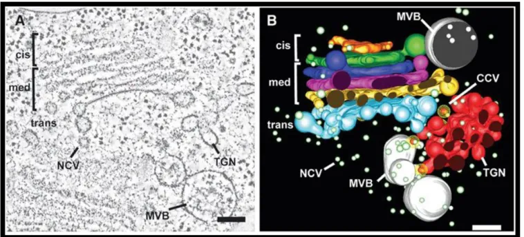
26
Figure 2: Electron tomographic reconstruction of a Golgi stack and associated structures of developing Arabidopsis seed.
(A) Transmission electron microscopy (TEM) image of a prepared tomographic slice (4.3 µm thick) through
a region of the Golgi complex. (B) Three dimensional reconstitutions of the same region as in A.
Compartments are colour-coded: cis-Golgi (orange and green), medial-Golgi (blue, purple, yellow), trans-Golgi (blue) and the trans-trans-Golgi-network (TGN) (red).
A CCV, non-coated vesicle (NCV) and multivesicular bodies (MVB) are also labelled. Scale bars: 100 nm. Figure reprinted from Otegui et al. (Otegui et al. 2006).
27
7. Vesicle trafficking
In all eukaryotic cells, transport between the different compartments of the secretory system is provided by vesicular trafficking. Vesicles are small compartments enclosed by a lipid bilayer which bud from a donor compartment and fuse with an acceptor compartment. Vesicle formation, recognition of and fusion with the acceptor organelles are not passive events but require several factors (Bassham et al. 2008; Bassham and Blatt 2008; Jürgens 2004; Sanderfoot et al. 1999).
.
7.1. Coat proteins and G-proteins: vesicle formation
Specific cargoes located in the lumen of the donor organelles are first recognized, by receptors. Coat proteins are involved in the membrane deformation and scission from the donor compartment to produce the vesicle. There are different types of coat proteins related to vesicle formation at specific organelles: COPII, COPI, CCV, retromer. COPII vesicles are involved in anterograde ER-Golgi transport, while COPI (Coatomer) vesicles are required for retrograde GA-ER transport (Barlowe et al. 1994). Specific small GTPases (G-proteins) initiate the budding process, the incorporation of the cargoes into the evaginated membrane and the scission of the vesicle from the donor organelle. In plant cells, there are several such G-proteins. ARFs are required for COPI and CCV formation, while SAR1 is involved in COPII vesicle formation (Bassham and Blatt 2008; Jürgens 2004; Sanderfoot et al. 1999). Coat formation implies the coordinated recruitment of activated G-proteins, specific phosphatidylinositides, membrane protein cargoes as well as receptors for soluble luminal cargoes and a layer of adaptor proteins. Finally an external coat layer is recruited (Sec13/Sec31 for COPII, the B subcomplex for COPI and the clathrin triskelion for CCV), which contributes to the membrane deformation required for vesicle formation.
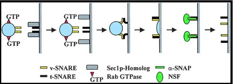
28 7.2. Machines of membrane fusion
Vesicle formation is followed by several events: coat disassembly, trafficking to the target organelle, attachment to it by tethering and docking factors and fusion (Bassham and Blatt 2008; Jürgens 2004; Sanderfoot et al. 1999). Once detached from the donor membrane in the cytosol, the vesicles move through the cytosol by association with cytoskeletal motors or other docking factors. Through the action of docking , tethering factors and members of the SNARE family, the vesicles identify their target compartment and then fuse with it to deliver their cargoes (fig. 3). Rabs are large family of small GTPases involved in the recruitment of motor proteins and of tethering and docking factors (Bassham and Blatt 2008; Jürgens 2004; Sanderfoot et al. 1999). Two groups of SNAREs have been classified: v-SNAREs, t-SNAREs. The v-SNAREs are found in vesicles originating from the donor compartment. The vesicles harbor v-SNAREs and a Rab-GTP. The second type of SNAREs: t-SNAREs, are localized in the membrane of the target compartment (Bassham and Blatt 2008; Jürgens 2004; Sanderfoot et al. 1999). The target organelle contains a t-SNARE complex associated with a Sec1 which maintains the t-SNARE in an inactive form. Sec1 proteins (6 family members in
A.thaliana) control the formation of this bundle (Hanson and Stevens 2000; Sanderfoot 2007)
(fig. 3).
When the vesicle and the target organelle interact, interaction with the Rab-GTP removes the Sec1 protein and exposes the t-SNARE, which becomes active. The t-SNAREs are then able to interact with the v-SNAREs : this is the “docking process” (Chen et al. 1999). Formation of the form-helix bundle of v- and t-SNAREs pulls the two membranes together, causing their fusion. Membrane fusion follows. Subsequently, NSF catalyzes the disassembly of v-/t-SNARE complex upon hydrolysis of ATP, and the released v-SNARE is recycled back to the donor compartment, while the t-SNAREs stay associated with a Sec 1 protein and are ready for another cycle of vesicle fusion (fig. 3) (Hay et al. 1997; Rothman and Söllner 1997). α-SNAP specifically binds to the v-/t-SNARE complex, recruits the NSF factor to the SNARE complex. This ATPase dissociates the v/t-SNARE complex to allow the recycling of v-SNAREs to their starting compartment (Malhotra and Rothman 1988; Sato et al. 1997).
29
Figure 3: The SNARE mechanism of vesicle fusion
Vesicle fusion is mediated by SNARE proteins, a Rab GTPase, a Sec1p-Homolog, α-SNAP and NSF. The v-SNARE and a Rab-type GTPase associated with the vesicular membrane recognize the t-SNARE and a Sec1p homolog on the surface of the target membrane. Sec1p maintained the t-SNARE in an inactive form. After recognition (docking), the Rab-GTP displaces the Sec1p homologs and exposes the t-SNARE which becomes active to interact with the v-t-SNARE and mediates vesicle fusion. α-SNAP and NSF work in collaboration to disassociate the v-SNARE/t-SNARE bundle to prepare for another fusion event. Figure copied from (Sanderfoot, Kovaleva et al. 1999).
30
8. Endocytosis process
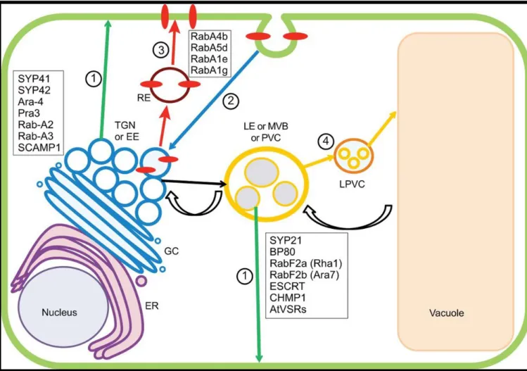
The endocytosis machinery of eukaryotes involved in the internalization of protein cargoes to endosomes/prevacuoles is well conserved in plants. Internalization of cargo proteins is mediated by specific membrane receptors. The cargo/receptor complex is packaged into vesicles and then delivered to EE which have been identified as TGN in plants (Russinova et al. 2004). Another endocytotic pathway involving lipid rafts has been proposed: cargo proteins are internalized into cells at plasma membrane micro-domains (Borner et al. 2005; Murphy et al. 2005; Samaj et al. 2005). Mono-ubiquitination is an endocytic signal for many membrane receptors and in some cases it plays a role in sorting into internal vesicles of MVB (Mukhopadhyay and Riezman 2007). ESCRT complexes (endosomal sorting complexes required for transport) are involved in the internalization of ubiquitylated proteins. The cargo proteins can be transported from the TGN to MVBs (identified as late endosomes). The complexes named ESCRT-I, -II and III (endosomal sorting complexes required for transport) collaborate step by step to achieve transport of proteins. The role of ESCRT-0 is to cluster ubiquitylated cargoes, ESCR-I and ESCRT-II form membrane invaginations and ESCRT-III causes vesicle scission (Bassereau 2010). ESCRT complexes are well characterized in animal and yeast and are likely to have a conserved role in all eukaryotic cells including plants (Bassham and Blatt 2008). Figure 4 (Contento and Bassham 2012) indicates the components of the plants endocytic pathways and the sorting pathways.
31
Figure 4: The sorting and endocytic pathway in plants.
The endomembrane is constituted of the nucleus, endoplasmic reticulum (ER), Golgi complex (GC), trans-Golgi network (TGN) or early endosome (EE), late endosome (LE) or multivesicular body (MVB) or prevacuolar compartment (PVC), late prevacuolar compartment (LPVC), vacuole (VAC) surrounded by the tonoplast, recycling endosome (RE), plasma membrane (PM). The TGN or EE is the start point of four principals sorting and endocytotic pathways.
-Pathway 1 (green arrows) indicates a sorting route to the PM that passes through the TGN and the MVB. -Pathway 2 (blue arrow) is the endocytotic process from plasma membrane to the TGN.
-Pathway 3 (red arrow) represents a recycling pathway, by which PM proteins (red ovals) returned to the PM. -Pathway 4 (orange arrow) shows the sorting route to the VAC. Transport from the TGN to MVBs is indicated
by a black arrow and the retrograde transport from the VAC to the MVB is showed by curved black arrows. Proteins associated with the different organelles are listed in the figure: TGN (Bassham et al., 2000; Chow et al., 2008; Lam et al., 2007; Sanderfoot et al., 2001; Ueda et al., 1996), with the RE (Geldner et al., 2009; Preuss et al., 2006;Rutherford and Moore, 2002), with MVBs (Bottanelli et al., 2012; Jiang and Rogers, 1998; Lee et al., 2004; Li et al., 2002; Paris et al., 1997;Sanderfoot et al., 1998;Spitzer et al., 2009).
32
9. Vacuoles
Vacuoles are the endpoint of the secretory system for soluble vacuolar proteins. Plant vacuoles vary in size, function and content in the different tissues and cell types. Most plant cells possess a large central vacuole, which can occupy more than 90% of the total cell volume. Plant cells can also contain distinct vacuole types at particular stages of development (Marty 1999). Vacuoles are highly dynamic compartments surrounded by the tonoplast. Vacuoles assume fundamental physical and chemical functions indispensable for cell viability such as mechanical support by turgor, which is also involved in cell growth. Moreover vacuoles participate in homeostasis of ions and water, degradation, storage of many plant compounds such as ions, pigments, secondary metabolites, enzymes involved in defense (Frigerio 2008; Marty 1999). After pathogen infection, vacuoles may respond by eliminating the pathogen through autophagy. If the cell is not able to recover a normal physiological function, it activates a specific program of cell death which leads to mobilization of hydrolytic enzymes in the cytosol to permeabilize the tonoplast (Gietl and Schmid 2001; Greenwood et al. 2005; Hara-Nishimura et al. 2005). Vacuoles are also involved in programmed cell death (PCD), which is an active process of selective elimination of certain cells upon biotic or abiotic stresses. Two mechanisms of PCD involving vacuoles in plants have been described : (i) disruption of the tonoplast causing the release of vacuolar contents in the cytoplasm, efficient against intracellular pathogens, (ii) and fusion of the tonoplast with the plasmalemma, releasing vacuolar contents in the cell wall, efficient against extracellular pathogens (Hara-Nishimura and Hatsugai 2011).
33
34 1.
Vacuole types
Regarding all the diverse functions the vacuole assumes, the theory of multiple vacuole types was proposed. The presence of LV and PSV was demonstrated by different studies. However, recently this model has been challenged by a series of studies. Existence of distinct vacuole types was provided by using pH-sensitive dyes, such as Neutral Red (Di Sansebastiano, Paris et al. 1998). Upon staining, acidic and neutral compartments were detected within a single cell. For instance, the mesophyll cells of Mesembryanthemum
crystallinum, presented two different vacuoles, an acidic and a neutral vacuole. The acidic
vacuole had a maximal volume at the end of the night and shrunk during the day, as malic acid was retrieved (Epimashko et al. 2004).
At the molecular level, two soluble vacuolar GFP markers have been described : the GFP-Chi and the Aleu-GFP possessing different vacuolar sorting determinants (VSD) from tobacco chitinase A or barley aleurain. They were localized in PSV and in LV respectively (Di Sansebastiano et al. 1998; Di Sansebastiano et al. 2001). During cell senescence, small acidic vacuoles named senescence-associated vacuoles (SAV) appeared in soybean mesophyll and guard cells. SAVs were characterized by the presence of senescence-specific cysteine protease SAG12 (Otegui et al. 2005).
The presence of distinct vacuoles was also demonstrated using fluorescent reporters of tonoplastic aquaporins, TIPs (Tonoplast Intrinsic Proteins). TIPs were used to characterize
A.thaliana vacuoles which contain 10 TIP isoforms: 3 γ-TIPs, 3 δ-TIPs, 1 α-TIPs, 1 β-TIPs, 1
ε-TIPs, 1 TIPs (Johanson et al. 2001). Based on tissue expression analysis, the ε-TIP and δ-TIP are preferentially expressed in roots, and in floral organs. γ-δ-TIP3 and δ-δ-TIP are expressed in flowers, whereas α-TIP and β-TIP were expressed during seed maturation, and TIP1, δ-TIP2, γ-TIP1 and γ-TIP2 are expressed during early stages of seed development (Frigerio et al. 2008). δ-TIP3 is also called DIP (Dark-Induced Protein). It has been found in root tip cells and developing seeds (Culianez-Macia and Martin 1993). In barley and pea root tip cells, two different vacuoles were characterized: PSV was characterized by the presence of α-TIP and the storage protein barley lectin; and the LV was characterized by the presence of γ-TIP and aleurain (Paris et al. 1996). Immunolocalisation experiments showed that α-, δ- and γ-TIPs did not colocalise in some plant cells (Jauh et al. 1999). In barley aleurone protoplasts, treated with ABA (Absissic Acid) or gibberellic acid, a second kind of vacuole was generated
35
containing α-TIP which was physically separated from the PSV (Swanson et al. 1998). In 1999, Jauh et al. showed that α-, δ- and γ-TIP did not colocalise in some plant cells, therefore some vacuoles were identified with two different TIPs.
1.1. The lytic vacuole
The LV is an acidic compartment that contains enzymes analogous to the lysosomal enzymes of animal cells. This vacuole is important to maintain turgor pressure for the storage of metabolites, and to sequester xenobiotic compounds (Taiz 1992). The barley cysteine protease aleurain was used as soluble reporter for this vacuole (Di Sansebastiano et al. 1998; Flückiger 1999; Paris and Rogers 1996). Several studies supported that the LV tonoplast contains the aquaporin γ-TIP (Barrieu et al. 1998; Höfte and Chrispeels 1992; Jiang et al. 2000; Marty-Mazars et al. 1995; Paris and Rogers 1996). However, Hunter et al., in 2007 failed to detect γ-TIP in LV of meristem and stele cells of pea root tips or in cells of developing Arabidopsis embryos.
1.2. The protein storage vacuole
Seed cells can have up to three different vacuoles with different functions in one single cell: a LV, a vegetative storage vacuole (neutral) and a seed PSV (Di Sansebastiano et al. 1998; Di Sansebastiano et al. 2001; Hoh et al. 1995; Paris and Rogers 1996).
The vegetative storage vacuole was found in specialized vegetative cells in response to wounding or to developmental switches (Jauh et al. 1998). δ-TIP was identified as its tonoplast marker (Jauh et al. 1998; Neuhaus and Rogers 1998; Park et al. 2004).
The PSV was found in cells from storage tissue of seeds and fruits. It major function was the storage of proteins such as 7S lectin, 2S albumin and 11S globulins (Herman and Larkins 1999; Muntz 1998; Okita and Rogers 1996). PSV can be labeled with α- and δ- TIPs antibodies (Jiang et al. 2000; Paris et al. 1996; Swanson et al. 1998). PSV can contain subcompartments, globoids and large crystalloids. Globoids contain for example aleurain, while the crystalloids are composed of crystalline 11S globulin. The matrix compartment contains a mixture of 7S lectins and 2S albumins. DIP was discovered to be associated with the crystalloid membranes (Jiang et al. 2000).
36 1.3. Evidence supporting unique vacuole
In root tips, only one vacuole containing storage proteins was characterized by both α-TIP and γ-α-TIP (Olbrich et al. 2007b). Data supporting the presence of several vacuoles in the same cell is rather an exception, limited to some particular stages of development, or in some tissue types (Frigerio et al. 2008). For instance, it has been shown that when soybean plants were subjected to changed physiological conditions, the γ-TIP marker diminished while the δ-TIP marker became present in the tonoplast indicating the conversion of a lytic vacuole to a vegetative storage vacuole (Murphy et al. 2005). In a study, the different fluorescent TIPs (α-, γ and δ-TIP) detected the same vacuoles in A.thaliana. Indeed, even the developing embryos which contain only a PSV, all three markers localized in its tonoplast. The expression profile of the TIP markers was established: γ-TIP and δ-TIP were limited to vegetative tissue except for root tips, while α-TIP was specifically expressed during seed maturation (Hunter et al. 2007).
2. Vacuolar sorting determinants
Plant vacuolar proteins are synthesized as precursors with propeptides, which are proteolytically removed upon targeting to the vacuole. These propeptides may also contain the VSD (Matsuoka and Nakamura 1999; Neuhaus and Rogers 1998). Three different types of VSD have been described: sequence-specific (ssVSD), C-terminal (ct-VSD) and condensation dependent (conVSD) (Neuhaus and Rogers 1998).
2.1. The ssVSD
Proteins carrying an ssVSD are addressed to LV (Holwerda et al. 1992; Koide et al. 1999; Matsuoka and Nakamura 1991). The barley aleurain and sweet potato sporamin have both been identified with an N-terminal propeptide that contains an NPIR motif. A sporamin mutant lacking the propeptide was secreted supporting the role of the propeptide in vacuolar targeting. The isoleucine (Ile) amino acid played a central role, however it can be replaced only with a leucine (Leu) (Kirsch et al. 1996; Matsuoka and Nakamura 1999; Matsuoka and Neuhaus 1999). Other proteins have been identified with a ssVSDs with no NPIR motif, but an essential Ile or Leu could be identified. The ssVSD can be localized in N-terminal, C-terminal (castor bean 2S albumin, (Brown et al. 2003), or in an internal parts of vacuolar proteins (Frigerio et al. 2001).
37 2.2. The c-terminal VSD
A C-terminal propeptide was found to be necessary for vacuolar targeting of several proteins. Ct-VSDs were identified at the C-terminal end of these propeptides of several vacuolar proteins: barley lectin (Bednarek et al. 1990), tobacco chitinase A (Neuhaus et al. 1991a), phaseolin (Frigerio et al. 1998). A consensus sequence could not be identified, but these sequences seem to be enriched in hydrophobic amino-acids (Matsuoka and Neuhaus 1999). Complete deletion of ct-VSD leads to protein secretion but many single point mutations or partial deletions of tobacco chitinase or barley lectin ct-VSDs had no effect on protein sorting (Dombrowski et al. 1993; Neuhaus et al. 1994). However, terminal glycines or terminal N-glycosylation caused secretion of the proteins. Therefore, no critical motif is important in ct-VSD for the correct targeting of the protein, but its three-dimensional structure might be involved in binding to a vacuolar receptor (Nielsen et al. 1996).
2.3. The psVSD
The third class of vacuolar sorting determinant is the condentation-dependant (conVSD) (Neuhaus and Rogers 1998). It has been described for the phytohemagglutinin of common bean and for legumin-like proteins. The conVSD signal was localized in different regions of the polypeptides or in the PSI (Protein-Specific Insert) domain of phytepsin (Kervinen et al. 1999; Tormakangas et al. 2001) supporting an important role for 3D structures (Tague et al. 1990; Vitale and Raikhel 1999). Therefore, the aggregation process was proposed as a possible mechanism for vacuolar sorting of these proteins (Vitale and Chrispeels 1992).
38
3. Vacuolar sorting receptors
3.1. The VSRs
A vacuolar receptor involved in trafficking of ssVSD proteins was identified from pea CCVs and was called BP-80 (Binding Protein of 80kD) (Kirsch et al. 1994; Paris et al. 1997). This receptor was able to bind barley aleurain and sporamin ssVSDs and the C-terminal propeptide of Brazil nut 2S albumin in a pH-dependent manner (Kirsch et al. 1994; Kirsch et al. 1996). In A.thaliana, there are seven homologues of BP80 the AtVSR multigene family (Ahmed et al. 2000; Neuhaus and Paris 2005).
3.1.1. Structure of VSR
VSR proteins are class I membrane proteins. They are constituted of a large N-terminal luminal domain, a transmembrane domain (TM) and a cytosolic tail domain. The luminal domain contains a PA (Protease-Associated) domain, followed by a large VSR-specific domain and three Cys-rich EGF (Epidermal Growth Factor) repeats (fig. 5).
3.1.2. Localisation of VSR
Antibodies produced against pea BP-80 detected VSRs in the PVC in pea and tobacco cells (Li et al. 2002). Different homologs of BP80 were identified such as PV72 found in pumpkin PAC (Shimada et al. 1997) and AtELP was identified in A.thaliana (Da Silva Conceiçao et al. 1997; Paris et al. 1997; Sanderfoot et al. 1998). Another study localized AtVSRs mainly in PVC and to a minor extent in TGN (Miao et al. 2006).
3.1.3. Function of VSR
Several studies supported the involvement of VSRs in protein sorting to LV. BP80 is able to bind in vitro barley aleurain ss-VSD and prosporamin from sweet potato (Kirsch et al. 1994). However the receptor does not bind to the ct-VSD of barley lectin (Kirsch et al. 1996). A chimeric reporter protein was constructed using a mutated form of proaleurain (lacking ss-VSD) connected via its C-terminus to the BP-80 TM domain and cytoplasmic tail. When expressed in tobacco cells, the construct was correctly addressed to the PVC, where the proaleurain moiety was processed to mature form (Jiang and Rogers 1998).
39
It has been proposed that VSRs are involved also in storage proteins targeting to PSV. The AtVsr1 null mutant leads to secretion of some seed storage proteins. However, the effect on storage protein transport is partial (Shimada et al. 2003a). This has been interpreted as evidence for VSRs as salvage receptors for stray storage protein that escaped from their classic route (Craddock et al. 2008; Hinz et al. 2007). More recently, Zouhar et al., (2010) observed a mistargeting of storage proteins in several simple VSR mutants, VSR1, VSR3 and VSR4, indicating that VSR could also be receptors for specific storage cargoes in seeds and in vegetative tissues (Zouhar et al. 2010).
The short cytosolic tail of VSRs contains a tyrosine motif (YMPL) which interacts with the muA component of adaptor protein type 1 complex (AP-1), that is involved in the formation of CCVs (Happel et al. 2004). Mutations of this domain resulted in accumulation of VSR2 in the vacuole in Nicotiana tabacum, indicating that recycling of VSR is impaired (Foresti et al. 2010; Saint-Jean et al. 2010). The mutations also showed that VSRs use two pathways to target vacuolar proteins: the major route leading directly to the lytic vacuole, and a minor route retrieving missorted ligands from the apoplast, a pathway requiring dipeptide Ile-Met motif in the cytosolic tail (Saint-Jean et al. 2010).
3.2 The RMRs
A second family of putative receptors was identified by their homology to the PA domain of the VSR proteins. They were named Receptor-Membrane-RingH2 or RMR (Cao et al. 2000; Jiang et al. 2000). The genome of Arabidopsis harbours six homologs (AtRMR1 to AtRMR6) (Park et al. 2005).
3.2.1. Structure of RMRs
These proteins are composed of an N-terminal luminal domain restricted to the PA domain, but lacking EGF-repeats, a transmembrane domain, and a cytosolic tail with a RING-H2 (Really Interesting New Gene, with two His) domain, and most have also a C-terminal serine-rich region (lacking in AtRMRs 1, 5, 6) (Jiang et al. 2000; Park et al. 2005) (fig. 5). The luminal PA domain shared with VSR proteins is known in other protein families to participate in ligand binding (Cao et al. 2000; Mahon and Bateman 2000). It constitutes a protein-protein interaction domain in addition to mediating substrate recognition for peptidases. The Ring-H2 domain found in RMRs is of the C3H2C3 type. This type of domain
40
is likely to be associated with an E3 ligase activity (Joazeiro and Weissman 2000; Tranque et al. 1996).
3.2.2. Localisation of RMR
Only few studies were performed on RMR localization. In Arabidopsis and tomato seeds, antibodies against RMR detected the same organelles as antibodies against DIP. DIP is associated with the crystalloid precursors of PSV during seeds development. The DIP-positive organelle was proposed by Jiang as PVC for PSV which fuse to it to deliver internal proteins (Jiang, Phillips et al. 2000).
Studies performed by Park et al., (2005) focused on the localization (by immunohistochemistry) and the biological function of AtRMR1 in leaf protoplasts. They found that endogenous AtRMR1 and AtRMR1-HA colocalized with DIP but not with AtPEP12p (a marker of the PVC). The colocalization of AtRMR1-HA and phaseolin in leaf protoplasts was supported by the specific in vitro binding of the luminal part of AtRMR1 to the phaseolin ct-VSD. Their results suggested that AtRMR1 functions as cargo receptor by interacting with the ct-VSD of phaseolin, which is transported to the PSV through the DIP/AtRMR1-positive organelle (Park et al. 2005).
AtRMR2 binds strongly to the chitinase ct-VSD but only if presented with a free C-terminus and only weakly to the proaleurain ssVSD. Immunogold labeling with AtRMR2 specific antibodies revealed a similar distribution to cruciferin in the GA stacks, and in DVs. RMR may associate with protein aggregates carrying ct-VSD during transit from the GA to the vacuole by DVs (Park et al. 2007). This pathway would be distinct from the CCVs pathway for ssVSD targeting through the VSR. This model is consistent with the results provided by Hinz et al., (2007) who localized AtRMR2 in the early stack of GA and in DVs (Hinz et al. 2007).
Additionnally, it has been recently demonstrated that OsRMR1 proteins localized in GA, TGN, PSV and in an organelle identified as PVC for PSV called (PVCs) in both rice cultured cells and in developing rice seeds. This localization seems to be conserved since OsRMR1 proteins were also found in Golgi, TGN, and PVCs in BY-2 and Arabidopsis cultured cells (Shen et al. 2011; Wang et al. 2011).
41
3.2.3 Function of RMRs
RMR proteins were found in strategic places: in the crystalloid membrane in PSV Arabidopsis seeds (Jiang et al. 2000) and in cis-Golgi stacks and DVs (Hinz, Colanesi et al. 2007). This localization is consistent with the studies of Castelli and Vitale (2005), which showed that the aggregation of the storage protein phaseolin depended on the presence of the ct-VSD and was an early event in the cis-Golgi stacks. RMRs have been detected in the PSV crystalloids whereas neither the lytic nor the storage protein tonoplast contained VSRs (due to recycling) (Jiang et al. 2000). Therefore, RMRs have characteristics of protein receptors which traffic through the Golgi complex and PVC to PSV.
It has been shown that AtRMR2 (protein JR702) and its ligands were not dissociated at low pH (Park et al. 2007). This observation suggested that RMRs either use a different mechanism to release their ligands, or are not recycled from an acidic PVC and thus accompany their cargos into the PSV. Two hypotheses were proposed: individual storage proteins could bind to RMR proteins and act as nuclei for further condensation of storage proteins. Alternatively, storage protein could form microaggregates which would expose on their surface free ct-VSDs accessible for RMRs-cargo interaction. Therefore, RMRs could function as storage protein receptor or assemble factors without being recycled. (Hinz et al. 2007).
However the role as a receptor was recently questioned since simple KO mutants of each AtRMRs did not show any missorting of storage proteins. A system to screen Arabidopsis mtv mutants (for „modified transport to the vacuole‟) which are affected in the trafficking of the marker VAC2 was used in simple KO RMR mutants (Sanmartin et al. 2007). VAC2 is a fusion protein constituted of the ligand CLAVATA3 fused to the barley lectin ct-VSD (Rojo et al. 2002). If VAC2 is correctly transported to the vacuole, where it is inactive, plants develop normal meristems. If VAC2 is not targeted to the vacuole it will be secreted causing premature termination of shoot apical meristems and flower meristems, allowing identification of the mtv mutants. Simple KO AtRMR mutants had a normal meristem development, indicating that VAC2 was correctly addressed to the vacuole. There was also no accumulation of precursor forms of 12S globulins and 2S albumins, indicating that their transport was not affected either. Double AtRMR KO mutants of AtRMR1&2 and AtRMR3&4 also did not show any seed storage protein phenotype (Zouhar et al. 2010).
42
Figure 5: Structure of plant vacuolar receptors
- (A) VSRs have a Protease –Associated domain (PA red), a VSR-specific domain (purple), three Cys-Rich EGF Repeat (Epidermal Growth Factor) (green), and a single trans-membrane domain (TM), the cytosolic domain of AtVSRs is restricted to a small Tail (purple).
- (B) RMRs have a Protease-Associated Domain (PA) (in red), a single TM. At the cytosolic part includes a Ring-H2 Domain (blue) and for most RMRs a Ser-Rich Domain (grey).
43
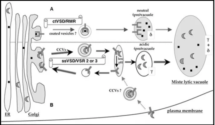
4. Vacuolar sorting pathways
For vegetative cells, Neuhaus and Paris (2005) proposed two main pathways which could be identified in vegetative cells (fig. 6) (Neuhaus and Paris 2005). The route of proteins to the LV starts after recognition of their ssVSD ligands in the Golgi by the VSR receptors. The complex ligand-VSR complex is then transported by CCVs to the PVC (fig. 6B). Once in the PVC, the acidic pH causes ligand release and enables receptors recycling. Finally, the PVC fuses with the acidic vacuole (Jiang et al. 2000). In the other pathway, ct-VSD proteins are thought to be recognized by RMRs and sent to a storage vacuole. Mechanisms involved in the RMR pathways are unknown and their implication remains to be clarified (Neuhaus and Rogers 1998; Park et al. 2004) (fig. 6A). Recently, a new study supported the existence of particular PVC for PSV. In rice cultured cells and developing seeds, OsRMR1 was found in GA, TGN, PSV and a distinct organelle proposed to be a storage PVC (sPVC) (Shen et al. 2011). The effect of wortmannin (a specific inhibitor of mammalian phosphatidylinositol 3-kinases) was also indicative of these two different protein sorting pathways to the vacuole. In tobacco cells, treatment with 33 µM wortmannin caused the inhibition of the ct-VSD proteins transport to vacuoles, while the ss-VSD protein targeting were not affected (Matsuoka et al. 1995).
Since some vacuoles appeared labeled with both TIP isoforms (∂- and ɣ-TIP) (Jauh et al. 1999), it has been proposed that the LV and the PSV can fused to form a hybrid vacuole. It have been shown that the ct-VSD of barley lectin and ss-VSD of sporamin were both addressed to the same vacuole in transgenic tobacco plants (Schroeder et al. 1993). The hybrid vacuole model allows a possible switch of the vacuole from storage to lytic functions (Murphy et al. 2005). In Arabidopsis seedlings, two pathways for TIP targeting to the tonoplast were proposed: one pathway (taken by TIP1;1) which is Brefeldin A (BFA) and therefore Golgi-dependent sensitive and another pathway (route taken by TIP3;1 and TIP2;1) which is Golgi-independent and BFA insensitive, but sensitive to C834 a newly discovered drug (Rivera-Serrano et al. 2012).
44
Figure 6: Model of vacuolar sorting pathways in vegetative cells.
(A) In the biogenesis of neutral vacuoles, RMR receptors bind the ctVSD of proteins in the Golgi,
and then the complex is transported to the neutral prevacuole. These neutral prevacuoles have a tonoplast with δ-TIP isoforms. They then may fuse with a preexisting large hybrid vacuole.
(B) In the biogenesis of lytic vacuoles, VSRs (subfamily VSR2 or VSR3) bind the ssVSD of proteins
in the Golgi, the complex is then transported by CCVs to the acidic prevacuolar compartments. The acidic pH causes ligand release and the receptors are recycled to the Golgi. These acidic prevacuoles have a tonoplast with γ -TIP. They then fuse with a preexisting large acidic or hybrid vacuole. Figure copied from (Neuhaus and Paris 2005)
45
5. De novo vacuole biogenesis
De novo vacuole biogenesis is still not characterized, however, it has been postulated
that vacuoles can be generated from small pre-existing vacuoles during cell development (Zouhar and Rojo 2009). For instance globoids, with characteristics of LV, could correspond to preexisting LVs that are incorporated inside of PSV (Frigerio et al. 2008; Jiang et al. 2001).
Autophagy could be an important mechanism of vacuolar enlargement. In meristematic daughter cells, the LV was formed from small vacuoles which enlarged by autophagy (Inoue and Moriyasu 2006). A KO mutant of VCL1 gene blocked the LV formation and showed an accumulation of autophagosomes during embryogenesis. Homologues of the
VCL1 gene in yeast and mammals are involved in autophagosomes fusion with the vacuole
(Zouhar and Rojo 2009).
The PVC origin of the PSV was supported by a study in seeds which showed that the
vamp727/syp22 double mutant presented a fragmented PSV and partial secretion of storage
proteins in seeds (Ebine et al. 2008). VAMP727 and SYP22 are known to be involved in SNARE complex formation which plays a role in the fusion between PVC and PSV (Ueda et al. 2004; Uemura et al. 2004).
47
48 1.
P.patens a simple system
1.1 The life cycle of P.patens
One of the advantages of moss is its relatively simple developmental pattern and the dominance of the haploid gametophyte in the life cycle (Cove et al. 1997). Studies have mostly been performed on species like Funaria hygrometrica, Ceratodon purpureus, and
Physcomitrella patens. The main advantage who lead scientist to choose P.patens for genetic
approaches was the possibility to perform in vitro crosses. P.patens is monoecious meaning that both sexual organs can be present on the same plant. This advantage, among many others, allows to grow it in very simple laboratory conditions to complete its life cycle (Cove et al. 1997). Like other land plants, P.patens shows an alternation between a haploid phase and a diploid phase. However, in contrast to ferns and seed plants, in mosses the haploid phase is the dominant. The haploid phase produces gametes through the generation of the gametophyte and the diploid phase produces haploid spores by meiosis through the generation of the sporophyte (fig. 7).
The gametophyte stage starts from a spore‟s germination which produces the protonema. This comprises network of filamentous cells displaying one-dimensional apical growth. The protonema extends by division and produces side branches. Most side branches develop into filaments, but some develop into the leafy shoots called gametophores. Once mature, gametophores give rise to sexual organs at their apex. The male gametes, or antherozoids, are produced in antheridia, whereas female gametes or egg cells are produced in archegonia. Self-fertilization is usual since both organs differentiate on the same shoot. The antherozoid reaches the archegonia, through water flux, and fuse with the egg cell to form a diploid embryo, the sporophyte. The sporophyte stage starts at fertilization and the zygote develops into a sporophyte, constituted of a diploid spore capsule which can contain up to 5000 haploid spores (fig. 7). Finally, spores germinate to produce the protonema of the gametophyte stage. P. patens develops in early summer in temperate zones. It grows along lakes and rivers on soil that has been exposed by falling water levels. Reproductive organ development is induced by short day. Fertilization needs temperatures below 18oC (Engel et
al., 1968). As a result, sporophytes are produced in the late summer, and spores during winter.
Several accessions are recognized, and the Gransden wild-type strain is widely used is by the hole community.
49
Figure 7: The life cycle of Physcomitrella patens
The P.patens life cycle is characterized by an alternation of two generations; the haploid gametophyte that produces gametes (yellow line) and the diploid sporophyte (red line). (A) Spores, (B) Protonema filaments composed of two types of cells; the caulonema (C) and the chloronema (D). Some side branches from caulonema cells can differentiate into buds (E) which give rise to gametophores (F) a meristematic structures bearing leaf-like structures, rhizoids and the sexual organs. When water is available flagellate sperm cells can swim from the antheridia (G) to an archegonium (H) and fertilize the eggwithin. The resulting diploid zygote
(I) originates a sporophyte composed of a foot, seta and capsule, spores (A) are produced by meiosis of spore