HAL Id: hal-03165375
https://hal.univ-lorraine.fr/hal-03165375
Submitted on 10 Mar 2021
HAL is a multi-disciplinary open access
archive for the deposit and dissemination of
sci-entific research documents, whether they are
pub-lished or not. The documents may come from
teaching and research institutions in France or
abroad, or from public or private research centers.
L’archive ouverte pluridisciplinaire HAL, est
destinée au dépôt et à la diffusion de documents
scientifiques de niveau recherche, publiés ou non,
émanant des établissements d’enseignement et de
recherche français ou étrangers, des laboratoires
publics ou privés.
Clinical profile and midterm prognosis of left ventricular
thrombus in heart failure
Anne-Iris Lemaitre, François Picard, Vincent Maurin, Maxime Faure, Pierre
dos Santos, Nicolas Girerd
To cite this version:
Anne-Iris Lemaitre, François Picard, Vincent Maurin, Maxime Faure, Pierre dos Santos, et al.. Clinical
profile and midterm prognosis of left ventricular thrombus in heart failure. ESC Heart Failure, Wiley,
2021, �10.1002/ehf2.13211�. �hal-03165375�
Clinical pro
file and midterm prognosis of left
ventricular thrombus in heart failure
Anne-Iris Lemaître
1*, François Picard
1, Vincent Maurin
1, Maxime Faure
1, Pierre Dos Santos
1,2,3†and
Nicolas Girerd
4†1Bordeaux University Hospital (CHU), Bordeaux, France;2Centre de Recherche Cardio-Thoracique de Bordeaux, Université de Bordeaux, Bordeaux, France;3IHU Liryc, Electrophysiology and Heart Modelling Institute, Foundation Bordeaux Université, Bordeaux, France;4Université de Lorraine, Inserm, Centre d’Investigations Cliniques Plurithématique1433, Inserm U1116, CHRU Nancy, and F-CRIN INI-CRCT (Cardiovascular and Renal Clinical Trialists), Nancy, France
Abstract
Aims We documented the midterm prognosis of left ventricular thrombus (LVT) in heart failure (HF) patients with dilated cardiomyopathy (DCM) and ischaemic cardiomyopathy (ICM). We aimed to characterize patients with LVT in the context of HF with reduced (≤40%) left ventricular ejection fraction and evaluate their risk for death and/or embolic events, overall, and specifically in patients with ischaemic or non-ischaemic aetiology. We also intended to identify risk factors for LVT in pa-tients with DCM.
Methods and results We included all HF patients (N = 105, age 56 ± 13) admitted from 2005 to 2018 in our institution for LVT without significant valve disease/prosthesis, heart transplant/left ventricular assist device, congenital heart disease, or acute myocardial infarction. Our primary endpoint was the 1 year risk of the composite of all-cause mortality (ACM) and symp-tomatic embolic events. Mean left ventricular ejection fraction was 23 ± 9%, and median BNP was 1795 pg/mL. Most (97%) patients were treated with vitamin K anticoagulants, and 64% had ICM. Symptomatic embolic events and/or ACM occurred in 20% of the population [embolic events (all within 30 days of LVT diagnosis) 15% and ACM 6%] and was similarly frequent in DCM or ICM (P> 0.05). Suspected/transient embolic events were more frequent in DCM (overall 13%; 29% in DCM vs. 5% in ICM, P< 0.01). Major bleeding occurred in 5% of patients. Left ventricular reverse remodelling occurred in 65% of patients, more frequently in DCM (86% in DCM vs. 65% in ICM, P = 0.02). In a case–control analysis matching DCM patients, BNP level was the only factor significantly associated with LVT (2447 pg/mL in LVT vs. 347 pg/mL, P < 0.001).
Conclusions Patients with LVT have markedly high natriuretic peptides and experience a 20% 1 year risk for embolic events and/or death following diagnosis despite anticoagulant treatment. Most patients have favourable remodelling/recovery. As all symptomatic embolic events occurred within 30 days of LVT diagnosis, a very careful initial management is warranted.
Keywords Left ventricular thrombus; Heart failure, systolic; Dilated cardiomyopathy; Thromboembolism; Stroke; Echocardiography
Received:7 September 2020; Revised: 7 December 2020; Accepted: 2 January 2021
*Correspondence to: Anne-Iris Lemaître, Hôpital Cardiologique du Haut-Lévêque,1 Avenue Magellan, 33600 Pessac, France. Email: anne-iris.lemaitre@chu-bordeaux.fr †Both authors contributed equally to this manuscript.
Introduction
Left ventricular thrombus (LVT) is a dreaded complication of left ventricular (LV) dysfunction or myocardial infarction. While its incidence after myocardial infarction has declined in the era of primary percutaneous intervention and with im-provements of peri-procedural care, its reported occurrence ranges from 4% to 15% after an ST-elevation myocardial infarction.1 Importantly, the occurrence of systemic embolic events has been reported to be as high as 29%.2In matched
analysis, the presence of LVT identified with magnetic reso-nance imaging (MRI) was associated with a four-fold higher long-term incidence of embolism.3
Heart failure (HF) with reduced LV ejection fraction (LVEF), due to an ischaemic (ICM) or non-ischaemic dilated cardiomy-opathy (DCM), is an emerging source of LVT.4In the setting of HF, several elements of Virchow’s triad can be present; blood stasis is favoured by akinetic myocardial wall or severely de-creased ventricular contraction, endothelium damage is favoured by necrosis or inflamed myocardium and increased
ESC Heart Failure (2021)
shear stress, and hypercoagulable state can be increased by systemic inflammation, thrombophilia, and prothrombotic factor activation. As previous large studies did not specifically focused on HF patients,3the incidence of LVT in HF, patients’ characteristics, and prognosis are mostly unknown. Given this limited evidence, there are no clear recommendations re-garding the diagnosis modalities and treatment of LVT in HF. We aimed to characterize patients with LVT in the context of HF with reduced (≤40%) LVEF and evaluate their risk for death and/or embolic events, overall, and specifically in pa-tients with ischaemic or non-ischaemic aetiology. We also intended to identify risk factors for LVT in patients with DCM.
Methods
Cohort study population
We extracted electronic medical records labelled with the di-agnosis‘intra-cardiac thrombosis’ or ‘intra-ventricular throm-bosis’ admitted in our institution from January 2005 to July 2018. This database query extracted a list of 1223 hospital stays matching 658 patients. We included patients diagnosed in our institution with LVT using transthoracic echocardiogra-phy (TTE), MRI, or computed tomograechocardiogra-phy (CT) scan. Patients with thrombosis limited to the atria and right cavities were excluded. In addition, we used the following exclusion criteria: ST-elevation or non-ST-elevation myocardial infarc-tion<1 month, concomitant severe valvular stenosis or me-chanic valves, heart transplant or LV assist device, complex congenital heart diseases, and takotsubo.
The date of inclusion in this retrospective cohort was the date of LVT diagnosis. Clinical data at diagnosis, including TTE and MRI data, were reviewed and collected in a comput-erized database. We retrieved information regarding all-cause mortality, cardiovascular mortality, and symptom-atic, transient, or suspected embolic events from routine clin-ical follow-up within 1 year of LVT diagnosis.
Cohort study outcomes
The composite primary endpoint was the occurrence of all-cause mortality and/or symptomatic embolic event within 1 year of LVT diagnosis reported in their medicalfile. Symp-tomatic embolic events were defined as persistent clinical symptoms suggestive of embolism (neurological deficit, isch-aemic symptoms, and abdominal, limb, or chest pain) con-firmed by imaging.
A number of secondary endpoints were considered. Suspected embolic events were defined as an ‘accidental’ dis-covery of emboli (ischaemic lesions on a CT scan) after diag-nosis of LVT, without any reported symptoms. Transient embolic events were transient ischaemic attack (TIA) defined
as a transient neurological deficit without ischaemic lesion on cerebral imaging. Major bleeding was defined as a bleeding event requiring hospitalization and clinically overt major bleeding events, that is, associated with a decrease in the haemoglobin level of ≥2 g/dL, transfusion of ≥2 units of packed red cells or whole blood, involving a critical site (intra-cranial, intraspinal, intraocular, pericardial, intra-articular, intramuscular with compartment syndrome, and retroperito-neal), or related to a fatal outcome. LV reverse remodelling was defined as an increase in LVEF of more than 10% or a de-crease in indexed LV end-diastolic diameter or volume of more than 10%.
Case
–control dilated cardiomyopathy study
To identify risk factors for LVT in the context of DCM, we matched patients with both LVT and DCM with patients with DCM but without LVT, admitted for the first time to our HF unit between May 2011 and April 2015. Patients were matched for age, sex, New York Heart Association (NYHA) class, and LVEF measured by TTE. In controls, baseline charac-teristics, BNP, NYHA class, echocardiographic data at 0 and 6 months, initial MRI data, and all-cause mortality were re-trieved from medicalfiles.
Statistical analysis
All analyses were performed using R software (The R Founda-tion for Statistical Computing). The two-tailed significance cri-terion was set at P < 0.05. Continuous variables are described as mean ± standard deviation or median (first to third quartile). Comparison between groups was performed using Student’s t-test or Mann–Whitney U test as appropri-ate. Categorical variables are presented as number (percentage) and compared using χ2 or Fisher’s exact test as appropriate. Kaplan–Meier method was used to describe the risk for all-cause mortality over time, and survivals were compared using log-rank test.
Results
Description of the study population
Among the 105 included patients, 38 (36%) had a non-ischaemic DCM (Table 1).
The majority (56%) of LVT were diagnosed by TTE (followed by MRI or CT scan), and LVT invisible in TEE was di-agnosed with MRI (37%) and CT scan (7%).
Among DCM patients, 27 (71%) were incident HF patients, meaning their cardiomyopathy was diagnosed less than a month ago. Aetiologies of DCM were as follows: idiopathic
2 A.-I. Lemaître et al.
ESC Heart Failure (2021) DOI: 10.1002/ehf2.13211
(N = 19), alcoholic (N = 8), post-chemotherapy (N = 4), non-compaction (N = 2), hereditary with identified mutation (N = 1), Friedreich ataxia (N = 1), Becker myopathy (N = 1), ar-rhythmia induced cardiomyopathy (N = 1), and post-myocar-ditis (N = 1).
Among ICM patients, 15 (22%) were incident HF patients (coronary disease diagnosed at the same time as the LVT). In prevalent patients (78%), mean delay between ICM diag-nosis and LVT was 6.3 years.
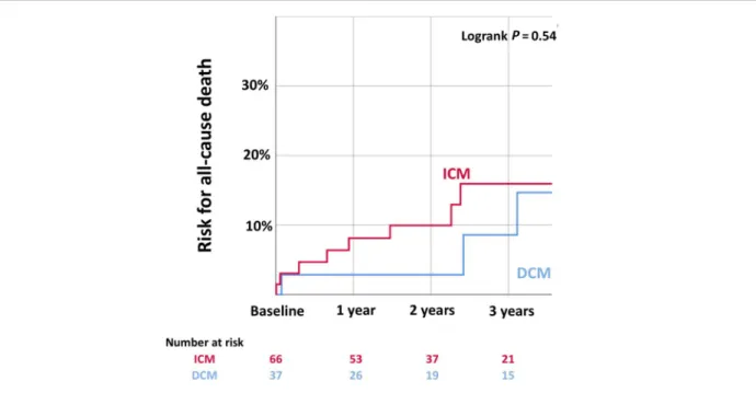
Mean age at diagnosis of LVT was 55.9 ± 12.9 years, and most patients were male (85%) (Table1). DCM patients were significantly younger (51.2 ± 14.4) than ICM patients (58.6 ± 11.2, P< 0.01, Table 1). Diabetes and hypertension were more frequent in ICM patients, whereas alcohol abuse was more frequent in DCM patients. ICM patients had a higher HASBLED score, in relation with a higher proportion of anti-platelet agents, and a higher CHA2DS2VASC score.
NYHA class was higher in DCM patients than in ICM patients (76% vs. 39% NYHA 3–4, P < 0.001). DCM patients had more severe initial presentations (cardiogenic shock 34% in DCM vs. 6% in ICM, P < 0.001), higher BNP values (2447 vs.
1401 ng/L), and lower LVEF (21 ± 6% vs. 29 ± 8%, P < 0.001, Table 1). LVT had an apical localization in a vast majority of cases (98 patients, 93% of cases).
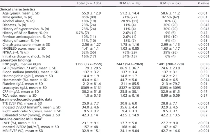
One year prognosis of left ventricular thrombus
During thefirst year, the primary endpoint of all-cause mor-tality and/or symptomatic embolic event occurred in 20% of patients (Figure1), all-cause mortality in 5.7%, and CV mor-tality in 4.8% (five patients—four due to a cardiogenic shock and one to massive ischaemic stroke). One patient died from a non-CV cause during thefirst year (septic shock). The 1 year risks for these outcomes were not significantly different in ICM and DCM patients (primary endpoint P = 0.96; all-cause mortality P = 0.41).
Symptomatic embolic events occurred in 15% of patients (Figure 1). No DCM patient had disabling stroke (two had a complete recovery, one had a slight dysarthria, and none underwent thrombolysis therapy). Among ICM patients, one limb ischaemia lead to amputation, one had toe necrosis, Table 1 Baseline characteristics in the whole cohort and in the DCM and ICM groups
Total (n = 105) DCM (n = 38) ICM (n = 67) P value
Clinical characteristics
Age (years), mean ± SD 55.9 ± 12.9 51.2 ± 14.4 58.6 ± 11.2 <0.01
Male gender, % (n) 85% (89) 71% (27) 92.5% (62) <0.01 Alcohol abuse, % (n) 18% (19) 28.9% (11) 10% (7) 0.032 Diabetes, % (n) 23% (24) 11% (4) 30% (20) 0.043 History of hypertension, % (n) 23% (24) 11% (4) 30% (20) 0.043 History of AF orflutter, % (n) 6.7% (7) 2.6% (1) 9% (6) 0.42 Previous anticoagulation, % (n) 10% (11) 2.6% (1) 15% (10) 0.054 History of cancer, % (n) 11% (10) 18% (7) 6% (4) 0.93
Cha2ds2vasc score, mean ± SD 2.56 ± 1.27 1.78 ± 1.16 2.99 ± 1.13 <0.001
HASBLED score, mean ± SD 1.41 ± 1.1 1.03 ± 0.85 1.63 ± 1.17 <0.01
NYHA 3–4, % (n) 52% (55) 76% (29) 39% (26) <0.001
Initial cardiogenic shock, % (n) 16% (17) 34% (13) 6% (4) <0.001
Laboratoryfindings
BNP (ng/L), median (IQR) 1795 (377–2559) 2447 (947–2969) 1401 (288–1778) 0.036 GFR (mL/min/1.73 m2), mean ± SD 79 ± 29.5 86.9 ± 36.7 74.6 ± 23.9 0.075 Blood sodium (mmol/L), mean ± SD 137 ± 3.2 137 ± 3.4 137 ± 3.1 0.39 Haemoglobin (g/dL), mean ± SD 14.4 ± 1.9 14.8 ± 1.7 14.2 ± 2.1 0.091
Haematocrit (%), mean ± SD 43.4 ± 6.1 44.7 ± 5.0 42.6 ± 6.5 0.074
Platelets (g/L), mean ± SD 212 ± 81.4 211 ± 85.5 213 ± 79.7 0.91
Leucocytes (g/L), mean ± SD 8369 ± 3131 8327 ± 3235 8393 ± 3095 0.92
CRP (mg/L), mean ± SD 30.2 ± 51.6 25.8 ± 30.1 32.9 ± 61.3 0.47
TCA ratio, mean ± SD 1.0 ± 0.13 1.02 ± 0.16 0.99 ± 0.09 0.57
Baseline echocardiographic data
TTE LVEF (%), mean ± SD 25.9 ± 7.7 20.8 ± 6.0 28.8 ± 7.1 <0.001
Indexed LVEDD (mm/m2), mean ± SD 34.0 ± 4.6 35.6 ± 4.4 32.9 ± 4.5 <0.01
Right ventricular S′ (cm/s), mean ± SD 9.5 ± 3.2 9.4 ± 3.3 9.5 ± 3.1 0.87 Estimated SPAP (mmHg), mean ± SD 42.3 ± 14 42.5 ± 14.9 42.2 ± 13.5 0.82
Baseline cardiac MRI dataa
LVEF (%), mean ± SD 23.1 ± 9.1 17.7 ± 5.8 27.7 ± 9.0 <0.001
Indexed LVEDV (mL/m2), mean ± SD 157 ± 48 168 ± 46 147 ± 47 0.068 MRI RVEF (%), mean ± SD 32.9 ± 15.1 24.1 ± 9.04 42.7 ± 14.6 <0.001 AF, atrialfibrillation; BNP, brain natriuretic peptide; CRP, C-reactive protein; DCM, dilated cardiomyopathy; GFR, glomerular filtration rate; ICM, ischaemic cardiomyopathy; IQR, inter-quartile range; LVEDD, left ventricular end-diastolic diameter; LVEDV, left ventricular end-diastolic volume; LVEF, left ventricular ejection fraction; MRI, magnetic resonance imaging; NYHA, New York Heart Association; RVEF, right ventricular ejection fraction; SD, standard deviation; SPAP, systolic pulmonary artery pressure; TCA, tricyclic antidepressant; TTE, transthoracic echocardiography.
a
Figure 1 Death, embolic events, and management of heart failure (HF) patients with left ventricular thrombus (LVT). AC, anticoagulant; ACM, all-cause
mortality; CV, cardiovascular; DCM, dilated cardiomyopathy; ICM, ischaemic cardiomyopathy; LVAD, left ventricular assist device; TIA, transient isch-aemic attack.
4 A.-I. Lemaître et al.
ESC Heart Failure (2021) DOI: 10.1002/ehf2.13211
all others underwent Fogarty procedure without complica-tions, and two strokes had disabling consequences.
Suspected or transient embolic events occurred in 13% of patients, significantly more frequently in DCM patients (29% in DCM vs. 4.5% in ICM, P < 0.01). Major bleeding event rates were not significantly different across groups.
All reported symptomatic embolic events occurred during thefirst 30 days following the diagnosis of LVT. No significant difference was seen when comparing incident with prevalent subgroups.
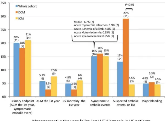
Long-term prognosis of left ventricular thrombus
During a median follow-up of 8.7 years, all-cause mortality occurred in 11 patients (10%) and was not significantly differ-ent in DCM and ICM (P = 0.63, Figure 2). Six patients died from cardiogenic shock, one from ventricular arrhythmia, one from ischaemic stroke, one from intracerebral bleeding, and two from septic shock.
We did not record any other embolic event during long-term follow-up (i.e. after the first year) attributed to an LVT.
Evolution and reverse remodelling
Left ventricular reverse remodelling was observed in most pa-tients after 12 months and was more frequent in DCM than in ICM patients (86% vs. 54% respectively, P = 0.02, Table 2). One year after diagnosis of LVT, DCM patients had higher
LVEF (40.2% vs. 33.9%, P = 0.04), lower BNP (195 vs. 596 ng/L, P< 0.01), but not significantly different hospitaliza-tion rates.
The 1 year LVT disappearance occurred in most patients, more frequently in DCM than in ICM (76.3% of LVT disappear-ance vs. 47.8%, respectively, P< 0.01, Table 2). Among all pa-tients, seven underwent LVT surgery or heart transplantation within thefirst year after diagnosis; three died while still hav-ing LVT and LVT’s persistence or disappearance was doubtful for six patients. Data were missing for nine patients (loss to follow-up, absence of imaging at 1 year, or available TTE of poor quality).
Risk factors and impact on prognosis of embolic
events
There was no significant difference for baseline characteris-tics between patients with symptomatic embolic events (N = 16) and without symptomatic embolic events (Supporting Information, Table S1). Mortality during follow-up, hospitalization rate for HF, and LV remodelling rates were not different among groups. Patients with or without the pri-mary endpoint also had similar baseline characteristics (Supporting Information, Table S2).
Management of left ventricular thrombus
A minority of patients (4.8%) underwent surgery for LVT: two DCM patients with suspected cerebral embolic events Figure 2 Risk of all-cause death of patients with left ventricular thrombus and non-ischaemic dilated cardiomyopathy (DCM) or ischaemic
(asymptomatic ischaemic lesions on a CT scan) had no size re-duction of a mobile LVT in spite of anticoagulant treatment for>5 days; one ICM patient had a symptomatic kidney is-chaemia and a voluminous and mobile residual LVT; one ICM patient had a voluminous LVT (maximum size 44 mm) and a TIA; and one ICM had coronary artery bypass and ven-tricle aneurysm resection at the same time.
The vast majority of patients (97.1%) received anticoagu-lant treatment (Figure 1, vitamin K antagonists in all cases). One DCM patient did not receive anticoagulant treatment cause of concomitant major bleed and two ICM patients be-cause of a very high estimated bleeding risk.
Anticoagulant treatment was maintained for more than 1 year in 80% of cases (66/81 patients with available data) and was more frequent in ICM than in DCM (87.5% vs. 65.4%, P = 0.04, Figure1). The majority (13/15) of patients in whom anticoagulation was stopped had clinically signi fi-cant reverse remodelling.
Ischaemic cardiomyopathy patients were more likely to re-ceive anti-platelet therapy and underwent implantable de fi-brillator implantation more frequently (Figure1).
Risk factors of left ventricular thrombus in dilated
cardiomyopathy
In our case–controls study, DCM patients with LVT were matched for age, sex, NYHA class, and LVEF with a cohort of 38 DCM patients without LVT. Patients with and without LVT had no significant differences in the proportion of diabe-tes and smoking, biological profile, and TTE/MRI characteris-tics. However, we found a significantly higher BNP level in patients with LVT (2447 ng/L, inter-quartile range 947–2969 vs. 347 ng/L, inter-quartile range 220–413, P < 0.001, Supporting Information, Table S3).
Discussion
Our study provides a detailed description of HF patients diag-nosed with LVT. Our mainfinding is that 1 year rate of events is sizable despite anticoagulant treatment (15% for embolic event and 5% for CV mortality). Of uttermost importance, all symptomatic events occurred within 30 days of LVT diag-nosis. We also show that most patients with DCM will expe-rience reverse remodelling (86% at 12 months). Despite the fact that DCM patients had more severe HF (as evidenced by higher BNP level, higher NYHA class and lower LVEF), their rate of embolic events was similar to those experienced by ICM patients.
Patients with HF have significant rates of embolic events, even in the absence of atrialfibrillation. The meta-analysis5 of four relevant prospective randomized controlled trials,6–9 including patients with HF with reduced LVEF in sinus rhythm, showed no significant reduction in the risk of all-cause death when taking warfarin vs. aspirin. However, warfarin signi fi-cantly reduced the risk of stroke (fatal and non-fatal ischae-mic and haemorrhagic strokes—relative risk of 0.59). Yet the recent large-scale COMMANDER-HF trial reported a neu-tral effect of rivaroxaban in ischaemic HF with reduced ejec-tion fracejec-tion in sinus rhythm.10 Oral anticoagulation is consequently currently not indicated for patients with HF with reduced LVEF in the absence of atrial fibrillation. How-ever, in a post hoc analysis of COMMANDER-HF, rivaroxaban significantly reduced by 32% the occurrence of all-cause strokes or TIA compared with a placebo.11This observation suggests that some patients could still benefit from an antico-agulant therapy, possibly when at high risk of LVT. For exam-ple, patients with non-compaction are known to have a propensity for thrombus formation and embolic events, and they may be candidates for lifelong anticoagulation even in the absence of thrombus. In addition, the occurrence of LVT is an indication for anticoagulants in HF.
Table 2 Imagingfindings, heart failure outcomes, and LVT evolution during follow-up
Total (105) DCM (n = 38) ICM (n = 67) P value
Imagingfindings
LV positive remodelling (n = 68), % (n) 65% (44) 86% (19) 54% (25) 0.02 TTE LVEF at 12 months (%), mean (±SD) 35.9 ± 11.1 40.2 ± 13.5 33.9 ± 9.3 0.04 RV S′ at 12 months (cm), mean (±SD) 11.2 ± 2.9 11.8 ± 3.4 10.9 ± 2.6 0.30 BNP at 12 months (pg/mL), median (IQR) 460 (68–528) 195 (25–204) 597 (101–797) 0.01
HF outcomes
Hospitalization for HF within the 1st year, % (n) 9.4% (8) 3.3% (1) 13% (7) 0.25 Hospitalization for HF during follow-up, % (n) 21% (18) 21% (6) 22% (12) 1
LVT evolution (n=84)
Confirmed LVT disappearance, % (n) 58.1% (51) 76.3% (29) 47.8% (32) 0.01 Confirmed LVT persistence, % (n) 14.3% (15) 2.6% (1) 20.9% (14) 0.02
Doubtful LVT persistence, % (n) 5.7% (6) 2.6% (1) 7.5% (5) 0.41
Death before LVT disappearance, % (n) 2.9% (3) 2.6% (1) 3.0% (2) 1
Missing data, % (n) 8.6% (9) 7.9% (3) 9.0% (6) 1
BNP, brain natriuretic peptide; DCM, dilated cardiomyopathy; HF, heart failure; ICM, ischaemic cardiomyopathy; IQR, inter-quartile range; LV, left ventricular; LVEF, left ventricular ejection fraction; LVT, left ventricular thrombus; RV, right ventricular; SD, standard deviation; TTE, transthoracic echocardiography.
6 A.-I. Lemaître et al.
ESC Heart Failure (2021) DOI: 10.1002/ehf2.13211
To our knowledge, our study is the largest contemporary cohort describing the clinical profile and outcomes of pa-tients with LVT in HF. Several studies before the MRI era tried to determine prevalence and embolic potential of LVT in HF of various etiologies12or in few patients with non-ischaemic DCM.13Surprisingly, these studies did notfind convincing as-sociation between LVT and the occurrence of embolic events.14,15 In contrast, we report herein that non-ischaemic DCM patients with an LVT have a high rate of embolic events within 30 days of diagnosis (possibly be-cause routine follow-up imaging was unable to identify thrombus prior emboli). However, these patients have few long-term consequences (death <3%, no disabling stroke) and a good recovery potential with 86% of patients experiencing 12 month reverse remodelling. Surprisingly, the only risk factors of LVT we identified in our dedicated case–control study are high BNP value. Importantly, other markers of severity and/or remodelling, not measured in our study, could also be predictive of LVT occurrence. More detailed evaluations could shade a new light on the LV mor-phological and functional determinants of LVT, possibly using MRI. Yet our paper indicates that a simple integrative marker (BNP) may be associated with LVT risk.
Our clinical management indicated LVT surgery only in ex-treme circumstances. In our view, patients with severely re-duced LVEF who undergo surgical thrombectomy are exposed to significant perioperative complications and high mortality. These patients may be precipitated towards LV as-sist device implantation or heart transplant after cardioplegia, with a high risk of organ failure due to ischae-mic injury. The consequence of this surgical approach may be unpredictable as the vintage (and subsequent stability) of the thrombus is by nature unknown and may largely de-pend on the frequency of echocardiographic follow-up of pa-tients. As a consequence, if no other emergent surgical procedure is indicated, our results call for a cautious manage-ment as the risk–benefit balance might not favour initial LVT surgery. Our‘wait-and-see’ approach seems to perform well in light of the low mortality rate of our cohort.
Clinical perspectives
Our results suggest that LVT is associated with a significant rate of embolic events, but the presence of LVT or embolic events does not seem to have an important impact on mor-tality or preclude reverse remodelling under medical treat-ment. From a clinical standpoint, these results suggest that LVT occurs in clinical situations that are likely to improve rather than in end-stage HF disease. LVT should in no way be perceived as a desperate condition. In addition, based on our report, a‘wait-and-see’ attitude based on vitamin K antagonist long-term anticoagulation appears as a safe and reasonable management of LVT in HF. Surgery was needed
only very rarely in our cohort. Most importantly, the very early phase of LVT management, using anticoagulants, ap-pears of key importance, as symptomatic embolic events oc-cur within 30 days of diagnosis. Importantly, most of the patients in whom anticoagulation was withdrawn after thrombus disappearance had significant reverse remodelling which was perceived to drastically decrease the risk of subse-quent thrombus reappearance.
Translational outlook
Our clinical study shows that natriuretic peptides are strongly associated with the presence of LVT. This finding suggests that HF severity is a determinant of the occurrence of LVT. Convincing evidence exist regarding the association of wors-ening HF with a procoagulant state.16Yet whether a specific coagulation profile underlies the genesis of LVT is unknown. Dedicated studies should be conducted to better understand this point, possible using a case–control strategy given the frequency of LVT in HF.
Limitations
This retrospective study has several limitations. We may have missed some cases because of clinicalfiles mislabelling. How-ever, this should not introduce significant bias in patients’ characteristics. In addition, we could not adequately report thrombus characteristics such as aspect, size, implantation, and mobility, which could all have an impact on the risk of emboli.
Accidental discovery of emboli was considered as an out-come in our study; this can overestimate event risk as other factors of emboli/thrombus (besides LVT) could be the actual cause.
Because of the retrospective design of this study, we did not have access to systematic whole-body imaging searching for asymptomatic systemic embolisms. In addition, it is possi-ble that a systematic use of CMR imaging could have modi-fied the persistence of LV thrombus as it is more sensitive than echocardiography.
This is a single-centre retrospective study, and the number of patients is moderate. Future larger studies should affirm the epidemiologicalfindings related to LVT in the specific set-ting of HF. Specifically, given the size of our population, we could not investigate the prognostic of LVT among different non-ischaemic DCM aetiologies. These larger studies might identify risk stratifier we were not able to detect because of limited statistical power. These studies should also determine whether regional dyskinesia/aneurism, significant regional contractility abnormalities, and/or fibrosis/morphology as assessed with CMR add to our understanding of LVT onset and prognosis.
The prognosis of LVT could also be different according to thrombus location within the left ventricle; this aspect should be evaluated in future reports.
As vitamin K antagonists remain the reference treatment of LVT, no non-vitamin K oral anticoagulants (NOACs) were used in this study. The prognosis of LVT treated with NOAC remains unknown, but some authors have recently called for their cautious use, because NOACs in patients with LVT were associated with a higher rate of strokes or systemic embolism in a large population of mixed LVT patients with reduced ejection fraction compared with warfarin.17
Conclusion
In conclusion, LVT is associated with a significant rate of embolic events, but the presence of LVT or embolic events does not seem to have an important impact on mortality or preclude reverse remodelling under medical treatment. The occurrence of LVT may be linked to the initial severity of HF as high BNP was a risk factor for LVT. Further investi-gations are needed to identify other risk factors of LVT and LVT-related embolic events. Meanwhile, a‘wait-and-see’ at-titude based on vitamin K antagonist long-term anticoa-gulation appears as a safe and reasonable management of LVT in HF. In all cases, as symptomatic embolic events occur
within 30 days of diagnosis, a very careful initial manage-ment is warranted.
Con
flict of interest
N.G. received honoraria from Novartis, AstraZeneka, Vifor and Boehringer.
Funding
None.
Supporting information
Additional supporting information may be found online in the Supporting Information section at the end of the article.
Table S1. Baseline characteristics and evolution of patients
with and without symptomatic embolic events.
Table S2. Baseline characteristics and evolution of patients
with and without the primary endpoint of all-cause death or embolic event.
Table S3. Baseline characteristics and evolution in the case–
control study of patients with DCM with and without left ven-tricular thrombus.
References
1. McCarthy CP, Vaduganathan M, McCarthy KJ, Januzzi JL Jr, Bhatt DL, McEvoy JW. Left ventricular thrombus after acute myocardial infarction: screening, prevention, and treatment.
JAMA Cardiol 2018;3: 642–649.
2. Lee JM, Park JJ, Jung HW, Cho YS, Oh IY, Yoon CH, Suh JW, Chun EJ, Choi SI, Youn TJ, Lim C, Cho GY, Chae IH, Park KH, Choi DJ. Left ventricular thrombus and subsequent thromboembolism, comparison of anticoagulation, surgical removal, and antiplatelet agents. J
Atheroscler Thromb 2013;20: 73–93.
3. Velangi PS, Choo C, Chen KA, Kazmirczak F, Nijjar PS, Farzaneh-Far A, Okasha O, Akçakaya M, Weinsaft JW, Shenoy C. Long-term embolic out-comes after detection of left ventricular thrombus by late gadolinium enhance-ment cardiovascular magnetic reso-nance imaging: a matched cohort study. Circ Cardiovasc Imaging 2019;
12: e009723.
4. McCarthy CP, Murphy S, Venkateswaran RV, Singh A, Chang LL, Joice MG, Rivero JM, Vaduganathan M, Januzzi JL, Bhatt
DL. Left ventricular thrombus: contem-porary etiologies, treatment strategies, and outcomes. J Am Coll Cardiol 2019;
73: 2007–2009.
5. Hopper I, Skiba M, Krum H. Updated meta-analysis on antithrombotic therapy in patients with heart failure and sinus rhythm. Eur J Heart Fail 2013; 15: 69–78.
6. Cleland JG, Findlay I, Jafri S, Sutton G, Falk R, Bulpitt C, Prentice C, Ford I, Trainer A, Poole-Wilson PA. The Warfa-rin/Aspirin Study in Heart failure (WASH): a randomized trial comparing antithrombotic strategies for patients with heart failure. Am Heart J 2004;
148: 157–164.
7. Cokkinos DV, Haralabopoulos GC, Kostis JB, Toutouzas PK, investigators H. Ef fi-cacy of antithrombotic therapy in chronic heart failure: the HELAS study.
Eur J Heart Fail 2006;8: 428–432.
8. Massie BM, Collins JF, Ammon SE, Armstrong PW, Cleland JG, Ezekowitz M, Jafri SM, Krol WF, O’Connor CM, Schulman KA, Teo K, Warren SR, WATCH Trial Investigators. Randomized
trial of warfarin, aspirin, and clopidogrel in patients with chronic heart failure: the Warfarin and Antiplatelet Therapy in Chronic Heart Failure (WATCH) trial.
Circulation 2009;119: 1616–1624.
9. Homma S, Thompson JL, Pullicino PM, Levin B, Freudenberger RS, Teerlink JR, Ammon SE, Graham S, Sacco RL, Mann DL, Mohr JP, Massie BM, Labovitz AJ, Anker SD, Lok DJ, Ponikowski P, Estol CJ, Lip GY, di Tullio MR, Sanford AR, Mejia V, Gabriel AP, del Valle M, Buchsbaum R, WARCEF Investigators. Warfarin and aspirin in patients with heart failure and sinus rhythm. N Engl J
Med 2012;366: 1859–1869.
10. Zannad F, Anker SD, Byra WM, Cleland JGF, Fu M, Gheorghiade M, Lam CSP, Mehra MR, Neaton JD, Nessel CC, Spiro TE, van Veldhuisen DJ, Greenberg B. Rivaroxaban in patients with heart fail-ure, sinus rhythm, and coronary disease.
N Engl J Med 2018;379: 1332–1342.
11. Mehra MR, Vaduganathan M, Fu M, Ferreira JP, Anker SD, Cleland JGF, Lam CSP, van Veldhuisen DJ, Byra WM, Spiro TE, Deng H, Zannad F, Greenberg
8 A.-I. Lemaître et al.
ESC Heart Failure (2021) DOI: 10.1002/ehf2.13211
B. A comprehensive analysis of the ef-fects of rivaroxaban on stroke or tran-sient ischaemic attack in patients with heart failure, coronary artery disease, and sinus rhythm: the COMMANDER HF trial. Eur Heart J 2019; 40: 3593–3602.
12. Gottdiener JS, Gay JA, VanVoorhees L, DiBianco R, Fletcher RD. Frequency and embolic potential of left ventricular thrombus in dilated cardio-myopathy: assessment by 2-dimensional echocardiography. Am J Cardiol 1983;
52: 1281–1285.
13. Ciaccheri M, Castelli G, Cecchi F, Nannini M, Santoro G, Troiani V, Zuppiroli A, Dolara A. Lack of
correlation between intracavitary thrombosis detected by cross sectional echocardiography and systemic emboli in patients with dilated cardiomyopathy.
Br Heart J 1989;62: 26–29.
14. Katz SD. Left ventricular thrombus and the incidence of thromboembolism in patients with congestive heart failure: can clinical factors identify patients at increased risk? J Cardiovasc Risk 1995;
2: 97–102.
15. Cioffi G, Pozzoli M, Forni G, Franchini M, Opasich C, Cobelli F, Tavazzi L, with the technical assistance of D. Rossi. Sys-temic thromboembolism in chronic heart failure: a prospective study in
406 patients. Eur Heart J 1996; 17: 1381–1389.
16. Popovic B, Zannad F, Louis H, Clerc-Urmès I, Lakomy C, Gibot S, Denis CV, Lacolley P, Regnault V. Endothelial-driven increase in plasma thrombin gen-eration characterising a new hypercoag-ulable phenotype in acute heart failure.
Int J Cardiol 2019;274: 195–201.
17. Robinson AA, Trankle CR, Eubanks G, Schumann C, Thompson P, Wallace RL, Gottiparthi S, Ruth B, Kramer CM, Salerno M, Bilchick KC. Off-label use of direct oral anticoagulants compared with warfarin for left ventricular thrombi. JAMA Cardiol 2020; 5: 685–692.