HAL Id: tel-02295014
https://tel.archives-ouvertes.fr/tel-02295014
Submitted on 24 Sep 2019HAL is a multi-disciplinary open access
archive for the deposit and dissemination of sci-entific research documents, whether they are pub-lished or not. The documents may come from teaching and research institutions in France or abroad, or from public or private research centers.
L’archive ouverte pluridisciplinaire HAL, est destinée au dépôt et à la diffusion de documents scientifiques de niveau recherche, publiés ou non, émanant des établissements d’enseignement et de recherche français ou étrangers, des laboratoires publics ou privés.
Neuronal properties and synaptic connectivity in rodent
presubiculum
Jean Simonnet
To cite this version:
Jean Simonnet. Neuronal properties and synaptic connectivity in rodent presubiculum. Neurons and Cognition [q-bio.NC]. Université Pierre et Marie Curie - Paris VI, 2014. English. �NNT : 2014PA066435�. �tel-02295014�
THÈSE DE DOCTORAT
DE L’UNIVERSITÉ PIERRE ET MARIE CURIE Spécialité Neurosciences
École doctorale Cerveau – Cognition – Comportement
Présentée par : Jean Simonnet
Pour obtenir le grade de
DOCTEUR DE L’UNIVERSITÉ PIERRE ET MARIE CURIE
Sujet de la thèse :
Neuronal properties and synaptic
connectivity in rodent presubiculum
Soutenue le 23.09.2014devant le jury composé de :
Dr Jean-Christophe Poncer Président Dr Dominique Debanne Rapporteur Dr Maria Cecilia Angulo Rapportrice
Pr Hannah Monyer Examinatrice
Dr Bruno Cauli Examinateur
Dr Desdemona Fricker Directrice de thèse
Université Pierre & Marie Curie - Paris 6 Tél. Secrétariat : 01 42 34 68 35 Bureau d’accueil, inscription des doctorants Fax : 01 42 34 68 40 et base de données Tél. pour les étudiants de A à EL : 01 42 34 68 41 Esc. G, 2ème étage Tél. pour les étudiants de EM à MON : 01 42 34 68 41 15 Rue de l’école de médecine Tél. pour les étudiants de MOO à Z : 01 42 34 68 51 75270 - PARIS CEDEX 06 E-mail : scolarite.doctorat@upmc.fr
Abstract
Cognitive functions rely on the generation and regulation of information in special-ized neuronal networks. The presubiculum, a cortical area located between the hip-pocampus and the entorhinal cortex, is involved in signaling the sense of orientation in animals as well as in humans. Most presubicular neurons are Head Direction Cells, that is, they fire as a function of directional heading. The presubiculum constitutes a crucial crossroad for spatial information. Very few data exist on the functional organization of the presubiculum, but its 6-layered cytoarchitecture suggests that signals are not passively relayed but rather actively integrated and refined.
During my PhD, I studied the microcircuit elements of rodent presubiculum in the slice preparation, linking structure and physiology using patch clamp records.
First, I focused on rat principal neurons and distinguished 3 groups: a homoge-neous population of regular spiking neurons in superficial layers, mostly pyramidal; intrinsically burst firing neurons of layer 4; and a very heterogeneous population of regular spiking neurons in deep layers. These populations constitute the primary el-ements for information processing in the presubiculum, and their diversity suggests a high computational power.
Then, I addressed the question of the inhibitory control in the presubiculum. Recordings were performed from slices of transgenic mouse strains that express fluo-rescent proteins in interneurons. We showed a continuum of diversity for parvalbumin-(PV) and somatostatin- (SST) containing interneurons, from the archetypical PV-positive fast spiking basket cells to the SST-PV-positive low-threshold spiking Martinotti cells. Regarding the inhibition, the presubiculum seems to possess the complexity of all cortical areas.
Finally, I investigated the synaptic interactions of pyramidal cells and Martinotti cells in superficial layers, using dual patch clamp recordings. Martinotti cells provide low amplitude but reliable inhibition onto pyramidal cell dendrites. I found that the strength at the excitatory synapse was enhanced following repetitive stimulation at high frequency. Consequently, dendritic inhibition by presubicular Martinotti cells may act as a homeostatic response to sustained excitation.
My PhD work brought essential knowledge about the presubicular microcircuit. It has shed light on the different populations of principal neurons and GABAergic interneurons and has uncovered a feedback inhibitory loop that is recruited during sustained but not transient activity.
Acknowledgments
First, I would like to thank the members of my Jury: Hannah Monyer, Jean Christophe Poncer, Bruno Cauli as well as Dominique Debanne and Maria Cecilia Angulo who have accepted to review this manuscript.
Je tiens à remercier Desdemona, qui m’a donné l’opportunité de travailler avec elle et qui m’a encadré tout au long de cette thèse. Tu as su me donner la liberté de travail dont j’avais besoin, et je te remercie de m’avoir permis d’exprimer mes idées et mes envies tout en ayant un oeil critique et avisé à chaque étape. Je pense que cela n’a pas été une chose facile avec mon caractère plutôt ombrageux. . . Difficile à convaincre notamment. Disons que nous avons eu un certain nombre de discussions passionnées sur la « bonne manip à faire » ou « la figure à montrer » ou « ce qu’il faut dire ». J’ai toujours pris cela dans le sens positif, à savoir que la confronta-tion de nos idées a toujours permis d’améliorer notre travail. Je voudrais aussi te remercier pour la compréhension dont tu as fait preuve, par rapport aux aléas de la vie qui ne concernent en rien le travail, mais qui l’impactent inévitablement. Enfin, je souhaite exprimer toute ma gratitude de m’avoir toujours soutenu tout au long de ces années.
Bien évidemment, cette thèse ne serait pas possible sans Richard, qui m’a ouvert les portes de son laboratoire et apporté son soutien quotidien. Vous avez su me prodiguer de très bons conseils durant toute cette thèse. S’il y a une chose que j’ai appris, c’est bien qu’un message simple est beaucoup plus fort. Faire des figures simples, des diapos simples, écrire des phrases courtes. « More is less ». Je tiens également à vous remercier de m’avoir toujours valorisé auprès de vos pairs.
Merci à tous les membres de l’équipe. Emmanuel, Michael, Ivan, Lim-Anna, Con-stanze, Roxanne, Caroline, Maja, Etienne, Juliane, Katia, Bertrand et Mérie. Vous m’avez tous beaucoup apporté sur le plan scientifique et humain. Je pense à Em-manuel qui m’a appris tout ce qu’il savait sur l’imagerie structurée ou l’immunohisto-chimie. Je pense à Caroline qui m’a beaucoup conseillé pour les doubles enreg-istrements. Je me rappelle du temps passé devant le poste d’enregistrement de Michael à regardé défiler les enregistrements. Je pense aussi aux bons moments passés autour d’une bière, ou lors des dégustations de vin. Une spéciale pour Mérie qui prend la relève auprès de Desdemona, et qui a du me supporter pendant cette dernière année, j’avoue que je n’ai pas toujours été très tendre et surement trop
exigent parfois.
Merci à Alberto et toute son équipe avec qui j’ai souvent interagi pour résoudre mes problèmes de souris, de tranches, de solution intra, ou d’oxygène. Je dois avoir une dette de café envers Charlotte qu’il faudra régler un de ces jours.
Merci à Claire et Stéphane ainsi qu’à leurs équipes, avec qui j’ai peut être moins interagi, mais qui ont apporté leur petit grain de sel de temps à autre.
Merci à Sean. Sean tu venais au labo pour travailler avec Desdemona et c’est vrai que nous discutions souvent de tes manips et j’ai pu t’aider de temps à autre quand tu avais des problèmes. Un jour, tu m’as simplement proposé de participer active-ment à ton projet. J’en ai été très touché. Cette collaboration m’a égaleactive-ment permis d’améliorer mon anglais et tu as été très pédagogue de ce point de vue là.
Merci à l’animalerie du 105, de l’ICM, aux plateformes de génotypage et d’histologie pour leur aide et support technique.
Merci aux Ajités. William, Pinar, Morgane P, Fabian, Kevin, Tristan, Morgane B, Alizée, . . . Je ne peux pas citer tous les noms, mais ils sauront se reconnaitre. On a passé de très bons moments tous ensemble et belle dynamique a été créée à l’ICM. Continuez s’il vous plait !
Je souhaite remercier Patricia Oliviero qui a superbement organisé, avec moi, la retraite des Doc et PostDoc en 2013. Merci pour toutes les petites choses que tu as faites pour moi, ne change rien tu es géniale !
Merci à la promo de master/thèse et associés. Carole, Audrey, Patrick, Esther, Nico, Raphael, Béné, Isa. . . j’en oublie c’est sur. . . On a passé de très bons moments, sou-vent de détente il faut le reconnaître, mais cela a été important pour tenir tout au long de ces années.
Carole. On était à la fac à Orléans en licence, on est venu à Paris avec la ferme intention de faire une thèse en neuro. On peut dire que c’est mission accomplie. Je pense que ca aurait été plus difficile sans toi sur le plan moral. Tu as su ètre présente au moment ou j’en avais besoin. Mème si l’on a fait des choses très différentes lors de nos thèses, nos discussions scientifiques m’ont aussi beaucoup apportées.
Merci à Y. Audrey. On travaillait sur des modèles similaires, avec les mèmes prob-lèmes. . . Enfin j’avais les problèmes, tu avais les solutions. Je suis venu te voir à plusieurs reprises quand j’avais des soucis de tranches ou de stéréotaxie, et tu as toujours été d’une aide plus que précieuse. Je pense que tu m’as aussi beaucoup influencé, sans que je ne me rende vraiment compte, dans ma façon de travailler. Merci aussi pour m’avoir aidé avec la rédaction.
J’aimerais remercier ma famille et mes proches qui ont toujours cru en moi et qui m’ont soutenu dans ma démarche.
Enfin, je tiens à remercier Eugénia du fond du cœur, pour m’avoir supporté et soutenu, surtout pendant la rédaction qui, je dois l’avouer, fut difficile, autant pour moi que pour elle.
Contents
Abbreviations 5
Presentation
7
I
Introduction
11
1 The presubiculum: Anatomy, function, microcircuit 15
1.1 The presubicular cortex . . . 15
1.1.1 Anatomy . . . 15
1.1.2 What kind of cortex? . . . 17
1.2 Presubiculum and spatial orientation . . . 20
1.2.1 Head direction cells of the presubiculum . . . 20
1.2.2 Head Direction Circuit . . . 22
1.2.3 The presubiculum is a major contributor of spatial represen-tation and memory . . . 26
1.3 Information processing in the presubicular microcircuit . . . 31
1.3.1 Anatomy and intrinsic excitability of presubicular neurons . . 31
1.3.2 Interlaminar, intralaminar and modular organization . . . 33
1.3.3 Input and output relays in the presubicular microcircuit . . . 35
2 How does a microcircuit work? 39 2.1 Many integrative levels in neuronal networks . . . 39
2.2 Neuronal intrinsic excitability . . . 42
2.2.1 Resting membrane potential . . . 42
2.2.2 Neuronal passive properties . . . 42
2.2.3 Action potentials . . . 43
2.3 Wiring a network: axonal conduction and regulation of information . 48 2.3.1 Axonal conduction velocity . . . 48 2.3.2 Analog information encoding in the axon . . . 49 2.4 Synaptic transfer and modulation of information in the presynaptic
terminal . . . 54 2.4.1 Basic mechanism of neurotransmitter release . . . 54 2.4.2 Synchronous versus asynchronous release of neurotransmitter . 56 2.4.3 Short term presynaptic plasticity . . . 58 2.4.4 Voltage dependent regulation of synaptic activation . . . 66 2.4.5 Regulation of presynaptic function by extrinsic factors . . . . 67
II
Methods
69
III
Results
79
ARTICLE 1. Cellular neuroanatomy of rat presubiculum 81
ARTICLE 2. Properties of presubicular neurons that project to lateral
mammillary nucleus or anterodorsal thalamus 101
ARTICLE 3. A continuum of diversity of Parvalbumin or Somato-statin expressing interneurons in mouse presubiculum 111 ARTICLE 4. Memory of past activity determines the recruitment
of a Martinotti cell-mediated inhibitory feedback loop in mouse
presubiculum 147
IV
Discussion
179
1 Building blocks of the presubiculum 183
1.1 Did we correctly addressed the whole diversity of principal neurons? . 183 1.2 Interneuron diversity . . . 185
2 Perspective: from neuronal diversity to function 187
3 Neurons that project to lateral mammillary (LMN) and anterodor-sal thalamus (ADN): implication for the visual update of the head
4 Memory of past activity at the pyramidal cell-to-Martinotti cell
synapse: properties and mechanisms 191
4.1 Better define the dynamics of the plasticity, its specificity and variability191 4.2 Mechanisms of activity dependent synaptic transfer at the pyramidal
cell to Martinotti cell synapse? . . . 192 4.2.1 Activity dependent action potential broadening . . . 193 4.2.2 Modulation at the synapse . . . 195 4.2.3 The transfer rate increase may results from a synergistic
mech-anism . . . 196
V
General conclusion
197
VI
Collaboration
201
ARTICLE. Cellular anatomy, physiology and epileptiform activity in the CA3 region of Dcx knockout mice: a neuronal lamination defect
and its consequences 203
List of figures
219
Abbreviations
Anatomy
ADN anterodorsal thalamus
AM anteromedial thalamus AV anteroventral thalamus CA corpus ammonis DG dentate gyrus dl dorso-lateral HF hippocampal formation
DTN dorso tegmental nucleus
LEA lateral entorhinal cortex
LDN laterodorsal thalamus
LMN lateral mammillary nucleus
MEA medial entorhinal cortex
PHR parahippocampal region
PaS parasubiculum
PER Perirhinal cortex
PrOS presubiculum
POR Postrhinal cortex
PoS postsubiculum = dorsal part of PrS
RSC retrosplenial cortex sub subiculum vm ventro-medial
Physiology
AP action potential DC direct current FS fast spiking LTS low-threshold spiking PTP posttetanic potentiation PPD paired-pulse depression PPF paired-pulse facilitation PPR paired-pulse ratio RP resting poolOthers
AMPA α-Amino-3-hydroxy-5-methyl-4-isoxazolepropionic acid
AIS axon initial segment
BAPTA 1,2-bis(2-aminophenoxy)ethane-N,N,N’,N’-tetraacetic acid
DTX dendrotoxin
EGTA ethylene glycol tetraacetic acid
GABA γ Amino-Butyrique Acid
GPCR G protein-coupled receptor
NMDA N-Methyl-D-aspartic acid
PV parvalbumin
PKC protein kinase C
SST somatostatine
The presubiculum is an understudied cortical area located in the parahippocam-pal region and involved in spatial navigation. My supervisor, Desdemona Fricker, therefore proposed this thesis project on information encoding at the level of the presubicular microcircuit.
The presubiculum contains head direction cells, which fire as a function of ani-mal’s directional heading. The head direction signal is generated from vestibular information, in subcortical areas that project to the presubiculum. Besides, pre-subiculum receives visual information from visual and retrosplenial cortices. The convergence of the two types of information in the presubiculum lead to the update of the head direction signal with visual cues. The presubiculum then distributes a visual landmark control to subcortical areas as well as a major drive to the down-stream entorhinal cortex. Consequently, the presubicular function deeply impacts the function of entorhinal cortex and hippocampus.
The six-layered organization of the presubiculum suggests a high computational power, implying that information is not passively relayed, but rather actively inte-grated and refined. But what happens when information enters the presubiculum has been unknown as the presubicular network and its components have never been studied in detail.
During my PhD, I studied the microcircuit elements of rodent presubiculum, using a model that allows a very precise investigation of neuronal and synaptic properties: the slice preparation. The underlying theme of my work was to use patch clamp records in whole cell configuration in order to obtain electrophysiological and mor-phological data on presubicular neurons and their connections.
A part of my work consisted in a description of the intrinsic electrophysiological and morphological properties of the different neurons in the presubiculum. My first project focused on principal neurons in rat, whereas the second study dealt with the diversity of GABAergic interneurons. I defined the "building blocks" of the presubiculum, an essential step in the understanding of information encoding in a network. I showed that the six layers of the presubiculum contain neurons with distinct biophysical properties and distinct dendritic and axonal arborizations, suggesting that different integrative capabilities exist within the network. Together with Mérie Nassar, we described the diversity of inhibitory interneurons, that are important elements for tuning cortical information. Our results suggest that the
presubiculum possesses all necessary elements for complex information treatment, as is the case in other cortical areas.
The last part of my work focused on the specific interactions between pyramidal cells and a subpopulation of interneurons, the Martinotti cells. I noticed an unusual form of plasticity at the excitatory synapse. The transfer of information was exquisitely low, as one action potential in the pyramidal cell rarely triggered a postsynaptic ex-citatory event in the Martinotti cell. However, sustained activity at high frequency was able to un-mute the synapse, which then became efficient to transfer information and summed events could fire the Martinotti cell. Presubicular head direction cells fire persistently as the head is turned in the cell’s preferred direction. The excita-tory synapses onto Martinotti cells of the presubiculum are therefore appropriately tuned to integrate and balanced the persistent intrinsic excitatory activity delivered by the presubicular microcircuit.
Two main parts in the introduction should acquaint the reader with the con-text of my research. The first part presents the presubiculum, its situation in the parahippocampal region, its anatomy and development. I then go over its function as a key relay for landmark information in the brain, and I eventually come back on the poor knowledge of its microcircuit. How does a microcircuit work? The second part reviews different elements that process the information in neuronal net-works, from ions channels responsible for excitability to some of the principles of computation in microcircuits. I mainly develop the regulation of information from the spike generation to the plasticity of synaptic release, because it provides useful information for the last study of my PhD.
1
|
The presubiculum: Anatomy, function,
microcircuit
1.1
The presubicular cortex
1.1.1
Anatomy
The presubiculum is a cortical region of the hippocampal-parahippocampal forma-tion in brain’s temporal lobe. Hippocampus proper is subdivided into the Dentate Gyrus, Ammon’s Horn (CA3, CA2, CA1) and Subiculum; The parahippocampal area is its continuation, composed of presubiculum, parasubiculum, entorhinal (me-dial and lateral parts), peri- and postrhinal cortices (Fig. 1;van Strien et al.,2009). The presubiculum corresponds to Brodmann’s area 27 and 48 (Brodmann,1909), following the temporoventral-to-septodorsal hippocampal axis in rodents (Fig. 1). The most dorsal part, corresponding to Brodmann’s area 48 is also called "Post-subiculum" (Brodmann, 1909; Rose and Woolsey, 1948; Blackstad, 1956). In the proximo-distal axis of the hippocampus (from dentate gyrus to subiculum), the pre-subiculum is located just next to pre-subiculum and is then followed by the parasubicu-lum; these 3 areas being classically grouped together into the "subicular complex". Eventually, in its retrodorsal part, the presubiculum is bordered by retrosplenial cortex.
The presubiculum is easily distinguishable from its neighboring areas regarding anatomical features such as cytoarchitecture and topography of afferent fibers (Fig. 2, Ramon y Cajal, 1899; Brodmann, 1909; Rose and Woolsey, 1948; Blackstad, 1956).
Presubiculum is a 6-layered cortex. Layers were already described by Ramon y Cajal according to their neuronal content and density, from the pial surface to the white matter (Fig. 2C; Ramon y Cajal, 1899). This description of layer holds true for non-human primates and rodent presubiculum.
Layer 1, the molecular layer, is almost empty and contains only few putative interneurons, (Cajal’s "short axon cells") and glial cells. Layer 2 is a thin layer of
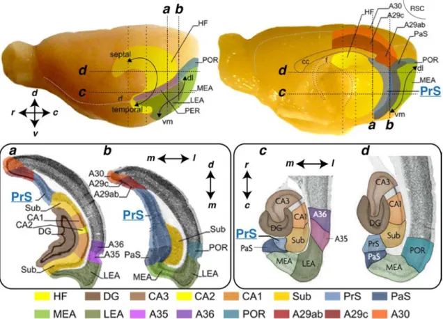
Figure 1: Representations of the hippocampal formation (HF), the parahippocampal
region (PHR) and retrosplenial cortex (RSC) in rat brain. Lateral (A) and midsagittal (B) views of the rat brain. Hippocampus contains dentate gyrus (DG), Ammon’s horn (CA1 to CA3) and subiculum (sub). Parahippocampal region is subdivided into pre-subiculum (PrS), parapre-subiculum (PaS), medial and lateral entorhinal areas (MEA and LEA) peri- and postrhinal cortices (PER and POR). Retrosplenial cortex is subdivided here in A29ab, A29c and A30 (Brodmann’s nomenclature). Hippocampus, PrS and PaS follow a dorsoseptal-to-ventrotemporal axis; Entorhinal cortices follow a dorsolateral-to-ventromedial (dl, vm); PER and POR are defined along a rostro-caudal axis. The dashed vertical (a, b) and horizontal (c, d) lines indicate levels of coronal and horizontal sections depicted in C. rf: rhinal fissure; cc: corpus callosum; f: fibria. Adapted fromSugar et al. (2011) andvan Strien et al. (2009)
approximately the same thickness as layer 1 and contains densely packed pyramidal and fusiform cells. In the most dorsal part of mouse presubiculum, layer 2 cell bodies tend to form clusters separated by fiber stripes (Slomianka and Geneser, 1991). Layer 3 is larger than layer 2 with a much lower neuronal density and is composed of pyramidal neurons. Layer 4 is also named "lamina dissecans" because it was described as a neuron free layer, containing only fibers and glial cells (Rose, 1926; Lorente De Nó, 1933). It is a convenient marker separating superficial layers (1, 2 and 3) from deep layers (5 and 6). Layer 5 is a layer with large to medium sized pyramidal cells whereas layer 6 contains smaller fusiform and pyramidal cells. In primate presubiculum, deep layers are separated in 3 sub-layers (5, 6, 7). This laminar organization has been observed with specific in situ hybridization stainings and is less clear in rodents (Ding, 2013).
The laminar organization of the presubiculum marks an abrupt transition with the adjacent subiculum, organized more like a cloud (even if subiculum may also be subdivided in different layers,O’Mara et al.,2001). An "extremely dense plexus formed by [ ] many afferent axons" in superficial layers of presubiculum distinguishes it from its neighbors, subiculum, parasubiculum and retrosplenial cortex (Ramon y Cajal, 1899; Blackstad, 1956). These terminals are more numerous in the dorsal part of presubiculum (area 48; Rose and Woolsey, 1948; Blackstad, 1956). The presubiculo-parasubiculum transition is marked by the absence of the densely packed layer 2 in parasubiculum, the cellular density of its superficial layers being more homogeneous. This transition is clearly visible with a specific marker of presubicular layer 2, calbindin (Boccara et al., 2010).
The dense presubicular layer 2 is also remarkably avoided by the characteristic plexus targeting the presubiculum (Fig. 2A and B, Ramon y Cajal, 1899). These dense afferent fibers define very well the limits of presubiculum, especially dorsally, where their interruption marks the border of presubiculum with retrosplenial cortex (Ramon y Cajal, 1899; Blackstad,1956).
1.1.2
What kind of cortex?
During development, radial migration of neuronal progenitors from the ventricu-lar zone shapes laminar compartments (Angevine and Sidman, 1961; Rakic, 1974). Then, subsequent change may occur to generate the adult cortical organization. Cortical areas may be classified according to the development of their laminar or-ganization and their aspect in the adult stage (Lorente De Nó, 1933; Filimonoff, 1947). These historic classifications can be criticized because they are based only on anatomy, but they are still of interest for defining different parts of the cortex.
Figure 2: Layers and afferent fibers in the presubiculum. A: Tionin-stained
horizontal section through the rat hippocampal formation. DG: dentate gyrus; S: subicu-lum; PrS: Presubicusubicu-lum; PaS: Parasubicusubicu-lum; EC : Enthorinal cortex; Note the obvious separation of superficial (1,2,3) and deep (5,6) layers by lamina dissecans (layer 4) in the presubiculum. Note that layer 2 is more dense than layer 3, and that presubicular deep layers appear as a continuation of the subiculum and entorhinal cortex deep layers. Adapted fromAmaral and Witter,1989. B: Drawing of a horizontal section correspond-ing to A, but uscorrespond-ing a 15 day old mouse, stained with the Golgi method. Adapted from Ramon y Cajal (1899). Note the dense "plexus" of afferent fibers in the presubiculum that partially avoid layer 2. C: Laminar organization of the human presubiculum. Nissl method, fromRamon y Cajal(1899). Cajal’s nomenclature (my interpretation): A, plexi-form layer (layer 1); B, small pyramidal and fusiplexi-form cell layer (layer 2); C, deep plexiplexi-form layer (layer 3); D, large to medium size pyramidal cell layer (layer 4 and 5); E, fusiform and triangular cell layer (layer 6).
The Isocortex (or Cortex Completus) comprise 6 layers whereas the Allocortex (or Cortex Incompletus) displays an incomplete structure (less than 6 layers) in develop-mental and adult stages. The Periallocortex (or Cortex Intermedius) physically lies between the two others and its structure changes between developmental and adult stage. Neocortex is Isocortex; hippocampus and subiculum constitute the Archicor-tex, which is part of the Allocortex; the presubiculum was lumped together with the entorhinal area and termed Periarchicortex, which is part of the Periallocortex (Lorente De Nó,1933; Filimonoff, 1947).
More recent findings (Bayer, 1980) have shown that embryogenesis is actually different between presubiculum and entorhinal cortex. First, neurogenesis occurs later in presubiculum. Second, deep layers are formed before superficial layers (like the classical cortical development) with a strong neurogenetic gradient. Indeed, deep layers appear at E15-18 whereas superficial layers appear at E17-20. A small gradi-ent also exists in gradi-entorhinal cortex but it occurs a little earlier (finished at E17 in deep layers and E18 in superficial layers). Another intriguing fact is that neurogene-sis timelines of presubiculum and subiculum are the same for deep layers but not for superficial layers. In the adult, it is interesting to look at the presubiculo-subiculum transition in horizontal slices (Fig 2A) to see that presubicular deep layers really appear to be a continuation of subiculum. From his studies on Marsupials, Brod-mann (Brodmann, 1909) even described this transition as an "abrupt interruption of layer II-V at the beginning of the subiculum, with only layer I and VI continuing into Ammon’s horn in greatly widened form".
All these developmental data showed that the six layers of presubiculum appear in a very specific and unique manner. However, functional consequences of this specific development, compared to neocortex or entorhinal cortex remain unknown.
1.2
Presubiculum and spatial orientation
To survive, mammals rely on their sense of orientation to get water, food, and mate, or to escape predators. This requires the innate ability to learn features of a novel environment as it is explored. This is spatial orientation and it uses two different cognitive processes: path integration and landmark navigation. Path integration uses a self derived representation of space using vestibular, proprioceptive and motor inputs; landmark navigation represents space using external cues such as visual, olfactory, auditory and somatosensory information. Among all the brain areas involved in these processes, the presubiculum encodes the head direction, one critical information for spatial cognition (Wiener and Taube,2005; Taube, 2007).
1.2.1
Head direction cells of the presubiculum
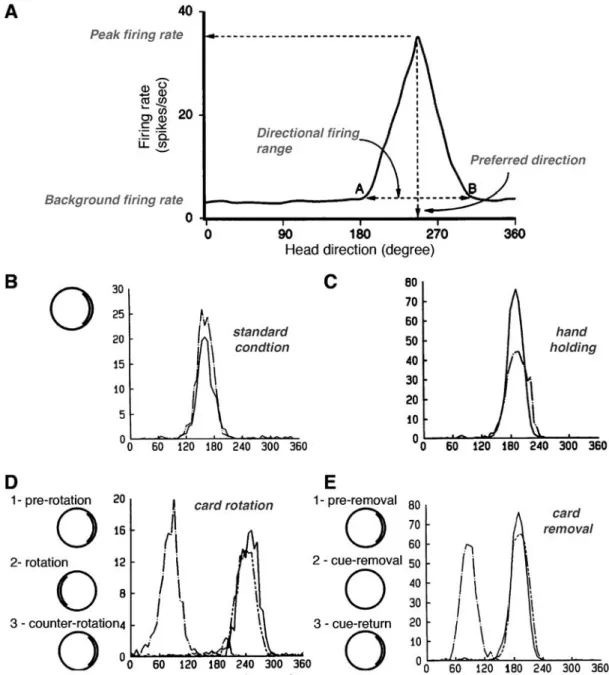
Extracellular recordings in freely moving rats have shown that 50-60 % of neurons in dorsal presubiculum (postsubiculum) are discharging as a function of animal’s directional heading (Ranck,1984;Taube et al., 1990a;Taube, 2007; Boccara et al., 2010). These neurons are called Head Direction Cells. Each head direction cell is characterized by a specific tuning curve of its firing rate as a function of the animal’s head direction (Fig. 3A).The cell’s preferred direction is defined as the one leading to the maximum firing rate. Basal firing rate is close to zero and increases only for directional ranges varying from 60◦ to 150◦ (average 90◦) with a triangular or Gaussian distribution of frequencies around the preferred direction (Blair and Sharp, 1995;Taube,1995). One cell is accurately tuned to only one head direction and the whole population allows a complete representation of orientation. Each neuron has a very stable tuning curve but the peak firing rate varies among presubicular neurons (from 5 to 115 Hz). Last, but not least, discharge persists without adaptation as long as the preferred direction is maintained (Taube et al.,1990a).
Head direction cells are not sensitive to the geomagnetic field but to environ-mental visual landmarks. Rotation of the major polarizing visual cue within the environment leads to a corresponding shift of the preferred direction (Fig. 3D). Head direction cell firing does not change in the absence of visual landmarks, but preferred direction can drift over time (Fig. 3E). Visual cues are used to control but not to generate the head direction signal. Furthermore, visual inputs exert a higher degree of control than other senses such as auditory or olfactory inputs (Goodridge et al.,1998). Motor activity seems to improve signal quality but is not necessary for its generation because preventing an animal from moving reduces peak firing rate but does not abolish head direction cell activity ((Fig. 3B),Taube et al., 1990b).
Figure 3: Basic features of presubicular head direction cells. A. Tuning curve
features of head direction cells (adapted fromTaube,1995): background firing rate is close to zero but increases within the directional firing range to reach the peak firing rate for the preferred direction. B. Stability of head direction cell firing across two recording sessions, one (dashed line) recorded 15 days after the other (solid line). In standard condition, a prominent cue card is disposed as a polarizing cue on one side of the open field wall.
C. Carrying the animal by hand and moving it around in the arena (dashed line) only
decreased peak firing rate compared to standard condition (solid line). D. Cue card
rotation causes a corresponding shift in preferred direction. Here, the same head direction was recorded in standard condition (1, solid line), after a 180◦ clockwise rotation of the cue card (2, dash-dot line) and after the equivalent counter rotation putting the card in its initial position (3, line with 2 short dashes). Animal has been returned to his home cage as environmental modifications were made. E. Drift of preferred direction following card removal. The same head direction was recorded in standard condition (1, solid line), after cue card removal (2, dash-dot line) and after cue card return to its initial position (3, line with 2 short dashes). Experimental results were adapted fromTaube et al. (1990a,b).
Properties of presubicular head direction cells show that an animal primarily uses path integration to keep track of changes in head direction but also landmark navigation to stabilize and correct the signal. The sense of head direction is com-puted, not only in the presubiculum, but through a head direction macrocircuit containing several interconnected brain areas.
1.2.2
Head Direction Circuit
Areas containing head direction cellsThe head direction circuit that generates and maintains the directional heading signal includes the dorsal tegmental nucleus (DTN) (Sharp et al., 2001b), lateral mammillary nucleus (LMN) (Stackman and Taube, 1998), anterior dorsal thala-mic nucleus (ADN) (Taube, 1995), lateral dorsal thalamus (LDN) (Mizumori and Williams, 1993), retrosplenial cortex (both granular and agranular regions) (Chen and Johnston,2004;Cho and Sharp,2001), entorhinal cortex (Sargolini et al.,2006) and the presubiculum. All these interconnected areas (Fig. 4; Table 1.1) contain head direction cells that differ in their specific tuning properties. One remarkable parameter is the directional range that is narrower for presubiculum and retrosple-nial cortex compared to ADN, LMN and DTN (Tuning curves, Fig. 4). In addition, subcortical head direction cells anticipate future head direction, that is, ADN and LMN tuning curves slightly vary between clockwise and counterclockwise head ro-tations (Fig. 4). Cortical neurons appear to be the most accurate in signaling head direction. This is explained by the hierarchy in the head direction circuitry, which was established mainly by doing lesioning of one area and looking at the conse-quences in others (Clark and Taube, 2012 for review). These studies have drawn attention to a sub-cortical generator using self-movement information; cortical areas may bring sensory information to increase stability and precision.
Subcortical origin of head direction signals
Head direction cell activity requires information generated by the vestibular labyrinth. The labyrinth is composed of the semicircular canal and the otolith organ that detect angular and linear acceleration respectively. Semicircular canal function is necessary for generating head direction cell activity in ADN (Muir et al., 2009) whereas the otolith organ is involved in signal robustness and stability (Yoder and Taube,2009). The vestibular signal is carried by angular head velocity cells, that fire as a function of head rotation speed and direction. These neurons are found all along the inte-grative pathway, from the vestibular organ to the Dorsal Tegmental Nucleus (DTN)
Figure 4: The Head direction circuit.
Left. Typical tuning curves showing firing rate (Hz) as a function of head direction
(de-gree) are shown for presubiculum (postsubiculum), retrosplenial cortex, ADN, left LMN and DTN. Solid lines and dashed lines represent tuning curves during clockwise and coun-terclockwise head turns, respectively. Adapted fromWiener and Taube (2005).
Right. Hypothethical landmark-processing circuit in rodents adapted fromYoder et al. (2011). On the one hand, the head direction signal is being generated by the reciprocal connections between the DTN and the LMN (dashed red lines) and then sent from the LMN to ADN, which projects to presubiculum (here PoS). On the other hand, visual information is conveyed to the presubiculum through different routes, including a direct connection from visual cortex, dorsal (red), ventral (purple), and tectal (orange) visual streams. These pathways target retrosplenial cortex, which has reciprocal connections with presubiculum. The presubicular signal is then sent, as a feedback control to upstream areas of the head direction system, LMN and ADN. But is also drives entorhinal cortex and therefore hippocampus. Abbreviations: ADN, anterodorsal thalamus; EC, entorhinal cortex; Hpc, hippocampus; LDN, lateral dorsal thalamus; LMN, lateral mammillary nuclei; Par, parietal cortex; PoR, postrhinal cortex; PoS, dorsal presubiculum / postsubiculum; Rsp, retrosplenial cortex; SC, superior colliculus; Vis, visual cortex.
and Lateral Mammilary Nucleus (LMN). These two last areas also contain head di-rection cells. Many experimental and modeling studies suggest that the DTN-LMN interactions would constitute the head direction cell generative circuit, converting angular velocity information in head direction information (Bassett et al., 2007; Clark and Taube, 2012).
The head direction signal is thought to be generated according to continuous attractor dynamics (see Fig. 5 ;Skaggs et al.,1995;Redish et al.,1996;McNaughton et al., 2006) and different versions exists for the head direction circuit (e.g. Sharp et al.,2001a versus Boucheny et al., 2005). Recent experimental findings reinforces the validity of these models in the generation of stable activity states ( Schmidt-Hieber and Häusser,2013;Domnisoru et al.,2013), such as the head direction signal. From LMN, the head direction signal is then relayed via the anterodorsal tha-lamus (ADN) (Fig. 4) that sends projections to cortical areas such as retrosplenial cortex (van Groen and Wyss,1990a) and presubiculum (van Groen and Wyss,1990c) driving cortical head direction cells. Functionally, ADN is a critical relay in the head direction circuit, its lesion disrupting head direction cells in cortical areas, including presubiculum (Goodridge and Taube, 1997), parasubiculum and entorhinal cortex (Clark and Taube,2012).
If head direction signal in ADN is not abolished by lesions of presubiculum (Goodridge and Taube, 1997), this last one plays a significant feedback control in refining the signal with visual information.
Visual landmark control of the head direction signal by the presubiculum Presubiculum is one entry point of visual information into the head direction sys-tem (Fig. 4). It receives direct projections of primary and secondary visual cortices (Vogt and Miller, 1983) and projections from retrosplenial cortex, relaying infor-mation from visual cortex (Vogt and Miller, 1983; van Groen and Wyss, 1990a; Jones and Witter,2007) and from associative visual cortical areas, such as posterior parietal and postrhinal cortices (Yoder et al.,2011). Visual information might also come from the laterodorsal thalamus (LDN) that sends direct projections to pre-subiculum (van Groen and Wyss,1992b). LDN receives visual inputs from pretectal areas and superior colliculus but it has no functional impact onto visual landmark dependent activity in presubiculum (Golob et al., 1998). LDN seems also to be associated with somatosensory inputs (Bezdudnaya and Keller, 2008), but head di-rection signal dependence upon somatosensory inputs has never been shown. By its direct projections to ADN (van Groen and Wyss, 1990c; Ishizuka, 2001; Yoder and Taube, 2011) and LMN (Allen and Hopkins, 1989; Gonzalo-Ruiz et al., 1992;
Figure 5: A continuous attractor network model of Head Direction (HD) signal generation. This network is graphically arranged in a ring with each HD cell (colored
circles) positioned according to their corresponding preferred tuning direction. Each HD cell drives nearby neurons more strongly than more distant neurons and feedback inhibition limits the overall activity (not shown here); a "hill" of high activity (warm points) emerges from these elements. This equilibrium is stable until the animal’s head turns, during which two additional signals are added to the circuit: an angular head velocity (AHV) (gray circle) and a conjunctive HD × AHV (black circle). (B) Following a head turn, conjunctive HD × AHV cells drive the activity hill in the appropriate HD. For example, a right head turn would engage HD × AHV neurons that are specifically sensitive to clockwise head turns (solid arrows). These neurons would in turn activate HD cells to the right of the hill and drive activity to the animal’s current HD. Adapted from Clark and Taube(2012); See also Sharp et al. (2001a) or McNaughton et al.(2006) for further information.
Yoder and Taube, 2011), the presubiculum appears like an ideal relay for carrying visual landmark information into subcortical generators of head direction signal. Indeed, presubiculum lesion impairs visual landmark control of a cell’s preferred direction in ADN (Goodridge and Taube, 1997) and LMN (Yoder et al., 2011). In other words, without the presubiculum, Head direction cells’ preferred directions in ADN and LMN are much less influenced by visual cues (Fig. 3D). This feedback visual control might be exerted in a larger extent in the whole head direction circuit, the presubiculum projecting also to the retrosplenial cortex (Wyss and van Groen, 1992), LDN (van Groen and Wyss, 1990b,c) or medial entorhinal cortex (Honda et al., 2008). Moreover, visual information transmitted via the presubiculum is also critical for the activity in the downstream hippocampus.
1.2.3
The presubiculum is a major contributor of spatial
representation and memory
The first evidence for the representation of space in the brain was the discovery of "place cells" in the hippocampus byO’Keefe and Dostrovsky(1971). Place cells fire as a function of the animal’s position within space, and they are believed to be the neuronal substrate of a spatial cognitive map. Since, spatial information processing has been shown to occur at the level of the whole hippocampal-parahippocampal area, especially through dialogue between the hippocampus and the medial entorhi-nal cortex.
Entorhinal-hippocampal connectivity
Interconnectivity within the hippocampal, parahippocampal and entorhinal cortices is depicted in figure 6. Entorhinal cortex sends many different projections to the hip-pocampus. Layer 2 neurons project to the dentate gyrus and also directly to CA3 (perforant path). Dentate gyrus granule cells excite CA3 pyramidal cells, which then contact CA1 pyramidal cells (Amaral and Witter, 1989) and also other CA3 pyramidal cells (Le Duigou et al., 2014). Entorhinal layer 3 cells also make direct contacts onto CA1 (Amaral and Witter, 1989; Kohara et al., 2013), Subiculum, and CA2 receives strong inputs from superficial entorhinal neurons; the originat-ing layer(s) beoriginat-ing debated: layer 2/3 (Chevaleyre and Siegelbaum, 2010) or solely layer 2 (Kohara et al., 2013). CA1 projects to subiculum. Both close the loop by projecting back to entorhinal cortex (Amaral and Witter, 1989). Subiculum is also interconnected with pre- and parasubiculum (Amaral and Witter, 1989; Kim and Spruston, 2011), CA1 projections to the dorsal part of the presubiculum have been
described (van Groen and Wyss, 1990c), but contradicted thereafter by another study (Cenquizca and Swanson, 2007).
Spatial neurons in entorhinal-hippocampal circuit
Hippocampus (CA1, CA3) contains place cells, that discharge for discrete locations (place fields) within the environment (O’Keefe and Dostrovsky, 1971). Grid cells were described in the entorhinal cortex (Fyhn et al., 2004) and in pre- and para-subiculum (Boccara et al., 2010). A grid cell discharges for multiple place fields disposed in a hexagonal grid manner within the environment. Entorhinal cortex also possesses head direction cells (Sargolini et al., 2006). Border cells, which are active only close to the environmental borders, were identified in the entorhinal cortex (Solstad et al., 2008) and the presubiculum (Boccara et al., 2010). Some cells encode a conjunctive representation of position, direction, and velocity in the entorhinal cortex (Sargolini et al., 2006) and presubiculum (Boccara et al., 2010). At the neuronal level, space is coded by place, grid, border, or head direction signal in the whole hippocampal-parahippocampal circuit.
The emergence of all these spatial signals in the hippocampal and parahippocam-pal areas is poorly understood. Grid cells could be a path integration signal in response to incoming linear and angular velocity signals; place cells were thought to derive from grid cell signal (see McNaughton et al., 2006; Moser and Moser, 2013). A recent study, showing entorhinal grid cells projecting directly onto the hippocampus supports this theory showing a possible direct influence onto place cells (Zhang et al., 2013). However, generation of place cells by grid cells has be-come a very controversial idea, entorhinal grid cells being impaired following lesions of hippocampus (Bonnevie et al.,2013). Today, some researchers consider these two systems as complementary processes of spatial cognition (Bush et al., 2014).
Compared to grid and place signals, head direction signal maturation occurs earlier during development (Langston et al., 2010; Wills et al., 2010) and they are not altered by hippocampal lesions (Golob and Taube, 1997). Thus, presubicular head direction cells do not require place and grid cells of the hippocampal-entorhinal circuit. In contrast, presubiculum function may be required for the generation of grid and place cells.
The entorhinal-hippocampal circuit relies on the presubicular directional signal
The presubiculum is an integrative relay for directional heading and visual infor-mation upstream to the entorhinal-hippocampal network (Yoder and Taube, 2011).
Figure 6: Entorhinal-hippocampal circuit and function. Top. Circuit of the main
excitatory connections. Colors of arrows represent different pathways: In red, the classical tri-synaptic circuit; in green, the direct cortical projection to CA1; in light blue, the direct cortical projection to CA2; in dark blue, the recurrent excitation in CA3; In purple, the reciprocity between presubiculum (PrS) and medial entorhinal cortex (MEC), with more projections from PrS to MEC. See section text for more details. Bottom. Neurons that code spatial information in the entorhinal-hippocampal circuit. FromMarozzi and Jeffery (2012). ab: angular bundle; fb: fimbria; dl: deep layers; hf: hippocampal fissure; pp: perforant path; sl: superficial layers; sc: Schaffer collaterals; sp: stratum pyramidale.
Inhibition of hippocampal place cell activity turns entorhinal grid cells into head direction cells (Bonnevie et al.,2013). This implies that the grid cell signal somehow contains a head direction signal. The only possible source of a directional signal for grid cells are the presubicular head direction cells (Fig 4). Ipsi- and contralateral projections from presubiculum reach layer 2/3 of entorhinal cortex (Honda et al., 2008), where grid cells are found.
Head direction cells, grid cells and place cells are equally influenced by visual landmarks. Rotation of a visual landmark produces an equivalent rotation of grid, place field and preferred direction (Fig. 7; Knierim et al., 1995; Sargolini et al., 2006). Lesions of anterior thalamus (ADN) and presubiculum degrade CA1 place fields (reduction of their spatial information content) and, interestingly, add them some directional information content (Calton et al.,2003). In addition, place fields of presubiculum-lesioned animals shift unpredictably and are barely controled by visual landmarks. These results show that (1) without head direction signal - disrupted by ADN lesion - the presubiculum is still able to exert a visual landmark control over place cells; (2) without the contribution of presubiculum, the head direction signal is not sufficient to completely control the hippocampal function; finally, (3) without upstream directional information processing, the hippocampus hijacks place signal and transforms it into a conjunctive head-direction/place signal.
The integration of information about visual landmarks and head directions by the presubiculum is a crucial step for subsequent spatial processing by entorhinal cortex and hippocampus and might be important for memorizing the explored environment. The head direction information delivered by presubiculum is very stable over time (Fig. 3B) and therefore behaves as a long term process that could be used for memory retrieval. Direct implication of head direction cells in memory has never been directly demonstrated, but, performance in landmark navigation dependent memory task was impaired in rats with presubicular lesion (Taube et al., 1992). This result of the Morris water maze task - where the rat has to escape milky-water pool by finding a hidden platform using a landmark navigation - reproduces quite well the effect of hippocampal lesions (Morris et al., 1982).
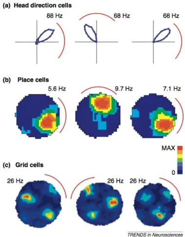
Figure 7: Landmark control of spatial signals. FromYoder and Taube(2011). Each panel displays the response of a different spatial cell type in rats to a 90◦ rotation of the salient visual landmark cue – a white sheet of cardboard attached along the inside wall of the enclosure (represented by a red arc in each panel). (a) The directional tuning curve of an anterior thalamic head direction cell, (b) the place field of a hippocampal place cell, and (c) the firing pattern of an entorhinal cortical grid cell show angular shifts of the spatial signal that approximate the amount of cue card rotation. Panel (a) is based on polar coordinates from Taube, 1995; (b) and (c) are based on data in Calton et al., 2003and Sargolini et al.,2006, respectively. Data shown in plots (b) and (c) have been smoothed to improve presentation. Peak firing rates are indicated for each plot.
1.3
Information processing in the presubicular
microcircuit
All spatial and non spatial information received by the presubiculum must be pro-cessed at the microcircuit level for building a local head direction signal. The con-tinuous attractor network model (Fig. 5) has often been considered for larger scales than microanatomy (see Sharp et al., 2001a) and few experimental data are avail-able to support or refute this model at the scale of the microcircuit (Taube, 2007). However, feedback inhibition (that might come from the local circuit) has been used in models to limit the overall neuronal activity (McNaughton et al., 2006). Fur-thermore, stellate cells in medial entorhinal cortex (putative grid cells) are mainly interconnected through disynaptic inhibition, which may constitute a recurrent in-hibitory attractor network able to generate grid cells dynamics (Couey et al., 2013). However, a recent study has cast doubt on this model (Buetfering et al., 2014), and suggests that interneurons may just control the gain of grid cell output.
The fact that head direction cells have more precise tuning properties in pre-subiculum than in sub-cortical areas (Tuning curves on Fig. 4), may reflect the refinement of the signal being relayed many times. Of course, the specificity of local head direction signals may be due to the specific features of information processing within each area, and cortical complexity may generate a more precise signal than subcortical nuclei. Head directional tuning properties could therefore depends on some features of the presubiculum, including neuronal electrophysiological intrinsic properties and morphologies, intra- and inter-laminar information flows, or putative modular organization such as cortical columns.
1.3.1
Anatomy and intrinsic excitability of presubicular
neu-rons
When I started my thesis work, little neuroanatomical data was available concerning the presubiculum. Funahashi and Stewart (1997a) partly characterized presubicu-lar neurons physiology and morphology, however, without providing an extensive description of neuronal diversity across all 6 layers (Fig. 8A). Pyramidal cells were found in layer 3 and 5, as well as stellate cells in layer 2 and 5 and all these cells were regular spiking neurons (Funahashi and Stewart, 1997a). However, this study did not give a clear view of presubicular diversity, due to a low number of recorded neu-rons. Another more recent study reported that deep layer neurons had a higher sag ratio (showing the Ih expression level) and that they adapted more than superficial
Figure 8: Cellular properties in presubiculum. A. Dendritic morphologies in
pre-and parasubiculum, fromFunahashi and Stewart(1997a). B. Intrinsic properties of deep and superficial layer neurons of dorsal presubiculum. These are regular spiking neurons; Ih expression seems higher in deep layer cells. C. Persistent activity can be induced in a presubicular neuron with an initial short depolarization that fires the cell, in the presence of a cholinergic agonist. B and C fromYoshida and Hasselmo(2009). D. A TTX-insensitive sodium current with slow activating and inactivating kinetics in presubicular principal neurons, fromFricker et al.(2009).
layer cells (Fig. 8B, Yoshida and Hasselmo, 2009).
Suprathreshold current pulses, in the presence of cholinergic receptor agonist carbachol (CCh; 10 µM) that "mimics" wakening, were able to trigger a persistent firing (less than 10 Hz) (Fig. 8C Yoshida and Hasselmo, 2009). The activation of ICAN (calcium-activated nonselective cationic current), was found to underly this
persisting firing in presubicular neurons. This study showed that head direction cell persistent firing may be supported at the cellular level, but its regulation should implicate an extrinsic inhibitory control, as the discharge persisted during tens of seconds.
Last, a TTX-insensitive sodium current with slow kinetics was revealed in super-ficial principal neurons, presumably expressed at distant sites from soma (Fig. 8D); Fricker et al., 2009). Such a current could support sustained firing in axon (Bean, 2007) and could amplify excitatory inputs in dendrites (Major et al., 2013).
1.3.2
Interlaminar, intralaminar and modular organization
Both interlaminar and intralaminar excitatory connection exist in the presubiculum (Funahashi and Stewart, 1997b). Recurrent excitation in deep layers (also in para-subiculum) can induce synaptic bursts in deep layers, but not in superficial layers, as few axonal collaterals are ascending (Funahashi and Stewart, 1997b, Fig. 9A, B). The lack of connectivity from deep to superficial layers was later confirmed with anterograde and retrograde tracings (Honda and Ishizuka, 2004, Fig. 9A, B). In contrast, many descending projections emerge from superficial layers and contact neurons in deep layers (Funahashi and Stewart, 1997b; Honda and Ishizuka, 2004, Fig. 9A, B, C). Axonal tracings of single layer 5 neurons showed that these cells had very diverse projection patterns including long septotemporal intrinsic projections (Honda et al.,2011, Fig. 9D). Different types of projections were highlighted: some of them were restricted to deep layers, hypothesized to send feedback information; other covered the whole presubicular plate and are thought to regulate the tempo-ral dynamics within a widespread neuronal population in the presubiculum (Honda et al.,2011).The presence of functional modules, such as cortical columns, was never demon-strated. Nevertheless, there are several peculiarities of the presubicular cortex in-dicating that this is a relevant question. Developmental cortical columns, clearly distinguishable during early post natal stage (Nishikawa et al., 2002), does not at-test that functional units exists in the adult. Nonetheless, anatomical modules were revealed in the adult. In monkey, "patches" were identified in superficial layers of presubiculum by labeling with several markers, including acetylcholinesterase,
cy-Figure 9: Presubicular intrinsic connectivity. A. Picrotoxin-induced burst revealed
by Local Field Potential (LFP) recordings in deep layers of the presubiculum. During this burst, deep layer neurons fire, whereas layer 2 cells remain silent, showing the non-propagation of activity from deep to superficial layers B. Antidromic stimulations revealed projections from superficial layers to deep layers in the presubiculum, but not from deep to superficial layers. aAP: antidromic action potential; rec: recording site; stim: stimulation. A and B were adapted from Funahashi and Stewart (1997b).C. Summary of associative and contralateral projections unraveled by Honda and Ishizuka (2004) using retrograde and anterograde tracings. D. A unique layer 5 pyramidal cell projection area along the septo-temporal axis which covered all layers of presubiculum. Sub: subiculum; PreS: presubiculum; ParS: parasubiculum; MEA: medial entorhinal area; LEA: lateral entorhinal area. FromHonda et al. (2011).
tochrome oxydase, myelin, calcium binding proteins paravalbumin, calbindin and calretinin (Ding and Rockland, 2001). The relevance of these patches is unclear, but they may be linked to some functional features. Indeed, a grid like arrangement of calbindin positive pyramidal cells exists in entorhinal cortex, and these neurons have been shown to be more theta modulated than the neighboring stellate, non-calbindin positive cells (Ray et al., 2014).
1.3.3
Input and output relays in the presubicular
microcircuit
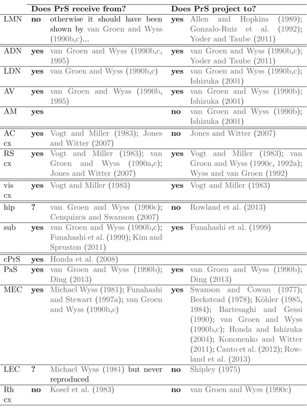
The microcircuit organization makes more sense when incoming and outgoing infor-mation pathways are understood. The long range connectivity of the presubiculum is summarized in table 1.1 and its known laminar organization is depicted on fig-ure 10. As the cellular neuroanatomy of the presubiculum has been unknown, it has been quite difficult to link the long range connectivity with the microcircuit elements. Some studies did identify input/output pathways in presubiculum. Only those identifying precise afferent targets or efferent populations will be presented here.
Presubicular superficial layer neurons constitute the major output toward ipsi-lateral and contraipsi-lateral medial entorhinal cortices (MEC), mainly targeting their superficial layers (Köhler, 1985; Honda and Ishizuka, 2004). Ipsilateral and con-tralateral projection originated from different neurons in superficial layers, and 20-30% of ipsilaterally projecting neurons are GABAergic (van Haeften et al.,1997). In addition, some projections emerge from deep layer neurons of the presubiculum and target deep layers and layer 1 of the ipsilateral MEC (Honda and Ishizuka, 2004). In presubiculum, layer 3 neurons that project to the entorhinal cortex receive di-rect inputs from retrosplenial cortex on their proximal dendrites (Kononenko and Witter,2011). Consequently, layer 3 neurons may relay the retroplenial information directly to the entorhinal cortex.
Two non-overlapping pathways toward lateral mammilary nucleus (LMN) and toward anterodorsal thalamus (ADN) have been identified in dorsal presubiculum (Yoder and Taube, 2011). The presubiculum-to-LMN projection originates exclu-sively from a thin layer of large pyramidal cells in layer 4, whereas cells that project to ADN are a heterogeneous population in deep layers. These pathways could con-stitute the cellular basis of landmark control of head direction cells in subcortical areas (see section 1.2.2;Yoder and Taube, 2011).
Figure 10: Laminar specificity of afferences and efferences in presubiculum.
See table 1.1 for references. B-F fromYoder and Taube(2011). Cholera toxin fluorophore conjugates were injected into LMN (B, Alexa fluor 488) and ADN (C, Alexa fluor 594) (scale bars=500 µm), migrated retrogradely in non-overlapping neuronal populations in presubiculum (D, scale bar=300 µm). Biotinylate dextran amines were injected into LMN or ADN, and retrogradely stained neurons were revealed in slice counterstained for thionin (E, scale bar=75µm and F, scale bar=50µm).
Does PrS receive from? Does PrS project to?
LMN no otherwise it should have been shown by van Groen and Wyss (1990b,c)...
yes Allen and Hopkins (1989); Gonzalo-Ruiz et al. (1992); Yoder and Taube (2011)
ADN yes van Groen and Wyss (1990b,c, 1995)
yes van Groen and Wyss (1990b,c); Yoder and Taube (2011)
LDN yes van Groen and Wyss(1990b,c) yes van Groen and Wyss (1990b,c); Ishizuka (2001)
AV yes van Groen and Wyss (1990b, 1995)
yes van Groen and Wyss (1990b); Ishizuka (2001)
AM yes no van Groen and Wyss (1990b);
Ishizuka (2001) AC
cx
yes Vogt and Miller (1983); Jones and Witter(2007)
no Jones and Witter (2007) RS
cx
yes Vogt and Miller (1983); van Groen and Wyss (1990a,c); Jones and Witter (2007)
yes Vogt and Miller (1983); van Groen and Wyss(1990c,1992a); Wyss and van Groen (1992) vis
cx
yes Vogt and Miller(1983) yes Vogt and Miller (1983) hip ? van Groen and Wyss (1990c);
Cenquizca and Swanson(2007)
no Rowland et al. (2013) sub yes van Groen and Wyss (1990b,c);
Funahashi et al.(1999);Kim and Spruston(2011)
yes Funahashi et al. (1999)
cPrS yes Honda et al.(2008)
PaS yes van Groen and Wyss (1990b); Ding(2013)
yes van Groen and Wyss (1990b); Ding (2013)
MEC yes Michael Wyss(1981);Funahashi and Stewart(1997a);van Groen and Wyss(1990b,c)
yes Swanson and Cowan (1977); Beckstead(1978);Köhler(1985, 1984); Bartesaghi and Gessi (1990); van Groen and Wyss (1990b,c); Honda and Ishizuka (2004); Kononenko and Witter (2011);Canto et al.(2012); Row-land et al.(2013)
LEC ? Michael Wyss (1981) but never reproduced
no Shipley (1975) Rh
cx
no Kosel et al.(1983) no van Groen and Wyss (1990c)
Table 1.1: Long range connectivity of the presubiculum (main connections). LMN:
lat-eral mammillary nucleus; ADN: anterodorsal thalamus; LDN: laterodorsal thalamus; AV: anteroventral thalamus ; AM: anteromedial thalamus; AC cx: anterior cingular cortex; RS: retrosplenial; vis: visual; hip: hippocampus; sub; subiculum; cPrS: contralateral pre-subiculum; PaS: parapre-subiculum; MEC: medial entorhinal cortex; LEC: lateral entorhinal cortex; Rh cx: perirhinal and postrhinal cortices.
2
|
How does a microcircuit work?
In the context of my PhD, this question would rather be “How does the presubic-ular microcircuit work?” Indeed, the network of the presubiculum has not been very well described so far. To understand how a network generates information, it is important to understand how the different elements of a given network partici-pate to information processing. The present chapter mainly focused on the current understanding of neuronal network physiology in a more general context.
My 3 first studies deal with neuronal properties: the morphology and intrinsic electrophysiology. I therefore go over the basis of neuronal excitability, explaining the biophysics behind the different parameters that I described in presubicular neu-rons (resistance, membrane time constant, the action potential shape. . . ) and why this is important for neuronal function.
In my last work, I am showing that the short-term dynamics of information transfer from pyramidal cells to Martinotti cells is uncommon. The synaptic trans-fer seems muted initially, but becomes efficient with repetitive high frequency stim-uli. This may involve a presynaptic form of plasticity in the axon and/or at the presynaptic terminals. I therefore reviewed the molecular mechanisms of plasticity present in the axons and at the presynaptic terminal in order to better discuss my results.
2.1
Many integrative levels in neuronal networks
The principle of a neuronal network was introduced for decades by Santiago Ramon y Cajal, who was the first to understand that neurons were anatomically and func-tionally distinct cellular units (Ramon y Cajal, 1899; Bullock, 2005; García-López et al., 2007). Drawings of Golgi stained neurons perfectly depicted the complexity of neuronal network anatomy, suggesting the direction of information flows between neurons and therefore providing a cartography of neuronal networks (Fig. 11, Ra-mon y Cajal,1899;Lorente de Nó,1934). However, knowing the diversity of neuronal morphologies, the location of neurons, and the anatomical pathway of information
Figure 11: Studying the network. Same neurons, different methods. A. Drawing
made by Ramon y Cajal of the hippocampal and parahippocampal network. (Ramon y Cajal, 1899). B. Lorente de No’s drawing of CA3 recurrent network (Lorente de Nó, 1934). The two authors deduced physiological pathways from their drawing, suggested with arrows. C. A way to address microcircuit connectivity through multiple patch clamp recordings, linking structure and physiology, fromCouey et al.(2013).
is only the first step to understand information processing in neuronal networks. These pathways are routes for information. As all the elements of the network pos-sess properties that can modulate the information, the nervous signal is not only passively transferred from one neuron to another.
Let’s take the example of sensory thalamo-neocortical projections. Excitatory information from the ventrobasal thalamus principally targets layer 4 (L4) neurons, which subsequently distribute intra-laminar (within L4) and inter-laminar (in layer 3 and 5) excitation (Lübke and Feldmeyer,2007). Ventrobasal thalamus neurons di-rectly project onto layer 4 fast spiking (FS) interneurons, which fire with very short latency due to the high amplitude of the synaptic responses and their fast integrative properties (Gabernet et al.,2005;Cruikshank et al., 2007, 2010); FS cells therefore provide feedforward inhibition onto L4 cells and enhance their temporal precision by defining an early and short window for excitation (Swadlow and Gusev, 2002; Cruikshank et al., 2007; Gabernet et al., 2005). In contrast, low-threshold spiking interneurons (LTS), putative dendrite-targeting interneurons, are not recruited by ventrobasal thalamic projections (Cruikshank et al., 2010), so they do not provide feedforward inhibition in L4, at least for the thalamic pathway. L4 excitatory neu-rons are able to activated LTS and PV cells, as a feedback inhibitory control ( Beier-lein et al.,2003), however their synaptic recruitment has very distinct dynamics. FS cells provide an initial feedback control that attenuates with time, whereas LTS are recruited later during high frequency stimuli (Beierlein et al., 2003). Moreover, as they target specific subcellular compartments they do not have the same impact, as somata targetting PV cells may directly regulate outputs whereas dendrite-targeting LTS neurons modulate inputs.
Even this oversimplified view of layer 4 activation by thalamic axons gives an idea of the complexity of computation in a microcircuit, depending on the targets of afferent axons, the intrinsic connectivity of local networks, the strength and dy-namics at a given synapse or the integrative properties of excitatory and inhibitory neurons.
2.2
Neuronal intrinsic excitability
Excitability properties allow neurons to continuously make decisions as they re-ceive information. All neurons are not alike and can be defined by their intrinsic excitability that reflects their responsiveness to incoming information. Knowledge of neuronal intrinsic excitability is, thus, essential for a better understanding of microcircuit information processing (Toledo-Rodriguez et al., 2005).
2.2.1
Resting membrane potential
Resting membrane potential (Vrest) is the neuronal membrane potential when the
neuron does not receive any information from afferences. Vrest is determined by
plasma membrane total conductance that mainly depends on K+, Na+ and Cl− ion movements. The membrane basal conductance is called leak conductance GL; Vrest
value can be expressed by the Goldman-Hodgkin-Katz equation:
Vrest = 58log10
pK[K+]e+ pN a[N a+]e+ pCl[Cl−]i
pK[K+]i+ pN a[N a+]i+ pCl[Cl−]e
This value is negative, generally close to -70 mV, and is mainly due to the high conductance of potassium ions, that brings the membrane potential close to EK+;
Na+ and Cl− ions also participate but their impact is more limited due to their lower permeability at this potential.
2.2.2
Neuronal passive properties
The elements of the neuronal plasma membrane can be seen as elements of an electrical circuit. The lipid bilayer is an insulator, separating external and internal conductive media; it is also a capacitor, its capacitance C being proportional to membrane surface. Currents can cross through the opened ionic channels: those are resistors and the total resistance equals the reciprocal of the sum of all specific conductances. At rest, membrane electrical properties can be modeled by a resistor (membrane resistance Rm) and capacitor (membrane capacitance Cm) put in parallel
and under the electromotive force Vrest. When a constant current Istim is applied to
the circuit, the parallel RC circuit passively reacts according to Rm and Cm values.
Istim= ICm+ IRm ; with ICm = Cm dV dt and IRm = V Rm Then, Istim= Cm dV dt + V Rm