The MIT Faculty has made this article openly available.
Please share
how this access benefits you. Your story matters.
Citation
Hassan, Musa A. et al. "Comparative ribosome profiling uncovers a
dominant role for translational control in Toxoplasma gondii." BMC
Genomics 18 (December 2017): 961 © 2017 The Author(s)
As Published
http://dx.doi.org/10.1186/s12864-017-4362-6
Publisher
BioMed Central
Version
Final published version
Citable link
http://hdl.handle.net/1721.1/114431
Terms of Use
Creative Commons Attribution
R E S E A R C H A R T I C L E
Open Access
Comparative ribosome profiling uncovers
a dominant role for translational control
in Toxoplasma gondii
Musa A. Hassan
1,2*, Juan J. Vasquez
3,8, Chew Guo-Liang
4, Markus Meissner
5,6and T. Nicolai Siegel
3,6,7Abstract
Background: The lytic cycle of the protozoan parasite Toxoplasma gondii, which involves a brief sojourn in the extracellular space, is characterized by defined transcriptional profiles. For an obligate intracellular parasite that is shielded from the cytosolic host immune factors by a parasitophorous vacuole, the brief entry into the extracellular space is likely to exert enormous stress. Due to its role in cellular stress response, we hypothesize that translational control plays an important role in regulating gene expression in Toxoplasma during the lytic cycle. Unlike
transcriptional profiles, insights into genome-wide translational profiles of Toxoplasma gondii are lacking.
Methods: We have performed genome-wide ribosome profiling, coupled with high throughput RNA sequencing, in intracellular and extracellular Toxoplasma gondii parasites to investigate translational control during the lytic cycle. Results: Although differences in transcript abundance were mostly mirrored at the translational level, we observed significant differences in the abundance of ribosome footprints between the two parasite stages. Furthermore, our data suggest that mRNA translation in the parasite is potentially regulated by mRNA secondary structure and upstream open reading frames.
Conclusion: We show that most of the Toxoplasma genes that are dysregulated during the lytic cycle are translationally regulated.
Keywords: Ribosome profiling, RNA-sequencing, Translation efficiency, Toxoplasma gondii, Apicomplexan Background
All living organisms are constantly exposed to a variety of biological stress, which may include limiting nutrient availability, oxidative stress, temperature shock, DNA damage and infection. Consequently, organisms show re-markable regulatory plasticity that allows them to thrive under different, sometimes harsh, environmental condi-tions [1, 2]. Historically, due to the relative ease of obtain-ing global transcript abundance estimates, most studies quantify fluctuations in mRNA abundance to gain insights into organismal response to stress [3, 4]. However, the catalogue of expressed genes and proteins is modulated at various steps, including mRNA splicing, export, stability,
translation, and protein degradation [5]. Consequently, transcript abundance rarely mirrors cellular protein levels [6]. Although the relative contribution of each of these steps in the gene-expression pathway is equivocal, mRNA translation is known to play a significant role in modulat-ing cellular protein levels [7, 8]. Indeed, translational con-trol of gene expression is known to provide opportunities for controlling spatial and temporal protein distribution [9]. Furthermore, because most eukaryotic mRNAs can be detected in cells at least 2 h after expression [10], com-pared to de novo transcription, regulating the translation of the available mRNAs provides a mechanism to rapidly adjust cellular protein levels in response to drastic changes in the environment or developmental stages [11, 12]. In fact, most translationally regulated mRNAs are known to encode proteins that regulate important cellular processes such as stress response, development, and synaptic trans-mission [7].
* Correspondence:musa.hassan@roslin.ed.ac.uk
1
Division of Infection and Immunity, The Roslin Institute, University of Edinburgh, Edinburgh, UK
2The Centre for Tropical Livestock Genetics and Health, The Roslin Institute,
University of Edinburgh, Edinburgh, UK
Full list of author information is available at the end of the article
© The Author(s). 2017 Open Access This article is distributed under the terms of the Creative Commons Attribution 4.0 International License (http://creativecommons.org/licenses/by/4.0/), which permits unrestricted use, distribution, and reproduction in any medium, provided you give appropriate credit to the original author(s) and the source, provide a link to the Creative Commons license, and indicate if changes were made. The Creative Commons Public Domain Dedication waiver (http://creativecommons.org/publicdomain/zero/1.0/) applies to the data made available in this article, unless otherwise stated.
Toxoplasma gondiiis an obligate intracellular apicom-plexan that infects virtually all warm-blooded verte-brates. In the definitive feline host, Toxoplasma undergoes sexual recombination, but reverts to asexual reproduction in the intermediate host, which includes humans. Asexual reproduction in Toxoplasma is charac-terized by the rapidly dividing tachyzoite stage. However, in response to host-derived stress factors, such as im-mune response, the rapidly dividing tachyzoites convert to the semi-dormant encysted bradyzoites and establish lifelong chronic infections in the central nervous system and muscle tissues of the host [13, 14]. Establishment of chronic infection is important for the re-entry of the parasite into the definitive host, and for horizontal trans-mission within intermediate hosts, through the preda-tion and consumppreda-tion of food products from chronically infected hosts, respectively [15]. The tachyzoite-to-bradyzoite conversion reportedly mirrors a stress re-sponse [2] and does not only involve significant changes in the parasite physiology and morphology, but also is accompanied by altered gene expression profiles [16]. During the lytic cycle the parasite invades a host cell, replicates, and then lyses out of the host cell before in-fecting a new host cell. This process temporarily exposes the parasite to the extracellular milieu. The extracellular viability of the parasite is reported to decrease dramatic-ally between 6 and 12 h after egress [17], indicating the level of biological stress induced on the parasite by host factors. Indeed, transcriptional data on most Toxoplasma strains have revealed stage-specific expression of several genes, such as surface antigens, stress response genes, virulence genes, and metabolic enzymes [13, 18, 19]. Consequently, regulating transcript abundance, and by extension their protein products, is key in regulating Toxoplasma developmental stages and intercellular transmission.
Translational regulation of gene expression has emerged as a key factor in the biology of apicomplexan parasites [20–23]. In Plasmodium, translational regula-tion is reported to modulate stage conversion and host-parasite interactions [20, 21]. For example, while Pb2 transcripts, a surface antigen, can be detected in Plasmo-dium bergheifemale gametocytes, the translation of Pb2 mRNA occurs only when the parasite is in the mosquito gut [24]. In Toxoplasma, genetic perturbation of the eukaryotic elongation factor 2 alpha (eIF2α), an import-ant component of the translation initiation complex, af-fects extracellular viability. Phosphorylated eIF2α is essential for transferring the initial methionyl tRNA (Met-tRNAi) to the 40S pre-initiation complex [25]. However, when phosphorylated at a regulatory serine (serine-51), eIF2α is unable to activate Met-tRNAi and global translation is diminished [25]. Toxoplasma para-sites expressing eIF2 (TgIF2α) with a mutation on the
regulatory serine (serine-71) are reported to exhibit de-creased extracellular viability [26]. Lower expression levels of eIF4, another translation initiation factor, has been observed in bradyzoites and attenuated Toxo-plasma strains [2, 23, 24, 26]. Finally, the endoplasmic reticulum (ER) stress response in Toxoplasma is charac-terized by preferential translation of a subset of genes, including the transcriptional regulator AP2 [23, 27]. This is particularly important since the integrity of the para-site ER is pivotal for the proper folding of essential pro-teins required for parasite invasion, immune evasion and the establishment of chronic infection [24]. Thus, it is plausible that the viability, pathogenesis, and transmission of Toxoplasma are dependent on its ability to recognize and translationally respond to host-derived stress.
Genome-wide insights on translational control and the underlying molecular factors that regulate mRNA trans-lation in Toxoplasma are largely unknown. Here, we ac-cess the translational landscape in Toxoplasma gondii and determine its impact on intercellular parasite trans-mission. To do this, we have used ribosome profiling to capture genome-wide translational profiles of intracellu-lar and extracelluintracellu-lar Toxoplasma parasites infecting hu-man foreskin fibroblasts. Our data reveal a putative role for translational control in regulating parasite gene expression during the lytic cycle. Additionally, our data revealed variable translational efficiency of several dys-regulated Toxoplasma mRNAs, such as the mRNAs en-coding dense granules, which are known to be spatially secreted during the lytic cycle. Finally, our data suggest that that mRNA secondary structure, putatively affect mRNA translation in Toxoplasma. These results not only provide greater insights into Toxoplasma gene regulation, but also provide a resource and template for elucidating the function of translational control in Toxoplasma biology. Finally, the ribosome footprints, will provide an additional resource for annotating Toxo-plasmatranscript features.
Results
Generation of mRNA profiles and ribosome footprints in
intracellular and extracellularToxoplasma
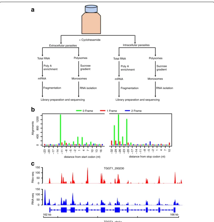
To investigate genome-wide transcriptional and transla-tional status in Toxoplasma during the lytic cycle, we performed RNA sequencing (RNA-seq) and ribosome profiling on two biological replicates of extracellular and intracellular parasites as previously described [4] (the ex-perimental layout is depicted in Fig. 1a).
The basic concept of ribosome profiling is that actively translated mRNAs are protected from ribonucleases by the decoding ribosomes. However, other classes of RNA-binding proteins can protect mRNA from nucleases. Therefore, the presence of sequencing reads derived
necessarily infer active translation. Since ribosomes decode mRNA by reading 3-nucleotides (3-nt) at a time, 3-nt periodicity on ribosome footprints is often used to distinguish ribosome protected RNA from other classes of nuclease resistant RNAs [3, 28–30]. Therefore, to in-crease coverage, we pooled ribosome-protected RNA
footprints from the two biological replicates for each sample, and used sub-codon resolution to call high con-fidence translated open reading frames (ORFs) in canon-ical Toxoplasma coding sequences. To do this, we used RiboTaper, a ribosome profiling analysis program that defines the peptidyl-site (P-site; the second tRNA entry
a
b
c
Extracellular parasites Poly A enrichment + Cyclohexamide Intracellular parasites Total RNA PolysomesSucrose gradient
Fragmentation RNA isolation Library preparation and sequencing
Monosomes mRNA
Total RNA Polysomes Poly A
enrichment
Sucrose gradient
Library preparation and sequencing Fragmentation RNA isolation Monosomes mRNA 162 kb 166 kb 0 50 100 150 Ribo-seq 0 50 100 150 RNA-seq TGGT1_chr1a TGGT1_293230 0 400 800 1200
distance from start codon (nt)
Alignments
−23 −20 −17 −14 −11 −8 −5 −2
1 4 7 10 13 16
distance from stop codon (nt)
−32 −29 −26 −23 −20 −17 −14 −11 −8 −5 −2
1 4 7 10
0 Frame 1 Frame 2 Frame
Fig. 1 Ribosome profiling of Toxoplasma gondii. a The experimental design. Cyclohexamide was added to the flasks for ~10 min prior to collecting the medium containing extracellular parasites and the host-cell monolayer was syringe lysed to release intracellular parasites. Chemically fragmented mRNA and sucrose gradient fractionated monosomes were used to prepare RNA-seq and Ribo-seq libraries, respectively, in parallel. b P-site tracks, colour-coded per frame, were created for all annotated exons in RiboTaper. c Ribo-seq (red peaks) and RNA-seq (blue peaks) read pile-up, presented as Fragment per kilobase exon per million reads (FPKM), on a multi-exonic gene. Exons are shown as blue blocks while introns are represented by blue lines
site linked to the growing polypeptide chain) of ribosome-protected RNA sequencing (Ribo-seq) reads mapping over annotated transcripts [3]. Henceforth, un-less otherwise stated, all analyses on extracellular or intracellular samples are based on pooled Ribo-seq or RNA-seq data. Because 3-nt periodicity often vary be-tween different Ribo-seq read lengths, we performed sub-codon resolution on 25–30-nt reads, which is within the range of 80S ribosome-protected RNA lengths [5]. We observed a strong 3-nt periodicity in 29-nt foot-prints, with up to 12-nt upstream of the AUG start site covered by ribosome footprints (12-nt offset) (Fig. 1b). Similar offsets were obtained using Riboprofiling [31], a Bioconductor package for processing Ribo-seq data (Additional file 1: Figure S1). Unlike RNA-seq reads that sometimes contain reads aligning to intronic regions, ribosome footprints mapped predominantly to annotated Toxoplasma protein coding regions (Fig. 1c). Therefore, the ribosome footprints in the current experiment are mostly derived from ribosome-protected nuclease-resistant mRNA fragments and can be used to accurately quantify translation in Toxoplasma.
Ribosome profiling confirms translation of annotated
CDSes and identifies novel translated ORFs inToxoplasma
RNA-seq alone cannot distinguish translated from non-translated transcripts. Additionally, it is not clear whether some annotated non-coding RNAs contain translated small open reading frames. These problems are exacerbated in non-model organisms, such as Toxo-plasma, with incompletely annotated genomes. Because Ribo-seq captures ribosome-engaged mRNAs, it is often used to not only estimate the translation efficiencies of annotated coding regions, but also to identify novel translated ORFs. Consequently, we used RiboTaper, as previously described [3], to identify translated ORFs based on 3-nt periodicity and P-site positions in the expressed Toxoplasma genes. Because the current anno-tation of Toxoplasma gene structures (ToxoDB.org; GT1 v28 [32]) is incomplete, and RiboTaper classifies ORFs based on known coding regions, we initially used RNA-seq reads (~500 million paired-end reads from this and a parallel study [33]) to update GT1 gene structures. To do this, we performed genome-guided transcript assem-bly using Trinity [34], followed by transcript structure resolution using the Program to Assemble Spliced Alignments (PASA) [35], as previously described [36]. Subsequently, we updated the structures of 6442 tran-scripts, mostly due to the addition of 5′ and 3′ UTRs (mean lengths of 435-nt and 508-nt, respectively) (Fig. 2a). Next, we used RiboTaper and identified 4224 ORFs in 4195 genes based on the updated transcript structures. Noteworthy, the identification of ORFs in RiboTaper is based on codon resolution on the Ribo-seq
reads that map to annotated transcript features rather than the simple presence of ribosome footprints. Thus, the number of translated ORFs identified by this ap-proach may be lower than the actual number of genes with ribosome footprints. Besides canonical ORFs, we identified 172 novel ORFs, mainly due to the alternative splicing of annotated transcripts (Fig. 2b), PASA-updated new transcripts structures (Fig. 2c), or novel transcripts (Fig. 2d). Therefore, by using ribosome foot-prints, we not only provide a greater resolution of the canonical ORFs to include alternative isoforms, but also identify novel ORFs in Toxoplasma (GT1).
Steady-state mRNA and translation efficiency in intracellular and extracellular parasites
We sought to evaluate global translational divergence in intracellular versus extracellular type I Toxoplasma parasites. Initially, we used HTSeq [37] to obtain raw read counts from the uniquely mapped RNA-seq and Ribo-seq reads, which were then normalized (Normal-ized Read Counts, NRC) in DESeq2 [38] to adjust for variation in sequencing depths across samples. Even though some genes were lowly expressed, approxi-mately 7065 genes (83% of the ~8460 genes annotated in the GT1 genome (v28) were expressed (average RNA-seq NRC > 5 across samples) (Additional file 2 A). Of the expressed genes, 6508 had ribosome footprints (average Ribo-seq NRC > 5 across samples), suggesting that 557 transcripts are non-coding or poorly translated in our parasite populations (Additional file 2 B). Inter-estingly, 274/557 (> 50% of the potentially non-coding or poorly translated genes) had an average RNA-seq NRC > 10 (mean NRC = 41.18; SEM = ±3.48), suggesting that these genes are expressed above background levels (which we arbitrarily set at NRC < 5) but are either translationally repressed in these parasite populations or non-coding. The protein products for most of these 274 genes are annotated in ToxoDB as “hypothetical”, but also included the KRUF proteins, which are encoded from a highly expanded gene family in the GT1 strain [39]. Also included in the 274 poorly trans-lated or non-coding genes was the Toxoplasma transla-tion initiatransla-tion factor 2 (TgIF2K-C), which is required for the parasites’ response to intracellular glutamine starvation in human cells [40]. These 274 transcripts were functionally enriched for, among others, “cell ad-hesion” (Bonferroni P value = 3.58e-4) and “microtubule motor activity” (Bonferroni P value = 9.96e-4). Although they are included in the current GT1 genome annota-tion, 27 of the 274 genes did not have any correspond-ing proteomic data in ToxoDB [32], suggestcorrespond-ing that they are non-coding. On the other hand, 83 transcripts exhibited low abundance with an average RNA-seq NRC < 5 (mean NRC = 3.14; SEM = ±0.14), but had
a
b
c
d
TGGT1_chrIX 4.423 mb 4.431 mb 0 200 400 Ribo-seq 0 200 400 RNA-seq TGGT1-292220 729 kb 731 kb 0 1000 2000 Ribo-seq 0 1000 2000 RNA-seq TGGT1_chrIb TGGT1_208450 4.463 mb 4.464 mb 0 100 200 Ribo-seq 0 100 200 RNA-seq TGGT1_292275 TGGT1_chrIX 2.5 kb 3.5 kb 0 1000 Ribo-seq 0 RNA-seq AAQM03000654 1000A PASA-updated transcript structure
Novel ORF arising from alternative splicing
Novel ORF arising from mis-annotated transcript
Novel ORF arising from a novel transcript
average Ribo-seq NRC > 5 (mean NRC = 13.17; SEM = ±3.52) (Additional file 2 C). These 87 genes included the SAG-related sequence (SRS) gene family that are implicated in Toxoplasma virulence in mice [41].
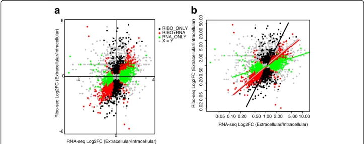
Next, we compared differences in mRNA abundance and ribosome occupancy between the intracellular and extracellular parasites. Using a Benjamini-Hochberg False Discovery Rate (FDR)≤ 10%, we identified three classes of differentially regulated genes: 1) 891 genes that varied both at the level of transcript abundance and ribosome occupancy i.e. concordant (RNA + RIBO) (Additional file 2 D), 2) 645 genes that varied only at
the levels of mRNA abundance (RNA-ONLY)
(Additional file 2 E), and 3) 1324 genes that varied only at the level of ribosome occupancy (RIBO-ONLY) (Fig. 3a and Additional file 2 F), indicating that many of the genes that are dysregulated in Toxoplasma during the lytic cycle are regulated at the translational level. To determine the overall contribution of translation in regulating gene expression during Toxoplasma’s lytic cycle, we used a standardized major-axis estimation
(SME) [42] analysis to calculate the slopes of fold changes in RNA-seq or Ribo-seq NRCs between intracellular and extracellular parasites. Unlike the RIBO + RNA transcripts, where the slope approached 1 (slope = 1.15), indicating the co-occurrence of changes in transcript abundance and ribosome occupancy, the slope for RIBO-ONLY transcripts (slope = 2.87) was sig-nificantly (P value < 2.22e-16) greater than 1 (Fig. 3b), confirming that many differences in gene expression between the intracellular and extracellular parasites occur at the translation level.
Next, we calculated differences in translation efficiency (TE) for each expressed transcript between extracellular and intracellular parasites using Ribodiff [43]. At a Benjamini-Hochberg FDR≤ 10%, we identified differen-tial TE in 834 genes in intracellular versus extracellular parasites (Additional file 2 G). Because of the potential variation in mRNA between intracellular and extracellu-lar parasites, which in the absence of a spike-in control during RNA sequencing may skew the data, we comple-mented the ribodiff protocol by ranking the genes based
(See figure on previous page.)
Fig. 2 Ribosome profiling reveals novel and annotated open reading frames. a Shown are examples of PASA-updated Toxoplasma transcript structure annotations (Black) and the corresponding current ToxoDB transcript annotation (Blue). A) The PASA-updated transcript is due to the addition of 5′/3′ UTRs (red arrows). Prediction of ORFs based on the PASA-updated transcript structures in RiboTaper identified canonical and novel ORFs. The novel ORFs were mainly due to; b alternative splicing of annotated transcripts (skipped exon, red arrow), c potentially mis-annotated transcripts structures, and d novel transcripts. The RiboTaper-predicted ORFs in B-D are presented as red blocks. In all the figures, Ribo-seq and RNA-seq read coverage on each transcript is shown in Fragment per kilobase exon per million reads (FPKM)
-6 0 6 -4 0 4
RNA-seq Log2FC (Extracellular/Intracellular)
Ribo-seq Log2FC (Extracellular/Intracellular)
X = Y RIBO_ONLY RIBO+RNA RNA_ONLY 0.05 0.10 0.20 0.50 1.00 2.00 5.00 10.00 0.02 0.05 0.20 0.50 2.00 5.00 20.00 50.00
RNA-seq Log2FC (Extracellular/Intracellular)
Ribo-seq Log2FC (Extracellular/Intracellular)
a
b
Fig. 3 Translation regulation is predominant in Toxoplasma gondii. a Although most transcripts showed no differential regulation (grey dots n = 4332) between the intracellular and extracellular parasites, several dysregulated genes were significantly (Benjamini–Hochberg FDR ≤ 0.1) regulated at the level of mRNA abundance only (green dots, RNA-ONLY, n = 650), ribosome occupancy only (black dots, RIBO-ONLY; n = 1339), and at the level of both mRNA abundance and ribosome occupancy (red dots, RIBO + RNA, n = 899). Differences in translational efficiency in intracellular versus extracellular parasites were calculated in Ribodiff on transcripts with average RNA-seq DESeq2 normalized read counts≥ 5. b The slope for red spots approached 1 (x = y), indicating equal fold changes in gene regulation at the level of mRNA abundance and ribosome occupancy between the intracellular and extracellular parasites (standardized major-axis estimation; SMA). Significant (P < 2.22e-16) divergence of black and red slopes demonstrate differences in gene regulation at the level of ribosome occupancy (RIBO-ONLY) and mRNA abundance (RNA-ONLY), respectively
on the z-scores of TE in each parasite population. We considered genes with RNA-seq NRC≥ 5 (7065 genes) and at least two standard deviations above or below the mean TE in each population as translationally up- or down- regulated, respectively. By this metric, 868 genes were translationally down-regulated while 119 genes were up-regulated in intracellular parasites. On the other hand, 1004 and 236 genes were down- and up-regulated, respectively, in extracellular parasites. Of the lated genes, 344 and 556 genes were exclusively dysregu-lated in intracellular and extracellular parasites, respectively (not deviating from the mean or dysregu-lated in the opposite directions in the two populations, e.g. up-regulated in intracellular but down-regulated in extracellular parasites). The “sporozoite development protein (TGGT1_257010)” and “BT1 family protein (TGGT1_236020)” genes were the most down- (z-score =−5.0; Log2TE =−6.03) and up-regulated (z-score = 4.79;
Log2TE = 4.15), respectively, in intracellular parasites. In
extracellular parasites, “the transporter, major facilitator family protein (TGGT1_266870)” and “CMGC kinase, CDK family (TGGT1_253580)” genes were the most down- (z-score =−5.85; Log2TE = 6.43) and up-regulated
(z-score = 6.13; Log2TE = 5.55), respectively. Although
dense granules are secreted by intracellular parasites [44], the translation efficiency for genes encoding these proteins (GRA1, GRA4, and GRA7) was up-regulated in extracellular parasites. Additionally, the translation of genes encoding the alveolin domain-containing inner membrane complex (IMC) proteins (IMC1, IMC4, IMC6, and IMC10), which are required during intracel-lular Toxoplasma cell division [45], were up-regulated in extracellular parasites.
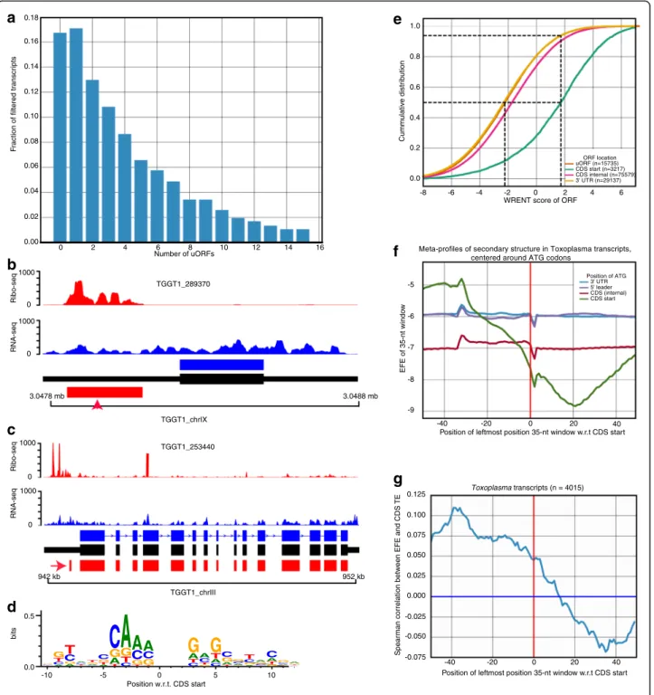
MostToxoplasma transcripts contain open reading frames
(ORFs) at their 5′ untranslated regions
Besides translation at canonical protein coding sequences (CDSes), ribosome profiling can reveal novel coding sequences, including coding sequences at the 5′ and 3′ untranslated regions (upstream and downstream ORFs, uORFs and dORFs, respectively) [3, 28]. Because the prevalence and translation regulatory potential of uORFs is largely unknown in Toxoplasma, we used a support vec-tor classifier [29] to identify translated uORFs. Based on the presence of a start and an in-frame downstream stop codon, we observed a high prevalence of uORFs in Toxo-plasma, with some transcripts having > 4 non-overlapping uORFs (Fig. 4a). From 4577 transcripts with annotated 5′ UTRs of lengths ≥ 20-nt, we identified uORFs in 3348 (73%). Similar abundance of uORFs has also been reported in Plasmodium falciparum [21]. We filtered the tran-scripts further to 2770 (translated uORFs) based on the presence of ribosome footprints, 3-nt periodicity on Ribo-seq reads, and a minimum level of expression of the
cognate transcript (Fragment per kilobase exon per mil-lion reads; FPKM≥ 1).
In other eukaryotes, uORFs are not only prevalent, but also regulate translation of cognate downstream CDSes [20, 28]. Consistent with the reported uORF-mediated repression of translation at canonical CDSes [46–48], we observed individual examples of highly-translated uORFs upstream of their cognate lowly-translated CDSes (Fig. 4b-c). Because, sequence and mRNA secondary structure can modulate translation [49–51], we per-formed linear regression with these features against translation efficiency in Toxoplasma, as previously de-scribed [28]. Briefly, we used annotated Toxoplasma CDSes lacking uORFs as a training set to define the se-quence motif that promotes translation initiation (initi-ation context), by weighting the contribution of position-specific scoring matrix (PSSM) to translation efficiency of individual transcripts (Fig. 4d). Next, we used the PSSM to score initiation sequences in individ-ual transcripts that contain uORFs (weighted relative en-tropy, WRENT) (See Methods). Relative to canonical CDSes, WRENT scores at uORFs were generally un-favourable to translation initiation (Fig. 4e). We then cal-culated the secondary structure ensemble free energy [52], using the ViennaRNA package [53], in a 35-nt slid-ing window across entire transcripts to evaluate the ef-fect of mRNA secondary structure on translation. Unlike humans and mice [28], Toxoplasma transcripts exhibited an unstable secondary structure before the CDS start codon and a more stable secondary structure after the CDS start codon (Fig. 4f ). Moreover, the stability of the secondary structure at these regions correlates with translation efficiency of the transcripts (Fig. 4g, and Additional file 3: Figure S2 and Additional file 4: Table S1). Thus, most uORFs in Toxoplasma are not efficiently translated and mRNA secondary structure putatively regulate translation efficiency in Toxoplasma.
Discussion
During the lytic cycle, Toxoplasma frequently transitions between an intracellular and extracellular niche, that is characterized by a variety of molecular changes in the parasite, including distinct transcriptional profiles [54]. Although components of the translation initiation complex, such as eIF2α, reportedly modulate stress re-sponse, extracellular survival and, virulence in Toxo-plasma [23, 26], global translational changes during Toxoplasma lytic cycle are largely unknown. Here, we used ribosome profiling to reveal that translational regu-lation of gene expression is prominent during Toxo-plasmas’ lytic cycle. Additionally, our data suggest mRNA secondary structure potentially regulate transla-tion in Toxoplasma. Even though most of the genes expressed during the lytic cycle are known to exhibit a
0.16 0.14 0.12 0.10 0.08 0.06 0.04 0.02 0.00 0 2 4 6 8 10 12 14 16 Number of uORFs F raction of filtered tr anscr ipts 0.18 3.0478 mb 3.0488 mb 0 1000 Ribo-seq 0 1000 RNA-seq TGGT1_289370 TGGT1_chrIX 942 kb 952 kb 0 1000 Ribo-seq 0 1000 RNA-seq TGGT1_253440 TGGT1_chrIII Position w.r.t. CDS start 0.0 0.5 bits -10 A CT G A G CTA GT -5 A T G C T A G
C
C T GA
T G CA
G CA
0 CT AG
G T CA 5 C A TG
T A G C A C G G CT C G A 10 GT A C GA ORF location uORF (n=15735) CDS start (n=3217) CDS internal (n=75579) 3' UTR (n=29137)Meta-profiles of secondary structure in Toxoplasma transcripts, centered around ATG codons
Position of ATG 3' UTR 5' leader CDS (internal) CDS start Spear m an correlation betw
een EFE and CDS
TE 0.125 0.100 0.075 0.050 0.025 0.000 -0.025 -0.050 -0.075
Position of leftmost position 35-nt window w.r.t CDS start
Toxoplasma transcripts (n = 4015)
-40 -20 0 20 40
Position of leftmost position 35-nt window w.r.t CDS start
EFE of 35-nt windo w -40 -20 0 20 40 -5 -6 -7 -8 -9
WRENT score of ORF
-8 -6 -4 -2 0 2 4 6 1.0 0.8 0.6 0.4 0.2 0.0 Cumm ulativ e distr ib ution
a
b
c
d
e
f
g
Fig. 4 Open reading frames in 5′ leader sequences (uORFs) are abundant in Toxoplasma. a Based on the presence of a start and a downstream in-frame stop codon, some Toxoplasma transcripts contain more than 14 non-overlapping uORFs. b-c Individual Toxoplasma transcripts with an upstream translated uORFs (red arrow) exhibit reduced ribosome occupancy at the cognate canonical protein coding sequences. Ribo-seq (red peaks) and RNA-seq (blue peaks) read pile-up on ToxoDB (blue) and PASA-updated (black) transcript structures. d Weighted position-specific scoring matrices (PSSMs) around (±10 nucleotides) annotated ATG of transcripts lacking uORFs. Vertical axis represents weighted relative entropy (WRENT). e Cumulative WRENT scores around ATG start codons along the coding transcripts. Dotted lines indicate median WRENT scores of uORF and CDS, and the proportion of uORFs (~90%) with WRENT scores less than the median CDS WRENT score. f Predicted secondary structure ensemble free energies (EFEs) in a sliding 35-nt window on 5′ UTRs, CDSes, and 3′ UTRs. A more negative EFE indicates more stable secondary structures. g The secondary structure EFE correlates with translation efficiency in CDSes. See also Additional file 3: Figure S2 and Additional file 4: Table S1
cyclic expression pattern coinciding with the different cell cycle stages [55], we show that the expression and translation of most of these genes are not temporally or spatially synchronized during the lytic cycle. However, since parasite replication and egress is not synchronized among individual parasites, it is impossible to decipher the translational changes that occur as the parasite adapts to the extracellular microenvironment. Thus, it is not clear whether the differences in translation efficien-cies between intracellular and extracellular parasites ob-served in this study are maintained throughout the lytic cycle. With single-cell or time course analysis of the Toxoplasma“translatome”, we may be able to show fluc-tuations in translation as the parasites egress or re-infect host cells.
Interestingly, Toxoplasma transcripts exhibited less stable RNA secondary structure before the ATG start site. Similar reduction in RNA secondary structure have been reported in zebrafish [28]. In contrast to CDSes, this switch from unstable to stable secondary structure around the initiation codon was not observed in uORFs. This distinction in the initiation context of uORFs and CDSes, in terms of both sequence and secondary struc-ture suggests that these two feastruc-tures are important for start site selection in Toxoplasma. uORFs have been shown to be prevalent and regulatory in a variety of organisms, including Apicomplexans [21, 56]. However, their prevalence and regulatory potential in Toxoplasma is largely unknown. We show that, while uORFs are prevalent in Toxoplasma, their translation is not favoured, probably due to selection at their initiation contexts (sequence and secondary structure). Neverthe-less, we observed individual cases where high translation at an uORF correlates with weak translation at a cognate downstream CDS, which raises interesting questions that are worthy of further investigations. For example, is the translation of uORF unfavourable at all the developmen-tal stages? How is the translation of uORFs regulated in Toxoplasma? Additionally, the mechanisms that regulate translation efficiency in Toxoplasma, which are equivo-cal, are worthy of further investigation. High ribosome occupancy may not be related to high rates of translation but rather ribosome pausing [57], which can be caused by long stretches of rare codons, high mRNA secondary structure, or interactions of the growing polypeptide chain with the ribosome [58, 59]. Overall, it is worthy in-vestigating the role of translational control in modulat-ing Toxoplasma strain differences in virulence, adaption to variable host genetic background or host cell activa-tion status.
Conclusion
The results presented in this work reveals key aspects of translational control in Toxoplasma gondii during the
lytic cycle. We show that many dysregulated genes are translationally regulated during intercellular parasite transmission and that uORFs are prevalent, although not translationally favoured in Toxoplasma gondii. We an-ticipate that this work will be the basis for future re-search on translational regulation in the different development stages of the parasite and host cell microenvironments.
Methods
Parasite culture, ribosome isolation and, sequencing libraries
Toxoplasma gondii was maintained in the laboratory by serial passage on human foreskin fibroblasts (HFFs), ac-cording to standard procedures [36]. For ribosome pro-filing, HFF monolayers in T175 flasks were infected with a high inoculum of a type I (RH) Toxoplasma strain. After 2 h of infection, the cell culture medium was re-moved, the monolayer rinsed with ice cold Phosphate saline buffer (PBS) to remove any extracellular parasites, fresh cell culture medium added, and the parasites let to replicate and lyse for ~18 h. 10 mins before harvest, cyclohexamide (100 μg/ml) was added to the cell cul-ture. Cell culture supernatant, containing lysed out extracellular parasites, was harvested and passed through 5 μm filters to remove HFFs. The remaining HFF monolayer, containing intracellular parasites, was rinsed with PBS to remove any extracellular parasites, scrapped, syringe lysed using 27G needles, and passed through 5μm filters. The parasites were pelleted by cen-trifugation at 1700 × g, 4 °C for 7 min. The parasite pel-lets (intracellular and extracellular) were washed with polysome buffer and processed for ribosome profiling, as previously described [4].
Pre-processing of Ribo-seq and RNA-seq data
Ribo-seq and RNA-seq reads were stripped from adapter sequences and aligned to the GT1 Toxoplasma genome (v28) using the split-aware aligner STAR [60], allowing up to 4 mismatches and discarding reads shorter than 20 nt. P-site locations and read length off-sets were in-ferred from the Ribo-seq data as previously described [3]. Normalized read counts (NRC) values for Ribo-seq and RNA-seq data were calculated in DESeq2 [61] based on counts data generated using HTseq [37]. P-site posi-tions, RNA-seq coverage, RNA-site positions for differ-ent de novo assembled gene structures were created in RiboTaper [3]. All the raw and processed data can be ob-tained from NCBI using the are GEOarchive accession number GSE99395.
Exon level annotation and ORF identification
First, GT1 transcripts were reconstructed de novo in Trinity [34] and PASA [62] guided by the GT1 genome (ToxoDB v28) [32]. Next, we used RiboTaper to identify
ORFs as previously described [3]. Briefly, we used the annotated canonical coding sequences (CCDS) in Tox-oDB to distinguish de novo assembled exons that; 1) overlap annotated exons in ToxoDB (CCDS), 2) do not overlap any exons inside CCDS-containing genes (non-CCDS) and, 3) overlap non-CCDS containing genes or do not overlap any annotated gene (non-CCDS). The non-CCDS included novel 5′/3′ UTRs, alternatively spliced exons, and novel exons. Next ORFs were defined based on the presence of an AUG start codon and an in-frame stop codon, after training the pipeline with 1000 CCDS from ToxoDB and random shuffling of the P-sites. Next, every transcript with a pair of consecutive start-stop codons (ORFs) was tested for 3-nt periodicity (P≤ 0.05) and all ORFs with less than 50% of in-frame P-sites discarded (Refer to [3] for a detailed description of the RiboTaper pipeline). Translation initiation con-text, RNA-secondary structure, upstream open reading frames (uORFs) repressiveness and, uORF positional fre-quencies and biases were identified and modelled as pre-viously described [28].
Additional files
Additional file 1: Figure S1. 3-nucleotide periodicity of ribo-seq reads as predicted in the Bioconductor package, Riboprofiling for all reads and 26–30-nt reads. The numbers in red show read offsets from the AUG start site. (AI 145 kb)
Additional file 2: Dataset. A) Average DESeq2 normalized RNA-seq and Ribo-seq read counts for all the transcripts tested for differential expression and ribosome occupancy. B) Genes exhibiting low ribosome occupancy but above background mRNA abundance. C) Genes exhibiting low mRNA abundance but above background ribosome occupancy. D) Genes that are differentially regulated both at the level of mRNA abundance and ribosome occupancy (RNA + RIBO). E) Genes that are differentially regulated only at the level of mRNA abundance (RNA-Only). F) Genes that are differentially regulated only at the level of ribosome occupancy (RIBO-Only). G) Genes that exhibit significant differences in translation efficiency between in intercellular and extracellular parasites. (XLS 4356 kb)
Additional file 3: Figure S2. A scatter-plot of the correlations between secondary EFE and CDS TE in all filtered transcripts. (AI 7943 kb) Additional file 4: Table S1. Individual correlations of sequence features with CDS TE for various transcript sets. (DOC 36 kb)
Abbreviations
CCDS:Canonical coding DNA sequences; dORF: Downstream Open Reading
Frame; FDR: False Discovery Rate; HFF: Human foreskin fibroblasts; NRC: Normalized Read Counts; ORF: Open reading frame; PASA: Program to Assemble Spliced Alignments; PBS: Phosphate Buffered Saline;
PSSM: Position-specific scoring matrix; SME: Standardized major-axis estima-tion; TE: Translation efficiency; uORF: Upstream open reading frame; UTR: Untranslated region; WRENT: Weighted relative entropy
Acknowledgements
The authors wish to thank Lorenzo Calviello for his help with troubleshooting RiboTaper for use in Toxoplasma.
Funding
This work was funded partly by a Wellcome Trust (http://www.wellcome.ac.uk) Recruitment Enhancement award to MAH. MAH is supported by a University of Edinburgh Chancellors’ Fellowship. TNS was funded by a Young Investigator Program of the Research Center for Infectious Diseases (ZINF) of the University
of Würzburg, and The German Research Foundation DFG (grant SI 1610/3-1). MM is a Wellcome Trust Senior fellow. The Roslin Institute receives strategic support from the Biotechnology and Biological Sciences Research Council (BBSRC). The funders had no role in the study design, data collection and analysis, decision to publish, or preparation of the manuscript.
Availability of data and materials
All the raw and processed data referenced in this manuscript are available in the GEOarchive under accession number GSE99395
(https://www.ncbi.nlm.nih.gov/geo/query/acc.cgi?acc=GSE99395).
Authors’ contributions
MAH, TNS and MM conceived and designed the experiment. MAH and JJV performed the experiments and revised the manuscript. MAH and CGL analysed, interpreted data and revised the manuscript. MAH drafted the original manuscript. All authors read and approved the final manuscript.
Ethics approval and consent to participate Not applicable
Consent for publication Not applicable.
Competing interests
The authors declare that they have no competing interests.
Publisher’s Note
Springer Nature remains neutral with regard to jurisdictional claims in published maps and institutional affiliations.
Author details
1Division of Infection and Immunity, The Roslin Institute, University of
Edinburgh, Edinburgh, UK.2The Centre for Tropical Livestock Genetics and
Health, The Roslin Institute, University of Edinburgh, Edinburgh, UK. 3
Research Centre for Infectious Diseases, University of Wuerzburg, Wuerzburg
97080, Germany.4Computational Biology Program, Basic Sciences and Public
Health Sciences Division, Fred Hutchinson Cancer Research Centre, Seattle,
WA 98105, USA.5Wellcome Centre for Molecular Parasitology, University of
Glasgow, Glasgow, UK.6Department of Veterinary Sciences, Experimental
Parasitology, Ludwig-Maximilians-Universität, München, 80802 Munich,
Germany.7Biomedical Center Munich, Physiological Chemistry,
Ludwig-Maximilians-Universität München, Planegg-Martinsried 82152,
Germany.8Present address: Department of Biological Engineering,
Massachusetts Institute of Technology, Cambridge, MA 02139, USA.
Received: 5 July 2017 Accepted: 1 December 2017
References
1. Ingolia NT, Brar GA, Rouskin S, McGeachy AM, Weissman JS. The ribosome profiling strategy for monitoring translation in vivo by deep sequencing of ribosome-protected mRNA fragments. Nat Protoc. 2012;7(8):1534–50. 2. Narasimhan J, Joyce BR, Naguleswaran A, Smith AT, Livingston MR, Dixon
SE, Coppens I, Wek RC, Sullivan WJ Jr. Translation regulation by eukaryotic initiation factor-2 kinases in the development of latent cysts in Toxoplasma gondii. J Biol Chem. 2008;283(24):16591–601.
3. Calviello L, Mukherjee N, Wyler E, Zauber H, Hirsekorn A, Selbach M, Landthaler M, Obermayer B, Ohler U. Detecting actively translated open reading frames in ribosome profiling data. Nat Methods. 2016;13(2):165–70. 4. Vasquez JJ, Hon CC, Vanselow JT, Schlosser A, Siegel TN. Comparative
ribosome profiling reveals extensive translational complexity in different Trypanosoma brucei life cycle stages. Nucleic Acids Res. 2014;42(6):3623–37. 5. Steitz JA. Polypeptide chain initiation: nucleotide sequences of the three
ribosomal binding sites in bacteriophage R17 RNA. Nature. 1969;224(5223):957–64. 6. Vogel C, Marcotte EM. Insights into the regulation of protein abundance from
proteomic and transcriptomic analyses. Nat Rev Genet. 2012;13(4):227–32. 7. Loya CM, Van Vactor D, Fulga TA. Understanding neuronal connectivity through the post-transcriptional toolkit. Genes Dev. 2010;24(7):625–35. 8. Kong J, Lasko P. Translational control in cellular and developmental
9. Besse F, Ephrussi A. Translational control of localized mRNAs: restricting protein synthesis in space and time. Nat Rev Mol Cell Biol. 2008;9(12):971–80. 10. Sharova LV, Sharov AA, Nedorezov T, Piao Y, Shaik N, Ko MS. Database for mRNA
half-life of 19 977 genes obtained by DNA microarray analysis of pluripotent and differentiating mouse embryonic stem cells. DNA Res. 2009;16(1):45–58. 11. Anderson P, Kedersha N. RNA granules. J Cell Biol. 2006;172(6):803–8. 12. Brant-Zawadzki PB, Schmid DI, Jiang H, Weyrich AS, Zimmerman GA, Kraiss LW.
Translational control in endothelial cells. J Vasc Surg. 2007;45(Suppl A):A8–14. 13. Cleary MD, Singh U, Blader IJ, Brewer JL, Boothroyd JC. Toxoplasma gondii asexual development: identification of developmentally regulated genes and distinct patterns of gene expression. Eukaryot Cell. 2002;1(3):329–40. 14. Kim S-K, Karasov A, Boothroyd JC. Bradyzoite-specific surface antigen SRS9 plays a
role in maintaining Toxoplasma gondii persistence in the brain and in host control of parasite replication in the intestine. Infect Immun. 2007;75(4):1626–34. 15. Tenter AM, Heckeroth AR, Weiss LM. Toxoplasma gondii: from animals to
humans. Int J Parasitol. 2000;30(12–13):1217–58.
16. White MW, Radke JR, Radke JB. Toxoplasma development - turn the switch on or off? Cell Microbiol. 2014;16(4):466–72.
17. Khan A, Behnke MS, Dunay IR, White MW, Sibley LD. Phenotypic and gene expression changes among clonal type I strains of Toxoplasma gondii. Eukaryot Cell. 2009;8(12):1828–36.
18. Walker R, Gissot M, Huot L, Alayi TD, Hot D, Marot G, Schaeffer-Reiss C, Van Dorsselaer A, Kim K, Tomavo S. Toxoplasma transcription factor TgAP2XI-5 regulates the expression of genes involved in parasite virulence and host invasion. J Biol Chem. 2013;288(43):31127–38.
19. Blader IJ, Manger ID, Boothroyd JC. Microarray analysis reveals previously unknown changes in Toxoplasma gondii-infected human cells. J Biol Chem. 2001;276(26):24223–31.
20. Bunnik EM, Chung DW, Hamilton M, Ponts N, Saraf A, Prudhomme J, Florens L, Le Roch KG. Polysome profiling reveals translational control of gene expression in the human malaria parasite Plasmodium falciparum. Genome Biol. 2013;14(11):R128.
21. Caro F, Ahyong V, Betegon M, DeRisi JL. Genome-wide regulatory dynamics of translation in the plasmodium falciparum asexual blood stages. elife. 2014;3 22. Jackson RJ, Hellen CU, Pestova TV. The mechanism of eukaryotic translation
initiation and principles of its regulation. Nat Rev Mol Cell Biol. 2010;11(2):113–27. 23. Joyce BR, Tampaki Z, Kim K, Wek RC, Sullivan WJ Jr. The unfolded protein
response in the protozoan parasite Toxoplasma gondii features translational and transcriptional control. Eukaryot Cell. 2013;12(7):979–89.
24. Zhang M, Joyce BR, Sullivan WJ Jr, Nussenzweig V. Translational control in Plasmodium and toxoplasma parasites. Eukaryot Cell. 2013;12(2):161–7. 25. Sonenberg N, Hinnebusch AG. Regulation of translation initiation in
eukaryotes: mechanisms and biological targets. Cell. 2009;136(4):731–45. 26. Joyce BR, Queener SF, Wek RC, Sullivan WJ Jr. Phosphorylation of eukaryotic
initiation factor-2{alpha} promotes the extracellular survival of obligate intracellular parasite Toxoplasma gondii. Proc Natl Acad Sci U S A. 2010;107(40):17200–5. 27. Radke JB, Lucas O, De Silva EK, Ma Y, Sullivan WJ Jr, Weiss LM, Llinas M,
White MW. ApiAP2 transcription factor restricts development of the Toxoplasma tissue cyst. Proc Natl Acad Sci U S A. 2013;110(17):6871–6. 28. Chew GL, Pauli A, Schier AF. Conservation of uORF repressiveness and
sequence features in mouse, human and zebrafish. Nat Commun. 2016;7:11663. 29. Ji Z, Song R, Regev A, Struhl K. Many lncRNAs, 5′UTRs, and pseudogenes are
translated and some are likely to express functional proteins. elife. 2015;4:e08890. 30. Fields AP, Rodriguez EH, Jovanovic M, Stern-Ginossar N, Haas BJ, Mertins P,
Raychowdhury R, Hacohen N, Carr SA, Ingolia NT, et al. A regression-based analysis of ribosome-profiling data reveals a conserved complexity to mammalian translation. Mol Cell. 2015;60(5):816–27.
31. Popa A, Lebrigand K, Paquet A, Nottet N, Robbe-Sermesant K, Waldmann R, Barbry P. RiboProfiling: a bioconductor package for standard Ribo-seq pipeline processing. F1000Res. 2016;5:1309.
32. Gajria B, Bahl A, Brestelli J, Dommer J, Fischer S, Gao X, Heiges M, Iodice J, Kissinger JC, Mackey AJ, et al. ToxoDB: an integrated Toxoplasma gondii database resource. Nucleic Acids Res. 2008;36(suppl 1):D553–6.
33. Gras S, Jackson A, Woods S, Pall G, Whitelaw J, Leung JM, Ward GE, Roberts CW, Meissner M. Parasites lacking the micronemal protein MIC2 are deficient in surface attachment and host cell egress, but remain virulent in vivo. Wellcome Open Res. 2017;2:32.
34. Guttman M, Garber M, Levin JZ, Donaghey J, Robinson J, Adiconis X, Fan L, Koziol MJ, Gnirke A, Nusbaum C, et al. Ab initio reconstruction of cell type-specific transcriptomes in mouse reveals the conserved multi-exonic structure of lincRNAs. Nat Biotechnol. 2010;28(5):503–10.
35. Haas BJ, Delcher AL, Mount SM, Wortman JR, Smith Jr RK, Hannick LI, Maiti R, Ronning CM, Rusch DB, Town CD, et al. Improving the Arabidopsis genome annotation using maximal transcript alignment assemblies. Nucleic Acids Res. 2003;31(19):5654–66.
36. Hassan MA, Melo MB, Haas B, Jensen KD, Saeij JP. De novo reconstruction of the Toxoplasma gondii transcriptome improves on the current genome annotation and reveals alternatively spliced transcripts and putative long non-coding RNAs. BMC Genomics. 2012;13:696.
37. Anders S, Pyl PT, Huber W. HTSeq–a python framework to work with high-throughput sequencing data. Bioinformatics. 2015;31(2):166–9.
38. Anders S, Huber W. Differential expression analysis for sequencing count data. Genome Biol. 2010;11:R106.
39. Reid AJ, Vermont SJ, Cotton JA, Harris D, Hill-Cawthorne GA, Konen-Waisman S, Latham SM, Mourier T, Norton R, Quail MA, et al. Comparative genomics of the apicomplexan parasites Toxoplasma gondii and Neospora caninum: Coccidia differing in host range and transmission strategy. PLoS Pathog. 2012;8(3):e1002567.
40. Konrad C, Wek RC, Sullivan WJ Jr. A GCN2-like eukaryotic initiation factor 2 kinase increases the viability of extracellular Toxoplasma gondii parasites. Eukaryot Cell. 2011;10(11):1403–12.
41. Wasmuth JD, Pszenny V, Haile S, Jansen EM, Gast AT, Sher A, Boyle JP, Boulanger MJ, Parkinson J, Grigg ME. Integrated bioinformatic and targeted deletion analyses of the SRS gene superfamily identify SRS29C as a negative regulator of Toxoplasma virulence. MBio. 2012;3(6)
42. Wanton D, Duursma R, Falster D, Taskinen S. smatr 3 - an R package for estimation and inference about allometric lines. Methods Ecol Evol. 2012;3:257–9.
43. Zhong Y, Karaletsos T, Drewe P, Sreedharan VT, Kuo D, Singh K, Wendel HG, Ratsch G. RiboDiff: detecting changes of mRNA translation efficiency from ribosome footprints. Bioinformatics. 2017;33(1):139–41.
44. Carruthers VB, Sibley LD. Sequential protein secretion from three distinct organelles of Toxoplasma gondii accompanies invasion of human fibroblasts. Eur J Cell Biol. 1997;73(2):114–23.
45. Dubey R, Harrison B, Dangoudoubiyam S, Bandini G, Cheng K, Kosber A, Agop-Nersesian C, Howe DK, Samuelson J, Ferguson DJP, et al. Differential roles for inner membrane complex proteins across Toxoplasma gondii and Sarcocystis neurona development. mSphere. 2017;2(5)
46. Barbosa C, Peixeiro I, Romao L. Gene expression regulation by upstream open reading frames and human disease. PLoS Genet. 2013;9(8):e1003529. 47. Wethmar K. The regulatory potential of upstream open reading frames in
eukaryotic gene expression. Wiley Interdiscip Rev RNA. 2014;5(6):765–78. 48. Somers J, Poyry T, Willis AE. A perspective on mammalian upstream open
reading frame function. Int J Biochem Cell Biol. 2013;45(8):1690–700. 49. Hinnebusch AG. Molecular mechanism of scanning and start codon
selection in eukaryotes. Microbiol Mol Biol Rev. 2011;75(3):434–67. first page of table of contents
50. Kozak M. Pushing the limits of the scanning mechanism for initiation of translation. Gene. 2002;299(1–2):1–34.
51. Wethmar K, Barbosa-Silva A, Andrade-Navarro MA, Leutz A. uORFdb–a comprehensive literature database on eukaryotic uORF biology. Nucleic Acids Res. 2014;42(Database issue):D60–7.
52. International HapMap C, Frazer KA, Ballinger DG, Cox DR, Hinds DA, Stuve LL, Gibbs RA, Belmont JW, Boudreau A, Hardenbol P, et al. A second generation human haplotype map of over 3.1 million SNPs. Nature. 2007;449(7164):851–61. 53. Lorenz R, Bernhart SH, Honer Z, Siederdissen C, Tafer H, Flamm C, Stadler
PF, Hofacker IL. ViennaRNA Package 2.0. Algorithms Mol Biol. 2011;6:26. 54. Szatanek T, Anderson-White BR, Faugno-Fusci DM, White M, Saeij JP,
Gubbels MJ. Cactin is essential for G1 progression in Toxoplasma gondii. Mol Microbiol. 2012;84(3):566–77.
55. Behnke MS, Wootton JC, Lehmann MM, Radke JB, Lucas O, Nawas J, Sibley LD, White MW. Coordinated progression through two subtranscriptomes underlies the tachyzoite cycle of Toxoplasma gondii. PLoS One. 2010;5(8):e12354. 56. Amulic B, Salanti A, Lavstsen T, Nielsen MA, Deitsch KW. An upstream open
reading frame controls translation of var2csa, a gene implicated in placental malaria. PLoS Pathog. 2009;5(1):e1000256.
57. Ingolia NT, Lareau LF, Weissman JS. Ribosome profiling of mouse embryonic stem cells reveals the complexity and dynamics of mammalian proteomes. Cell. 2011;147(4):789–802.
58. Dimitrova LN, Kuroha K, Tatematsu T, Inada T. Nascent peptide-dependent translation arrest leads to Not4p-mediated protein degradation by the proteasome. J Biol Chem. 2009;284(16):10343–52.
59. Doma MK, Parker R. Endonucleolytic cleavage of eukaryotic mRNAs with stalls in translation elongation. Nature. 2006;440(7083):561–4.
60. Dobin A, Davis CA, Schlesinger F, Drenkow J, Zaleski C, Jha S, Batut P, Chaisson M, Gingeras TR. STAR: ultrafast universal RNA-seq aligner. Bioinformatics. 2013;29(1):15–21.
61. Love MI, Huber W, Anders S. Moderated estimation of fold change and dispersion for RNA-seq data with DESeq2. Genome Biol. 2014;15(12):550. 62. Haas B, Zeng Q, Pearson DM, Cuomo AC, Wortman JR. Approaches to
fungal genome annotation. Mycology. 2011;2(3):118–41.
• We accept pre-submission inquiries
• Our selector tool helps you to find the most relevant journal
• We provide round the clock customer support
• Convenient online submission
• Thorough peer review
• Inclusion in PubMed and all major indexing services
• Maximum visibility for your research Submit your manuscript at
www.biomedcentral.com/submit