HAL Id: tel-00766423
https://tel.archives-ouvertes.fr/tel-00766423
Submitted on 18 Dec 2012HAL is a multi-disciplinary open access
archive for the deposit and dissemination of sci-entific research documents, whether they are pub-lished or not. The documents may come from teaching and research institutions in France or abroad, or from public or private research centers.
L’archive ouverte pluridisciplinaire HAL, est destinée au dépôt et à la diffusion de documents scientifiques de niveau recherche, publiés ou non, émanant des établissements d’enseignement et de recherche français ou étrangers, des laboratoires publics ou privés.
Maria Molina-Calavita
To cite this version:
Maria Molina-Calavita. Huntingtin and mitosis. Agricultural sciences. Université Paris Sud - Paris XI, 2012. English. �NNT : 2012PA114845�. �tel-00766423�
UNIVERSITÉ PARIS‐SUD 11
ÉCOLE DOCTORALE :
INNOVATION THÉRAPEUTIQUE : DU FONDAMENTAL À L’APPLIQUÉ
PÔLE : PHYSIOPATHOLOGIE MOLECULAIRE ET CELLULAIRE
DISCIPLINE : Biologie cellulaire et moléculaire
ANNÉE 2011‐2012 SÉRIE DOCTORAT N°1199
THÈSE DE DOCTORAT
soutenue le 22/10/2012
par
Maria MOLINA‐CALAVITA
Huntingtine et mitose
Directeur de thèse : Sandrine HUMBERT DR2 INSERM (Institut Curie)
Composition du jury :
Rapporteurs : Arnaud ECHARD DR2 CNRS (Institut Pasteur)
Sonia GAREL DR2 INSERM (IBENS)
Examinateurs : Annie ANDRIEUX DR1 CEA (CEA)
Renata BASTO DR2 CNRS (Institut Curie)
Anselme PERRIER CR1 INSERM (I-STEM)
Alla nonna
Acknowledgements
J’aimerais remercier tout d’abord les membres de mon jury de thèse ‐ Arnaud Echard,
Sonia Garel, Annie Andrieux, Anselme Perrier, Renata Basto ‐ d’avoir accepté de lire et
juger mon travail et de m’accompagner dans la dernière étape de ma thèse.
Merci à l’Institut Curie pour avoir financé ma thèse.
J’aimerais remercier chaleureusement Sandrine Humbert, ma directrice de thèse. Merci
pour m’avoir accueillie dans son laboratoire. Tout au long de ces années, elle a su orienter
mes recherches. J’ai particulièrement apprécié la grande liberté de fonctionnement au
cours de ma thèse qui m’a permis de choisir les projets qui me tenaient à cœur et m’a
donné la capacité de m’exprimer de façon personnelle dans mon travail. Merci encore
pour le soutien et les conseils !
Je suis très redevable envers Frédéric Saudou, avec qui j’ai travaillé au début de ma thèse.
J’ai beaucoup apprécié sa créativité scientifique. Son grand enthousiasme pour mes
résultats m’a toujours motivée à continuer.
Je voudrais aussi remercier tous les membres du labo, présents ou passés. Grand merci
pour votre gentillesse, vos conseils, les bonnes soirées, les discussions tant scientifiques
qu’amicales et la bonne humeur ! Merci pour cette atmosphère unique au labo, pour cet
esprit de groupe et pour cette bonne ambiance quotidienne. C’était vraiment sympa de
travailler avec vous ! Sans les oublier, mention spéciale aux membres de l’unité 1005.
Je tiens sincèrement à remercier à ma famille, qui a toujours soutenu toutes mes
décisions, cette thèse n’aura pas été possible sans vous. Vi ringrazio di cuore per essermi
sempre stati vicini, anche se siamo separati da un oceano intero vi porto sempre con me.
Grazie ! Y a vos David, por haberme soportado y apoyado siempre en los mejores y
peores momentos de esta etapa.
Merci à tous !
Abbreviations
µm µM +TIP ADP APC aPKC Arp 2/3 ATP bHLH BDNF BSA CAP‐Gly Cdc42 Cdk Cnn CLIP170 DIC DNA EB3 ECM FAT GFP GTP HAP1 Micrometer Micromolar Microtubule plus‐end tracking proteins Adenosin diphosphate Adenomatosis polyposis coli Atypical protein kinase C Actin related protein 2/3 Adenosin triphosphate Basic helix loop helix Brain derived neurotrophic factor Bovine serum albumin Cytoskeleton‐associated protein glycine‐rich Cell division cycle 42 Cyclin dependent kinase Centrosomin Cytoplasmic linker protein of 170 kDa Dynein intermediate chain Deoxyribonucleic acid End‐binding protein 3 Extra cellular matrix Fast axonal transport Green fluorescent protein Guanosine triphosphate Huntingtin associated protein 1HEAT HIP1 HD HeLa HTT HTT Htt htt IGF‐1 IT15 JNK LGN LIMK1 Min MT myo NB nm NES NESC NLS NuMA PAR
Huntingtin, Elongation factor 3, protein phosphatase 2A , and the yeast kinase TOR1 Huntingtin interacting protein 1 Huntington’s disease Human adenocarcinoma epithelial cell line Huntingtin Human and primates HTT gene symbol Mouse and rat HTT gene symbol Fly HTT gene symbol Insulin growth factor 1 Interesting transcript 15 c‐Jun N‐terminal kinase Leucine‐Glycine‐Asparagine enriched protein LIM‐kinase 1 Minute Microtubule Myosin Neuroblast Nanometer Nuclear export signal Neural epithelial stem cell Nuclear localisation signal Nucleus and Mitotic Apparatus protein Partitioning defective
PAR3 PCM1 Pins pH PLL‐PEG PtdIns(3,4,5)P3 PI3K Rho RhoA RGPC RNA s siRNA TBP UV Partitioning defective 3 Peri‐centriolar molecule‐1 Partner of inscuteable Power of hydrogen
PLL(20)‐g[3.5]‐PEG(2): poly‐L‐lysine (20kDa)‐ grafted with‐poly(ethyleneglycol)(2kDa) Phosphatidylinositol‐3,4,5‐triphosphate Phosphoinositide 3‐kinase Ras homologue Ras homologue A Radial glial progenitor cell Ribonucleic acid Second Small interfering RNA TATA binding protein Ultraviolet
Acknowledgements ... 5 Abbreviations ... 7
INTRODUCTION
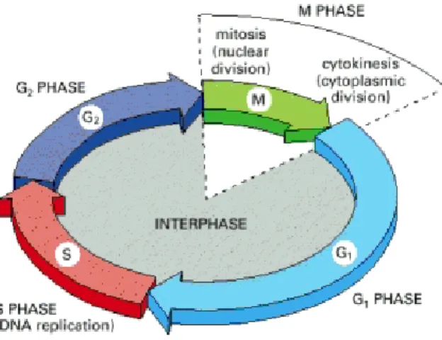
... 15 1 Huntington’s disease ... 17 1.1 Overview and introduction to the disease ... 17 1.2 Neuropathology ... 17 1.3 Symptoms ... 19 1.4 The HTT gene and the mutation ... 20 1.5 Normal HTT ... 21 1.5.1 Overview ... 21 1.5.2 Expression and structure ... 22 1.6 Functions of wild‐type HTT ... 23 1.6.1 Embryonic development ... 24 1.6.2 Antiapoptotic ... 24 1.6.3 Pro‐survival factor ... 25 1.6.4 Synaptic activity... 26 1.6.5 Transcriptional factor ... 26 1.6.6 Axonal and vesicular transport ... 27 1.7 Mutant HTT and its down‐stream effect: HTT‐mediated toxicity ... 29 1.7.1 Cleavage by caspases and nuclear translocation ... 30 1.7.2 Aggregation and toxicity ... 30 1.7.3 Transcriptional deregulation ... 31 1.7.4 Excitotoxicity ... 34 1.7.5 Mitochondrial‐based defects, energy and trafficking ... 35 1.7.6 Intracellular transport alteration ... 36 1.7.7 Synaptic dysfunction ... 37 1.8 Post‐translational modification ... 38 1.9 Mechanisms of cell death ... 39 1.9.1 Autophagy ... 40 1.9.2 Apoptosis ... 41 2 Mitosis ... 43 2.1 Overview ... 432.2 Cell cycle ... 43 2.3 The mitotic spindle ... 45 2.4 Positioning of the mitotic spindle ... 46 2.4.1 Spindle orientation in development and tissue organization ... 46 2.4.2 Spindle orientation in cell fate specification ... 48 2.5 Key players in mitotic spindle orientation ... 50 2.5.1 Cell shape ... 50 2.5.2 Cell‐cell junctions (cadherins) and focal adhesions (integrins) ... 51 2.5.3 Microtubules: overview ... 54 2.5.4 Microtubule post‐translational modifications ... 55 2.5.5 Microtubule plus‐end tracking proteins (+TIP) ... 57 2.5.6 The actin cortex ... 60 2.5.7 Spatial reorganization of the mitotic cell ... 62 2.5.8 Cortex polarization ... 64 2.5.9 Microtubule‐actin interaction in mitosis ... 65 2.6 Determination of the division plane ... 67 3 Neural development ... 69 3.1 Overview ... 69 3.2 Brain development ... 69 3.2.1 Neural tube formation ... 69 3.2.2 Brain development ... 70 3.3 Neurogenesis ... 71 3.3.1 Progenitors cells ... 72 3.3.2 Molecular control of neurogenesis ... 74 3.3.3 Cell division of neural progenitors and cell fate ... 76 3.3.4 Interkinetic nuclear migration ... 78 3.4 Mechanisms of mitotic spindle orientation in vertebrate neural progenitor cells ... 81 4 Context of the project ... 83
RESULTS AND DISCUSSION
... 85 5 pARIS‐htt: a new tool to study huntingtin functions ... 87 5.1 Study presentation ... 87 5.2 Article I ... 875.3 Discussion ... 107 6 Role of HTT during mitosis ... 109 6.1 Study presentation ... 109 6.2 Article II ... 109 6.3 Discussion ... 127 7 Mutant HTT and mitosis ... 129 7.1 Study presentation ... 129 7.2 Article III ... 129 7.3 Discussion and perspectives ... 179
8
GENERAL DISCUSSION
... 183 8.1 The importance of studying full‐length HTT ... 183 8.2 HTT and mitosis ... 184 8.2.1 HTT dynamic localization during cell division ... 184 8.2.2 HTT as a transport‐mediator during mitosis ... 185 8.3 HTT and neurogenesis ... 186 8.3.1 HTT and corticogenesis ... 186 8.3.2 The role of HTT in cell fate ... 187 8.3.3 HTT and neuronal migration ... 187 8.3.4 Is HD a cortical developmental disorder? ... 188 9 Annexe I: Rodent HD models ... 191 10 Annexe II: Other HD models ... 197 11 Annexe III: Huntington’s disease signalling ... 203 12 Annexe IV: HD therapeutic strategies and biomarkers ... 207 13 Annexe V: Huntingtin is required for mitotic spindle orientation and mammalian neurogenesis (supplemental information) ... 215 14 Annexe VI: pARIS‐htt: an optimised expression platform to study huntingtin reveals functional domains required for vesicular trafficking (supplemental information) ... 257 15 Bibliography ... 269INTRODUCTION
1 Huntington’s disease
1.1 Overview and introduction to the disease
Huntington’s disease (HD) is a neurodegenerative disorder first characterized as a hereditary, late‐ onset form of chorea by George Huntington in 1872 (Huntington, 2003). In his work (Figure 1), the description of the clinical aspects of HD is remarkably complete. The adult onsets, relentless progression, fatal outcome, mental involvement alongside the movement disorder are all recognized.
Figure 1. The title page of George Huntington’s original paper "On Chorea". (1872).
In 1968, Milton Wexler started the Hereditary Disease Foundation (HDF) when his wife was diagnosed with HD. The HDF enrolled a number of scientists to work in the disease. The discovery of the gene responsible for HD was achieved by Nancy S Wexler, Milton Wexler’s daughter. In 1976 the team conducted a twenty yearlong study in a Venezuelan town near Lake Maracaibo in which they collected blood samples and documented different individuals to work out a common pedigree. This work led to the development of a chromosomal test to identify potential sufferers. The HDF recruited and supported a large group composed for more than 100 scientists from all over the word named the Huntington Disease Collaborative Research Group (HDCRG). In 1993 the group reported the discovery of the gene responsible for HD and of its associated mutation (The Huntington’s Disease Collaborative Research Group, 1993).
1.2 Neuropathology
The histopathology in HD is characterized by the atrophy of the caudate and putamen in the basal ganglia, as well as the cerebral cortex, reducing brain weight by up to 25‐30% (Aylward, 2007; Rosas et al., 2002; Vonsattel & DiFiglia, 1998)(Figure 2).
Figure 2. Atrophy of the striatum and cortex in HD brain. Post mortem brains from a HD patient (left) and healthy individual (right).
Particularly evident is the loss of cells in the caudate (c) and putamen (p), resulting in enlarged lateral ventricles in HD brain.
The most affected neurons are GABAergic type II medium spiny projection neurons, which constitute about 80% of striatal neurons, and large neurons in layers III, IV, and V of the cortex (Hedreen et al., 1991). Interestingly, striatal interneurons are not affected (Zucker et al., 2005). Medium spiny neurons receive glutamatergic signals from the cerebral cortex, thus defects in the basal ganglia‐thalamocortical pathways involved in motor control may contribute to the choreiform disorders seen in HD (Albin et al., 1990). Globus pallidus, thalamus, subthalamic nucleus, substantia nigra, white matter, and the cerebellum can be markedly affected (Vonsattel & DiFiglia, 1998). Recent work has also indicated that the hypothalamus can be significantly atrophied in HD patients (Kassubek et al., 2004; Politis et al., 2008).
The most commonly used grading system to assess the severity of HD degeneration is based on macroscopic and microscopic criteria. The pattern of striatal degeneration in post mortem tissues classifies HD cases into five different severity grades going from 0, as a non discernible neuropathology, to 4, with 95% of neuronal loss in caudate nuclei (Vonsattel et al., 1985).
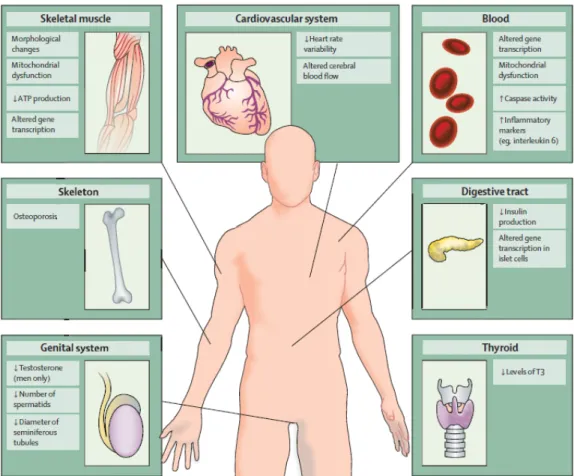
Most clinical features of HD can be attributed to central nervous system (CNS) degeneration, but some aspects of the disease could be linked to defects outside the CNS (Figure 3). Indeed, weight loss, skeletal‐muscle atrophy, cardiac failure, osteoporosis, testicular atrophy and dysfunction of blood‐derived cells are observed in patients (Van Der Burg, 2009). These features of the disease are clinically important as they reduce quality of life and, in some cases, correlate with disease progression and contribute to early death.
Figure 3. Peripheral pathology in patients with Huntington’s disease. (Adapted from Van Der Burg et al., 2009).
1.3 Symptoms
HD symptoms comprise adult‐onset personality changes, generalized motor dysfunctions, and cognitive decline. The peak age of adult‐onset HD is between 35 and 50 years. A small percentage of patients (10%) develop symptoms before age 20. This corresponds to the juvenile variant of the disease usually resulting from paternal transmission. Early onset is associated with increased severity as well as with a more rapid disease progression (Beighton & Hayden, 1981).
In the early stages, HD is associated with progressive emotional, psychiatric, and cognitive disturbances such as anxiety, irritability and depression. Choreiform movements and loss of motor coordination are observed. Commonly reported symptoms in HD include progressive weight loss, alterations in sexual behaviour, and disturbances in the wake‐sleep cycle that occur very early in the course of the disease and may partly be explained by hypothalamic dysfunction (Politis et al., 2008). With the progression of the disease patients show motor speech disorder (dysarthria) and difficulty in swallowing (dysphagia). Decreased movement referred as hypokinesia are also present. In the later stages, HD is characterized by progressive dementia, or gradual impairment of the mental processes involved in comprehension, reasoning, judgment, and memory (Rosenblatt, 2007). Motor problems worsen reaching a final phase of inability to initiate movements (akinesia).
Patients with advanced HD become unable to care for themselves. Life‐threatening complications may result from injuries related to serious falls, poor nutrition, infection, choking, and inflammation. Death occurs 15‐20 years after onset of the first symptoms. At present, there is no cure. The majority of therapeutic strategies currently used in HD are designed to ameliorate the primary symptoms of HD condition (see Annexe IV). Treatments or drugs have limited benefits, and do not delay or halt disease progression.
A top priority in the HD field is the identification of biological markers, or biomarkers. Several candidate HD biomarkers have emerged during the last years (see Annexe IV). A combination of clinical, neuroimaging, and biochemical biomarkers will be necessary to enhance the accuracy, specificity, and sensitivity in tracking disease onset and progression. Moreover, biomarkers will be important to assess the efficiency of future HD treatments.
1.4 The HTT gene and the mutation
In 1983, after Nancy Wexler expedition to Venezuela, the HD gene was mapped on the tip of chromosome 4 (Gusella et al., 1983). Since then, 4p16.3 was identified as the most likely position of the HD gene (Bates et al., 1991; MacDonald, Haines, et al., 1989; MacDonald, Cheng, et al., 1989; Snell et al., 1992). The HDCRG published the discovery of a new gene called IT15 (interesting transcript 15) with a polymorphic CAG (cytosine‐adenine‐guanine) triplet expansion in the first exon (The Huntington’s Disease Collaborative Research Group, 1993). When researchers examined this region of IT15 in non‐HD controls, they found that the number of CAG repeats varied from 6 to 35. Analysis of the same region in the IT15 gene in individuals with HD showed that those always had 40 or more CAG repeats. It was concluded that the trinucleotide repeat expansion was responsible for HD. The IT15 gene is now renamed the HTT gene (HTT) because of the name assigned to the protein. The discovery of the causal HD gene has stimulated research, and work is now focusing on molecular mechanisms of disease.
The gene encoding HTT in vertebrates is composed of 67 exons spanning over 170 kb (Figure 4). (CAG)n repeats are located in the first exon. Normal alleles are polymorphic for the CAG repeat, containing 11 to 35 CAG repeats. When the repeats reach 41 or more the disease is fully penetrant (Mcneil et al., 1997; Rubinsztein et al., 1996). Incomplete penetrance happens with 36–40 repeats I am not sure about this range, we need to check: these individuals do not develop HD but are at risk of transmitting the disease to their children, known as “genetic anticipation”. This phenomenon is explained by the fact that the expanded CAG repeats are not stable and tend to expand from generation to generation specifically when the disease gene is inherited from the father (Ranen et al., 1995). Extremely large CAG repeats of 60 or greater are often associated with a disease onset during childhood or adolescence (juvenile).
Figure 4. Human HTT gene. The 5' end of HTT has a sequence of (CAG)n repeats. 35 or less repeats are not associated with the disorder. Pathological threshold is defined by 41 or more CAG repetitions. Incomplete penetrance happens with 36 to 41 repeat. 60 or more repeats are often associated with a juvenile onset (before 20 years old). HTT is located on the short (p) arm of chromosome 4 at position 16.3.
There is a strong inverse correlation between CAG repeats length and age at onset of motor symptoms (Andrew et al., 1997). A recent work shows that normal allele CAG length, interaction between expanded and normal alleles and presence of a second expanded allele do not influence age at onset of motor manifestations, indicating that the rate of HD pathogenesis leading to motor diagnosis is determined by a completely dominant action of the longest expanded allele (Lee et al., 2012). However, the number of CAG repeats accounts for about 60% of the variation in age of onset and disease manifestations. This observation suggests the existence of environmental and genetic modifiers. Several studies revealed that a large set of genes distinct from the HD locus itself could contribute to modify disease onset and progression (Chattopadhyay et al., 2003; Jian‐ liang Li et al., 2003; Wexler, 2004). All of these modifiers relate to various mechanisms implicated in HD pathology as excitotoxicity, dopamine toxicity, metabolic impairment, transcriptional deregulation, protein misfolding, and oxidative.
Most of the patients are heterozygous for the mutant allele. Homozygous cases of the disorder show the same age of onset of the disease that heterozygous HD cases, but the rate of progression can be enhanced (Squitieri et al., 2003).
Of note, other neurodegenerative diseases are also caused by an abnormal CAG expansion in a single causative gene. This results in protein aggregation, late‐onset neurodegeneration, and selective vulnerability of a subset of neurons. This includes nine other diseases with expansions in polyQ tracts: spinal and bulbar muscular atrophy (SBMA), dentatorubral and pallidoluysian atrophy (DRPLA), and spinocerebellar ataxias (SCA) 1,2,3,6,7,12,17.
1.5 Normal HTT
1.5.1 Overview
HTT gene encodes huntingtin protein (HTT), a large soluble protein with a molecular weight of
350kD (Figure 5). (CAG)n are translated in a glutamine tract. HTT has little homology to other proteins but is well conserved from D. melanogaster to mammals, suggesting a central role in cell homeostasis. Three putative domains of HTT have been identified in multialignment
corresponding to human HTT amino acids 1–386 (HTT1), 683–1,586 (HTT2), and 2,437–3,078 (HTT3). In particular, comparison of more divergent orthologs and quantification of evolutionary pressure on the three blocks revealed that the NH2‐terminal fragment (HTT1) is the most recently evolved part of HTT, while the COOH‐terminal part represents the most conserved portion among all animals, from sea urchin to insects and mammals (Zuccato etal., 2010). HTT is a 350kDa protein. This high molecular weight hampers the production of crystals and mass spectrometry studies to elucidate its structure. Up to date, there are no clear data on the structure of the protein. 1.5.2 Expression and structure HTT is ubiquitously expressed throughout the body; higher levels are found in brain and testis. In the brain, HTT can be found at highest levels in the cerebellar cortex, the neocortex, the striatum and hippocampus (Trottier et al., 1995). At subcellular levels, HTT has a large distribution. HTT is associated with a variety of organelles, including the nucleus, endoplasmic reticulum (ER), Golgi complex, and mitochondrion (Hilditch‐Maguire et al., 2000; Hoffner et al., 2002; Kegel et al., 2002; Panov et al., 2002; Strehlow et al., 2007). HTT is also found within neurites and at synapses, where it associates with various vesicular structures such as clathrin‐coated vesicles, endosomal compartments and microtubules (MT) (Difiglia et al., 1995; Hilditch‐Maguire et al., 2000; Hoffner et al., 2002; Velier et al., 1998).
Figure 5. HTT amino acid sequence. (Q)n indicates the polyglutamine tract, which is followed by the polyproline sequence (P)n; the red
emptied rectangles indicate the three main groups of HEAT repeats (HEAT group 1, 2, 3). The small green rectangles indicate the caspase cleavage sites and their amino acid position (513, 552, 586), while the small pink triangles indicate the calpain cleavage sites and their amino acid positions (469, 536). Boxes in yellow: B, regions cleaved preferentially in the cerebral cortex; C, regions of the protein cleaved mainly in the striatum; A, regions cleaved in both. Posttranslational modifications: ubiquitination (UBI) and/or sumoylation (SUMO) sites (green); palmitoylation site (orange); phosphorylation at serines 13, 16, 421, and 434 (blue); acetylation at lysine 444 (yellow). The nuclear pore protein translocated promoter region (TPR, azure) is necessary for nuclear export. (Adapted from Zuccato et al., 2010)
At NH2 terminus, glutamine stretch in human HTT begins at the 18th amino acid. A recent study suggests that the (Q)n tract may modulate a normal function of HTT (Zheng et al., 2010). In vitro studies on fibroblasts indicated that the (Q)n contributes in modulating longevity and energy status. Moreover, mice lacking (Q)n stretch live significantly longer than wild‐type mice.
In mammals, the (Q)n region is followed by a polyproline ((P)n or polyP) stretch. It was suggested that the (P)n function may reside in the stabilization of the (Q)n tract by keeping it soluble (Steffan et al., 2004). This domain, together with Src homology 3 (SH3) region, would be implicated in protein‐protein interaction.
HTT is also enriched in consensus sequences called huntingtin, elongation factor 3, protein phosphatase 2A, and TOR 1 (HEAT) repeats that are organized into protein domains important for protein‐protein interactions. The repeats are well conserved in HTT through evolution. A recent study of HEAT repeats number and distribution revealed a total of 16 HEAT repeats in HTT, which are organized into 4 clusters (Tartari et al., 2008). Recombinant full‐length HTT purified from insect cells has high helical content, is an elongated molecule, and remains intact despite extensive proteolytic nicking (Li et al., 2006). This preliminary characterization of HTT is consistent with a structure composed entirely of HEAT repeats, folded via a continuous hydrophobic core into a single superhelical solenoid.
A functionally active COOH‐terminal nuclear export signal (NES) sequence and a less active nuclear localization signal (NLS) are present in HTT, which might indicate that the protein (or a portion of it) is involved in transporting molecules from the nucleus to the cytoplasm (Xia et al.,2003). In concordance with this, it has been show that the nuclear pore protein TPR has a role in the nuclear export of N‐terminal HTT (Cornett et al., 2005). PolyQ expansion reduces this nuclear export to cause the nuclear accumulation of HTT.
HTT is subjected to several post‐translational modifications that regulate its wild‐type functions but also participate to HD pathology. This will be discussed in chapter 1.8 of this manuscript.
1.6 Functions of wild‐type HTT
HTT ubiquitary expression and widespread subcellular localization does not facilitate the determination of its functions. However, identification of HTT interactors (see Annexe III) and development of different animal models (see Annexe I and II) put on evidence the following proposed roles for normal HTT.
1.6.1 Embryonic development
HTT is required for normal embryogenesis, as knock‐out (KO) mice for HTT (Hdh ‐/‐) die at an early developmental stage, E7.5. The basis of this effect appears to be increased apoptosis in the embryonic ectoderm shortly after the onset of gastrulation (Duyao et al., 1995; Nasir et al., 1995; Zeitlin et al., 1995). The inactivation of HTT gene (htt) does not reveal a phenotype in D.
melanogaster embryos; htt is dispensable for D. melanogaster development but is crucial for aged
adults (Zhang et al., 2009). This discrepancy might be due to intrinsic differences between mouse and fly embryogenesis as early lethality in mouse is likely to result from the absence of HTT in extraembryonic tissues (Dragatsis et al., 1998). In agreement with this hypothesis is the observation that mice deleted for Htt in adult stages show neurodegeneration (Dragatsis et al., 2000) and, HTT‐ko adult flies show a compromised mobility and reduced viability (Zhang et al., 2009).
Conditional inactivation of the Htt gene in the midbrain and hindbrain in Wnt1 cell lineage results in congenital hydrocephalus (Dietrich et al., 2009). These results implicate HTT also in the regulation of cerebral spinal fluid (CSF) homeostasis.
To bypass the early lethality induced by the absence of HTT and analyze the role of the latter after gastrulation, mice expressing less than 50% of the normal of the protein were generated. These mice present defects in the formation of the precursor of the epiblast, as well as malformations of the cortex and striatum, and die shortly after birth (White et al., 1997). Analyses of chimeras created by blastocyst injection of Htt ‐/‐ ES cells suggest that HTT plays a specific role in neuronal survival, neuroblasts need to synthesize HTT if they are to progress in development and differentiation (Reiner et al., 2001; Reiner et al., 2003).
Finally, studies using developing zebrafish showed that a reduction in Htt levels affects the formation of most of the anterior regions of the neural plate (Henshall et al., 2009). These data indicate that HTT is required at different steps of embryonic development and that its total absence or 50% reduced presence generates a very early phenotype in mice. 1.6.2 Antiapoptotic
Overexpression studies have demonstrated that HTT has a role in maintaining cell viability in response to acute toxic stimuli and excitotoxicity. Wild‐type HTT can suppress apoptosis in vitro in response to exogenous toxic stimuli such as 3‐nitropropionic acid, which selectively damages the striatum, presumably by preventing activation of proaspase‐9 (Rigamonti et al., 2000, 2001). Similarly, HTT can protect against excitotoxicity after quinolic acid injection in vitro and in vivo, by mediating apoptosome complex activity and inhibiting caspase activation downstream of cytochrome‐c release (Rigamonti et al., 2000; Leavitt et al., 2006). Cells depleted of wild‐type HTT
are more sensitive to apoptotic cell death and show increased level of caspase‐3 activity, with respect to control cells (Zhang et al., 2006). HTT is also implicated in anti‐apoptotic pathways due to its interaction with HIP1, which reduces HIP1 and Hippi’s ability to mediate procaspase‐8 cleavage and apoptosis (Gervais et al., 2002).
More recently, the anti‐apoptotic role of HTT has been highlighted also in non mammalian models. In fact, apoptotic cell death is observed in zebrafish embryos in which Htt is knocked‐down by morpholino technology (Diekmann et al., 2009). Htt morpholino‐injected zebrafish show a massively increased cell death as indicated by caspase‐3 activity especially in the midbrain/hindbrain region of the developing zebrafish embryo. This increased apoptosis is accompanied by a severe underdevelopment of the CNS.
1.6.3 Pro‐survival factor
Several studies have shown that HTT regulates the cellular levels of brain‐derived neurotrophic factor (BDNF) (Gauthier et al., 2004; Zuccato et al., 2001). BDNF is a neurotrophin that is particularly important for the survival of striatal neurons and for the activity of the cortico‐striatal synapses. However, striatal neurons depend on the supply of BDNF from the cortical afferents (Baquet et al., 2004) . Indeed, BDNF is produced by cortical neurons and transported to striatal neurons. Wild‐type HTT regulates both BDNF transcription and intracellular dynamics. HTT was found to bind a BDNF transcription repressor element, REST/NRSF, and retain it in the cytoplasm permitting transcription of BDNF (Zuccato et al., 2001, 2003). HTT has also been shown to regulate BDNF delivery from the cortex to the striatum by promoting its transport through its interaction with HAP1 (Gauthier et al., 2004; Wu et al., 2010).
Further studies revealed that wild‐type HTT is a substrate for the serine/threonine kinase Akt. Upon IGF‐1 activation, Akt phosphorylates a number of substrates thus activating prosurvival pathways by stimulating the expression of prosurvival genes, whereas death genes such as BAX or Bcl‐2 are repressed. In particular, Akt mediates prosurvival effect through HTT phosphorylation. Akt directly phosphorylates HTT at serine 421 (Emilie Colin et al., 2008; Zala et al., 2008). This phosphorylation stimulates BDNF anterograde transport in wild‐type conditions. In HD, by rescuing the deficient BDNF transport, phosphorylation of serine 421 reduces mutant HTT‐induced toxicity.
HTT has also been implicated in the stress response system because phosphorylation of HTT by CDK5 in response to DNA damage prevents HTT from inducing p53 dependent apoptosis (Anne et al., 2007). Additionally, phosphorylation of the N17 region of HTT regulates nuclear entry and association with chromatin in response to heat shock (Atwal et al., 2011).
1.6.4 Synaptic activity
Synapses are essential to neuronal function. Wild‐type HTT interacts with cytoskeletal and synaptic vesicles proteins essential for exo‐ and endocytosis at the synaptic terminals, thus participating in the control of synaptic activity in neurons (Smith et al., 2005). HTT is localized post‐ synaptically with PSD‐95 (post‐synaptic density 95), a membrane‐associated guanylate kinase (MAGUK) protein that binds the NMDA and kainate receptors at the postsynaptic density (Fan et al., 2009; Sun et al., 2001). HTT is also localized pre‐synaptically through its interactions with synaptic vesicles, HIP1, and HIP14, suggesting roles in synaptic signaling and vesicle recycling (Stowers et al., 2007; Waelter et al., 2001). Furthermore, HTT can bind to PACSIN1/syndapin, syntaxin, and endophilin A, which collectively play a key role in synaptic transmission, as well as in synaptic vesicles and receptor recycling (Smith et al., 2005).
1.6.5 Transcriptional factor
(Q)n and (P)n regions have been demonstrated to form polar zipper structures and interact with DNA directly, suggesting HTT may function as a transcriptional activator (Gerber et al., 1994; Perutz et al., 1994). Several studies have shown interactions between HTT and various transcription factors such as the cAMP response‐element binding protein (CBP) (Mccampbell et al., 2007; Steffan et al., 2000), p53 (Mccampbell et al., 2007; Steffan et al., 2000), Sp1, TAFII130 (Dunah et al., 2002), N‐CoR, and Sin3A (Boutell et al., 1999; Luthi‐Carter et al., 2000). By binding to these trancription factors, HTT was reported to act as a transcription activator or repressor. HTT activates the transcirption of genes encompassing RE1 sequence, a conserved 21–23 base pair DNA Repressor element 1 (also known as the neuron‐restrictive silencer element, NRSE). This sequence is recognized by the RE1‐silencing transcription factor (REST; also known as neuronal restrictive silencing factor, NRSF) transcriptional regulator, which acts as a transcriptional silencer (Cattaneo et al., 2005). Thus wild‐type HTT has a broad role in regulating neuronal gene transcription: cells and mice expressing increased wild‐type levels also show higher levels of mRNAs transcribed from RE1/NRSE‐containing neuronal genes (Zuccato et al., 2003). In particular, BDNF exon II promoter contains a RE1 sequence. Wild‐type HTT thus promotes Bdnf transcription because it sequesters the available REST/NRSF in the cytoplasm, thereby preventing it from forming the nuclear co‐repressor complex at the RE1/NRSE nuclear site and allowing gene transcription (Zuccato et al., 2003).
1.6.6 Axonal and vesicular transport
The function of HTT as a facilitator of long‐ and short‐range transport along MT is documented in mammalian cells, D. melanogaster and mouse models. Much of the information gleaned from analysis of HTT interacting proteins suggests a role in intracellular transport (Figure 6).
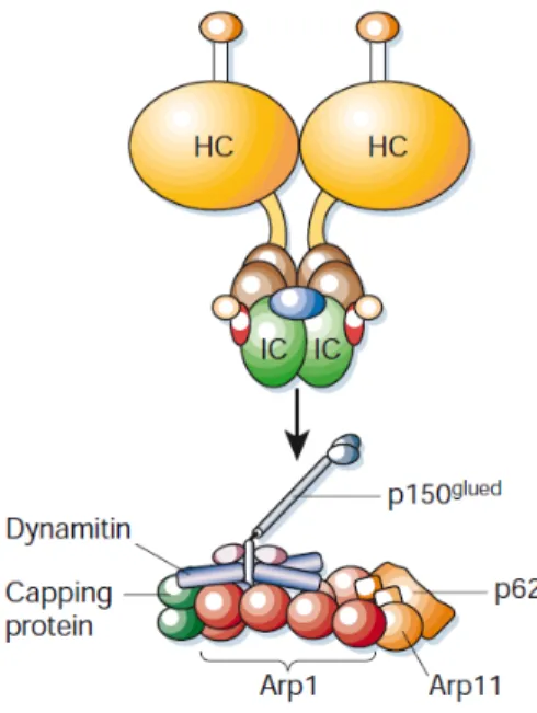
Yeast two‐hybrid screens have implicated HTT in binding HAP1 (X. J. Li et al., 1995), which is involved in axonal transport via its interaction with kinesin (Mcguire et al., 2006), dynein (Engelender et al., 1997; Rong et al., 2007), dynactin subunit p150Glued (Li et al., 1998) and HIP1 and HIP14, which have roles in endocytosis (Kalchman et al., 1997; E E Wanker et al., 1997), suggesting a role for HTT in various aspects of MT and actin based intracellular transport. Dynactin is an essential co‐factor for the transport of membranous organelles by the minus‐end‐directed microtubule motor cytoplasmic dynein. Figure 6. Schematic of HTT. The N‐terminal membrane localization signal (aa1–18) is shown in green and the polyglutamine repeat region (beginning at aa17) is shown in red. The light blue region (aa172–372) has been shown to associate with acidic phospholipids at the plasma membrane. The site of palmitoylation of Cys214 by HIP14 is indicated by a blue lollipop and phosphorylation of Ser421 by Akt is indicated by a yellow lollipop. The myosin VI linker protein optineurin is known to associate with the N‐terminal region of HTT. HAP1 interacts with the N‐terminal region of normal HTT and the expanded polyglutamine repeat found in mutant HTT has been shown to enhance binding. HAP1 also interacts with the plus‐end‐directed MT motor kinesin and dynactin. The minus‐end‐directed MT motor dynein interacts with HTT (aa600–698) and with the dynein activator dynactin. HAP40, an effector of the small GTPase Rab5, binds to the C‐terminal region of HTT (Adapted from Caviston & Holzbaur, 2009).
Additionally, we and others have shown a direct interaction between HTT and dynein. The HTT/dynein interaction was initially described by yeast two‐hybrid system. This interaction was functionally validated in cell culture, by demonstrating that reduced HTT expression leads to disruption of Golgi stacks, a hallmark dynein‐mediated defect (Caviston et al., 2007; Pardo et al., 2010). Mapping experiments identify a binding site for the dynein intermediate chain to residues 601–698 of HTT. Immunoprecipitation of endogenous proteins from brain extract with an anti‐ dynein antibody demonstrates the co‐precipitation of a complex that includes cytoplasmic dynein, dynactin, HTT, kinesin and HAP1 (Caviston et al., 2007; Colin et al., 2008). In line with this, full‐ length versions of HTT lacking the interaction domain for dynein (‐DYN) and HAP1 (‐HAP1) fail to transport BDNF vesicles in neuronal cells (Pardo et al., 2010). These observations will be discussed in detail in the results section 5 of this thesis.
HTT has been suggested to act as a ‘molecular switch’ in controlling anterograde and retrograde transport of BDNF (Figure 7). Experiments in cell culture measuring transport of BDNF demonstrate that phosphorylation of serine 421 (S421) favors anterograde transport, while unphosphorylated S421 favors retrograde transport (Colin et al., 2008). A similar ‘switching’ mechanism has been attributed to HTT by its interaction with HAP40, a Rab5 associated protein, suggesting that HTT functions to transfer endosomes from MT to F‐actin tracks (Pal et al., 2008; Peters & Ross, 2001). HTT has also been implicated in actin‐based transport by its interaction with optineurin, which may physically link HTT to the actin motor myosin‐VI (Sahlender et al., 2005). Interestingly, ablation of HTT phosphorylations at S1181/S1201 leads to increased anterograde and retrograde velocities via an increased attachment of motor proteins and BDNF vesicles to MT (Ben M’Barek et al., manuscript in preparation). Figure 7. Proposed model of the directionality switch induced by phosphorylation of HTT. HTT binds to HAP1 and to the dynein complex. HAP1 interacts with p150Glued and kinesin‐1. When HTT is not phosphorylated, the kinesin‐1 interaction with motor complex is weak, kinesin‐1 is detached from MTs and vesicles leading to retrograde transport. When HTT is phosphorylated, the kinesin‐1 association with motor complex is increased and kinesin‐1 is recruited to vesicles, therefore inducing a switch to anterograde transport. (Adapted from Colin et al., 2008).
A recent publication indicates that wild‐type HTT is essential for protein trafficking to the centrosome and normal ciliogenesis. Ciliogenesis is regulated by an HTT‐HAP1‐peri‐centriolar molecule‐1 (PCM1) pathway. Depletion of HTT or HAP1 leads to dispersion of PCM1 from centrosomes and reduced ciliogenesis in cells (Keryer et al., 2011).
Fast axonal transport (FAT) requires consistent energy over long distances to fuel the molecular motors that transport vesicles. A recent study from our laboratory (Zala et al., manuscript in preparation) shows that FAT depends on glycolytic but not on mitochondrial ATP. Indeed, FAT
would use ATP that is directly generated on vesicles from glyceraldehyde 3‐phosphate dehydrogenase (GAPDH) and phosphoglycerate kinase (PGK) activities. GAPDH is a very abundant protein present both in the cytoplasm and the nucleus and localizes on fast moving vesicles within axons. Consistent evidence show that would HTT scaffolds GAPDH on vesicles with depletion of HTT reducing GAPDH attachment to vesicles.
1.7 Mutant HTT and its down‐stream effect: HTT‐mediated toxicity
Down‐stream amino acid 17, HTT has a polymorphic (Q)n/(P)n rich domain. The abnormal expansion of (Q)n (polyQ) stretch encoded by the nucleic acids (CAG)n causes HD. HD is an autosomal dominant disorder. This suggests that the mutation leads to a toxic gain of function. Accordingly, several cellular and animals models were developed by over‐expressing mutant HTT. But there is evidence that a loss of protective function of HTT could act synergistically with the gain of toxic functions (Figure 8). Mutant HTT is found in both the nucleus and the cytoplasm of HD brain (Benn et al., 2005; Gutekunst et al., 1999). Hypothesis about the nuclear effects of mutant HTT focuses mainly on transcriptional dysregulation, while toxicity of HTT in the cytoplasm involves ubiquitin/proteasome dysfunction, aberrant caspase activity and cell death, synaptic dysfunction, excitotoxicity, mitochondrial dysfunction, autophagy and impaired axonal transport. Figure 8. Key cellular pathogenic mechanisms in HD. (A): the mutation in HTT causes a conformational change of the protein that leads to partial unfolding or abnormal folding of the protein, which can be corrected by molecular chaperones. Full‐length mutant HTT is cleaved by proteases in the cytoplasm. In an attempt to eliminate the toxic HTT, fragments are ubiquitinated and targeted to the proteasome for degradation. However, the proteasome becomes less efficient in HD. Induction of the proteasome activity as well as of autophagy protects against the toxic insults of mutant HTT proteins by enhancing its clearance. (B): NH2‐terminal fragments containing the polyQ strech accumulate in the cell cytoplasm and interact with several proteins causing impairment of calcium signaling and homeostasis (C) and mitochondrial dysfunction (D). (E): N‐terminal mutant HTT fragments translocate to the nucleus where they impair gene transcription or form intranuclear inclusions. (F): the mutation in HTT alters vesicular transport and recycling. (Adapted from Zuccato et al., 2010).
1.7.1 Cleavage by caspases and nuclear translocation
HTT is found cleaved in cellular and mouse HD models and in HD patients. The cleavage events occur both in normal and mutant huntingtin, but the latter is more susceptible to proteolysis and generates N‐terminal fragments that are found in the cytoplasm and nucleus of neuronal and non neuronal cells.
HTT cleavage is a key event in the pathology. Indeed, inhibition of HTT cleavage prevents neurodegeneration and toxicity in vivo (Gafni et al., 2004; Graham et al., 2006). Further supporting the importance of mutant HTT cleavage, expression of different N‐terminal fragments with expanded polyQ is sufficient to induce a HD‐like pathology, whereas longer fragments are less toxic in cellular and animals models (de Almeida et al., 2002; Karpuj et al., 1999; Lunkes et al., 1998; Saudou et al., 1998).
Several consensus cleavage sites have been identified in HTT (Figure 5). Caspases 1,3,6,7 and 8 as well as calpain can cleave HTT in vivo and in vitro (Difiglia, 2002; Gafni et al., 2004; Gafni & Ellerby, 2002; Hermel et al., 2004; Mende‐Mueller et al., 2001). In particular, studies have strengthened the evidence for a role of caspase‐6‐mediated cleavage in the disease process. Activation of caspase‐6 may be a primary event in the proteolytic process of mutant HTT. This would then lead to the activation of additional proteolytic caspase activities (for example, to activation of caspase‐ 2 and ‐3), exacerbating neurodegeneration and contributing to the appearance of the disease phenotype.
Once HTT is cleaved, N‐terminal fragments are translocated to the nucleus. This nuclear translocation of mutant HTT induces neuronal apoptosis (Saudou et al., 1998) Accumulation of small N‐terminal fragments could be in part a result of a diminution in the interaction of HTT with the nuclear pore protein TPR (translocated promoter region) in a mutant context (Cornett et al., 2005). The decreased mutant HTT‐TPR interaction would reduce the export toward the cytoplasm of mutant fragments.
PTM are suggested as important regulators of huntingtin proteolysis (see below). In fact, HTT phosphorylation at S434 by Cdk5 prevents the cleavage of the protein, while phosphorylation at S421 reduces huntingtin cleavage by caspase‐6 and the nuclear accumulation of caspase‐6 fragments (Luo et al., 2005; Warby et al., 2005, 2009).
1.7.2 Aggregation and toxicity
Progressive accumulation of abnormal protein aggregates associated with neuronal loss is a common molecular event observed in al polyQ diseases, but also in other neurodegenerative diseases such as Parkinson’s disease (PD), Alzheimer disease (AD) amyothrophic lateral sclerosis (ALS). HTT fragment length and amount, as well as the length of the (Q)n, are critical factors in
determining the aggregation process (Chen et al., 2002; Hackam et al., 1998; Li et al., 2001; Li et al., 1998).
Two major aggregation pathways are in competition with each other and explain how the polyQ expansion can facilitate aggregation: The first pathway is mediated by aggregation of the polyQ stretch. PolyQ aggregation displays kinetics of nucleated‐growth polymerization with a prolonged lag‐phase required for forming an aggregation nucleus, followed by a fast extension phase during which additional polyglutamine monomers rapidly join the growing aggregate (G. Bates, 2003; Paoletti et al., 2008; Erich E Wanker, 2000). The second pathway depends on the first 17 N‐ terminal amino acids and involves intermediate structures. It is characterized by the formation of oligomers having the first 17 amino acids of the protein in their core and polyQ sequences exposed on the surface (Thakur et al., 2009).
Toxicity of nuclear mutant HTT aggregates is still under debate. However, the field is reaching a consensus. The absence of correlation between nuclear aggregates and cell death show that HTT inclusions are not pathogenic per se but rather an attempt of the cells to sequester toxic soluble fragments (Arrasate et al., 2004; Gutekunst et al., 1999; Saudou et al., 1998). In agreement, inhibition of ubiquitination increases mutant HTT toxicity while decreasing aggregates formation. However, while aggregates are not directly responsible for the induction of cell death they are physically interfering with key cellular functions such as intracellular transport and transcription thus leading to neuronal dysfunction.
Misfolded mutant HTT could impair with chaperone and proteasome systems. This would lead to the accumulation of misfolded or damaged proteins and aggregates formation, inducing a cellular stress response that leads to cell death. Indeed, earlier studies have provided evidence that misfolded toxic polyQ HTT protein induces a global impairment of the ubiquitin–proteasome system (UPS) that is pathogenic in HD (Bennett et al., 2005). However, other studies showed an accumulation of polyubiquitinated proteins in HD in the absence of a general UPS impairment (Bett et al., 2006; Bett et al., 2009; Bett et al., 2009; Maynard et al., 2009). This suggested that the UPS is generally functional in HD, but that pathogenic HTT may impair selectively the ubiquitination process of specific substrates. A recent study from our lab exposed that HTT binds to ‐catenin and to the destruction complex by interacting with ‐TrCP and axin. The presence of an abnormal polyQ expansion in mutant HTT leads to a decreased binding to ‐catenin therefore impairing the binding of ‐catenin to the destruction complex and subsequently resulting in ‐ catenin accumulation (Godin et al., 2010). Finally, recruitment of chaperones to polyQ aggregates would induce mutant HTT accumulation with an abnormal conformation (Hay et al., 2004). 1.7.3 Transcriptional deregulation DNA transcription is a highly regulated cellular process that is impaired in HD, resulting in altered levels of expression for a number of genes. Both wild‐type and mutant HTT have been shown to
interact with a range of transcription factors, giving rise to the hypothesis that abnormal interactions between mutant HTT and proteins involved in transcription lead to transcriptional deregulation, which is an early event in HD pathogenesis (Sugars & Rubinsztein, 2003)
Wild‐type and polyQ HTT are cleaved by caspases, resulting in N‐terminal fragments that enter the nucleus and alter transcription (Difiglia et al., 1995; Steffan et al., 2000). PolyQ tracts and glutamine rich regions are common in transcription factors, arguing that wild‐type HTT may act in a similar manner, and the expanded polyQ could alter its endogenous interactions with transcription factors and DNA.
Wild‐type HTT is known to bind the RE1‐silencing transcription factor/neuron‐restrictive silencer factor (REST/NRSF) complex (Figure 9). This interaction is likely mediated by HAP1, dynactin, and the REST/NRSF‐interacting LIM domain protein (RILP) (Shimojo, 2008). The interaction between HTT and REST/NRSF is reduced in HD, allowing translocation of REST/NRSF into the nucleus. This reduces the transcription of BDNF, as well as of many other neuronal genes under the control of the REST complex, which is a global regulator of neuronal gene transcription (Zuccato et al., 2007, 2008; Zuccato & Cattaneo, 2007). Figure 9. The transcription factor REST/NRSF binds to RE1/NRSE elements in neuronal gene promoters such as in the BDNF gene. Wild‐ type HTT sustains the production of BDNF by interacting with REST/NRSF in the cytoplasm, thereby reducing its availability in the nucleus to bind to RE1/NRSE sites. Under these conditions, transcription of BDNF and of other RE1/NRSE regulated neuronal genes is promoted. Mutant HTT fails to interact with REST/NRSF in the cytoplasm, which leads to increased levels of REST/NRSF in the nucleus. Under these conditions, REST/NRSF binds avidly to the RE1/NRSE sites, suppressing the transcription of BDNF and of other RE1/NRSE regulated neuronal genes. (Adapted from Zuccato et al., 2010).
TAFII‐130, a cofactor for CREB dependent transcription accumulates in mutant HTT nuclear aggregates (Figure 10). PolyQ tract, impairs the soluble association of TAFII130 with Sp1, and directly interfers with the binding of Sp1 to DNA (Dunah et al., 2002; M. Shimohata et al., 2005; T. Shimohata et al., 2000). A role for Sp1 is supported by the downregulation of two relevant gene promoters in cell models: dopamine D2 receptor and nerve growth factor receptor. Sp1 disruption appears to occur early in human HD pathogenesis, being detectable even in presymptomatic grade 1 postmortem tissue (Dunah et al., 2002; M. Shimohata et al., 2005; T. Shimohata et al., 2000).
Figure 10. Mutant HTT represses transcription of Sp1‐dependent promoters (i.e., dopamine receptors D1 and D2 genes) by abnormally interacting with specific transcription cofactors such as Sp1 itself, TFIIF, and TFII130. (Adapted from Zuccato et al., 2010). Transcriptional dysregulation in HD is also linked to energy deficits (Figure 11). Particularly, mutant HTT inhibits expression of PGC‐1, a master regulator of mitochondrial biogenesis and function, by interfering with CREB/TAF4 at the PGC‐1 promoter (Cui et al., 2006). Figure 11. The transcription factor cAMP‐responsive element (CRE)‐binding protein (CREB) binds to DNA elements that contain a CRE sequence, as in the promoter of the PGC1‐ gene, a master regulator of genes involved in mitochondrial function and energy metabolism. Mutant HTT interferes with CREB and TFIID, leading to reduced activation of PGC1‐ gene, reduced PGC1‐ protein levels, and consequently, downregulation of its mitochondrial target genes. (Adapted from Zuccato et al., 2010).
The transcription of genes involved in cholesterol and lipid metabolism was also reported to be affected in HD. HD cells showed reduced sterol responsive element binding protein (SREBP) nuclear translocation and reduced activity of a SRE‐reporter gene in the presence of mutant HTT (Figure 12) (Valenza et al., 2005). These results imply that less SREBP reaches the transcriptionally active sites in the nucleus causing reduced expression of SRE‐regulated genes involved in cholesterol and lipid metabolism. Figure 12. SREBP binds to SRE to regulate the transcription of genes involved in the cholesterol biosynthesis pathway. Under physiological conditions, SREBP is transported from the endoplasmic reticulum to the Golgi region, where it is cleaved to obtain a fragment that enters the nucleus and activates cholesterogenic genes. In the presence of mutant HTT, this mechanism is impaired, which leads to the reduced expression of SREBP‐dependent genes and decreases the biological effects of cholesterol biosynthesis. (Adapted from Zuccato et al., 2010).
Mutant HTT may also cause transcriptional deregulation by inhibiting the action of histone acetylases such as CBP, p300, and P/CAF through binding of the expanded polyQ tract to acetyltransferase domains (Steffan et al., 2001) (Figure 13). Acetylation of histones through
histone acetyltransferase activity facilitates unwinding of chromatin, rendering it transcriptionally active; conversely, inhibition of histone acetylase (HAT) activity results in repression of gene transcription. In agreement, administration of histone deacetylase (HDAC) inhibitors rescues neurodegeneration in cellular, fly, and mouse models of HD (Ferrante et al., 2003; Hockly et al., 2003; Steffan et al., 2001). Figure 13. Levels of histone acetylation at specific lysine residues are determined by concurrent reactions of acetylation (Ac) and deacetylation, which are mediated by histone acetylases (HATs) and histone deacetylases (HDACs). Histone acetylation is vital for establishing the conformational structure of DNA‐chromatin complexes suitable for transcriptional gene expression. Mutant HTT leads to disruptions in HAT and HDAC balance, leading to general transcriptional repression. (Adapted from Zuccato et al., 2010). 1.7.4 Excitotoxicity Excitotoxicity occurs from either overactive glutamate release, reduced glutamate uptake by glial cells, or glutamate hypersensitivity of NMDA receptors (NMDAR) or downstream signaling pathways in the striatal cells. As striatal cells depend on glutamatergic activation from cortical cells, excitotoxicity is an appealing mechanism to explain mutant HTT‐mediated toxicity in striatal cells. Early evidence for this came from human patient brain slices demonstrating reduced NMDAR binding specifically in the striatum (Young et al., 1988). YAC128 mouse models expressing full‐ length mutant HTT also display excitotoxic sensitivity in striatal cells (Benn et al., 2007).
Possible mechanisms for this effect include changes in NMDAR protein levels or post‐translational modifications (Ali & Levine, 2006; Cepeda et al., 2001). Defective glutamate clearance from the synaptic cleft by glia has also been suggested as a possible cause of excitotoxicity. The transporter GLT1, a glial glutamine transporter, appears down‐regulated in mouse models of HD, increasing levels of glutamine in the synaptic cleft (Estrada‐Sánchez et al. , 2009). Mutant HTT induced excitotoxicity could also be the result of a decrease interaction with PSD‐95. PSD‐95 recruits NMDA receptors and facilitates their activation, but in a HD pathological context, PSD‐95 is not properly recycled and NMDA receptors become hypersensitive (Sun et al., 2001).
In addition to glutamate, other neurotransmitter systems that control the activity of the corticostriatal synapse contribute to render striatal neurons more sensitive to excitotoxic stimuli (Bamford et al., 2004). Adenosine A2 receptors (A2AR) and cannabinoid receptors (CB1R) are particularly abundant on the corticostriatal terminals, where, when activated, they increase glutamate release. A crucial input to the striatum comes from the substantia nigra pars compacta, whose fibers represent the main striatal source of dopamine. Dopamine can directly regulate glutamate release from corticostriatal terminals by stimulating the D2 receptors (D2R) located on the cortical afferents
Impaired clearance of glutamate from the synaptic cleft may contribute to enhance excitotoxic neurodegeneration in HD (Tzingounis & Wadiche, 2007). Glial cells may play important roles through cell‐cell interactions. For example, decreased glutamate uptake in glial cells by GLT‐1, the Na+dependent glial transporter of glutamate, contributes to increased neuronal vulnerability and neuronal excitotoxicity in neurons. 1.7.5 Mitochondrial‐based defects, energy and trafficking Many studies have indicated that mitochondrial dysfunction may contribute to neurodegenerative diseases, including HD. Mitochondria are the ‘powerhouses’ of the cell, generating adenosine‐5'‐ triphosphate (ATP) and maintaining cellular homeostasis. Neurons have intense energy demands that are met by mitochondria: ATP is essential in neurons to fuel ionic pumps and ATP‐dependent enzymes (Johri et al., 2011). Mitochondria also buffer intracellular calcium levels and sequester apoptotic factors, playing a vital role in neuronal function and survival. Dysfunction of mitochondria can lead to metabolic insufficiency, oxidative damage, excitotoxicity, and neurodegeneration (Hollenbeck & Saxton, 2005).
There is extensive evidence for bioenergetic deficits and mitochondrial dysfunction in HD: such as a pronounced weight loss despite sustained caloric intake; nuclear magnetic resonance spectroscopy showing increased lactate in the cerebral cortex and basal ganglia; decreased activities of oxidative phosphorylation (OXPHOS) complexes II and III, and reduced aconitase activity in the basal ganglia; abnormal mitochondrial membrane depolarization in patient lymphoblasts; abnormal ultrastructure of mitochondria in cortical biopsies obtained from patients with both juvenile and adult‐onset HD; pathologic grade dependent reductions in numbers of mitochondria in HD postmortem brain tissue; and in striatal cells from mutant HTT knock‐in mice: both mitochondrial respiration and ATP production are significantly impaired (Johri & Beal, 2012). HD patient post mortem brain slices and mouse models demonstrate decreased levels of cAMP and ATP/ADP ratios, indicating defective mitochondrial energy metabolism (Gines et al., 2003). Positron emission tomography (PET) imaging in the striatum of patients during presymptomatic or early stages of HD revealed increased oxygen over glucose utilization, suggesting early defects in glycolysis (Powers et al., 2007). The R6/2 mouse model shows increased oxygen consumption and increased uncoupling protein‐2 mRNA levels in the brown adipose tissue, which suggests inefficient coupling of the electron transport and ATP synthesis (van der Burg et al., 2008). Similarly, HD patients often demonstrate severe chorea‐independent weight loss (Djoussé et al., 2002). Defects in the mitochondrial respiratory chain complex II and III have been observed specifically in the caudate and putamen, but not cerebellum or cortex of HD patients (Brennan et al., 1985; Gu et al., 1996; Tabrizi et al., 1999). Together these results point to cell specific defects associated with mitochondrial energy production in areas of the brain prone to neurodegeneration in HD. Because many of these effects are seen pre‐symptomatically, defects may represent early events in pathogenesis which could provide useful targets for therapeutics.