HAL Id: hal-02074621
https://hal.archives-ouvertes.fr/hal-02074621
Submitted on 8 Mar 2020
HAL is a multi-disciplinary open access
archive for the deposit and dissemination of sci-entific research documents, whether they are pub-lished or not. The documents may come from teaching and research institutions in France or abroad, or from public or private research centers.
L’archive ouverte pluridisciplinaire HAL, est destinée au dépôt et à la diffusion de documents scientifiques de niveau recherche, publiés ou non, émanant des établissements d’enseignement et de recherche français ou étrangers, des laboratoires publics ou privés.
Chimpanzee with Acute Flaccid Paralysis, from the
Tchimpounga Chimpanzee Rehabilitation Center,
Republic of Congo
Illich Manfred Mombo, Nicolas Berthet, Alexander Lukashev, Tobias Bleicker,
Sebastian Brünink, Lucas Léger, Rebeca Atencia, Debby Cox, Christiane
Bouchier, Durand Patrick, et al.
To cite this version:
Illich Manfred Mombo, Nicolas Berthet, Alexander Lukashev, Tobias Bleicker, Sebastian Brünink, et al.. First Detection of an Enterovirus C99 in a Captive Chimpanzee with Acute Flaccid Paralysis, from the Tchimpounga Chimpanzee Rehabilitation Center, Republic of Congo. PLoS ONE, Public Library of Science, 2015, 10 (8), pp.e0136700. �10.1371/journal.pone.0136700�. �hal-02074621�
First Detection of an Enterovirus C99 in a
Captive Chimpanzee with Acute Flaccid
Paralysis, from the Tchimpounga Chimpanzee
Rehabilitation Center, Republic of Congo
Illich Manfred Mombo1,2*, Nicolas Berthet1,3, Alexander N. Lukashev4, Tobias Bleicker5, Sebastian Brünink5, Lucas Léger2, Rebeca Atencia6, Debby Cox6, Christiane Bouchier7,
Patrick Durand2, Céline Arnathau2, Lionel Brazier2, Joseph N. Fair8, Bradley S. Schneider8, Jan Felix Drexler5, Franck Prugnolle1,2, Christian Drosten5,
François Renaud2,9, Eric M. Leroy1,2‡, Virginie Rougeron1,2‡
1 Centre International de Recherche Médicale de Franceville, BP769, Franceville, Gabon, 2 Laboratoire MIVEGEC UMR 224–5290 CNRS-IRD-UM1-UM2, IRD, Montpellier, France, 3 Centre National de la Recherche Scientifique, UMR3569, 25 rue du docteur Roux, 75724, Paris, France, 4 Chumakov Institute of Poliomyelitis and Viral Encephalities, Moscow, Russia, 5 Institute of Virology, University of Bonn Medical Centre, Bonn, Germany, 6 The Jane Goodall Institute, Suite 550, 1595 Spring Hill Rd, Vienna, Virginia, 22182, United States of America, 7 Institut Pasteur, Genomic platform, 28, rue du Docteur Roux, F-75724, Paris, France, 8 Metabiota, Inc., 1 Sutter Street, Suite 600, San Francisco, California, 94104, United States of America, 9 CHRU de Montpellier, Montpellier, France
‡ These authors co-managed this work. *mombo.illich@gmail.com
Abstract
Enteroviruses, members of the Picornaviridae family, are ubiquitous viruses responsible for mild to severe infections in human populations around the world. In 2010 Pointe-Noire, Republic of Congo recorded an outbreak of acute flaccid paralysis (AFP) in the humans, caused by wild poliovirus type 1 (WPV1). One month later, in the Tchimpounga sanctuary near Pointe-Noire, a chimpanzee developed signs similar to AFP, with paralysis of the lower limbs. In the present work, we sought to identify the pathogen, including viral and bac-terial agents, responsible for this illness. In order to identify the causative agent, we evalu-ated a fecal specimen by PCR and sequencing. A Human enterovirus C, specifically of the EV-C99 type was potentially responsible for the illness in this chimpanzee. To rule out other possible causative agents, we also investigated the bacteriome and the virome using next generation sequencing. The majority of bacterial reads obtained belonged to commensal bacteria (95%), and the mammalian virus reads matched mainly with viruses of the Picorna-viridae family (99%), in which enteroviruses were the most abundant (99.6%). This study thus reports the first identification of a chimpanzee presenting AFP most likely caused by an enterovirus and demonstrates once again the cross-species transmission of a human path-ogen to an ape.
OPEN ACCESS
Citation: Mombo IM, Berthet N, Lukashev AN, Bleicker T, Brünink S, Léger L, et al. (2015) First Detection of an Enterovirus C99 in a Captive Chimpanzee with Acute Flaccid Paralysis, from the Tchimpounga Chimpanzee Rehabilitation Center, Republic of Congo. PLoS ONE 10(8): e0136700. doi:10.1371/journal.pone.0136700
Editor: Juan C. de la Torre, The Scripps Research Institute, UNITED STATES
Received: May 22, 2015 Accepted: August 6, 2015 Published: August 24, 2015
Copyright: © 2015 Mombo et al. This is an open access article distributed under the terms of the
Creative Commons Attribution License, which permits unrestricted use, distribution, and reproduction in any medium, provided the original author and source are credited.
Data Availability Statement: The EV-C99 isolate IJC04 sequence files are available from the GenBank database (accession number KP793035). Funding: This work was supported by the Gabonese Government, Total Gabon and the‘Institut National d’Ecologie et d’Evolution’ of CNRS (France). The work was also supported by a fellowship BSTD of the Institut de Recherche pour le Développement (IRD), France, as well as the generous support of the American people through the United States Agency for International Development (USAID) Emerging
Introduction
In the last few years, many pathogens capable of infecting humans, such as Ebola virus [1,2], simian immunodeficiency viruses [3], respiratory viruses [4,5], anthrax [6], herpes viruses [7] as well as enteroviruses [8,9] have been identified in great apes. Contacts between human pop-ulations and great apes as well as monkeys are frequent and increasing, due to commercial poaching, deforestation, and monitoring [10–13]. The direct consequence is the increased like-lihood of pathogens exchanges and thus of emerging zoonoses and antropozoonoses [10–13].
Enteroviruses (EVs) are small non-enveloped viruses with positive single strand RNA genomes that belong to the Picornaviridae family. Their genome, about 7.5 kb in length, encodes four structural proteins (VP1-VP4) and seven non-structural proteins (2A to 2C and 3A to 3D). EVs consist of more than 300 serotypes and, based on the VP1 sequence, are classi-fied into nine species EV-A to EV-H and EV-J [14]. EVs infecting humans belong to species EV-A to EV-D, formerly termed Human Enterovirus A-D, while the other species contain viruses infecting bovines (EV-E and EV-F), porcines (EV-G) and monkeys (EV-H and EV-J) [14]. EV-C is associated with various human illnesses, such as the common cold, acute hemor-rhagic conjunctivitis, aseptic meningitis and acute flaccid paralysis (AFP). Human EV A-D have been detected in non-human primates, captive or wild Old World monkeys (such as Macaca sp, Cercopithecus sp, Cercocebus and Papio sp) and wild African great apes (Gorilla gorilla and Pan troglodytes) [8,15–17]. Importantly, both known and novel types of human enteroviruses could be found in primates, indicating common cross-species transmission [18].
In mid-October 2010, a poliomyelitis outbreak occurred in the human population of Point-Noire and surrounded areas and villages in the Republic of Congo (RC). The causative agent of this outbreak was identified as the type 1 wild poliovirus (WPV1), while a novel enterovirus 105 (EV-C105) was detected in one patient. Both PV1 and EV-C105 are members of the EV-C species [19,20]. In November 2010, one month after the start of this outbreak, a chimpanzee (Pan troglodytes troglodytes) from the Tchimpounga Chimpanzee Rehabilitation Center devel-oped a syndrome compatible with AFP. The main objective of this study was to identify the pathogen responsible of this syndrome. We identified EV-C99 as a possible causative agent of this chimpanzee’s illness. This is the first identification and characterization of non-polio EV-C in a chimpanzee displaying AFP.
Materials and Methods
Case report and specimen collection
In November 2010, in the Tchimpounga Chimpanzee Rehabilitation Center located 50 km from Pointe-Noire, a juvenile female chimpanzee presented signs of AFP. This chimpanzee first developed a 38.8°C fever and four days after, she lost the ability of her lower limbs and became quadriplegic. The first clinical examination revealed absence of the osteotendinous reflex, a myasthenia and myalgia after flexion of the lower limbs, and dysphonia. This chim-panzee, a member of a group of twelve individuals, was the only one showing these symptoms of AFP. The other examination based on cerebral scan and x-ray photography did not permit to identify the cause of the disease. After the disease onset, the chimpanzee received treatments of B vitamin and glucose, survived, but did not recover the use of lower limbs, which is a typical sign of AFP caused by poliovirus in humans.
In order to identify the pathogen responsible for AFP, a specimen of feces was collected from this individual (named IJC04). The sample was subsequently stored at -80°C before being sent to the International Centre for Medical Research (CIRMF, Franceville, Gabon) for diag-nostic investigations.
Pandemic Threats PREDICT. The funders had no role in study design, data collection and analysis, decision to publish, or preparation of the manuscript. The contents are the responsibility of the authors and do not necessarily reflect the views of USAID or the United States Government. Metabiota, Inc. provided support in the form of salaries for authors BSS and JNF, but did not have any additional role in the study design, data collection and analysis, decision to publish, or preparation of the manuscript. The specific roles of these authors are articulated in the‘author contributions’ section.
Competing Interests: The authors have the following interests. This work was partly supported by Total Gabon. Joseph N. Fair and Bradley S. Schneider are employed by Metabiota, Inc. There are no patents, products in development or marketed products to declare. This does not alter the authors' adherence to all the PLOS ONE policies on sharing data and materials, as detailed online in the guide for authors.
Ethics
The permission to take sample was approved by the Tchimpounga Chimpanzee Rehabilitation Center and the Ministry of Health of Republic of Congo. The fecal sample was collected using a non-invasive method [11].
EV detection
RNA was extracted from the fecal sample using EZ1 Virus Mini kit (Qiagen, Hilden, DE), according to the manufacturer’s recommended procedure. EV detection was performed by amplifying the 5’-untranslated region (5’-UTR) via a one-step real-time reverse transcription (RT)-PCR, as described in Dierssen et al. [21]. The positive sample was amplified using nested RT-PCR targeting the capsid gene VP1 [22]. Positive amplicons were sequenced in both direc-tions (SEQLAB GmbH, DE) to confirm the results and allow for genetic characterization.
EV type assignment and phylogenetic analysis
The complete capsid gene VP1 sequence obtained in this study was compared to a database of complete sequences of most serotypes available on GenBank, in order to determine whether this sequence was genetically related to a known EV serotype. Multiple alignments were per-formed using the ClustalW algorithm and phylogenetic trees were constructed by the neigh-bor-joining method using the Kimura two-parameter model [23], with 10,000 generated trees based on the MEGA program (version 5.2.2) [24].
EV full-length genome amplification and metavirome analysis
In order to (i) obtain the full-length EV genome and (ii) validate that EV-C99 was the causative agent, the viral metagenome was obtained via next generation sequencing (NGS).
Illumina library preparation and sequencing. RNA was re-extracted with QIAmp Viral RNA Mini kit (Qiagen) according to the manufacturer’s instruction and treated with Turbo DNAse (Life Technologies). RNA was reverse transcribed into cDNA with SuperScript III reverse transcriptase using random hexamer primers (Life Technologies). Generated cDNA was then amplified with the Phi29 enzyme, as described previously [25]. A barcoded library was prepared using the Nextera XT kit (Illumina). Amplified DNA was fragmented into 350 bp and adapters were added for multiplexing samples within the same channel. Sequencing was performed with Illumina Hi-seq 2000 sequencer (Illumina) as recommended by the manufacturer.
Bioinformatic and recombination event analyses. All reads were filtered according to quality (Phred score>20). Each viral read was selected using viral database and only the region that matched viral genome was considered. In order to obtain the full-length of viral genome, all reads were initially assembled to contigs with ABYSS software [26] with different values of k, and contigs were then assembled into a‘super assembly’ with the CAP3 program [27]. To look at potential recombination events, similarity plot and bootscanning analysis were con-ducted using Simplot V3.5.1 software [28]. Similarity plot was done using the Kimura substitu-tion model [23] and the bootscanning analysis was carried out using the neighbor-joining method [29] with a sliding window of 400 nt moving in steps of 20 nt.
16S rRNA amplification and pyrosequencing. Given that bacteria such as Clostridium botulinum have been known to be responsible of symptoms comparable to AFP, we evaluated the bacterial community present in the fecal sample of the infected chimpanzee by NGS.
Ion Torrent library preparation and sequencing. DNA was extracted from frozen fecal samples using the DNA stool Mini kit (Qiagen), following the manufacturer’s instructions.
DNA concentrations were measured using QuBit 2.0 Fluorometer kit (Life Technologies). Bac-terial 16S ribosomal RNA (rRNA) genes contain nine“hypervariable regions” (named from V1 to V9) that demonstrate considerable sequence diversity among different bacteria. These regions of bacterial 16S rRNA genes were amplified by PCR using two sets of primers V2-4-8 and V3-6,7–9 available in the Ion 16S Metagenomics kit (Life Technologies). The PCR prod-ucts were purified using SPRI method (Solid Phase Reversible Immobilization) (Agencourt AMPURE XP). Each amplicon was then assessed for fragment size distribution and DNA con-centration using a Bioanalyser 2100 (Agilent Technologies, USA). Library preparation followed the Ion Plus Fragment Library (Life Technologies). Products were first end-repaired and puri-fied using SPRI method (Agencourt AMPURE XP). Then, library was bar-coded using the Ion Xpress Barcode Adapter 1–16 kit (Life Technologies) and sample was again purified using SPRI method. Finally, emulsion PCR and enrichment steps were carried out using the Ion PGM (Life Technologies). Sequencing was undertaken using 316 chips and Ion PGMSequen-cing 400 on the Ion Torrent Personal Genome Machine (PGM).
Bioinformatic analyses. All sequences were processed initially using the Ion Torrent plat-form-specific pipeline software Ion Reporter (Life Technologies).
Viral and bacterial phylogenetic abundances. Phylogenetic abundances were calculated using the R software [30]. Reads of each sample were regrouped according to taxa. Box plots were done using the logarithm of percentages. Then, relative abundances were classified from the most to the least abundant taxa.
Results
Molecular detection and characterization of EV
The goal of this work was to identify the pathogen responsible for AFP in a sick chimpanzee (IJC04). Since enteroviruses are often responsible for AFP in humans [31] and since an out-break due to poliovirus occurred a year before in the human population in RC [19], 50 kilome-ters from the site where AFP was registered in a chimpanzee, we decided to first screen for the poliovirus. A real-time RT-PCR was positive for EV. Then, a nested RT-PCR allowed the acquisition of the VP1 sequence. This sequence was compared to a database of sequences con-taining almost all EV serotypes available in GenBank confirming that this chimpanzee was infected with an EV.
Phylogenetic analysis of the complete VP1 sequences of EV species A-D showed that IJC04 isolate was closely related to the Enterovirus C species (EV-C). IJC04 sequence grouped with EV-C99 serotype sequences and formed a cluster with three Cameroonian EV-C99 strains (JX417880, JX417882 and JX417883) [32] and two EV-C99 strains from Bangladesh BAN00 and BAN01 (EF015008 and EF015009) [33], with a bootstrap value of 100% (Fig 1). The VP1 sequence showed that IJC04 isolate sequence was close to the strains EV-C99 BAN00
(EF015008) and BAN01 (EF015009) isolated in Bangladesh, with 81.1% and 78.2% nucleotide (nt) sequence identities, or 93.7% and 94.7% amino acid (aa) sequence identity respectively and shared 79% to 80.1% nt (93.1% to 94.7% aa) identities with the Cameroonians strains. It is known that EV-C, for example poliovirus, accumulate about 1% of nucleotide substitutions per year [34]. In this context, 20% nt distance would correspond to divergence of human and chim-panzee isolates from a common ancestor about 10 years ago. Bayesian phylogenetic dating was attempted, but did not yield a precise dating of the common ancestor of human and chimpan-zee EV-C99 because of the limited sampling of EV-C99 sequences available in Genbank (data not shown).
EV-C99 full genome analysis
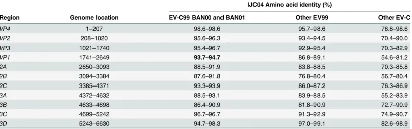
Nearly complete genome was successfully assembled, with 6,648 nucleotides obtained. Despite repetitive attempts, 5'UTR (749 nt) and 3'UTR (72 nt) were not obtained (Table 1). The ORF (Open Reading Frame) encodes for a polyprotein of 2,210 amino acids, and is presumably cleaved in three polyproteins P1 (VP1-VP4, 883 amino acids), P2 (2A-2C, 574 amino acids) and P3 (3A-3D, 753 amino acids) (Table 1).
Similarity plot analysis was used to evaluate nucleotide distance between IJC04, the geneti-cally closest EV-C99 BAN00 and BAN01 strains over the genome (Fig 2). In the P1 and P2 cod-ing regions IJC04 most similar to EV-C99 BAN00 (EF015008) and BAN01 (EF015009). In the P3 there was no single enterovirus sequence clearly most similar to IJC04 (including other Genbank sequences, data not shown).
NGS overview, viruses and bacteria flora
A total of 5,049,883 RNA reads were obtained: 11.7% representing 590,472 sequences reads matched RNA viruses and 88.3% of the other reads corresponded to the host genome (the
Fig 1. Phylogenetic tree constructed using the entire VP1 sequences (approximately 900 bp) of all EV-C serotypes. Study sample is indicated in blue (IJC04) and EV-C99 serotype sequences in red. Accession numbers and information concerning the date of collection and the host species (Hu for human and Cpz for Chimpanzee) are shown. The bootstrap values correspond to 10,000 replicates. Only values above 0.6 are indicated at the nodes. The scale bar represents nucleotide substitutions per site.
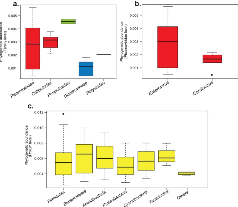
chimpanzee, Pan troglodytes troglodytes). Within the RNA viral reads, 98.95% mapped to mam-malian viruses (584,283 reads), mainly to Picornaviridae (576,942 reads, 98.75%) and the Calici-viridae (7,341 reads, 1.25%) families (Fig 3A). Concerning the other reads, 6,189 (1.02%) matched to plant viruses of the Pospiviroidea (6006 reads) and the Potyviridae families (35 reads) (Fig 3A). Finally, 148 reads (0.02%) matched to insect viruses of the family Dicistroviridae (Fig 3A).
Within the Picornaviridae family level, reads were distributed among two genera: the Enterovirus genus, represented by 576,801 reads (99.6%), and the Cardiovirus genus, repre-sented by 141 reads (0.4%) (Fig 3B). Within the Enterovirus genus, the EV-C species was the
Table 1. Pairwise amino acid sequences identities between IJC04 isolate and other EV-C prototype strains.
IJC04 Amino acid identity (%)
Region Genome location EV-C99 BAN00 and BAN01 Other EV99 Other EV-C
VP4 1–207 98.6–98.6 95.7–98.6 76.8–98.6 VP2 208–1020 95.6–96.3 93.4–94.5 70.4–90.0 VP3 1021–1740 95.4–96.7 92.9–95.4 70.3–82.9 VP1 1741–2649 93.7–94.7 86.8–89.1 54.6–81.2 2A 2650–3093 88.5–91.9 83.8–88.5 70.3–85.8 2B 3094–3384 87.6–91.8 76.8–80.4 56.7–80.4 2C 3385–4371 93.3–93.9 86.0–87.2 76.3–86.9 3A 4372–4632 88.5–93.1 83.9–88.5 55.2–83.9 3B 4633–4698 86.4–90.9 81.8–90.9 72.7–90.9 3C 4699–5242 96.7–96.7 91.3–92.9 74.9–90.7 3D 5243–6630 94.7–98.3 97.0–99.1 82.6–98.9 doi:10.1371/journal.pone.0136700.t001
Fig 2. Similarity plot of complete coding region of EV-C99 chimpanzee isolate IJC04 with two EV-C99 sequences from Bangladesh (BAN00 and BAN01), one from China (HT-XEBGH09F), and the next closest serotype Coxsackievirus (CVA24). Similarity plot was performed using a sliding window of 400 nt moving with 20 nt-steps. The coding region structure is provided above the plot.
most represented in the sample with 541,915 reads (93.95%). The resting reads corresponded to the three other following species EV-A, B and D. Noteworthy, enterovirus 5’ non-translated region (ca. 750 nt, or 10% of the genome) is not species-specific, therefore this finding does not assume presence of additional enteroviruses in the sample.
For the microbiome, out of the total of 589,899 reads obtained, 299,078 (50.7%) reads remained after eliminating unreliable (non-bacterial) or poor quality reads. Sequences were then assembled and a total of 292,948 consensus sequences were obtained. Among these reads,
Fig 3. Phylogenetic abundance variations of viral and bacterial reads obtained by NGS in the sample IJC04, a. Viral families phylogenetic abundances, b. Viral genus phylogenetic abundances within the Picornaviridae family, c. Bacterial phylogenetic phyla abundances. Box plots are classified from the most to the least abundant group. Boxes represent interquartile range between first and third quartiles and the line inside represents the median. Squares represent outliers beyond the whiskers. The y-axis represents the logarithms of percentage to normalize the values.
31 reads did not map to any known bacteria sequences. The rest matched Gram-positive bacte-ria phyla including Firmicutes (232,734 reads, 79.4%), Actinobactebacte-ria (22,380 reads, 7.9%) and Gram-negative bacteria phyla such as Bacteroidetes (27,899 reads, 9.5%), Proteobacteria (4,443 reads, 1.51%) of and Tenericutes (2,313 reads, 0.79%) (Fig 3C). Finally, 2,397 reads (0.81%) matched photosynthetic bacteria belonging to the phylum of Cyanobacteria.
Discussion
In this study, we report the first identification of an EV-C associated with AFP in a chimpan-zee. Enterovirus was detected in fecal sample only from the sick chimpanzee, but not other 11 apes in the group (data not shown). Phylogenetic analysis of the complete capsid gene allowed the identification of the potential causative agent as EV-C99.
In order to rule out other etiologic agents, we investigated the presence of other viral and bacterial agents by mapping metagenomic reads obtained from NGS to available reference genomes. The virus phylogenetic composition of the fecal sample showed that reads corre-sponded mainly to mammalian viruses belonging to Caliciviridae and Picornaviridae families. The rest of the reads corresponded to plant and insect viruses, which are very likely derived from chimpanzee's diet and could not be associated to AFP syndrome. Within the Picornaviri-dae family, the Enterovirus genus (99%) was the principal representative in terms of reads cov-erage, followed by the Cardiovirus genus with only 0.4% of reads detected. Members of the Cardiovirus genus have been associated in non-polio AFP in human populations [35]. How-ever, we could not consider these viruses as the causative agent of the AFP in this chimpanzee. Indeed, a study showed that the viral load was correlated to the number of reads detected by sequencing [36]. The Caliciviridae family (1.25%) is composed of viruses mainly responsible for gastroenteritis in human and animal (such as pigs and bats) populations [37–39]. It has to be noticed that in a recent study, we characterized a sapovirus isolated from the same chimpan-zee [40]. However, in the current study, this virus was not considered as the causative agent of the AFP observed in this chimpanzee because there are no reports of caliciviruses causing neu-rological manifestations in humans or primates. Concerning the bacteria flora, mainly com-mensal bacteria have been identified (95–96%), the resting reads corresponding to telluric bacteria. Indeed, the results showed that the Firmicutes, the Bacteroidetes and the Actinobac-teria phyla constituted the vast majority of this chimpanzee’s gut microbiota (Fig 3C). The Fir-micutes phylum is composed of bacteria with various functions and some are known to be responsible of diseases in human and other vertebrates such as Clostridium botulinum (causing a neuroparalytic disease called botulism) [41]. None of the reads obtained in this study matched with Clostridium botulinum. Interestingly, the same bacterial flora composition in terms of abundance and diversity is commonly observed in humans [42], as well in gorillas and chimpanzees gut microbiota [43]. In summary, it seems that none of the bacteria detected in this analysis could be responsible of the AFP in this chimpanzee.
As a summary, considering the abundance of EV-C in the fecal sample, neuropathogenic potential of poliovirus and (albeit rarely) of other enteroviruses, EV-C99 is the possible causa-tive agent of the AFP this chimpanzee. However, due to a lack of available serum, detection of antibodies targeting EV-C99 could not performed to better assume this hypothesis. This is the first report of a great ape presenting AFP symptoms associated with a human non-polio EV, member of EV-C species. Indeed, historically, the first strain of EV-C99 was identified in Ban-gladesh in 2000 [33]. Subsequently several other EV-C99 were identified or isolated during poliovirus surveillance from healthy humans or AFP patients in Asia, Europe, North America and Africa [44]. Since this detection, no outbreak has been recorded and no confirmed symp-tomatic cases were reported. Otherwise, EV-C99 has been identified in one synanthropic
rhesus macaque (Macaca mulatta) in Bangladesh [18]. In 1964, the Yerkes Regional Primate Research Center (Florida, USA) reported clinical manifestations of AFP in apes caused by poliovirus type 1 [45]. Two years later, another epidemic of paralytic poliomyelitis occurred among chimpanzees of the Gombe National Park (Tanzania), months after a polio outbreak in human population (cited by Dowdle, 1997) [46]. However, laboratory investigations were not performed to determine the exact causative pathogen involved.
The identification of an EV99, commonly circulating in humans, in a captive chimpanzee suggests interspecies transmission from human to chimpanzee. This finding is another exam-ple consistent with studies, which demonstrated that non-human primates living in close con-tact with humans are exposed to the risk of infection with human pathogens [18,47,48]. Even though chimpanzees are known to be infected by human and simian EVs, it has to be noticed that this is the first observation of an AFP in a chimpanzee associated to a human EV strain.
Acknowledgments
We are grateful to the people who helped us, especially Luc Abate and Majoline Tchioffo in the research unit MIVEGEC (IRD, France).
Author Contributions
Performed the experiments: IMM TB SB CB VR RA DC PD CA LB. Analyzed the data: IMM ANL LL NB. Contributed reagents/materials/analysis tools: EML FR JFD CD FP JNF BSS. Wrote the paper: IMM ANL VR EML.
References
1. Formenty P, Hatz C, Le Guenno B, Stoll A, Rogenmoser P, Widmer A. Human infection due to Ebola virus, subtype Cote d'Ivoire: clinical and biologic presentation. Journal of Infectious Diseases. 1999; 179(Supplement 1):S48–S53.
2. Leroy EM, Rouquet P, Formenty P, Souquiere S, Kilbourne A, Froment JM, et al. Multiple Ebola virus transmission events and rapid decline of central African wildlife. Science. 2004; 303(5656):387–90. doi:
10.1126/science.1092528PMID:14726594.
3. Keele BF, Van Heuverswyn F, Li Y, Bailes E, Takehisa J, Santiago ML, et al. Chimpanzee reservoirs of pandemic and nonpandemic HIV-1. Science. 2006; 313(5786):523–6. doi:10.1126/science.1126531
PMID:16728595; PubMed Central PMCID: PMC2442710.
4. Kaur T, Singh J, Tong S, Humphrey C, Clevenger D, Tan W, et al. Descriptive epidemiology of fatal respiratory outbreaks and detection of a human-related metapneumovirus in wild chimpanzees (Pan troglodytes) at Mahale Mountains National Park, Western Tanzania. American journal of primatology. 2008; 70(8):755–65. doi:10.1002/ajp.20565PMID:18548512.
5. Kondgen S, Schenk S, Pauli G, Boesch C, Leendertz FH. Noninvasive monitoring of respiratory viruses in wild chimpanzees. EcoHealth. 2010; 7(3):332–41. doi:10.1007/s10393-010-0340-zPMID:
20865440.
6. Leendertz FH, Lankester F, Guislain P, Neel C, Drori O, Dupain J, et al. Anthrax in Western and Central African great apes. American journal of primatology. 2006; 68(9):928–33. doi:10.1002/ajp.20298
PMID:16900500.
7. Gilardi KV, Oxford KL, Gardner-Roberts D, Kinani JF, Spelman L, Barry PA, et al. Human herpes sim-plex virus type 1 in confiscated gorilla. Emerging infectious diseases. 2014; 20(11):1883–6. doi:10. 3201/eid2011.140075PMID:25341185; PubMed Central PMCID: PMC4214296.
8. Harvala H, Sharp CP, Ngole EM, Delaporte E, Peeters M, Simmonds P. Detection and genetic charac-terization of enteroviruses circulating among wild populations of chimpanzees in Cameroon: relation-ship with human and simian enteroviruses. J Virol. 2011; 85(9):4480–6. doi:10.1128/JVI.02285-10
PMID:21345956; PubMed Central PMCID: PMC3126250.
9. Harvala H, Van Nguyen D, McIntyre C, Ahuka-Mundeke S, Ngole EM, Delaporte E, et al. Co-circulation of enteroviruses between apes and humans. The Journal of general virology. 2014; 95(Pt 2):403–7. doi:10.1099/vir.0.059048–0PMID:24189620.
10. Gillespie TR, Nunn CL, Leendertz FH. Integrative approaches to the study of primate infectious dis-ease: implications for biodiversity conservation and global health. American journal of physical anthro-pology. 2008;Suppl 47:53–69. doi:10.1002/ajpa.20949PMID:19003885.
11. Leendertz FH, Pauli G, Maetz-Rensing K, Boardman W, Nunn C, Ellerbrok H, et al. Pathogens as driv-ers of population declines: the importance of systematic monitoring in great apes and other threatened mammals. Biological Conservation. 2006; 131(2):325–37.
12. Woodford MH, Butynski TM, Karesh WB. Habituating the great apes: the disease risks. Oryx. 2002; 36 (02):153–60.
13. Pusey AE, Pintea L, Wilson ML, Kamenya S, Goodall J. The Contribution of Long‐Term Research at Gombe National Park to Chimpanzee Conservation. Conservation Biology. 2007; 21(3):623–34. PMID:
17531041
14. Knowles NJ, Hovi T, Hyypiä T, King AMQ, Lindberg AM, Pallansch MA, et al. Family Picornaviridae. In Virus Taxonomy: Classification an Nomenclature of Viruses: Ninth Report of the International Commit-tee on Taxonomy of Viruses. 2012:855–80. Ed: King A.M.Q., Adams M.J., Carstens E.B. and Lefkowitz E.J. San Diego: Elsevier.
15. Hoffert WR, Bates ME, Cheever FS. Study of enteric viruses of simian origin. Am J Hyg. 1958; 68 (1):15–30. PMID:13559208.
16. Kalter SS. Animal "orphan" enteroviruses. Bull World Health Organ. 1960; 22:319–37. PMID:
14404195; PubMed Central PMCID: PMC2555328.
17. Nix WA, Jiang B, Maher K, Strobert E, Oberste MS. Identification of enteroviruses in naturally infected captive primates. J Clin Microbiol. 2008; 46(9):2874–8. doi:10.1128/JCM.00074-08PMID:18596147; PubMed Central PMCID: PMC2546737.
18. Oberste MS, Feeroz MM, Maher K, Nix WA, Engel GA, Hasan KM, et al. Characterizing the picornavi-rus landscape among synanthropic nonhuman primates in Bangladesh, 2007 to 2008. J Virol. 2013; 87 (1):558–71. doi:10.1128/JVI.00837-12PMID:23097448; PubMed Central PMCID: PMC3536389. 19. Grard G, Drexler JF, Lekana-Douki S, Caron M, Lukashev A, Nkoghe D, et al. Type 1 wild poliovirus
and putative enterovirus 109 in an outbreak of acute flaccid paralysis in Congo, October-November 2010. Euro surveillance: bulletin Europeen sur les maladies transmissibles = European communicable disease bulletin. 2010; 15(47). PMID:21144443.
20. Lukashev AN, Drexler JF, Kotova VO, Amjaga EN, Reznik VI, Gmyl AP, et al. Novel serotypes 105 and 116 are members of distinct subgroups of human enterovirus C. The Journal of general virology. 2012; 93(Pt 11):2357–62. doi:10.1099/vir.0.043216–0PMID:22894922.
21. Dierssen U, Rehren F, Henke-Gendo C, Harste G, Heim A. Rapid routine detection of enterovirus RNA in cerebrospinal fluid by a one-step real-time RT-PCR assay. Journal of clinical virology: the official pub-lication of the Pan American Society for Clinical Virology. 2008; 42(1):58–64. doi:10.1016/j.jcv.2007. 11.016PMID:18164234.
22. Oberste MS, Maher K, Kilpatrick DR, Pallansch MA. Molecular evolution of the human enteroviruses: correlation of serotype with VP1 sequence and application to picornavirus classification. J Virol. 1999; 73(3):1941–8. PMID:9971773; PubMed Central PMCID: PMC104435.
23. Kimura M. A simple method for estimating evolutionary rates of base substitutions through comparative studies of nucleotide sequences. J Mol Evol. 1980; 16(2):111–20. PMID:7463489.
24. Tamura K, Peterson D, Peterson N, Stecher G, Nei M, Kumar S. MEGA5: molecular evolutionary genet-ics analysis using maximum likelihood, evolutionary distance, and maximum parsimony methods. Mol Biol Evol. 2011; 28(10):2731–9. doi:10.1093/molbev/msr121PMID:21546353; PubMed Central PMCID: PMC3203626.
25. Berthet N, Reinhardt AK, Leclercq I, van Ooyen S, Batejat C, Dickinson P, et al. Phi29 polymerase based random amplification of viral RNA as an alternative to random RT-PCR. BMC Mol Biol. 2008; 9:77. doi:10.1186/1471-2199-9-77PMID:18771595; PubMed Central PMCID: PMC2535778. 26. Simpson JT, Wong K, Jackman SD, Schein JE, Jones SJ, Birol I. ABySS: a parallel assembler for short
read sequence data. Genome Res. 2009; 19(6):1117–23. doi:10.1101/gr.089532.108PMID:
19251739; PubMed Central PMCID: PMC2694472.
27. Huang X, Madan A. CAP3: A DNA sequence assembly program. Genome Res. 1999; 9(9):868–77. PMID:10508846; PubMed Central PMCID: PMC310812.
28. Lole KS, Bollinger RC, Paranjape RS, Gadkari D, Kulkarni SS, Novak NG, et al. Full-length human immunodeficiency virus type 1 genomes from subtype C-infected seroconverters in India, with evidence of intersubtype recombination. J Virol. 1999; 73(1):152–60. PMID:9847317; PubMed Central PMCID: PMC103818.
29. Saitou N, Nei M. The neighbor-joining method: a new method for reconstructing phylogenetic trees. Mol Biol Evol. 1987; 4(4):406–25. PMID:3447015.
30. Team RC. R: A language and environment for statistical computing. 2012.
31. Kelly H, Brussen KA, Lawrence A, Elliot E, Pearn J, Thorley B. Polioviruses and other enteroviruses isolated from faecal samples of patients with acute flaccid paralysis in Australia, 1996–2004. Journal of paediatrics and child health. 2006; 42(6):370–6. doi:10.1111/j.1440-1754.2006.00875.xPMID:
16737480.
32. Sadeuh-Mba SA, Bessaud M, Massenet D, Joffret ML, Endegue MC, Njouom R, et al. High frequency and diversity of species C enteroviruses in Cameroon and neighboring countries. J Clin Microbiol. 2013; 51(3):759–70. doi:10.1128/JCM.02119-12PMID:23254123; PubMed Central PMCID: PMC3592076.
33. Brown BA, Maher K, Flemister MR, Naraghi-Arani P, Uddin M, Oberste MS, et al. Resolving ambiguities in genetic typing of human enterovirus species C clinical isolates and identification of enterovirus 96, 99 and 102. The Journal of general virology. 2009; 90(Pt 7):1713–23. doi:10.1099/vir.0.008540–0PMID:
19264596.
34. Jorba J, Campagnoli R, De L, Kew O. Calibration of multiple poliovirus molecular clocks covering an extended evolutionary range. J Virol. 2008; 82(9):4429–40. doi:10.1128/JVI.02354-07PMID:
18287242; PubMed Central PMCID: PMC2293050.
35. Blinkova O, Kapoor A, Victoria J, Jones M, Wolfe N, Naeem A, et al. Cardioviruses are genetically diverse and cause common enteric infections in South Asian children. Journal of virology. 2009; 83 (9):4631–41. doi:10.1128/JVI.02085-08PMID:19193786; PubMed Central PMCID: PMC2668475. 36. Logan G, Freimanis GL, King DJ, Valdazo-Gonzalez B, Bachanek-Bankowska K, Sanderson ND, et al.
A universal protocol to generate consensus level genome sequences for foot-and-mouth disease virus and other positive-sense polyadenylated RNA viruses using the Illumina MiSeq. BMC genomics. 2014; 15:828. doi:10.1186/1471-2164-15-828PMID:25269623; PubMed Central PMCID: PMC4247156. 37. Hansman GS, Saito H, Shibata C, Ishizuka S, Oseto M, Oka T, et al. Outbreak of gastroenteritis due to
sapovirus. J Clin Microbiol. 2007; 45(4):1347–9. doi:10.1128/JCM.01854-06PMID:17267629; PubMed Central PMCID: PMC1865854.
38. Scheuer KA, Oka T, Hoet AE, Gebreyes WA, Molla BZ, Saif LJ, et al. Prevalence of porcine norovi-ruses, molecular characterization of emerging porcine sapoviruses from finisher swine in the United States, and unified classification scheme for sapoviruses. Journal of clinical microbiology. 2013; 51 (7):2344–53. doi:10.1128/JCM.00865-13PMID:23678065; PubMed Central PMCID: PMC3697660. 39. Tse H, Chan WM, Li KS, Lau SK, Woo PC, Yuen KY. Discovery and genomic characterization of a
novel bat sapovirus with unusual genomic features and phylogenetic position. PloS one. 2012; 7(4): e34987. doi:10.1371/journal.pone.0034987PMID:22514697; PubMed Central PMCID: PMC3325917. 40. Mombo IM, Berthet N, Bouchier C, Fair JN, Schneider BS, Renaud F, et al. Characterization of a
gen-ogroup I sapovirus isolated from chimpanzees in the republic of congo. Genome announcements. 2014; 2(4). doi:10.1128/genomeA.00680-14PMID:25035327; PubMed Central PMCID: PMC4102864.
41. Simpson LL. Molecular pharmacology of botulinum toxin and tetanus toxin. Annual review of pharma-cology and toxipharma-cology. 1986; 26:427–53. doi:10.1146/annurev.pa.26.040186.002235PMID:3521461. 42. Arumugam M, Raes J, Pelletier E, Le Paslier D, Yamada T, Mende DR, et al. Enterotypes of the human
gut microbiome. Nature. 2011; 473(7346):174–80. doi:10.1038/nature09944PMID:21508958; PubMed Central PMCID: PMC3728647.
43. Moeller AH, Shilts M, Li Y, Rudicell RS, Lonsdorf EV, Pusey AE, et al. SIV-induced instability of the chimpanzee gut microbiome. Cell host & microbe. 2013; 14(3):340–5. doi:10.1016/j.chom.2013.08. 005PMID:24034619; PubMed Central PMCID: PMC3802538.
44. Bessaud M, Joffret ML, Holmblat B, Razafindratsimandresy R, Delpeyroux F. Genetic relationship between cocirculating Human enteroviruses species C. PloS one. 2011; 6(9):e24823. doi:10.1371/ journal.pone.0024823PMID:21931857; PubMed Central PMCID: PMC3171481.
45. Allmond BW Jr, Froeschle JE, Guilloud NB. Paralytic poliomyelitis in large laboratory primates. Viro-logic investigation and report on the use of oral poliomeylitis virus (OPV) vaccine. American journal of epidemiology. 1967; 85(2):229–39. PMID:6067015.
46. Dowdle WR, Birmingham ME. The biologic principles of poliovirus eradication. The Journal of infectious diseases. 1997; 175 Suppl 1:S286–92. PMID:9203732.
47. Kondgen S, Kuhl H, N'Goran PK, Walsh PD, Schenk S, Ernst N, et al. Pandemic human viruses cause decline of endangered great apes. Current biology: CB. 2008; 18(4):260–4. doi:10.1016/j.cub.2008.01. 012PMID:18222690.
48. Prugnolle F, Rougeron V, Becquart P, Berry A, Makanga B, Rahola N, et al. Diversity, host switching and evolution of Plasmodium vivax infecting African great apes. Proceedings of the National Academy of Sciences of the United States of America. 2013; 110(20):8123–8. doi:10.1073/pnas.1306004110