HAL Id: hal-01325368
https://hal.sorbonne-universite.fr/hal-01325368
Submitted on 2 Jun 2016
HAL is a multi-disciplinary open access
archive for the deposit and dissemination of
sci-entific research documents, whether they are
pub-lished or not. The documents may come from
teaching and research institutions in France or
abroad, or from public or private research centers.
L’archive ouverte pluridisciplinaire HAL, est
destinée au dépôt et à la diffusion de documents
scientifiques de niveau recherche, publiés ou non,
émanant des établissements d’enseignement et de
recherche français ou étrangers, des laboratoires
publics ou privés.
SRSF2-p95 hotspot mutation is highly associated with
advanced forms of mastocytosis and mutations in
epigenetic regulator genes
Katia Hanssens, Fabienne Brenet, Julie Agopian, Sophie Georgin-Lavialle,
Gandhi Damaj, Laure Cabaret, Maria-Olivia Chandesris, Paulo de Sepulveda,
Olivier Hermine, Patrice Dubreuil, et al.
To cite this version:
Katia Hanssens, Fabienne Brenet, Julie Agopian, Sophie Georgin-Lavialle, Gandhi Damaj, et al..
SRSF2-p95 hotspot mutation is highly associated with advanced forms of mastocytosis and mutations
in epigenetic regulator genes. Haematologica, Ferrata Storti Foundation, 2014, 99 (5), pp.830-835.
�10.3324/haematol.2013.095133�. �hal-01325368�
Introduction
Mastocytosis is a rare and clonal hematopoietic disorder described as the accumulation of abnormal mast cells in the bone marrow (BM).1,2In adults, mastocytosis most often pres-ents as a persistent systemic disorder of variable course and prognosis.3,4 Disease phenotypes range from indolent to aggressive and are defined by WHO criteria: mainly B- and C-findings that describe the extent of organ and tissue damage resulting from systemic mast cell infiltration. In approximate-ly 40% of cases, systemic mastocytosis is diagnosed in con-junction with associated clonal hematologic non-mast cell lin-eage diseases (AHNMD) which include myelodysplastic syn-dromes (MDS), myeloproliferative neoplasm (MPN), as well as both acute and chronic forms of myeloid leukemia (AML, CML, CMML).4,5
It remains unclear why in some cases mastocytosis evolves aggressively while in other cases the disease remains indo-lent. Efforts to discriminate and to predict the clinical course of mastocytosis have uncovered genetic mutations that figure prominently in this disease. KITD816V mutation is the most common (>80% of mastocytosis cases) and is thought to drive the expansion of affected clones towards the mast cell lineage,6 but does not segregate with advanced disease. In contrast, TET2 mutation, found in approximately 20% of patients, is associated with aggressive forms of mastocytosis.7
Mutations in other epigenetic modifiers have been described, but so far they have not been clearly associated to any partic-ular form of disease and, overall, their prognostic relevance is not clear.8 More recently, a hotspot mutation in SRSF2, a com-ponent of the RNA splicing machinery, has been identified and associated with leukemic transformation.9,10 Among myelodysplastic syndromes and other hematologic disorders, SRSF2 mutation is most frequent in CMML, with reports ranging from 28.4% to 47.2%.11Like TET2, SRSF2 mutation occurs early in disease ontogeny and has been dubbed a founder mutation.12As such, SRSF2 mutation is thought to pre-dispose early progenitor cells to malignant selection, per-haps via its role in the acetylation/phosphorylation network and as an important regulator of DNA stability and mRNA splicing.13We have now sequenced for SRSF2 mutation in our cohort of mastocytosis patients, previously characterized for both KIT and TET2 mutations,7and have revealed a striking association between SRSF2 mutation and advanced disease types.
Methods
Patients’ data
Seventy-two patients (35 men; 37 women) with mastocytosis diag-nosis as defined by the WHO criteria14were enrolled in a prospective
national multicenter study between 2005 and 2013. The cohort
con-©2014 Ferrata Storti Foundation. This is an open-access paper. doi:10.3324/haematol.2013.095133. The online version of this article has a Supplementary Appendix.
Manuscript received on July 16, 2013. Manuscript accepted on January 2, 2014. Correspondence: erinn.soucie@inserm.fr
Mastocytosis is a rare and chronic disease with phenotypes ranging from indolent to severe. Prognosis for this dis-ease is variable and very few biomarkers to predict disdis-ease evolution or outcome are currently known. We have performed comprehensive screening in our large cohort of mastocytosis patients for mutations previously found in other myeloid diseases and that could serve as prognostic indicators. KIT, SRSF2-P95 and TET2 mutations were by far the most frequent, detected in 81%, 24% and 21% of patients, respectively. Where TET2 and SRSF2-P95 mutation both correlated with advanced disease phenotypes, SRSF2-P95 hotspot mutation was found almost exclusively in patients diagnosed with associated clonal hematologic non-mast cell disease. Statistically, TET2 and SRSF2-P95 mutations were highly associated, suggesting a mechanistic link between these two factors. Finally, analysis of both clonal and sorted cell populations from patients confirms the presence of these mutations in the mast cell component of the disease, suggests an ontological mutation hierarchy and provides evidence for the expansion of multiple clones. This highlights the prognostic potential of such approaches, if applied systematically, for delineating the roles of specific mutations in predisposing and/or driving distinct disease phenotypes.
SRSF2-p95 hotspot mutation is highly associated with advanced forms
of mastocytosis and mutations in epigenetic regulator genes
Katia Hanssens,1,2Fabienne Brenet,1Julie Agopian,1,2Sophie Georgin-Lavialle,2,3,4Gandhi Damaj,5 Laure Cabaret,2,6 Maria Olivia Chandesris,2,6Paulo de Sepulveda,1 Olivier Hermine,2,3,6Patrice Dubreuil,1,2*§and Erinn Soucie1*§
1Inserm U1068, Centre de Recherche en Cancérologie de Marseille, Signalisation, Hématopoïèse et Mécanismes de l’Oncogenèse,
Centre de Référence des Mastocytoses, Institut Paoli-Calmettes, Aix-Marseille Université UM 105, CNRS UMR7258, Marseille;
2Centre de Référence des Mastocytoses, Hôpital Necker-Enfants-Malades, Université Paris Descartes, Sorbonne Paris Cité, Paris;
3CNRS UMR 8147, Hôpital Necker-Enfants-Malades, Université Paris Descartes, Sorbonne Paris Cité, Paris; 4Service de Médecine
Interne, Hôpital Tenon, Université Paris 6, Pierre et Marie Curie, Paris; 5Département d'Hématologie, Centre Hospitalier
Universitaire, Amiens; and 6Service d'Hématologie Adulte, Hôpital Necker-Enfants-Malades, Université Paris Descartes, Sorbonne
Paris Cité, France
sists of patients diagnosed with cutaneous mastocytosis (CM), CM (type TMEP), indolent SM (ISM), systemic mastocytosis with AHNMD (SM-AHNMD), aggressive SM (ASM), mast cell leukemia (MCL) and mast cell sarcoma (Online Supplementary Table
S1).1-3 AHNMD diagnosis for each patient has been indicated
(Figure 1), and this is detailed in the Online Supplementary Table S1. Patients were further grouped using the operational terms “advanced” and “non-advanced” to account for the number of patients in certain classifications, e.g. MCL and mast cell sarcoma that were not large enough for statistical analysis. Statistical analy-sis of predictive factors (anemia, blast count, thrombocytopenia, hypoalbuminemia etc.), KIT and TET2 mutation for this cohort have been presented elsewhere.7All patients were included in a
mastocytosis pathophysiological study which started in 2003 and is sponsored by the Association For Initiative and Research on Mast cell and Mastocytosis (AFIRMM). The study was approved by the Necker Hospital ethical committee, and carried out accord-ing to the Declaration of Helsinki. Each patient provided informed consent.
Mutation screening
Mutation analysis for KIT and NRAS, KRAS and TET2 has been described previously.15,16 Other methods have been previously
described as follows: DNA Sanger-sequencing of exon-coding sequences of ASXL1, CBL, DNMT3A, IDH1, IDH2, JAK2 and
EZH2,17SRSF2, U2AF1, ZSRSR2 d,11and SF3B1.18
Clonal analysis
Leukocytes were purified using FicollR(Sigma) from fresh bone
marrow from patients and plated at low density in methocult medium (H4035 without Epo, StemCell Technologies). Individual colonies were isolated at Day 10-12 of culture and DNA was iso-lated for mutation screening.
FACS sorting
Fresh or frozen whole bone marrow biopsy material from patients was stained with the following antibody cocktail: anti-humanCD3-ECD, anti-humanCD14-alexa647, anti-humanCD25-PE and anti-humanFcepsilonGR1a-FITC (all from BD Biosciences). Cells were sorted on an LSRII and DIVATM (Becton-Dickinson)
software was used. Sorted cell populations were directly lysed and DNA was isolated for mutation screening.
Statistical analysis
Statistical comparisons for predictive factors and mutations were based on Fisher’s exact test. All reported P values were two-tailed with confidence intervals of 95%. Survival data were ana-lyzed using GraphPad Prism software version 5.01 (GraphPad Software Inc., San Diego, CA, USA) and both the log rank (Mantel-Cox) and Gehan-Breslow-Wilcoxon tests were applied to survival curves to determine significant differences.
Results
SRSF2P95 hotspot mutation is highly correlated
with advanced forms of mastocytosis
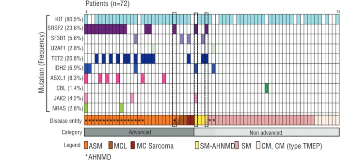
Total bone marrow or peripheral blood was collected from a cohort of 72 patients with mastocytosis, catego-rized according to the WHO classification and for advanced or non-advanced disease (Online Supplementary
Table S1). Biopsied material from these patients was
screened for mutation in genes commonly mutated in mastocytosis or other myeloid diseases (Figure 1). After
KIT (81%), the SRSF2-P95 hotspot mutation was the most
frequent mutation found in these patients (17 of 72; 23.6%). We detected four different mutations of the NSRSF2-P95 codon in patients: SRSF2-P95H, SRSF2-P95L, SRSF2-P95T and SRSF2-P95R (Online Supplementary Table
S1). We have previously reported an association between
TET2 and advanced systemic mastocytosis7 and, like TET2, we also saw a significant correlation between
SRSF2-P95 mutation and advanced disease (P<0.0001) (Table 1). Unlike TET2 mutation, that was found only in patients positive for KIT mutation,73 patients who were negative for KIT mutation were positive for SRSF2-P95 mutation. SRSF2 mutation also coincides significantly with the presence of AHNMD (15 of 17 patients with SRSF2-P95 mutation present with AHNMD), but not with any particular form of AHNMD (Table 2).
Advanced forms of mastocytosis are associated
with short overall survival
Survival curves were generated to compare the survival of mastocytosis patients with advanced disease versus non-advanced disease (Figure 2A). We found a significant reduction in the survival time at diagnosis for patients diagnosed with advanced forms of mastocytosis (P>0.001). Since the vast majority of patients with SRSF2-P95 mutation have advanced disease types, to eliminate this bias, we next generated survival curves for patients diagnosed with advanced disease types only to compare survival time of patients with or without SRSF2-P95 muta-tion (Figure 2B). We found no significant difference between the overall survival of patients with advanced disease and SRSF2-P95 MUT compared to patients with advanced disease and SRSF2-P95 WT (P>0.5) (Figure 2B). Taken together, advanced disease diagnosis and not SRSF2-P95 mutation is associated with ASM and AHNMD
Table 1.SRSF2-P95 mutation is correlated with advanced mastocyto-sis.
Non-advanced Advanced Total P
SRSF2 mutation <0.0001 (F)
N. 41 31 72
WT 39 (95%) 16 (52%) 55 (76%)
P95H/L/R/T 2 (5%) 15 (48%) 17 (24%)
(F) Fisher’s exact test.
Table 2.Contingency analysis for SRSF2-P95 mutation and AHNMD.
SRSF2 WT SRSF2-P95 Total P Associated disease <0.0001 (F) N. 55 17 72 no AHNMD 46 (84%) 2 (12%) 48 AHNMD 9 (16%) 15 (88%) 24 Type of AHNMD 0.4731 (C) N. 9 15 24 CML 1 0 1 AML 1 2 3 CMML 0 4 4 MDS 2 3 5 MSD (AREB) 3 2 5 MPN (SMG myeloid) 1 2 3 MPN/MDS 1 1 2 Waldenstrom 0 1 1
SRSF2-P95 mutation appears to be the dominant factor for predicting shorter overall survival times in patients.
Interestingly, 3 patients positive for SRSF2-P95 mutation at the original time of diagnosis developed an AHNMD during the course of this study (within 2 years). Patient n. M40 originally diagnosed with SM developed AHNMD (MDS), Patient n. 1318 originally diagnosed with ISM pro-gressed to SM-AHNMD, and Patient n. D60 originally diagnosed with ASM developed MCL (Figure1, dotted boxes, and Online Supplementary Table S1).
SRSF2-P95 mutation is significantly associated with
mutations in epi-regulators
In addition to KIT and SRSF2-P95, amplicon screening was also performed for other known mutations: SF3B1,
U2AF1 and ZRSR2 (splicing factors), TET2, IDH1, IDH2, AXSL1, DNMT3A, EZH2 (epigenetic regulators), as well as JAK2, CBL, NRAS and KRAS (Figure 1 and Online Supplementary Table S1).
Generally, more mutations were found in patients with advanced disease than patients with non-advanced dis-ease. After KIT, SRSF2-P95 and TET2 mutation were by far the most frequent mutations found in patients: 24% and 21%, respectively. Interestingly, statistical analysis shows that TET2 mutation is significantly correlated to SRSF2-P95 mutation in these patients (P<0.01) (Table 3), and an even more significant correlation is found when mutations in epigenetic factors are considered as a whole (mutation of SRSF2 in combination with at least one epi-genetic factor; P<0.0001).
Consistent with a previous report showing that SF3B1 mutations are infrequent in mast cell diseases,19 SF3B1 mutations were detected in only 4 (5.6%) of our patients: 2 patients had mutations in exon 14 (codon 666 and the other at codon 663), and 2 patients had mutations in exon 15. We also identified 2 patients with mutation in U2AF1 (both at codon 101 in exon 2). Interestingly, mutations in
splicing factors SRSF2, SF3B1 and U2AF1 are mutually exclusive in these patients (Figure 1 and Online
Supplementary Table S1).
SRSF2-P95 mutation is present in mast cells
Given the strong association between SRSF2 mutation and AHNMD, to ensure that the SRSF2 mutation was present in the mast cell component of the disease, we sort-Figure 1.The frequencies and distribution of 10 gene mutations in 72 patients with mastocytosis. Each column represents one individual patient with mutated gene(s) shown by different colored bars. The last two rows describe the disease classification for each patient. Half boxes reveal evolved diagnosis during the course of this study and dotted lines highlight those patients with SRSF2-P95 mutation and disease evolution. No mutations were found for ZRSR2, KRAS, IDH1, DNMT3A or EZH2.
Figure 2.Survival proportions of patients with advanced forms of mastocytosis. (A) Percent survival of patients with advanced disease type and non-advanced disease type in years post-disease diagnosis. (B) Percent survival of patients with advanced disease phenotype, with or without SRSF2-P95 mutation (Adv SRSF2-P95 MUT or Adv SRSF2-P95 WT), in years post-disease diagnosis. P-values were cal-culated using the log rank (Mantel-Cox) test.
A
B
Survival among aggressive forms: survival proportions Advanced Non-advanced
Patients (n=72)
ASM MCL MC Sarcoma
Advanced Non advanced
M u ta ti o n ( F re q u e n c y ) *AHNMD
SM-AHNMD SM CM, CM (type TMEP)
Legend Category Disease entity P e rc e n t s u rv iv a l P e rc e n t s u rv iv a l P<0.001 Years Years 0 5 10 15 20 25 0 5 10 15 P>0.05 Adv SRSF2-P95 MUT Adv SRSF2 WT Survival of data 1: survival proportions
150 100 50 0 150 100 50 0 KIT (80.5%) SRSF2 (23.6%) SF3B1 (5.6%) U2AF1 (2.8%) TET2 (20.8%) IDH2 (6.9%) ASXL1 (8.3%) CBL (1.4%) JAK2 (4.2%) NRAS (2.8%)
ed cell populations from total bone marrow of 7 patients with advanced disease phenotypes for genotype analysis (Figure 3A). Genomic DNA from the three collected pop-ulations, T cells, monocytes and mast cells was isolated and genotyped for KIT, TET2 and SRSF2 mutations (Figure 3B). In all 7 patients for whom sorted populations,
KIT, SRSF2 and TET2 mutations, were detected in whole
bone marrow biopsies (Online Supplementary Table S1) as well as in the mast cell and monocyte compartments. In all patients, KIT, TET2 and SRSF2 mutations were variably present in T cells, depending on when, during hematopoi-etic differentiation, the individual mutations were acquired. For patient M40, in whom KIT mutation was not detected, we detected JAK2 V617F mutation in mast cells and monocytes, but not in T cells. Finally, in patients presenting with two different TET2 mutations, both mutations were present in positive cell populations. Interestingly, in one patient (Patient n. 1445), we detected
SRSF2 mutation in T cells where TET2 and KIT mutations
were present only in mast cells and monocytes.
SRSF2-P95 mutation can occur early or late during
clonal evolution
To determine the relative timing of SRSF2 mutation dur-ing clonal evolution, we examined individual colonies derived from total bone marrow of 2 patients with
advanced disease and KITD816V, TET2 and SRSF2-P95 mutations. Mutational analysis of single colonies distin-guished three different patterns of mutation accumulation (Figure 3C, arrows). In both patients, we identified clones harboring TET2 mutation alone, TET2 and SRSF2, TET2 and KIT or all three mutations. These data are consistent with TET2 mutation preceding both SRSF2 and KIT muta-tion in both patients, and either SRSF2 or KIT mutamuta-tion occurring next. Interestingly, where mutation screening of whole bone marrow for Patient n. F50 identified SRSF2-P95 mutation is associated with ASM and AHNMD
Table 3. Contingency analysis for SRSF2-P95 mutation and Epi-regu-lator mutations. SRSF2 WT SRSF2-P95 Total P Tet2 mutation 0.0049 (F) N. 55 17 72 WT 48 (87%) 9 (53%) 57 MUT 7 (13%) 8 (47%) 15 Epi-regulators* <0.0001 (F) N. 55 17 72 at least 1 9 (16%) 13 (77%) 22 0 46 (84%) 4 (23%) 50
(F) Fisher’s exact test; *TET2, IDH1/2, ASXL1.
Figure 3. SRSF2, TET2 and KIT status of sorted cell populations for 7 patients with ASM. (A) Total bone marrow of 7 patients with advanced disease phenotypes were FACS sort-ed using lineage-specific antibodies. Figure shows a representative FACS profile from one patient. (B) DNA was isolated from the three collected pop-ulations, T cells, monocytes and mast cells and genotyped for KIT, TET2 and SRSF2 mutation. (C) Analysis of sin-gle colonies for mutations in SRSF2, TET2 and KIT. Single colony-forming units derived from mononuclear cells of whole bone marrow, were isolated and analyzed individually for the pres-ence of SRSF2, TET2 and KITD816V mutations. Each colony is represent-ed by a dot that is placrepresent-ed in boxes according to genotype. The unique patient numbers and the diagnoses are shown above the corresponding boxes. Light gray arrows indicate the suggested order of mutation events. A black dot indicates that SRSF2-P95H mutation was detected rather than SRSF2-P95L. Monocytes CD3 Patient: F50 (ASM-AHNMD) SRSF2P95L SRSF2P95H SRSF2 P95H SRSF2WT SRSF2WT Patient: V20 (ASM-AHNMD) FceRa1 T-cells C D 1 4 C D 2 5 Mast cells A B C
KITWT KITWT KITD816V
TET2WT TET2Q1389*TET2Q1389*
KITWT KITWT KITWT KITWT KITD816BV
TET2WT TET2Q734*TET2M1800DTET2M1800DTET2M1800D
KITD816V, TET2 Q1389* and SRSF2-P95L mutations (Online Supplementary Table S1), clonal analysis revealed 5 of 14 colonies harboring a distinct SRSF2-P95H mutation. Both SRSF2-P95L and SRSF2-P95H colonies harbored TET2 Q1389* mutation, but only SRSF2-P95L colonies were positive for KITD816V mutation.
Discussion
We now report that the SRSF2-P95 hotspot mutation is highly correlated with advanced forms of mastocytosis, and, in contrast to TET2, is associated almost exclusively with, and might predict, the onset of AHNMD.
We also find a small number of patients negative for the SRSF2-P95 mutation but harboring a mutation in another splicing factor, either SF3B1 or U2AF1. Combined, spliceosome mutations were found in as many as 32% of patients, pointing to a pathogenic role for abnormal RNA splicing in SM. In addition to its role as an SR protein in promoting alternative exon inclusion and integrating other steps in RNA metabolism,20a unique role for SRSF2 has been described in regulating PolII pausing and elongation at promoters.21,22SF3B1 is part of a much larger complex associated with the catalytic activity of the spliceosome, and U2AF1 is an auxiliary U2-factor involved in splice-site recognition. So far, among these factors, only U2AF1 has been directly associated with the deregulated splicing of a specific, cancer relevant target: EZH2.23Future studies to address the global or specific role of gene splicing in hema-tologic diseases should be informative in dissecting the molecular contribution of SRSF2 and other splicing muta-tions in these contexts.
Extensive mutation analysis of this cohort has also revealed an association between SRSF2 mutation and mutation in genes whose products function in modifying the epigenome (epi-regulators), including TET2. This is consistent with a previous report showing an association between spliceosome mutations and mutations in epige-netic modifiers.24Mechanistically, splicing is often tightly coupled with transcription and recent work suggests that alternative splicing might be affected by chromatin struc-ture and histone modification.25Together with their role in regulating DNA stability, it is interesting to speculate that
SRSF2 and epigenetic modifier mutations may act
syner-gistically to promote advanced disease. At present, how-ever, it is unclear why these mutations coincide with such high frequency.
Interestingly, from a clinical perspective, 3 patients with SRSF2-P95 mutation were re-diagnosed with more severe disease and AHNMD during the course of this study. Pursuant to reports of an association between SRSF2 mutation, poor prognosis and leukemic progression of MPNs,26,27these cases may provide support for a prognos-tic relevance of SRSF2-P95 mutation in mast cells and in predicting advanced disease progression. A longer follow up for the 2 patients with SRSF2-P95 mutation and no AHNMD, as well as screening in all new patients for mutation in splicing factor genes, will be necessary to val-idate this hypothesis. Indeed, here we have valval-idated our sequencing results from whole bone marrow using sorted primary mast cells for a subset of patients. However, the advent of deep sequencing methods will in the future be important and allow for more sensitive screening for mutations in mastocytosis patients using whole bone mar-row, even when the mast cell burden in the bone marrow
is low. By this approach, we may also reveal a larger num-ber of mutations in patients with non-advanced disease to better address issues of specific mutations and their prog-nostic relevance.
Importantly, where SRSF2 mutation has previously been associated with diseases relevant to AHNMD,26 mutation analysis of sorted cell populations from the bone marrow of mastocytosis patients shows that SRSF2-P95 mutation is present in both mast cells and monocytes from the bone marrow, supporting a role for SRSF2-P95 mutation in mast cell transformation and possibly a clonal relationship between the ASM and myeloid AHNMD components of this disease.
Finally, our previous analyses using clonal and sorted cell populations isolated from patient bone marrow, sug-gested that TET2 mutation occurs prior to KIT mutation during clonal evolution of advanced forms of mastocyto-sis.7 By this same approach, we now find that SRSF2 mutation can occur relatively late during the ontogeny, while in other cases this can precede both TET2 and KIT mutation. We find evidence to support both: 1) a clone harboring two mutations TET2, KITD816V but wild type for SRSF2 was isolated from total bone marrow of a patient with advanced disease and positive for all three mutations in the mast cell compartment (Figure 3B and C; Patient n. F50); and 2) SRSF2-P95 mutation was detected in all cell types from a patient in whom TET2 and KIT mutation were detected in only mast cells and monocytes (Figure 3B; Patient n. 1445). In this case, for SRSF2-P95 mutation to be present in T cells, the mutation must have occurred either independently or else in a common pro-genitor, upstream of both TET2 and KIT mutation. Since all three mutations are detected in both the mast cell and monocyte (AHNMD) components of the disease, it is unclear from these results whether clonal evolution favors one pattern over the other in different cell types. Finally, not only do these results reveal different patterns of muta-tion hierarchy and potentially multiple clones in a single patient, but they also suggest the possibility of parallel development of different diseases (mastocytosis and AHNMD).
Overall, we have performed a comprehensive screen for mutations previously associated with myeloproliferative disorders within our large cohort of mastocytosis patients. We report that, in addition to TET2 mutation, mutation of the spliceosome factor SRSF2, is also frequent and corre-lates to advanced disease. In contrast to previous studies, our patient cohort contains a significant number of both advanced and non-advanced cases, and as such this study has a high clinical importance. Moreover, statistically, these two mutations are strongly associated, suggesting that in addition to their known functions during differen-tiation, a mechanistic link between spliceosome and epi-genetic regulators could promote transformation in vivo.
Funding
This work was financed by INSERM, la Ligue Nationale Contre le Cancer (équipe labellisée PD), INCA (PD), La Fondation de France (ES) and La Fondation de Recherche Médicale (FB).
Authorship and Disclosures
Information on authorship, contributions, and financial & other disclosures was provided by the authors and is available with the online version of this article at www.haematologica.org.
SRSF2-P95 mutation is associated with ASM and AHNMD
References
1. Sánchez-Muñoz L, Alvarez-Twose I, García-Montero AC, Teodosio C, Jara-Acevedo M, Pedreira CE, et al. Evaluation of the WHO criteria for the classification of patients with mastocytosis. Mod Pathol. 2011;24(9):1157-68.
2. Pardanani A. Systemic mastocytosis in adults: 2012 update on diagnosis, risk strati-fication, and management. Am J Hematol. 2012;87(4):401-11.
3. Sperr WR, Valent P. Diagnosis, progression patterns and prognostication in mastocyto-sis. Expert Rev Hematol. 2012;5(3):261-74. 4. Lim KH, Tefferi A, Lasho TL, Finke C,
Patnaik M, Butterfield JH, et al. Systemic mastocytosis in 342 consecutive adults: Survival studies and prognostic factors. Blood. 2009;113(23):5727-36.
5. Wang SA, Hutchinson L, Tang G, Chen SS, Miron PM, Huh YO, et al. Systemic masto-cytosis with associated clonal hematological non-mast cell lineage disease: Clinical signif-icance and comparison of chomosomal abnormalities in SM and AHNMD compo-nents. Am J Hematol. 2013; 88(3):219-24. 6. Wilson TM, Maric I, Simakova O, Bai Y,
Chan EC, Olivares N, et al. Clonal analysis of NRAS activating mutations in KIT-D816V systemic mastocytosis. Haematologica. 2011;96(3):459-63.
7. Soucie E, Hanssens K, Mercher T, Georgin-Lavialle S, Damaj G, Livideanu C, et al. In aggressive forms of mastocytosis, TET2 loss cooperates with c-kitd816v to transform mast cells. Blood. 2012;120(24):4846-9. 8. Vainchenker W, Delhommeau F,
Constantinescu SN, Bernard OA. New muta-tions and pathogenesis of myeloproliferative neoplasms. Blood. 2011; 118(7):1723-35. 9. Kar SA, Jankowska A, Makishima H,
Visconte V, Jerez A, Sugimoto Y, et al. Spliceosomal gene mutations are frequent events in the diverse mutational spectrum of chronic myelomonocytic leukemia but large-ly absent in juvenile myelomonocytic leukemia. Haematologica. 2013;98(1):107-13. 10. Makishima H, Visconte V, Sakaguchi H,
Jankowska AM, Abu Kar S, Jerez A, et al. Mutations in the spliceosome machinery, a novel and ubiquitous pathway in leukemo-genesis. Blood. 2012;119(14):3203-10. 11. Yoshida K, Sanada M, Shiraishi Y, Nowak D,
Nagata Y, Yamamoto R, et al. Frequent path-way mutations of splicing machinery in myelodysplasia. Nature. 2011; 478(7367):64-9.
12. Mian SA, Smith AE, Kulasekararaj AG, Kizilors A, Mohamedali AM, Lea NC, et al. Spliceosome mutations exhibit specific asso-ciations with epigenetic modifiers and proto-oncogenes mutated in myelodysplas-tic syndrome. Haematologica. 2013;98(7): 1058-66.
13. Edmond V, Moysan E, Khochbin S, Matthias P, Brambilla C, Brambilla E, et al. Acetylation and phosphorylation of SRSF2 control cell fate decision in response to cisplatin. EMBO J. 2011;30(3):510-23.
14. Valent P, Horny HP, Escribano L, Longley BJ, Li CY, Schwartz LB, et al. Diagnostic criteria and classification of mastocytosis: A consensus proposal. Leuk Res. 2001; 25(7):603-25.
15. Bo C, Hermine O, Palmérini F, Yang Y, Grandpeix-Guyodo C, Leventhal PS, et al. Pediatric mastocytosis is a clonal disease associated with D816V and other activating c-kit mutations. J Invest Dermatol. 2010;130 (3):804-15.
16. Delhommeau F, Dupont S, Della Valle V, James C, Trannoy S, Massé A, et al. Mutation in TET2 in myeloid cancers. N Engl J Med. 2009;360(22):2289-301. 17. Brecqueville M, Rey J, Bertucci F, Coppin E,
Finetti P, Carbuccia N, et al. Mutation analysis of ASXL1, CBL, DNMT3A, IDH1, IDH2, JAK2, MPL, NF1, SF3B1, SUZ12, and TET2 in myeloproliferative neo-plasms. Genes Chromosomes Cancer. 2012;51(8):743-55.
18. Papaemmanuil E, Cazzola M, Boultwood J, Malcovati L, Vyas P, Bowen D, et al. Somatic SF3B1 mutation in myelodysplasia with ring sideroblasts. N Engl J Med. 2011; 365(15): 1384-95.
19. Visconte V, Tabarroki A, Rogers HJ,
Hasrouni E, Traina F, Makishima H, et al. SF3B1 mutations are infrequently found in non-myelodysplastic bone marrow failure syndromes and mast cell diseases but, if present, are associated with the ring siderob-last phenotype. Haematologica. 2013;98(9): e105-7.
20. Zhong XY, Wang P, Han J, Rosenfeld MG, Fu XD. SR proteins in vertical integration of gene expression from transcription to RNA processing to translation. Mol Cell. 2009; 35(1):1-10.
21. Lin S, Coutinho-Mansfield G, Wang D, Pandit S, Fu XD. The splicing factor SC35 has an active role in transcriptional elonga-tion. Nat Struct Mol Biol. 2008;15(8):819-26. 22. Ji X, Zhou Y, Pandit S, Huang J, Li H, Lin CY, et al. SR proteins collaborate with 7SK and promoter-associated nascent RNA to release paused polymerase. Cell. 2013; 153(4):855-68.
23. Khan SN, Jankowska AM, Mahfouz R, Dunbar AJ, Sugimoto Y, Hosono N, et al. Multiple mechanisms deregulate EZH2 and histone H3 lysine 27 epigenetic changes in myeloid malignancies. Leukemia. 2013; 27(6):1301-9.
24. Visconte V, Makishima H, Maciejewski JP, Tiu RV. Emerging roles of the spliceosomal machinery in myelodysplastic syndromes and other hematological disorders. Leukemia. 2012;26(12):2447-54.
25. Brown SJ, Stoilov P, Xing Y. Chromatin and epigenetic regulation of pre-mrna process-ing. Hum Mol Genet. 2012;21(R1):R90-6. 26. Zhang SJ, Rampal R, Manshouri T, Patel J,
Mensah N, Kayserian A, et al. Genetic analy-sis of patients with leukemic transformation of myeloproliferative neoplasms shows recurrent SRSF2 mutations that are associat-ed with adverse outcome. Blood. 2012;119 (19):4480-5.
27. Lasho TL, Jimma T, Finke CM, Patnaik M, Hanson CA, Ketterling RP, et al. SRSF2 mutations in primary myelofibrosis: signifi-cant clustering with IDH mutations and independent association with inferior over-all and leukemia-free survival. Blood. 2012; 120(20):4168-71.