HAL Id: pasteur-03247207
https://hal-pasteur.archives-ouvertes.fr/pasteur-03247207
Preprint submitted on 2 Jun 2021HAL is a multi-disciplinary open access archive for the deposit and dissemination of sci-entific research documents, whether they are pub-lished or not. The documents may come from teaching and research institutions in France or abroad, or from public or private research centers.
L’archive ouverte pluridisciplinaire HAL, est destinée au dépôt et à la diffusion de documents scientifiques de niveau recherche, publiés ou non, émanant des établissements d’enseignement et de recherche français ou étrangers, des laboratoires publics ou privés.
Distributed under a Creative Commons Attribution - NonCommercial - NoDerivatives| 4.0 International License
Yasaman Karami, Aracelys López-Castilla, Andrea Ori, Jenny-Lee
Thomassin, Benjamin Bardiaux, Therese Malliavin, Nadia Izadi-Pruneyre,
Olivera Francetic, Michael Nilges
To cite this version:
Yasaman Karami, Aracelys López-Castilla, Andrea Ori, Jenny-Lee Thomassin, Benjamin Bardiaux, et al.. Computational and biochemical analysis of type IV Pilus dynamics and stability. 2021. �pasteur-03247207�
Computational and biochemical analysis of type IV Pilus dynamics and
1
stability
2
Yasaman Karami1, Aracelys López-Castilla1,2, Andrea Ori3, Jenny-Lee Thomassin3, Benjamin Bardiaux1, Therese 3
Malliavin1, Nadia Izadi-Pruneyre1,2, Olivera Francetic3 and Michael Nilges1,4* 4
1Structural Bioinformatics Unit, Department of Structural Biology and Chemistry, Institut Pasteur, CNRS UMR3528, Paris, France. 5
2NMR of Biomolecules Unit, Department of Structural Biology and Chemistry, Institut Pasteur, CNRS UMR3528, Paris, France. 6
3Biochemistry of Macromolecular Interactions Unit, Department of Structural Biology and Chemistry, Institut Pasteur, CNRS UMR3528, 7 Paris, France. 8 4Lead Contact 9 *Correspondence: [email protected] (M.N.) 10 SUMMARY 11
Type IV pili (T4P) are distinctive dynamic filaments at the surface of many bacteria that can rapidly extend, retract and 12
withstand strong forces. T4P are important virulence factors in many human pathogens, including Enterohemorrhagic 13
Escherichia coli (EHEC). The structure of the EHEC T4P has been determined by integrating Nuclear Magnetic Resonance
14
(NMR) and cryo-electron microscopy data. To better understand pilus assembly, stability and function, we performed a total 15
of 108 µs all-atom molecular dynamics simulations of wild-type and mutant T4P. Extensive characterization of the 16
conformational landscape of T4P in different conditions of temperature, pH and ionic strength was complemented by targeted 17
mutagenesis and biochemical analyses. Our simulations and NMR experiments revealed a conserved set of residues defining 18
a novel calcium-binding site at the interface between three pilin subunits. Calcium binding enhanced T4P stability ex vivo and 19
in vitro, supporting the role of this binding site as a potential pocket for drug design.
20
INTRODUCTION
21
In bacteria, multiple molecular machineries perform a wide variety of biological functions necessary for their survival, social 22
organization, motility, and capacity of infecting host cells. Bacterial interactions with their environment often rely on surface 23
polymers called pili, which are assembled through a regular repetition of one or few protein subunits called pilins. In some 24
cases, pilins are irreversibly or covalently linked to each other, building fibers with high stability and mechanical resistance. 25
This is the case of type I pili from Gram-negative (Hospenthal, Costa, and Waksman 2017) and sortase-dependent pili from 26
Gram-positive bacteria (Kang and Baker 2012). Other pili are maintained by noncovalent interactions between subunits to 27
combine mechanical strength with flexibility and dynamics. Remarkable representatives of this class are type IV pili (T4P), 28
most notably the T4aP subclass that are able to rapidly assemble and disassemble (Craig, Forest, and Maier 2019). T4P 29
dynamics is crucial for their biological functions, which include adhesion to host cells, twitching motility, DNA uptake, protein 30
secretion, and microcolony formation. These fibers can reach several micrometers in length and range between 6-9 nm in 31
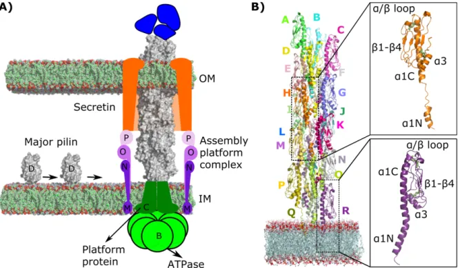
diameter. The machinery that assembles T4P is formed by several protein sub-complexes that span the bacterial envelope 32
(Figure 1a). T4P are anchored in the inner membrane (IM) and extend beyond the outer membrane (OM) of Gram-negative 33
bacteria through a channel called the secretin. Dedicated ATPases at the cytoplasmic base of the complex transmit motions to 34
the assembly platform IM complex to drive fiber extension and retraction. 35
T4Ps are present in Enterohemorrhagic Escherichia coli (EHEC), an important human pathogen. The major pilin PpdD (or 36
HcpA) is one of the EHEC virulence factors (Xicohtencatl-Cortes et al. 2007) and its sequence is highly conserved in 37
Enterobacteriaceae (Luna Rico et al. 2019). The structure of the periplasmic domain of EHEC PpdD determined by NMR 38
(Amorim et al. 2014; Bardiaux et al. 2019), combined with the cryo-EM density map of the EHEC pilus at 8 Å resolution, led 39
to an atomistic model of the T4P filament (Bardiaux et al. 2019). A variety of structural data is available for other T4P, with 40
major contributions coming from cryo-EM (Wang et al. 2017; Kolappan et al. 2016; Hartung et al. 2011; Craig et al. 2006; 41
Craig et al. 2003; Parge et al. 1995). Despite the recent progress in cryo-EM and integrative structural biology, the high 42
flexibility of this family of fibers often limits the resolution of the structure. Modeling is therefore a necessary step in structure 43
determination, even for the highest resolution cryo-EM maps obtained to date, such as those of the Thermus thermophilus T4P 44
(Neuhaus et al. 2020). Moreover, given the T4P flexibility and ability to rapidly extend and retract, a single structural model 45
is not sufficient to fully understand their behavior, nor to design new ways to regulate and interfere with their functions. A 46
crucial ingredient currently missing is a comprehensive and accurate characterization of the dynamic properties of these 47
systems. 48
49
Figure 1. Schematic representation of the EHEC T4P assembly system. (A) Major pilin PpdD (gray) and minor pilins (blue) are depicted.
50
Cytoplasmic ATPases (B, light green) transmit the conformational changes to the IM platform protein (C, dark green) and to the assembly
51
complex (M, N, O, P, purple shades), which is connected with the secretin (orange) in the OM. (B) The starting structure for the MD
52
simulations is depicted. T4P subunits (A-R) are shown as cartoon models in different colors. The POPE membrane atoms are shown with
53
spheres colored in green (carbon), red (oxygen), and blue (nitrogen). The insets highlight the structure of two subunits with two conformations
54
for the linker: as a coil (in orange), and modelled as a helix (in purple). The disulfide bonds are highlighted with green sticks.
55
The main objective of the present study is to investigate internal dynamics of T4P to be able to propose strategies that regulate 56
T4P behavior by directly targeting the pilus. With the aim to reach a deeper understanding of T4P dynamics and its functional 57
role, we performed a total of 108 µs all-atom molecular dynamics (MD) simulations of EHEC pili. This allowed us to reveal, 58
at atomistic detail, the network of dynamic interactions between subunits and the role of different regions in modulating 59
filament flexibility. This in-depth characterization of the conformational dynamics can also help explain the physical-chemical 60
basis of the molecular mechanisms behind the filament formation. We also analyzed the effect of specific structural features 61
of pilin subunits on pilus dynamics and stability. While most T4a pilins have one disulfide bridge, defining the C-terminal loop 62
(Amorim et al. 2014), and homologous type II secretion pseudopilins have none, enterobacterial T4P has an additional, 63
conserved disulfide bond connecting the N-terminal a helix with the globular domain (Luna Rico et al. 2019). In this study, 64
by combining in silico analysis with mutagenesis and biochemical assays, we analyzed the effects of these disulfide bonds on 65
pilin stability and dynamics, as well as on the assembly of the pilus. Finally, we studied the role of ions. Other filaments of the 66
T4P superfamily, such as archaeal flagella and the type II secretion pseudopili, are stabilized by calcium or other cations 67
(Meshcheryakov et al. 2019; Lopez-Castilla et al. 2017; Korotkov et al. 2009), however their effect on T4aP has not been 68
investigated. Here, by using NMR we showed that PpdD subunit binds calcium and identified the residues involved in this 69
interaction. We combined MD analysis with targeted mutagenesis and functional assays to characterize the interaction of 70
calcium and other ions within the assembled pilus and understand their role in fiber biogenesis and dynamics. Together, our 71
MD analysis and ex vivo and in vitro assays allowed us to identify the key structural elements required for the stability and 72
assembly of T4P. 73
RESULTS
74
We performed a series of all-atom MD simulations listed in Table 1. To elucidate the role of ions in pilus dynamics, we carried 75
out MD simulations of T4P in the absence of ions and in the presence of Ca2+, Na+, Mg2+, Mn2+ and at different salt 76
concentrations. The effect of Ca2+ was studied at different temperatures: 36.85°C (310 K), 60°C (333.15 K) and 80°C (353.15 77
K). We also evaluated the role of specific subunit features in pilus stability and function, notably the two disulfide bridges 78
present in PpdD (by analyzing double substituted PpdD variants C50C60S and C118C130S), and the fully conserved residue 79
E5 (variant E5A). Finally, we studied the effects of different histidine protonation states for the essential residue H54: i) HSE 80
(ε), ii) HSD (d), and iii) HSP (ε−d). In total, we performed 108 µs MD simulations of 16 different systems, with three replicates 81
for each. For all analyses, we considered the union of the results from the replicates. A protonated ε nitrogen of H54, and a salt 82
concentration of 100mM NaCl were assumed for all studied systems, unless stated otherwise. In the simulated pilus, the 83
subunits were labeled from A (the most distant from the membrane) to R (the closest to the membrane) (Figure 1B). To reduce 84
the edge effects, we excluded from the analyses the 4 top (A-D) and 4 bottom (O-R) subunits and only considered the 10 85
intermediate protomers (E-N), hereafter designated as “bulk” subunits. 86
Impact of pilin flexibility on length fluctuations
87
T4P are highly flexible fibers that undergo substantial length fluctuations (Biais et al. 2010) and show remarkable resistance 88
to force (Biais et al. 2008). We first studied in detail the relationship between individual residue fluctuations of EHEC pilin 89
subunits and the overall flexibility of the pilus bulk subunit in MD simulations of wild-type pilus in the presence of calcium 90
(WTCa2+). 91
Residue fluctuations. The PpdD fold includes a long N-terminal a helix and a b-sheet globular domain (Figure 1B). The 92
middle of the N-terminal helix (residues G11 to P22) is unstructured in assembled pilins and is hereafter referred to as the 93
linker. Similar to most T4a pilins, a disulfide bond stabilizes the C-terminal loop (C118 - C130) of PpdD, in addition to a 94
disulfide bond between residues C50 and C60, conserved in the enterobacterial T4aP subclass (Figure 1B). The dynamics of 95
every region, calculated as the root mean square fluctuations (RMSF) of every residue with respect to the average conformation 96
during the final 2.5 µs from the three MD replicates, is shown in Figure 2A. The maximum fluctuations (RMSFmax) mapped 97
on the structure (Figure 2B) revealed several highly flexible regions (RMSFmax > 4 Å): (i) G62-V81 (a/b loop and b1), (ii) 98
E92-N95 (b2/b3 loop), (iii) W105-T112 (b3/b4 loop), and (iv) D137-N140, the C-terminal region. 99
Filament dynamics. PpdD has the longest linker of any other pilus of this class, providing more flexibility to the filament 100
(Bardiaux et al. 2019). To further explore its role, we investigated how the a1 helix and filament length changes correlate with 101
the dynamics of the linker region. In principle, changes of filament length could be achieved by re-arrangements of the pilins 102
as more or less rigid blocks, or by conformational changes within the pilins. We found that, while the filament length changes 103
along the simulation were large, the changes of the bulk subunits were subtle, with an average length decrease of 1.7 Å (Figure 104
2C). We calculated all changes of distance as the difference between the instantaneous values along the MD simulations and 105
the initial values in the cryo-EM structure. The changes of distance along the linker region and a1 (see Methods) in the three 106
replicates along every subunit show an overall reduction of length for both the linker and a1 (Figure 2D). We also measured 107
the angles formed at the two ends of the linker region: G11 and P22 (see Methods for angle definition), and observed an overall 108
increase along the simulations (Figure 2E), indicating bending of a1N and a1C (Figure 2F). Hence, the overall reduction of 109
the filament length in the simulations is predominantly a consequence of the high flexibility of the linker and an increase of 110
the angles formed between the two segments of a1 and the linker. The linker was the main region responsible for overall length 111
fluctuations of the filament, with a correlation coefficient of 0.8 (Figure S1). The overall reduction of length along the pilus 112
was accompanied by a decrease in the separation between globular domains of the bulk subunit (Figure 2G and Figure S1). 113
In T4aP, due to the helical symmetry, every subunit (S), makes contacts with six other subunits (S−4, S−3, S−1, S+1, S+3, S+4), 114
along the right-handed 1-start, left-handed 3-start and right-handed 4-start helices (Bardiaux et al. 2019). We observed a larger 115
reduction of the S distance to subunits S+3 and S+4 (Figure 2G). Consequently, the overall length reduction along the pilus long 116
axis brings pilin subunits closer to each other, resulting in compaction and strengthening their contacts. 117
Exposed segments. To understand how pilus dynamics modifies solvent exposure of different regions during MD simulations, 118
we divided the PpdD primary sequence into contiguous pentapeptide segments and compared their solvent accessibility 119
(SASA, Å2) to the starting cryo-EM structure (Figure 2H). We classified segments with a SASA of greater than 250 Å2 as 120
exposed. The results show that residues in the linker and a1 helix become more buried, while the residues of the globular 121
domain b1−b4 become more exposed. The differences between the average SASA from MD simulations and the initial values 122
from the cryo-EM structure were mapped on the pilin structure (Figure 2I). For two segments in particular –89TGQES93 and 123
104GWDNA108 – highlighted with red and green arrows, respectively, the average SASA increased strongly over the 124
simulations, indicating that the b2/b3 and b3/b4 loops (Figure 2I) become more exposed to the solvent. Interestingly, the 125
104GWDNA108 peptide on the b3/b4 loop also showed the highest degree of fluctuations from the RMSF analysis (Figure 2B). 126
Previous NMR studies identified the same region as highly dynamic in the PpdD monomer in solution (Bardiaux et al. 2019). 127
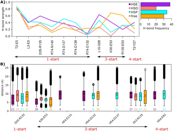
128
Figure 2. Residue fluctuations of the wild-type T4P from the MD simulations. (A) The per-residue RMSF of atomic coordinates are
129
measured from the MD simulations, with respect to the average conformation. The maximum, minimum and average RMSF values were
130
calculated over the bulk subunits from the three replicates and plotted as pink, orange, and green curves, respectively. The secondary
131
structures are indicated (size of the rounds proportional to the persistence of the secondary structure along the MD trajectories). The starting
132
structure of a single subunit is shown with the α helices and β strands colored in purple and cyan, respectively. The two disulfide bridges are
shown as yellow sticks. (B) The maximum RMSF values are mapped on the structure. (C) The average filament distance change is measured
134
over the three replicates for the full-length pilus (in orange), and bulk subunit (in green). (D) The changes of distances between the Ca atoms
135
of the first and last residues of the linker (G11 and P22, in cyan), and a1 (F1 and E53, in magenta) are reported for bulk subunit with respect
136
to their initial size, over the last 2.5 µs of each replicate simulation. The average values for each subunit (letters E to N) are shown with
137
yellow stars. (E) The changes of angles formed at the G11 (in magenta), and P22 (in cyan) are averaged over bulk subunit and the replicates.
138
Their initial values from the cryo-EM structure are shown with horizontal lines. (F) The segments used for the calculations of angles in (E)
139
are shown on the structure. (G) Changes of distance between the center of mass of bulk subunit globular domains (b1−b4) are averaged over
140
the replicates. (H) The average SASA of 5-mers over the subunits of pilus are shown in shades of blue for the replicates, and pink for the
141
initial structure (PDB code: 6GV9). The gray line corresponds to the threshold used for the predictions of exposed regions. The arrows point
142
to the segments that become exposed. (I) The difference between average SASA from the MD and the initial structure is mapped on the
143
structure, with colors from blue (buried) to red (exposed). Arrows indicate the loops 89TGQES93 (orange) and 104GWDNA108 (green) that are 144
the most exposed to the solvent.
145
Influence of Hydrogen-bonding network on filament formation
146
The analysis of MD trajectories in terms of inter-subunit Hydrogen-bonds (H-bonds) and salt bridges revealed the network of 147
interactions between the subunits (Figure 3). To analyze H-bonds, we recorded the inter-subunit interactions present for more 148
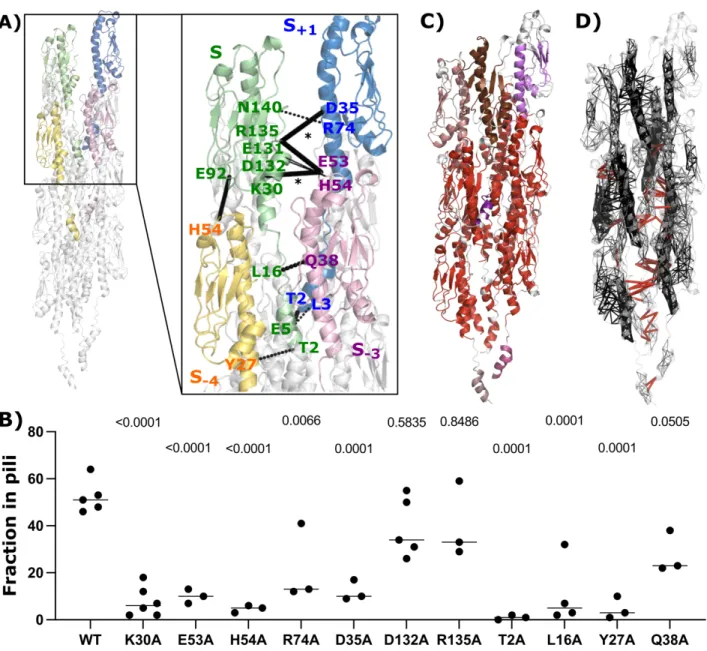
than 50% of the simulation time in at least one replicate and that are observed between at least three pairs of subunits along 149
the pilus symmetry (Figure S2). Salt bridges were studied by measuring distances between the center of mass of oxygen atoms 150
from the acidic side chains and the center of mass of nitrogen atoms from the basic side chains, for all bulk subunits (see 151
Methods). The results were mapped on one set of subunits within the helical symmetry (S−4 S−3 S S+1) (Figure 4A), where the 152
thickness of the lines indicates the number of subunits for which the interactions are present. We observed 4 distinct pairs of 153
H-bonds and salt bridges at the S-S+1 interface, 5 pairs at the S-S+3 interface, and 2 pairs at the S-S+4 interface. A strong network 154
of H-bonds is present along the 1-start helix, involving mostly the a1 helix, whereas the majority of salt bridges are formed 155
along the 3-start helix. Two sets of residues were pivotal for those interactions: i) K30, D35, E53, H54, R135, E92, E131, 156
D132, involved in salt bridges, and ii) T2, L3, E5, L16, Y27, Q38, R74, N140, forming H-bonds. Some residues formed both 157
salt bridges and H-bonds, e.g., D35-R135 and K30-E53. To experimentally validate the predicted importance of these 158
interactions for pilus stability, we generated alanine substitutions and performed piliation assays. In our assay conditions, 159
around 50% of total wild-type PpdD was assembled into pili on average (Figure 4B). In comparison, piliation was fully or 160
partially impaired for a large number of these variants including T2A, E5A, L16A, Y27A, K30A, D35A, E53A, H54A, and 161
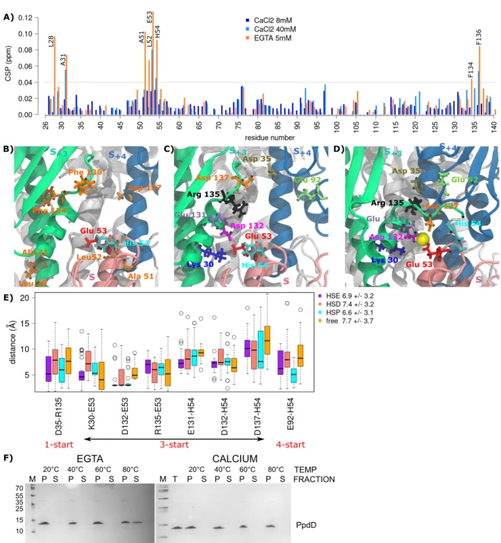
R74A, supporting the role of these residues in assembly or stability of EHEC pili. 162
We also measured the distances between all pairs of residues forming inter-subunit hydrophobic contacts for at least 70% of 163
the simulation time (averaged over the helical symmetry) and recorded those within 3 Å (Figure S3). As expected, the majority 164
of such residues belong to the a1 helix and form strong hydrophobic contacts. 165
Applying COMMA2 analysis (Karami et al. 2018) to the wild-type bulk subunit allowed us to identify a set of communication 166
blocks (CBspath)in the pilus (see Methods). The residues within a CBpath are linked by communication pathways built by 167
transitivity. A communication pathway is defined as a chain of residues displaying correlated motions and linked by stable 168
non-covalent interactions, hence representing an efficient route to transmit information through physical interactions. The 169
COMMA2 analysis revealed nine different CBspath (Figure 4C), the largest of which (in red) contains residues from all 10 bulk 170
subunits. To estimate the overall communication, we computed the number of pathways longer than 3 residues, and mapped 171
intra-subunit (in black) and inter-subunit pathways (in red) on the structure of the pilus (Figure 4D). We found that the a1-172
helices play an important role in communications both within and between the subunits. Only 1% of the pathways constitute 173
the inter-subunit communications (1547 pathways out of 202884), mainly involving the α1 helix residues. These results 174
highlight the essential role of the α1 helix in pilus stability and allowed us to trace the communication route across the pilus. 175
176
Figure 3. Inter-subunit interaction network. (A) The strength of bonds, computed as the percentage of conformations in which the
H-177
bond is formed and the total number of H-Bonds formed along the pilus symmetry are reported for the calcium free and bound form pilus
178
with different protonation states of His 54: HSE (ε), HSD (d), and HSP (ε−d). (B) The distances (Å) between pairs of residues forming salt
179
bridges at the interface between subunits of the pilus are shown. The values above x-axis are the frequency of each salt bridge along the pilus
180
symmetry. The left right arrows below each plot highlight the symmetry type of the interacting pairs. The data regarding the calcium bound
181
(HSE, HSD, HSP) and free forms of the pilus are colored in orange, cyan, salmon and purple, respectively.
183
Figure 4. Inter-subunit interactions of the pilus. (A) Key residues involved in the interactions forming the pilus symmetry are highlighted
184
on the first set of four protein subunits (S+1, S, S−3, S−4), colored in blue, green, pink and yellow, respectively. Dashed lines correspond to
185
H-bonds and full lines to salt-bridges, where the thickness of the connections is proportional to their number of occurrences summed up
186
according to the subunit symmetry along the pilus. (B) Dot plot comparing piliation efficiency of PpdD and its variants carrying alanine
187
substitutions of individual residues. P values are shown above each graph as calculated in comparison with PpdD wild type piliation. (C)
188
Pathways-based communication blocks (CBspath) identified by COMMA2 are mapped on the structure, with different colors. (D) The set
189
of intra and inter-subunit pathways (> 3 residues) are displayed in black and red, respectively, as segments linking residues’ Ca atoms. The
190
thickness of black segments is proportional to the number of pathways linking the residue pair.
191
Calcium binding by PpdD modulates the stability of T4P
192
Calcium is important for function and stability of some filaments of the T4P superfamily, including type II secretion pseudopili 193
and archaeal flagella (Meshcheryakov et al. 2019; Lopez-Castilla et al. 2017; Korotkov et al. 2009). So far, however, its effect 194
on T4aP has not been investigated. Solution NMR analysis indicated weak calcium binding to the soluble periplasmic domain 195
of the PpdD pilin (residues 26–140) as shown by chemical shift perturbations (CSP) in the presence of EGTA (a calcium 196
chelating agent) or calcium (Figure 5A). Addition of EGTA resulted in the highest CSP values for residues L28, A31, 197
51ALEH54, F134 and F136. Among them, only E53 is negatively charged, located at the tip of the a1 helix, in a loop defined 198
by the disulfide bridge C50-C60. As calcium was not added to any of our buffers, this indicates that it was present in the protein 199
sample purified from the bacterial periplasm. Upon addition of up to 40 mM calcium, we observed the highest CSP for the 200
same region, notably for residues A31, E53, H54 and F136. Based on these results, we had placed one calcium ion close to 201
residue E53 in each PpdD subunit of the starting structure for the calcium-bound simulations (see Methods). Remarkably, 202
during the simulations, we observed a movement of calcium toward the interface of three subunits: S, S+3, and S+4. We explored 203
the interaction network in the vicinity of calcium, by comparing the calcium-bound and calcium-free simulations, and revealed 204
a set of residues surrounding the calcium: E53, H54 (in the S protomer), K30, E131, D132, R135, D137 (in S+3), and D35, E92 205
(in S+4) (Figure 5BC). Interestingly, this set of residues overlaps with the set crucial for the interactions between subunits 206
described above, forming H-bonds and salt bridges. Moreover, each calcium ion remained stable at the interface of three 207
subunits and resulted in the formation of stronger H-bonds and salt bridges (Figure 3). In the calcium-free simulations, residue 208
K30 (positively charged) interacts with residues E53 (negatively charged), through both H-bonds and salt bridges (Figure 5B). 209
However, the presence of calcium rearranged the interaction network due to its positive charge. In the calcium-bound 210
simulations we observed that calcium oscillates between E53 (negatively charged) and D132 (negatively charged) as shown in 211
Figure 5C. The distances between residues close to the calcium ion decreases overall by about 1 Å upon calcium binding, with 212
an average distance of 7 Å for the calcium-bound simulation (HSE), compared to 8 Å for the calcium-free one (Figure 5D). 213
While the distances decrease for most of the pairs, some exceptions are noted for K30-E53, R135-E53, and D132-H54. As 214
observed for the salt-bridges and H-bonds, this can be explained by the fact that calcium intercalates between these residues. 215
Although such rearrangements increase the distance between K30-E53 and R135-E53 and apparently weaken their contacts, 216
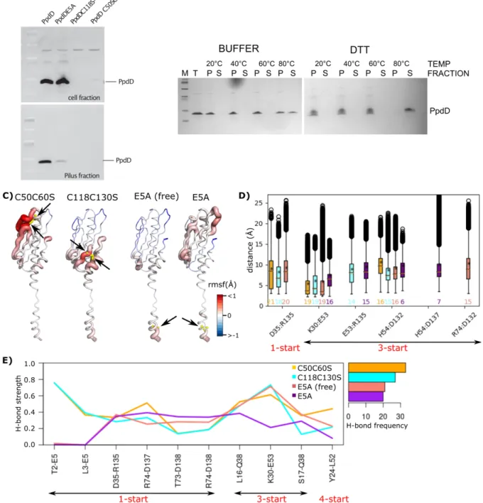
these interactions are actually reinforced through the presence of Ca2+ ion that bridges those residues and neutralizes their 217
charge. It is noteworthy that the side chain of H54 in its double protonation (HSP) state is closer to residue D137 (along the 3-218
start symmetry) and to E92 (along the 4-start symmetry), further strengthening the network of interactions. Importantly, 219
functional assays show the importance of E53 and H54 residues for T4P assembly (Figure 4B). 220
MD simulation analyses suggest that calcium should stabilize the pilus. To test this prediction, we designed biochemical 221
experiments to assess the effects of calcium on pilus stability in vitro. Purified EHEC pili were incubated for 5 minutes at 222
increasing temperatures in the presence or absence of calcium. Upon ultracentrifugation, intact pili were recovered in the pellet 223
(P) and the dissociated subunits remained in the supernatant (S) (Figure 5F). The experiments showed that EHEC pili are quite 224
resistant to temperatures up to 60°C, but at 80°C they start to disassemble in vitro. Pili incubated with buffer alone or buffer 225
supplemented with EGTA behaved similarly, indicating a low level of divalent cation contamination in the buffer. Strikingly, 226
the addition of calcium stabilized the pili at 80°C, fully preventing dissociation of PpdD from the filaments. To better 227
characterize this effect, we carried out MD simulations at different temperatures (60°C and 80°C), in the absence and presence 228
of calcium. While the pilus remained stable at both temperatures of 60°C and 80°C, these conditions induced high global 229
fluctuations across the pilus (Figure S4), and secondary structure perturbations specifically around the a3 helix (Figure S5). 230
Higher fluctuations were detected in the area of C50-C60 loop at 80°C compared to 60°C, possibly correlating with loss of 231
fiber stability. However, for the simulations in the presence of calcium, we observed that the Ca2+ ions were not stable at their 232
initial binding region and moved away after 50-200 ns in each simulation. 233
We also investigated the effects of ions and salt concentrations by performing MD simulations of T4P in 100 mM salt 234
concentration by placing sodium (Na+), magnesium (Mg2+) or manganese (Mn2+) ions in the calcium binding site, or by 235
increasing the salt concentration to 150 mM in the presence of calcium (Ca2+). In the simulations with 100 mM salt, the 236
positively charged divalent ions behaved similarly to the calcium and remained stably bound in the same pocket, while in the 237
simulations with 150 mM, the Ca2+ ions moved away from the binding site. 238
239
Figure 5. The effect of calcium binding on the overall interactions between subunits. (A) Histogram showing the CSP values of PpdD
240
backbone amide signals in the presence of calcium (8mM in dark blue and 40mM in cyan) and EGTA (5mM in orange), as a function of
241
residue number. Residues displaying highest CSP (> 0.04 ppm) are labeled. (B) Residues displaying significant spectral changes in the
242
presence of EGTA are labeled on the structure and colored according to their physico-chemical properties (hydrophobic in orange, positively
243
charged in blue and negatively charged in red). (C,D) Key residues involved in the interactions between subunits and calcium are highlighted
244
as sticks in different colors. The three subunits (S, S+3, S+4), are colored in pink, green and blue, respectively, while the rest of the pilus are
245
in white. The calcium ion is shown as a yellow sphere. (E) The comparison of distance variations between pairs of residues in the
246
neighborhood of the calcium, in the absence of the calcium (free), and presence of calcium (HSE, HSD, HSP). (F) Thermal stability of the
247
EHEC T4P. Aliquots of isolated pili were incubated for 5 min at 20°, 40°, 60° and 80°C in 50 mM HEPES 50 mM NaCl buffer supplemented
248
with either 10 mM EGTA, or 20 mM calcium. Total pili samples (T) were fractionated by ultracentrifugation (see Methods). Equivalent
249
volumes of pellet (P) and supernatant (S) fractions were analyzed by denaturing electrophoresis on 10% Tris-Tricin polyacrylamide gels and
250
stained with Coomassie blue.
Histidine 54 and the importance of its protonation
252
The single histidine (H54) at the tip of a1 helix, close to the calcium binding site is one of the key residues for the pilus 253
formation, conserved in the enterobacterial T4aP subclass (Luna Rico et al. 2019). Its mutation to alanine abolished piliation 254
(Figure 4B). To determine whether the protonation state of H54 affects pilus conformational stability, we performed three sets 255
of MD simulations at different pH around the pKa of H54: i) HSE (ε), ii) HSD (d), and iii) HSP (ε−d). First, we measured the 256
inter-subunit H-bonds over all the pairs of the bulk subunit, and recorded those that are present more than 50% of the simulation 257
time in at least one replicate and between 3 pairs of subunits (Figure 3A). This analysis suggests that both HSE and HSP 258
protonation states lead to stronger H-bond networks. However, the interactions are more persistent for HSE, occurring 38 259
times, compared to 34 for HSP. Interestingly, HSE results in the establishment of stronger H-bonds: (D35-R135) along the 1-260
start, (L16-Q38 and K30-E53) along the 3-start and (T2-Y27) along the 4-start symmetry. Also, the number and persistence of 261
salt bridges are smaller for HSD (Figure 3B, the values above the x-axis). For both HSE and HSP, we observed salt bridges 262
starting from H54 along the 3-start and 4-start symmetry. This analysis revealed a direct impact of H54 protonation state on 263
stabilizing inter-subunit interactions, specifically along the 3-start and 4-start helical symmetry. 264
The role of disulfide bridges and E5 on T4P structure and dynamics
265
Disulfide bridges. The presence of two disulfide bridges is conserved in enterobacterial T4aP subclass. Disrupting either of 266
the two disulfide bridges led to a complete degradation of PpdD in bacteria, presumably due to increased exposure of dynamic 267
loop regions to proteolysis in bacterial periplasm (Figure 6A). To assess the dependence of pilus stability on disulfide bonds, 268
we treated purified pili with the reducing agent (DTT). Adding DTT had little effect on fiber stability at 60°C and below 269
(Figure 6B). At 80°C, however, pili were fully dissociated, emphasizing the importance of these disulfide bonds. To gain 270
molecular insight into their role, we performed MD simulations of pili lacking C50-C60 or C118-C130 disulfide bond. We 271
observed that for both mutants, calcium ions were not stable and the fluctuations around the average increased with respect to 272
the wild-type simulations, in particular close to the mutation sites (Figure 6C, the regions colored in red on the first two 273
structures from the left). Similar perturbations were also observed at the level of secondary structures around the strands b1 274
for C50C60SCa2+, and b4 for C118C130SCa2+(Figure S5). The mutations induced changes in interaction networks, with a 275
strong decrease in the number of salt bridges, their persistency, and increase in the distances for the two mutants (Figure 6D). 276
The only exception was the salt bridge between K30 and E53, which was more frequently present in the mutants compared to 277
the wild type. This can be explained by the fact that the Ca2+ ions diffuse away in the simulations of mutants, allowing K30 278
and E53 to interact directly, compared to the wild-type simulations where this interaction was mediated by Ca2+ ion. 279
Furthermore, the H-bond D35-R135 along the 1-start symmetry of the pilus became less stable, with the average strength of 280
0.59 for the wild type compared to 0.33 for the C50C60SCa2+, and 0.28 for the C118C130SCa2+ (Figure 6E). 281
Role of residue E5. The Alanine substitution of residue E5 prevents PpdD assembly (Bardiaux et al. 2019) (Figure 6A). We 282
created the E5A variant in silico and carried out MD simulations in the absence and presence of calcium ions. For the 283
simulations in the presence of calcium, we observed that Ca2+ ions were not stable, and moved away from their initial binding 284
pocket. The comparison of local residue fluctuations with those of the wild-type pilus showed that the E5A substitution induces 285
local perturbations around the fifth position (Figure 6C), as well as at the level of secondary structures in the a1N region 286
(Figure S5). The mutation induced a major decrease in inter-subunit interaction strength and persistency (Figure 6D,6E). The 287
total number of H-bonds along the pilus was reduced by about 50% (21 and 20 for E5A and E5ACa, respectively, compared to 288
38 for wild type). On the other hand, intra-subunit interactions between E5 and F1 were increased from 51% in the simulations 289
of the wild type to 82% and 74% in the E5A and E5ACa simulations, respectively. This can be explained by the disruption of 290
the inter-subunit contacts of E5 along the 1-start helix. 291
292
Figure 6. Role of disulfide bonds and E5 in pilus stability. (A) Piliation assay with single and double mutant variants. (B) Thermal stability
293
of the EHEC T4P in the presence (BUFFER) and absence (DTT) of disulfide bridges. Total pili samples (T) were fractionated by
294
ultracentrifugation and equivalent volumes of pellet (P) and supernatant (S) fractions were analyzed (see Methods). (C) The differences
295
between the average per-residue RMSF are measured from the MD simulations of the C50C60SCa2+, C118C130SCa2+, E5A (free form), and 296
E5ACa2+ (from left to right), with respect to the wild-type (HSE) pilus. The differences are then mapped on the structure of each system. The 297
mutation positions are shown with yellow sticks and pointed to by black arrows. (D) The distances between pairs of residues forming salt
298
bridges, and (E) strength and frequency of H-bonds at the interface between subunits of the mutants are reported. The values above x-axis
299
are the frequency of each salt bridge. The left right arrows bellow each plot, highlight the symmetry type of the interacting pairs.
300
DISCUSSION
301
For this study of the conformational dynamics and stability of the EHEC T4P, we used a total of 108 µs MD simulations of 302
the wild-type T4P in different conditions, i.e., in presence and absence of different ions, protonation states, and salt 303
concentrations, as well as point mutations, validated by experimental studies. It enabled us to identify regions responsible for 304
the flexibility of the pilus, playing key roles for the overall length fluctuation of the filament. Moreover, our simulations 305
revealed the network of interactions governing the communications across T4P, and allowed us to characterize a novel type of 306
a calcium binding site that specifically stabilizes the assembled pilus polymer. 307
This study is to the best of our knowledge the most extensive study of the conformational dynamics and stability of a filament 308
of this class. It is also the first study that uses a complete structural model with the “linker region” in the N-terminal helix. The 309
loss of helical structure in a1N regions delimited by Gly and Pro residues was observed in the cryo-EM reconstructions of T4P 310
from N. meningitidis (Kolappan et al. 2016), N. gonorrhoeae (Wang et al. 2017), P. aeruginosa (Wang et al. 2017), and EHEC 311
(Bardiaux et al. 2019) in the 5-8 Å resolution range, in addition to two higher resolution structures for the wide and narrow 312
forms of T. thermophilus with the resolutions of 3.2 Å and 3.5 Å, respectively (Neuhaus et al. 2020), and the structure of the 313
homologous T2SS pseudopilus from Klebsiella oxytoca with a resolution of 5 Å (Lopez-Castilla et al., 2017). It has been 314
proposed that this conserved feature of T4P facilitates the integration of pilin subunits into the pilus (Kolappan et al. 2016), 315
allows T4P to withstand the extension under force, and to relax back to a native state when the force is removed (Wang et al. 316
2017). MD studies predating these structures, such as the atomistic and coarse-grain steered MD simulations to study the force-317
induced conformational changes of a T4P from Neisseria gonorrhoae (Zhao et al. 2017; Baker, Biais, and Tama 2013), were 318
based on structural models of T4P with a continuous N-terminal helix. It is interesting that in those steered MD simulations, 319
the pilus length variation was also linked to the particular region between G14 and P22 in the subunit. Among all the known 320
structures of pili, the EHEC T4P has the longest inker region in the α1 helix, extending from residues G11 to P22. Our analysis 321
identified in particular the role of the linker region in fiber flexibility: changes of its length show a strong correlation (0.8) with 322
the overall flexibility of the filament. Such high correlation could explain the role of the linker in the filament resistance under 323
force. 324
Despite the fact that in our simulations the pilus was in the “resting state” and absence of any forces, we observe an overall 325
reduction of length at different levels, in the linker, a1 helix, globular domains and consequently along the pilus. Residues in 326
the linker and a1 helix become more buried, while the residues of the globular domain b1−b4 become more exposed. This is 327
consistent with the decrease of separation between the subunits, resulting in residues along the a1 helix becoming more buried. 328
The reason for the length reduction in our simulations could be that we only simulate a small fraction of the pilus. Forces are 329
typically generated by adding or removing pilin subunits, respectively, at the base of the pilus, leading to the extension or 330
retraction of the pilus. These extension-attachment-retraction cycles are powered by the strongest linear motors (ATPase) 331
known to date, and T4Ps have remarkable resistance to forces in the range of 100 pN (Clausen et al. 2009; Maier et al. 2002; 332
Merz, So, and Sheetz 2000). It was also shown that the reversible force-induced conformational changes revealed the exposure 333
of a hidden epitope (Biais et al. 2010), and the resistance to force was linked to the packing of a1 helices, maintaining their 334
hydrophobic contacts within the core of the pilus (Baker, Biais, and Tama 2013). The N-terminal half of the α1 helix is highly 335
conserved in the T4P, and is almost completely made up of hydrophobic residues, with the exception of T2 and E5. Our 336
simulations highlight the role of hydrophobic contacts between the a1-helices in holding the core of the pilus together. 337
However, they also underline the essential role of polar residues forming H-bonds (T2, L3, E5, L16, Y27, Q38, R74, N140) 338
and salt bridges (K30, D35, E53, H54, R135, E92, E131, D132) at the interface of pilin subunits, along the 1-, 3-, and 4-start 339
helix. Our results are in agreement with previous charge inversion mutagenesis analysis, which showed that the majority of 340
charged EHEC T4P residues are required for pilus assembly (Bardiaux et al. 2019). At the same time, normal mode analysis 341
of EHEC T4P indicated the role of interactions along the 3-start helix in pilus rigidity (Bardiaux et al. 2019). Our study 342
highlights the crucial interplay of the linker region in a1, whose intrinsic flexibility induces long range dynamics along the 343
filament, while the hydrophobic and polar interactions between the tightly packed helices provide stability. 344
In bacterial cells disulfide bridges are essential for PpdD monomer stability and protease resistance precluding the detailed 345
analysis of their molecular role. MD data, coupled with the in vitro biochemical assays of an increase in temperature provided 346
a consistent picture of the role of the disulfide bridges in the stability of T4P. 347
Strictly conserved residue E5, is crucial for T4P assembly, since it contributes to integration of pilin subunits into the pilus 348
(Bardiaux et al. 2019; Nivaskumar et al. 2014; Lory and Strom 1997). The mutation E5A prevents the assembly at multiple 349
levels (Luna Rico et al. 2019), and probably pilin extraction from the membrane as suggested by studies of the effects of E5A 350
on the T2SS pseudopilin PulG (Santos-Moreno et al. 2017). Our MD data supports an additional role of this residue at the level 351
of inter-protomer interactions, mostly by stabilizing the interactions between T2 and L3 on one subunit and E5 on the adjacent 352
subunit, in agreement with previous work on other T4P (Craig et al. 2006; Wang et al. 2017). 353
MD simulations revealed the structural role of calcium in a member of the T4aP family, for which no classical calcium binding 354
site had been described. By combining NMR chemical shift perturbation analysis, biochemical assays and MD simulations we 355
revealed the molecular mechanism of calcium-mediated fiber stabilization. Simulations show how calcium bound at the tip of 356
the pilin monomer becomes coordinated in assembled pili by additional residues from subunits S+3 and S+4, acting as a 357
molecular glue. The set of residues involved in the interactions with calcium (K30, D35, E53, H54, E92, E131, D132, R135, 358
D137) largely overlaps with the set of residues involved the in the network of H-bonds and salt bridges. We observed that 359
calcium remains stable at the interface between pilin subunits, and results in the formation of stronger salt bridges and H-bonds 360
at the interface. We also showed that the residues in the proximity of the calcium-binding region became more solvent exposed 361
during the simulations. Complementary functional assays confirmed a subset of the residues in the calcium-binding region as 362
critical for T4P assembly. In addition, in vitro biochemical assays showed that pili are resistant to temperatures up to 60°C and 363
they start to disassemble only at 80°C. These results confirm previous findings regarding the thermal stability of meningococcal 364
pili, also members of the T4aP subclass (Li, Egelman, and Craig 2012). Strikingly, the addition of calcium stabilized pili even 365
at 80°C, fully preventing the dissociation of PpdD from the assembled filaments. Even though the simulations were too short 366
to capture pilus dissociation, large fluctuations and secondary structure perturbations are in line with the biochemical data on 367
fiber stability. Simulations with different ions support the requirement of positively charged divalent ions for stable binding. 368
The importance of calcium and the identification of a metal binding pocket at the interface between pilins in the simulations 369
raises the possibility that this pocket could be exploited for drug discovery, since small molecules could bind there, affecting 370
T4P assembly or function. T4P are virulence factors in many human pathogens. Newly emerging anti-virulence strategies have 371
been developed to target the ATPases of T4P at the cytoplasmic base, resulting in disassembly of the pilus (Denis et al. 2019; 372
Duménil 2019). Targeting the pilus directly rather than the basal body would have the advantage that potential drugs do not 373
need to traverse the bacterial membrane. 374
This extensive characterization of the conformational landscape of T4P allowed us to reveal, at atomistic detail, the network 375
of dynamic interactions between subunits and the role of different regions in modulating the filament flexibility. Such 376
characterization of the conformational dynamics can also explain the physico-chemical basis of the molecular mechanisms 377
behind the fluctuations in the overall length of the pilus. This valuable information has a profound impact at multiple different 378
levels. From a fundamental point of view, our study elucidated the physico-chemical interactions that drive the behavior of 379
large and dynamic molecular assemblies. These interactions must provide an optimal balance between structural integrity of 380
the filaments and the flexibility required to perform their biological functions and to resist under stress. Furthermore, the 381
acquired knowledge will guide rational strategies to modulate the behavior of these filaments that are critical for a variety of 382
bacterial functions related to virulence. 383
METHODS
384
PPDD CALCIUM BINDING MONITORED BY NMR
385
The soluble periplasmic domain of EHEC PpdD comprising residues 26–140 of the mature protein, was produced and purified 386
as previously reported (Bardiaux et al. 2019; Amorim et al. 2014). NMR data were acquired at 293 K on a Varian spectrometer 387
operating at a proton frequency of 500 MHz. Proton chemical shifts were referenced to 2,2-dimethyl-2-silapentane-5 sulfonate 388
(DSS) as 0 ppm. 15N were referenced indirectly to DSS (Wishart et al. 1995). The pulse sequences were from VnmrJ Biopack. 389
NMR data were processed with NMRPipe/NMRDraw (Delaglio et al. 1995) and analyzed with the CcpNmr Analysis software 390
package (Vranken et al. 2005). 1H–15N HSQC experiments were acquired on 0.4 mM PpdD in 50 mM Tris-HCl pH 7, 50 mM 391
NaCl supplemented with either EGTA at 5mM or CaCl2 from 8-40 mM. Chemical shift perturbations (CSPs) of PpdDp 392
backbone amide cross-peaks were quantified by using the equation CSP = [Δ δH2 + (Δ δN× 0.159)2]1/2, where Δ δH and Δ δN 393
are the observed 1H and 15N chemical shift changes between the two experimental conditions. The residues belonging to the 394
tag were not considered for this analysis. The 1H and 15N resonance assignments were from (Amorim et al. 2014). 395
PILUS THERMAL STABILITY ASSAY
396
The E. coli strain PAP5386 is a fimAB::kan DfliC derivative of BW25113 F’lacIQ (Datsenko and Wanner 2000). Bacteria were 397
transformed with plasmids pMS41 encoding EHEC T4P assembly system and pCHAP8565 carrying the major pilin gene ppdD 398
(Luna Rico et al. 2019). Bacteria were cultured for 5 days at 30°C on solid M9 glycerol agar plates containing 100 µg.mL-1 of 399
carbenicillin (Cb) and 25 µg.mL-1 of chloramphenicol (Cm), supplemented with 1 mM isopropyl-b-D-1-thiogalactopyranoside 400
(IPTG) to induce the expression of the T4P genes. Bacteria were harvested and pili were extracted by shearing and concentrated 401
by ultracentrifugation as described previously (Luna-Rico, Thomassin, and Francetic 2018). To assess pilus stability, the 402
equivalent amounts of pili were taken and resuspended in buffer (50 mM HEPES, 50 mM NaCl). The pilus samples were 403
supplemented with 10 mM EGTA, 10 mM EGTA + 20 mM CaCl2, or 40 mM DTT, incubated at indicated temperatures for 5 404
min and then placed on ice. Intact pili were separated from dissociated subunits by a 30-min centrifugation at 53 000 rpm in 405
the Beckman TLA55 rotor and table-top ultracentrifuge at 4°C. Equivalent amounts of total (T), pili-containing pellet (P) and 406
supernatant fractions containing dissociated pilin subunits (S) were analyzed on 10% Tris-Tricin SDS-PAGE and stained with 407
Coomassie blue. 408
SITE-DIRECTED MUTAGENESIS
409
Mutations were introduced into the PpdD gene carried on plasmid pCHAP8565 by a modification of the QuickChange method 410
for site-directed mutagenesis, as described previously (Bardiaux et al. 2019). The plasmids used in this study and mutagenic 411
oligonucleotide primers are listed in Tables S1 and S2. 412
PILUS ASSEMBLY ASSAYS
413
To analyze pilus assembly, bacteria of E. coli strain BW25113 F’lacIQ were transformed with plasmid pMS41 encoding the 414
EHEC pilus assembly genes and pCHAP8565 or its mutant derivatives encoding PpdD variants (Luna Rico et al. 2019). 415
Bacteria were cultured for 48-72 hours on M9 minimal agar plates (Miller 1972) containing Cb (100 µg.mL-1) and Cm (25 416
µg.mL-1). Bacteria were harvested and resuspended in LB medium at OD600nm of 1. The suspension was vortexed vigorously 417
for 1 min to detach pili from bacteria. Bacteria were pelleted by 5-min centrifugation at 16000 g at 4°C and resuspended in 418
SDS sample buffer at a concentration of 10 OD600nm per 1 mL. The supernatant containing pili was subjected to another round 419
of centrifugation for 10 min. Pili were precipitated with 10% tri-chloroacetic acid on ice for 30 min and pelleted by a 30-min 420
centrifugation at 16000g at 4°C. Pili pellets were washed twice with ice-cold acetone, air-dried and resuspended in SDS-sample 421
buffer at a concentration equivalent to that of the bacteria. The equivalent volumes of each fraction were analyzed by denaturing 422
SDS polyacrylamide gel electrophoresis (SDS-PAGE) on 10% polyacrylamide Tris-Tricine gels (Schägger and Von Jagow 423
1987). Proteins were transferred on nitrocellulose by Western blot and probed with PpdD antibodies and secondary anti-424
rabbit antibodies coupled to HRP as described (Luna Rico et al. 2019). The fluorescent signal was recorded with a Typhoon 425
FLA-9000 scanner (GE) and quantified by ImageJ. Data were processed and statistically analyzed with GraphPad Prism8 426
software. 427
MOLECULAR DYNAMICS SIMULATIONS
428
Studied systems. The 3D coordinates of EHEC T4P were retrieved from the Protein Data Bank (Berman et al. 2000) with 429
PDB code: 6gv9, residues 1 to 140, 8 Å resolution (Bardiaux et al. 2019). The published structure contains 14 subunits of the 430
major pilin PpdD; each subunit comprises (i) a long N-terminal helix (a1) that is broken into two helices (a1N and a1C) by 431
an extended linker from G11 to P22, (ii) four b strands forming a b sheet (b1−b2−b3−b4), (iii) a long loop (a/b loop) 432
connecting the a1 to the b domain, and (iv) another helix (a3) at the C-terminus (Figure 2A). Two disulfide bonds are present 433
in each subunit, between Cys residues (C50, C60) and (C118, C130). We performed the MD simulations of T4P in a 1-434
palmitoyl-2-oleoyl-sn-glycero-3-phosphoethanolamine (POPE) model membrane. The protein was initially placed near the 435
membrane with the linker and N-terminus of the first subunit partially inserted into the bilayer (Figure 1B). The number of 436
subunits was increased from 14 to 18 by applying the internal helical symmetry parameters. Moreover, the environment of the 437
histidine (H54) was manually checked, and consequently protonated with a hydrogen at the ε nitrogen. We also performed MD 438
simulations at two other protonation states: (i) a protonated d nitrogen, and (ii) two protonated ε and d nitrogens. In addition, 439
a single substitution (E5A) and two double substitutions (C50C60S and C118C130S) were studied in silico using the 440
CHARMM-GUI server (Jo et al. 2008). 441
Ion placement protocol. We identified the calcium binding site using NMR and by titration of T4P with calcium (see PpdD 442
calcium binding monitored by NMR). From this experiment four consecutive residues, 51ALEH54 showed the highest 443
chemical shift perturbation, among which only E53 is negatively charged. Consequently, we placed the calcium ion (Ca2+) 444
close to this residue, and performed minimization. In addition to calcium, we evaluated the effect of other ions, namely: sodium 445
(Na+), magnesium (Mg2+) and manganese (Mn2+). These ions were placed at the same position than the one chosen for Ca2+ 446
Preparation. All systems were prepared with CHARMM-GUI membrane builder server (http://www.charmm-gui.org/?doc-447
1?4input/membrane) and the CHARMM36m force field parameter set (Huang et al. 2017; MacKerell Jr, Feig, and Brooks 448
2004): (i) hydrogen atoms were added, (ii) the ions were positioned at the tip of a1C, following the procedure mentioned in 449
the previous paragraph, (iii) the POPE model membrane was used to build the inner bacterial membrane, (iv) the solute was 450
hydrated with a rectangular box of explicit TIP3P water molecules with a buffering distance up to 14 Å, (v) Na+ and Cl− 451
counter-ions were added to reproduce physiological salt concentration (100 mM and 150 mM solution of sodium chloride). 452
The following simulations were carried out: (i) T4P in the presence and absence of calcium with different protonation states: 453
WT, WTCa2+, WT(δ)Ca2+, WT(e−δ)Ca2+, (ii) T4P in the presence of different ions (calcium, sodium, magnesium, manganese) and 454
salt concentrations (100 and 150 mM): WTNaCl150mMCa2+, WTNa, WTMg, WTMn, (iii) T4P in the presence and absence of calcium 455
at different temperatures (60°C and 80°C): WT60°, WT 60°Ca2+, WT80°, WT 80°Ca2+ , and (iv) a set of single and double mutants of 456
T4P: E5ACa2+, C50C60SCa2+ and C118C130SCa2+. A salt concentration of 100mM NaCl was used for all systems and the ε 457
nitrogen of H54 was protonated, unless stated otherwise. For each system, three replicates of simulations were performed. 458
Typically, these systems are composed of ∼295,000 atoms, in a rectangular water box with dimensions of ∼100 Å × 100 Å × 459
308 Å. The details of all studied systems are reported in Table I. 460
Table 1. Details of the MD simulations. All studied systems, as well as their corresponding temperature, number of replicates and simulation
461
time are reported here.
462
System Name Temperature (K) # Replicates Simulation length (ns)
Wild type WT 310 3 3000 WTCa2+ 310 3 3000 WT(d)* Ca2+ 310 3 3000 WT(e-d) Ca2+ 310 3 3000 WTNaCl-150mM# Ca2+ 310 3 1000 WTNa 310 3 1000 WTMg 310 3 1000 WTMn 310 3 1000 Temperatures WT60º 333.15 3 3000 WT60º Ca2+ 333.15 3 3000 WT80º 353.15 3 3000 WT80º Ca2+ 353.15 3 3000 Substitutions C50C60SCa2+ 310 3 2000 C118C130SCa2+ 310 3 2000 E5A 310 3 2000 E5ACa2+ 310 3 2000 Total - 48 108000
*The protonation of ε nitrogen for H54. 463
#The salt concentration of 100mM NaCl were considered for all the studied systems, unless stated otherwise. 464
Production of the trajectories. The GROMACS 2019.4 package was used to carry out all simulations (Abraham et al. 2015). 465
The energy minimization was performed by steepest descent algorithm for 10000 steps, to minimize any steric overlap between 466
system components. This was followed by an equilibration simulation in an NPT ensemble at 310 K, allowing the lipid and 467
solvent components to relax around the restrained protein. All the protein and lipid non-hydrogen atoms were harmonically 468
restrained, with the constraints gradually reduced in 6 distinct steps with a total of 0.375 ns. The particle mesh Ewald algorithm 469
(PME) (Essmann et al. 1995) was applied to calculate electrostatic forces, and the van der Waals interactions were smoothly 470
switched off at 10-12 Å by a force-switching function (Steinbach and Brooks 1994). Production runs were performed in the 471
NPT ensemble. The time step was set to 2.0 fs, the temperature was kept at 310 K (except for the simulations at 333.15 K and 472
353.15 K), temperature was kept constant using the Nosé-Hoover thermostat (Melchionna, Ciccotti, and Lee Holian 1993) and 473
a constant pressure of 1 atm was maintained with the Parrinello-Rahman barostat (Parrinello and Rahman 1981). The SHAKE 474
algorithm (Kräutler, Van Gunsteren, and Hünenberger 2001) was used to freeze bonds involving hydrogen atoms, allowing for 475
an integration time step of 2.0 fs. The PME method (Darden, York, and Pedersen 1993) was employed to treat long-range 476
electrostatics. Half-harmonic potentials were applied at the tip of the pilus (subunit A-D), in order to prevent the dissociation 477
of the tip. PLUMED 2.6.0 (Tribello et al. 2014) and the PLUMED-ISDB (Bonomi and Camilloni 2017) module were used to 478
add lower and upper walls on the distance between the Ca atoms of four pairs of residues (T45A-L52B, T45B-L52C, T45C-L52D, 479
L49D-K30A), with a force constant of 1000 kcal/(mol Å). The choice of residues was made according to their fluctuations, and 480
those with low deviations were selected. Coordinates of the system were written every 10ps. For every system, two or three 481
replicates were performed, starting with different initial velocities as reported in Table 1. 482
Stability of the trajectories. Standard analyses of the MD trajectories were performed with the gmx module of GROMACS 483
2019.4. All analyses performed in this study were applied to the “bulk” subunit, i.e. remaining 10 intermediate subunits, after 484
excluding 4 subunits from the top (A-D) and bottom (O-R) of the pilus (E-N). The root mean square deviations (RMSD) of 485
backbone atoms (Ca, C, N, O) from the initial frame were recorded along each replicate (Figure S6). Based on the RMSD 486
profiles, we performed the subsequent analysis over the subset of simulations where the systems are fully relaxed, i.e. 487
considering the last 900 ns, 1500 ns and 2500 ns for the simulations of 1000 ns, 2000 ns and 3000 ns, respectively. The per-488
residue root mean square fluctuations (RMSF) were computed over the backbone atoms (Ca, C, N, O), with respect to the 489
average conformation (Figure S6). The secondary structures were assigned with DSSP (Kabsch and Sander 1983) and 490
averaged over the replicates (Figure S5). All studied systems remained stable along the MD trajectories. 491
Stability of the trajectories. Standard analyses of the MD trajectories were performed with the gmx module of GROMACS 492
2019.4. All analyses performed in this study were applied to the “bulk” subunit, i.e. remaining 10 intermediate subunits, after 493
excluding 4 subunits from the top (A-D) and bottom (O-R) of the pilus (E-N). The root mean square deviations (RMSD) of 494
backbone atoms (Ca, C, N, O) from the initial frame were recorded along each replicate (Figure S6). Based on the RMSD 495
profiles, we performed the subsequent analysis over the subset of simulations where the systems are fully relaxed, i.e. 496
considering the last 900 ns, 1500 ns and 2500 ns for the simulations of 1000 ns, 2000 ns and 3000 ns, respectively. The per-497
residue root mean square fluctuations (RMSF) were computed over the backbone atoms (Ca, C, N, O), with respect to the 498
average conformation (Figure S6). The secondary structures were assigned with DSSP (Kabsch and Sander 1983) and 499
averaged over the replicates (Figure S5). All studied systems remained stable along the MD trajectories. 500
Salt bridges: We used VMD to detect salt bridges if the distance between any of the oxygen atoms of acidic residues and the 501
nitrogen atoms of basic residues are within the cut-off distance of 3.2 Å in at least one frame (Humphrey, Dalke, and Schulten 502
1996). Moreover, for every salt bridge we recorded the distance between the center of mass of the oxygen atoms from the 503
acidic side chains and the center of mass of the nitrogen atoms from the basic side chains. Then we merged the distances from 504
the three replicates of each system, and calculated the average distance of every pair. 505
Hydrogen bonds: We identified hydrogen-bonds (bonds) with the HBPLUS algorithm (McDonald and Thornton 1994). H-506
bonds are detected between donor (D) and acceptor (A) atoms that satisfy the following geometric criteria: (i) maximum 507
distances of 3.9 Å for D-A and 2.5 Å for H-A, (ii) minimum value of 90° for D-H-A, H-AAA and D-A-AA angles, where AA 508
is the acceptor antecedent. For a given H-bond between residues i and j, an interaction strength is computed as the percentage 509
of conformations in which the H-Bond is formed between any atoms of the same pair of residues (i and j). We merged the 510
results from different replicates and assigned the maximum strength to every pair. 511
Distances and lengths: The length of the filament was measured as the distance between the center of mass of Ca atoms of 512
residues A51-E53 in the first four subunits and the center of mass of Ca atoms of residues F1-K30 in the last four subunits. 513
The length of the linker was defined as the distance between Ca atoms of G11 and P22, and the length of the a1 helix as the 514
distance between Ca atoms of F1 and E53. The separation between the globular domains were measured as the distance 515
between the center of mass of Ca atoms from the residues forming the b sheet (b1−b4) on each subunit. The changes of 516
distance were calculated as the difference between the observed values along the MD simulations and the initial values from 517
the cryo-EM structure. 518
Angles: We measured the angles at the two ends of the linker, i.e. G11 and P22. For that, we considered three segments over 519
the a1 helix: (i) residues F1-I10, (ii) I12-I21, and (iii) A23-E53, and measured the angles between them. The angle at G11 was 520
defined based on the lines that best fit to F1-I10 and I12-I21 segments. Similarly, the angle at P22 was calculated between the 521
lines that fit best to the I12-I21 and A23-E53 segments. 522
Residue burial: The average solvent accessible surface area (SASA, Å) was measured for residues in the wild-type pilus with 523
the gmx sasa module of GROMACS for the initial cryo-EM structure and over the replicates of MD simulations. We identified 524
segments of 5-residue long (5-mer) and calculated the SASA of every 5-mer as the sum of corresponding residual SASA 525
values. 526
COMMA ANALYSIS
527
COMMA2 (Karami et al. 2018) was applied to the replicates of MD simulations, and communication blocks were extracted. 528
COMMA2 identifies pathway-based communication blocks (CBspath), i.e. groups of residues that move together, and are linked 529
by non-covalent interactions, and clique-based communication blocks (CBsclique), i.e. groups of residues close in space that 530
display high concerted atomic fluctuations. Communication pathways and independent cliques are used to construct a colored 531
graph 𝑃𝐶𝑁(𝑁, 𝐸) defined by nodes N that correspond to the residues of the protein and edges E that connect residues adjacent 532
in a pathway or belonging to the same clique. COMMA2 extracts connected components from the graph by using depth-first 533
search (DFS) to identify the protein dynamical units. These units are referred to as “communication blocks” (see (Karami, 534
Laine, and Carbone 2016) for formal definitions and detailed descriptions). Communication pathways are chains of residues 535
that are not adjacent in the sequence, form stable non-covalent interactions (hydrogen-bonds or hydrophobic contacts), and 536
communicate efficiently. Communication efficiency or propensity is expressed as (Karami, Laine, and Carbone 2016): 537
𝐶𝑃(𝑖, 𝑗) =< -𝑑/0− 𝑑̅/03 4
> (1) 538
where dij is the distance between the Ca atoms of residues i and j and 𝑑̅/0 is the mean value computed over the set of 539
conformations. Two residues i and j are considered to communicate efficiently if CP(i,j) is below a communication propensity 540
threshold, CPcut. The strategy employed to set the value of CPcut is explained in (Karami, Laine, and Carbone 2016). However, 541
the algorithm is modified in this work by considering the definition of chains. Intuitively, it is expected that neighboring 542
residues in the sequence forming well-defined secondary structure, communicate efficiently with each other. Therefore, we 543
evaluate the proportion pss of residues that are in an a-helix, a b-sheet or a turn in more than half of the conformations. Then 544
for every residue i surrounded by 8 sequence neighbors (4 before and 4 after), we compute a modified communication 545
propensity MCP(i) as:
546
𝑀𝐶𝑃(𝑖) = 89∑/;< 0=/>< 𝐶𝑃(𝑖, 𝑗) 0?/;8A0AB;CA/A(B><)
(2) 547
where N is the total number of residues in each chain. CPcut is chosen such that the proportion pss of MCP values are lower than 548
CPcut. Whenever more than one replicate of MD trajectories is available, we measured the CPcut for each replicate and 549
considered the average value for the identification of pathways. 550
COMPUTATIONAL TOOLS
551
Trajectories generated by MD simulations were analyzed with gmx rms, gmx rmsf, gmx sasa utilities of GROMACS 2019.4 552
(Abraham et al. 2015). VMD (Humphrey, Dalke, and Schulten 1996) and PyMOL (DeLano 2002) were used for visualization 553
and plots were generated using the R software package (Team 2013), and python (Van Rossum and Drake Jr 1995). 554
LEAD CONTACT
555
Further information and requests for resources should be directed to and will be fulfilled by the Lead Contact, Michael Nilges 556
([email protected]). 557
DATA AVAILABILITY
558
Raw MD trajectories and data are available from the authors upon request. 559