HAL Id: inserm-02446160
https://www.hal.inserm.fr/inserm-02446160
Submitted on 20 Jan 2020
HAL is a multi-disciplinary open access
archive for the deposit and dissemination of
sci-entific research documents, whether they are
pub-lished or not. The documents may come from
teaching and research institutions in France or
abroad, or from public or private research centers.
L’archive ouverte pluridisciplinaire HAL, est
destinée au dépôt et à la diffusion de documents
scientifiques de niveau recherche, publiés ou non,
émanant des établissements d’enseignement et de
recherche français ou étrangers, des laboratoires
publics ou privés.
Proteomics based identification of KDM5 histone
demethylases associated with cardiovascular disease
Marika Mokou, Julie Klein, Manousos Makridakis, Vasiliki Bitsika, Jean-Loup
Bascands, Jean Sébastien Saulnier-Blache, William Mullen, Michael Sacherer,
Jerome Zoidakis, Burkert Pieske, et al.
To cite this version:
Marika Mokou, Julie Klein, Manousos Makridakis, Vasiliki Bitsika, Jean-Loup Bascands, et al..
Pro-teomics based identification of KDM5 histone demethylases associated with cardiovascular disease.
EBioMedicine, Elsevier, 2019, 41, pp.91-104. �10.1016/j.ebiom.2019.02.040�. �inserm-02446160�
Research paper
Proteomics based identi
fication of KDM5 histone demethylases
associated with cardiovascular disease
Marika Mokou
a,b, Julie Klein
c,d, Manousos Makridakis
a, Vasiliki Bitsika
a, Jean-Loup Bascands
e,f,
Jean Sebastien Saulnier-Blache
c,d, William Mullen
g, Michael Sacherer
h, Jerome Zoidakis
a, Burkert Pieske
i,j,k,l,
Harald Mischak
g,m, Maria G. Roubelakis
b, Joost P. Schanstra
c,d,⁎⁎
, Antonia Vlahou
a,⁎
aBiotechnology Laboratory, Centre of Basic Research, Biomedical Research Foundation of the Academy of Athens, Athens, Greece bLaboratory of Biology, University of Athens, School of Medicine, Athens, Greece
c
Institut National de la Santé et de la Recherche Médicale (INSERM), U1048, Institut of Cardiovascular and Metabolic Disease, Toulouse, France
d
Université Toulouse III Paul-Sabatier Toulouse, Toulouse, France
e
INSERM, U1188, Sainte Clotilde, La Réunion, France
f
Université de La Réunion, Sainte Clotilde, La Réunion, France
g
BHF Glasgow Cardiovascular Research Centre, University of Glasgow, Glasgow, United Kingdom
hDepartment of Cardiology, Medical University of Graz, Graz, Austria i
Department of Internal Medicine and Cardiology, Charité University Medicine, Berlin, Germany
j
German Center for Cardiovascular Research (DZHK), Partner Site, Berlin, Germany
k
Department of Internal Medicine and Cardiology, German Heart Center, Berlin, Germany
l
Berlin Institute of Health (BIH), Germany
m
Mosaiques Diagnostics GmbH, Hannover, Germany
a b s t r a c t
a r t i c l e i n f o
Article history: Received 14 January 2019
Received in revised form 11 February 2019 Accepted 18 February 2019
Available online 28 February 2019
Background: The increased prevalence of cardiovascular disease (CVD) indicates a demand for novel therapeutic approaches. Proteome analysis of vascular tissues from animal models and humans with CVD could lead to the identification of novel druggable targets.
Methods: LC-MS/MS analysis of thoracic aortas from three mouse models of non-diabetic and diabetic (streptozotocin (STZ)-induced) atherosclerosis followed by bioinformatics/pathway analysis was performed. Selectedfindings were confirmed by proteomics analysis of human vessels from patients with CVD as well as in vitro studies (migration, proliferation, angiogenesis assays) using endothelial (HUVEC) cells.
Findings: Comparative tissue proteomics of low density lipoprotein receptor deficient (Ldlr−/−) and diabetic Ldlr−/− (Ldlr−/−STZ) with wild type (WT) animals led to the identification of 284 differentially expressed pro-teins in both models. Among them, 177 propro-teins were also differentially expressed in diabetic apolipoprotein E deficient (ApoE−/−STZ) mice, suggesting expression changes associated with atherosclerosis independent of the model used. These proteins recapitulated the hallmarks of atherosclerosis. Comparison of thesefindings with differentially expressed proteins in human vessels with CVD enabled shortlisting of six commonly dysreg-ulated proteins. Among them, lysine-specific demethylase 5D (KDM5D) exhibited pronounced overexpression accompanied by a reduction in the protein levels of its substrate, the trimethylated lysine 4 of histone H3 (H3K4me3), in patients with CVD. Functional interference studies applying a KDM5 inhibitor on HUVEC reduced cell proliferation, migration and tube-forming ability in vitro.
Interpretation: This high-throughput proteomics strategy identified KDM5 histone demethylases being poten-tially involved in CVD, possibly by affecting H3K4 methylation.
Fund: [SysVasc, HEALTH-2013 603288], [ERA-CVD PROACT: ANR-17-ECVD-0006, 01KL1805], [FRM, DEQ20170336759].
© 2019 Published by Elsevier B.V. This is an open access article under the CC BY-NC-ND license (http:// creativecommons.org/licenses/by-nc-nd/4.0/). Keywords: Cardiovascular disease Atherosclerosis Diabetes Proteomics KDM5 H3K4 1. Introduction
The development and progression of atherosclerotic lesions is a complex process that includes endothelial cell dysfunction [1], in
flam-mation,fibrous cap and necrotic core formation as well as plaque
⁎ Correspondence to: A. Vlahou, Biomedical Research Foundation, Academy of Athens, 4 Soranou Ephessiou St., 115 27 Athens, Greece.
⁎⁎ Correspondence to: J. P. Schanstra, INSERM, Unit 1048, 1 Avenue J. Poulhes, 31432 Toulouse, France.
E-mail addresses:joost-peter.schanstra@inserm.fr(J.P. Schanstra), vlahoua@bioacademy.gr(A. Vlahou).
https://doi.org/10.1016/j.ebiom.2019.02.040
2352-3964/© 2019 Published by Elsevier B.V. This is an open access article under the CC BY-NC-ND license (http://creativecommons.org/licenses/by-nc-nd/4.0/).
Contents lists available atScienceDirect
EBioMedicine
destabilization and rupture [2]. Given the multifactorial phenotype of atherosclerosis, novel diagnostic and therapeutic approaches should be based on the study of multiple molecular features simultaneously [3]. High-throughput omics strategies including genomics, transcripto-mics, proteotranscripto-mics, lipidomics and metabolomics have been applied in atherosclerosis studies [4]. Among these omics approaches, proteomics produces a stable readout directly linked to cell function and phenotype. In addition, proteins can be pharmacologically addressed, and may
serve as biomarkers of disease [5]. Previous proteomics or
metabolomics-based efforts to delineate molecular mechanisms of early atherosclerosis, included among others, proteome and metabo-lome characterization of atherosclerotic rabbit models with subsequent investigation of translatability of thefindings into human disease using plasma or urine samples [6,7] as well as studies of comprehensive anal-ysis of the proteomic architecture of human early atherosclerotic arte-rial tissues [8]. Nevertheless many mechanisms still remain elusive.
Well established animal models of atherosclerosis have shown to be important tools for the elucidation of the molecular mechanisms that govern atherosclerosis [9]. Among those, we and others have shown that low density lipoprotein receptor deficient (Ldlr−/−) and apolipo-protein E deficient (ApoE−/−) mice on high cholesterol diet mimic major characteristics of human dyslipidemia [9] and metabolic changes [10], supporting their frequent use as preclinical models of atheroscle-rotic disease. In brief, in a previous study, we employedfive different models of cardiovascular disease (CVD) including the atherosclerotic
Ldlr−/− and ApoE−/− animal models, the klotho-hypomorphic mice
(kl/kl) and the stroke-prone spontaneously hypertensive (SHRSP) rats with or without salt feeding [10]. Comparison of the blood metabolite signature of these animals with the 26 metabolite- signature from pa-tients with CVD (represented as carotid intima media thickness (cIMT)), identified eleven and ten metabolites in the Ldlr−/− and the ApoE−/− animals respectively having the same statistical significant regulation trend with humans [10]. Among the common blood metabo-lites were several phospholipids and acylcarnitines. The lower coverage that was observed when comparing the other animal models with the human metabolite signature further suggested that the Ldlr−/− and ApoE−/− models better recapitulate the human cIMT signature [10]. Even more, diabetes-accelerated atherosclerosis, characterized cur-rently by increased prevalence and limited therapeutic options [11] can be mimicked in ApoE−/− and Ldlr−/− models by artificial induc-tion of diabetes using streptozotocin (STZ) [12]. As these models (ApoE −/− and Ldlr−/−) are characterized by different types of lipoproteins, Research in context
Evidence before this study
A number of high-throughput proteomic studies using different animal models have shed light on several key mechanisms of ath-erosclerosis. An in depth comparative characterization of athero-sclerotic animal models on different genetic backgrounds, promises to improve the validity of findings and lead to the identi-fication of key molecules involved in cardiovascular disease. Added value of this study
In this study, we provide a holistic, proteomics-based integrated approach using several animal models of atherosclerosis, human samples and in vitro experiments for the characterization of con-sistent protein changes associated with cardiovascular disease re-gardless of genetic background or disease aetiology. We report for the first time, a high-throughput proteomics analysis of the com-mon proteomic alterations in three different animal models of ath-erosclerosis from two different genetic backgrounds (Ldlr−/− and ApoE−/−), in the presence or absence of diabetes. The specificity of these findings was further improved by comparative analysis with proteomics data of human vascular tissues, which finally led to identification of proteins and pathways common in both species. The cross-species comparison provides evidence that KDM5D is a novel candidate with increased expression in cardio-vascular disease. This finding was further supported by a concom-itant decrease on the expression levels of the H3K4me3, a KDM5 substrate previously associated with cardiovascular disease. Implications of all the available evidence
Our data support the implication of KDM5 demethylases in cardio-vascular disease, possibly by dysregulating the methylation status of H3K4. Furthermore, high confidence proteomic datasets are
provided supporting previously identified
atherosclerosis-associated changes in widely employed animal models, for further use in systems biology approaches and model selection for pre-clinical studies.
Table 1
The top 15 (based on fold change) proteins in Ldlr−/−, Ldlr−/−STZ and ApoE−/−STZ mice that were found statistically significant upregulated compared to WT. Gene
Symbol
Protein Ldlr−/− vs WT Ldlr−/−STZ vs WT ApoE−/−STZ vs WT Rank Log2fold
change
P value Rank Log2fold
change
P value Rank Log2fold
change P value Stk25 Serine/threonine-protein kinase 25 1 9.316 0.011 2 9.28 0.011 1 9.55 0.032 Xrn1 5′-3′ exoribonuclease 1 2 9.310 0.0097 1 9.51 0.0097 2 9.46 0.026 Letm1 Mitochondrial proton/calcium exchanger protein 3 8.598 0.0097 3 9.04 0.0097 3 9.00 0.026 Kif26b Kinesin-like protein KIF26B 4 8.097 0.0097 4 8.12 0.0097 5 7.81 0.026
Cfl1 Cofilin-1 5 8.028 0.0097 5 7.68 0.0097 4 7.95 0.026
Hsd3b4 3 beta-hydroxysteroid dehydrogenase type 4 6 7.542 0.011 7 7.08 0.011 8 7.07 0.032 Zfyve1 Zincfinger FYVE domain-containing protein 1 7 7.529 0.0097 16 5.85 0.0097 11 6.96 0.026 Tiam1 T-lymphoma invasion and metastasis-inducing protein 1 8 7.30 0.044 46 3.91 0.018 12 6.75 0.026 Zmat1 Zincfinger matrin-type protein 1 9 7.28 0.0097 8 7.02 0.0097 7 7.21 0.026 Hectd3 E3 ubiquitin-protein ligase HECTD3 10 6.72 0.0097 6 7.23 0.0097 6 7.23 0.026 Cenpe Centromere-associated protein E 11 6.66 0.0097 71 3.00 0.031 14 6.30 0.026 Hpf1 Histone PARylation factor 1 12 6.54 0.011 12 6.37 0.0012 24 5.64 0.036 Kdm5d Lysine-specific demethylase 5D 13 6.47 0.0097 10 6.77 0.0097 9 7.07 0.026 Ice1 Little elongation complex subunit 1 14 6.47 0.011 11 6.61 0.034 13 6.58 0.032 Fzd6 Frizzled-6 15 6.31 0.0097 23 5.35 0.0097 19 6.03 0.026 Ndufa10 NADH dehydrogenase [ubiquinone] 1 alpha subcomplex subunit 10, mitochondrial 20 5.69 0.0097 9 6.91 0.0097 40 4.64 0.026 Iars2 Isoleucine‐‐tRNA ligase, mitochondrial 19 5.77 0.0097 13 6.29 0.0097 22 5.76 0.026 Afg1l AFG1-like ATPase 18 5.79 0.0097 14 6.23 0.0097 17 6.09 0.026 Ndrg2 Protein NDRG2 17 6.15 0.0097 15 6.11 0.0097 15 6.24 0.026 Lgals3 Galectin-3 31 5.07 0.0097 17 5.82 0.0097 10 7.02 0.026
the atherogenic mechanisms should ideally be investigated in both ge-netic backgrounds [13].
With the aim to identify potential novel targets in CVD, we applied proteome analyses on vascular tissue of these frequently used mouse models of atherosclerotic disease in the presence and/or absence of di-abetes. Potential targets for intervention identified with this approach were validated in human vascular tissue of patients with CVD and their impact was further assessed in vitro. Our results reveal dysregula-tion of histone modifiers in CVD and suggest an involvement of the KDM5 histone demethylases in vessel formation, being of potential therapeutic interest.
2. Materials and methods 2.1. Animals
Wild type (WT) (n = 5), Ldlr−/− (n = 5), Ldlr−/−STZ (n = 5) and
ApoE−/−STZ (n = 3) on C57BL/6 background male mice (Charles
River) were maintained in a pathogen-free environment under stan-dard laboratory conditions. To induce diabetes, 6 week-old ApoE−/− and Ldlr−/− mice received daily intraperitoneal injections of STZ (Sigma-Aldrich) at a dose of 50 mg/kg in sodium citrate buffer (pH 4.5) for 5 consecutive days. All mice were then fed a 1.25% choles-terol enriched diet (TD.96335, ssniff Spezialdiäten) for 17 weeks with unrestricted access to water. One week before sacrifice, the mice were placed in metabolic cages for 18 h to measure diuresis and to collect urine samples that were stored at−80 °C. For blood analyses, animals underwent a 6 h fasting period prior to the blood test. Glycemia was assessed by measuring the blood glucose concentration using a glucometer. An autoanalyzer was used to measure the total cholesterol and triglyceride concentrations. At the end of the experiment, mice were euthanized after mild anesthesia with isoflurane (1.5%) followed by cervical dislocation. The thoracic aortas were removed, frozen in liq-uid nitrogen and stored at−80 °C until processed. All animal proce-dures were in accordance with the guidelines from Directive 2010/63/ EU of the European Parliament on the protection of animals used for sci-entific purposes. The project was approved by the local (Inserm/UPS US006 CREFRE) and national ethics committees under the number 02604.02.
2.2. Human vascular tissues
The clinical samples used in this study were collected at the Medical University of Graz, Austria. Human vascular tissues from peripheral ves-sels (iliaca communis, iliaca externa, femoralis superficialis) and coro-nary arteries (left anterior descending artery) were collected from multiorgan donors without CVD background (controls, n = 20) and pa-tients with cardiovascular disease including typical cardiovascular risk factors (cases, n = 30). All patients had manifest vascular disease as key inclusion criterion (such as peripheral artery disease or coronary ar-tery disease) and their clinical data are presented in the results section below. Controls had no evidence of vascular disease. Further details on assessing CVD are given in Lygirou et al. [14]. More than one vessel type was available for some participants. Following harvesting, the vas-cular tissues were snap frozen in liquid nitrogen and stored at−80 °C until preparation for proteomics analysis. The study complied with the principles outlined in the Declaration of Helsinki. Sample collection was approved by the local ethics committee (approval number: 26–355 ex 13/14), and for all individuals there was a signed written in-formed consent.
2.3. Sample preparation
Approximately 10–20 mg (net weight) of animal or human tissue specimens, as applicable, were homogenized in lysis buffer containing 0.1 M Tris-HCl pH 7.6 supplemented with 4% SDS and 0.1 M DTE.
Homogenization was performed with the bullet blender homogenizer (Next Advance) [15]. After tissue homogenization the protein concen-tration of each sample was determined by the Bradford Assay. Ten mi-crograms of each protein sample were applied onto SDS-PAGE (5% stacking, 12% separating). Electrophoresis was stopped when samples just entered the separating gel. Gels werefixed with 30% methanol, 10% acetic acid for 30 min followed by 3 washes with water (5 min each) and stained with Coomassie Colloidal Blue overnight. Each band (one per sample) was excised from the gel and further sliced into small pieces (1–2 mm). Gel pieces were destained with 40%
Acetoni-trile, 50 mM NH4HCO3and then reduced with 10 mM DTE in 100 mM
NH4HCO3for 20 min at room temperature (RT). After reduction, sam-ples were alkylated with 54 mM Iodoacetamide in 100 mM NH4HCO3 for 20 min at RT in the dark. Samples were then washed with 100 mM NH4HCO3for 20 min at RT, followed by another wash with 40% Acetoni-trile, 50 mM NH4HCO3for 20 min at RT and afinal wash with ultrapure water under the same conditions was performed. Gel pieces were dried in a centrifugal vacuum concentrator and trypsinized overnight in the dark at RT, by adding 600 ng of trypsin per sample (trypsin stock solu-tion: 10 ng /μl in 10 mM NH4HCO3, pH 8.5). Peptides were extracted
after incubation with the following buffers: 50 mM NH4HCO3 for
15 min at RT followed by two incubations with 10% formic acid (FA), acetonitrile (1:1) for 15 min at RT. Peptides were eluted in afinal vol-ume of 600μl and filtered with 0.22 μm pore size PVDF membrane sy-ringefilters (Merck Millipore) before dried in a centrifugal vacuum concentrator. Dried peptides were reconstituted in mobile phase A buffer (0.1% formic acid, pH 3.5) and processed with LC-MS/MS analysis. 2.4. LC-MS/MS analysis
LC-MS/MS was performed as previously described [16]. Samples were injected into a Dionex Ultimate 3000 RSLS nanoflow system con-figured with a Dionex 0.1 × 20 mm 5 μm C18 nano trap column. Mobile phase was 2% ACN: 0.1% FA with aflow rate of 5 μl/min. The analytical
column was an Acclaim PepMap C18 nano column 75μm × 50 cm, 2
μm 100 Å at a flow rate of 300 nl/min. The trap and nano-flow column were maintained at 35 °C. The samples were eluted with a gradient starting at 1% B for 5 min rising to 5% B at 10 min then to 25% B at 360 min and 65% B at 480 min. Mobile phase A was 0.1% FA while mobile phase B was 80% ACN:0.1% FA. The column was washed and re-equilibrated prior to each sample injection. The eluent was ionized using a Proxeon nano spray ESI source operating in positive ion mode. For mass spectrometry analysis, an Orbitrap LTQ Velos (Thermo Finnigan) was operated in MS/MS mode, scanning from 380 to 2,000 m / z. Ionization voltage was 2.6 kV and the capillary temperature was 275 °C. The resolution of ions in MS1 was 60,000 and 7,500 for higher-energy collisional dissociation (HCD) MS2. The top 20 multiply charged ions were selected from each scan for MS/MS analysis using HCD at 35% collision energy. Dynamic exclusion was enabled with a re-peat count of 1, exclusion duration of 30 s.
2.5. MS data processing, quantification, illustration and functional annotation
Rawfiles were analyzed with Proteome Discoverer 1.4 software
package (Thermo Finnigan), using the SEQUEST search engine and the UniProt human (Homo sapiens) reviewed database, downloaded on May 30, 2016, including 20,204 entries (human samples) or the UniProt mouse (Mus musculus) reviewed database, downloaded on June 26, 2015, including 16,717 entries. The search was performed using carba-midomethylation of cysteine as static and oxidation of methionine as dynamic modifications. Two missed cleavage sites, a precursor mass tol-erance of 10 ppm and fragment mass toltol-erance of 0.05 Da were allowed. False discovery rate (FDR) validation was based on q value: target FDR (strict): 0.01, target FDR (relaxed): 0.05. Quantification was performed at the peptide level employing a clustering approach as previously
described [14]. Label free quantification was performed by utilizing the precursor area values of each sample as defined by the Proteome Dis-coverer 1.4 software package. Samples were analyzed individually (not pooled) and assigned to distinct groups. For a small number of pep-tide sequences where no peppep-tide precursor area could be retrieved (al-though the peptides were identified) the missing values were replaced by zero. If a peptide was not identified in a particular sample the missing values were replaced by zero. For the quantitation-statistical analysis, only the peptides which were present in at least 60% of the samples in at least one group were further considered. The precursor area values were subjected to the following normalization method within each
sample prior quantification analysis: normalized peak area = (peptide peak area/sum of peptides peak area) × 106
. Protein expression values were calculated as the sum of all the normalized peptide areas that were assigned for a given protein. P value≤0.05 was considered statisti-cally significant. Visualization of the proteomics findings in the form of
heat map was performed using the Heatmapper web server (http://
heatmapper.ca/) [17]. The gene names used in the heatmap, inTable 1 and Supplementary Tables (wherever indicated) were updated on De-cember 2018 based on UniProt database. The function of the proteins was extracted from UniProt. Functional analysis was performed with
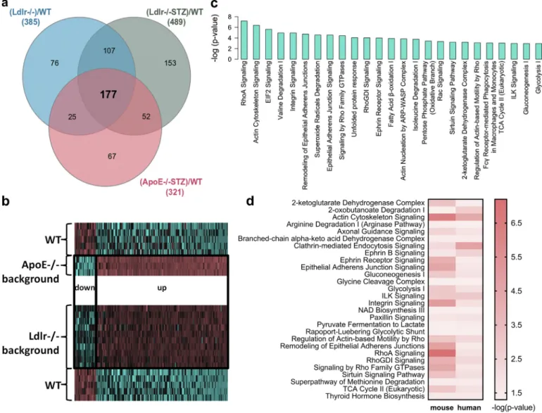
Fig. 2. Comparative proteomic analysis and functional annotation. (a) Comparative analysis of the proteomicfindings led to the identification of 177 differentially expressed proteins that are common in all disease models (Ldlr−/−, Ldlr−/−STZ, ApoE−/−STZ) when compared to WT controls. (b) The expression changes of these 177 proteins in disease are illustrated in the heatmap for each group, indicating the similar trend of expression of these proteins in all cases. (c) Ingenuity Pathway Analysis (IPA) mapping of the representative molecular pathways by the 177 differentially expressed proteins. (d) The common molecular pathways between mouse and human proteomicfindings based on IPA.
Fig. 1. Proteome analysis of the Ldlr−/− background mouse models. (a) Box-and-whiskers plots of main biochemical characteristics (cholesterol and glycemia levels) of the animal models included in the proteomic analysis. Statistical significance between the diseased (Ldlr−/−, n = 5; Ldlr−/−STZ, n = 5) and control (WT, n = 5) animals was determined using 2-tailed Mann-Whitney U tests (*Pb 0·05, **P b 0·01). (b) Principal component analysis of the thoracic aorta proteome was performed in an unsupervised fashion. The log2
transformed values (prior adding one) of the total protein identifications for each animal were used in this analysis. The scatter plot illustrates the first three principal component scores in a three-dimensional space. Three distinct clusters are inferred indicating the absence of outlying samples. (c) The differential expression analysis results are graphically represented with volcano plots; (i) Volcano plot of log2(fold change: (Ldlr−/−)/WT) versus –log10(Mann-Whitney p-value) and (ii) Volcano plot of log2(fold change: (Ldlr−/−STZ)/
WT) versus–log10(Mann-Whitney p-value). Proteins uniquely expressed per model werefiltered out. Red data points indicate 385 differentially expressed proteins when comparing
Ldlr−/− with WT animals and 489 proteins between Ldlr−/−STZ and WT that were detected based on at least two peptides identification, had a fold change of N1·5 (or b 1/1·5) and a p-value ofb0·05. Among these, (d) comparative proteomic analysis identified 284 proteins that were common in both group comparisons. (e) The heatmap illustrates the expression level changes of the 284 differentially expressed proteins among the WT, Ldlr−/− and Ldlr−/−STZ animals. (f) Correlation of the levels of the 284 differentially expressed proteins was analyzed using the 2-tailed Pearson correlation coefficient (P b 0·0001) after log2transformation of the values.
the Ingenuity Pathway Analysis software (QIAGEN Inc.) using the de-fault settings [18].
2.6. Cell culture and treatment with the inhibitor
Human Umbilical Vein Endothelial Cells (pooled HUVEC) were cul-tured in Endothelial Growth Medium-2 (EGM-2) (Lonza) and were used at passages 4–6. When required, HUVEC were starved in Endothe-lial Basal Medium-2 (EBM-2): EGM-2 (4∶1). For the inhibition experi-ments the KDM5 Histone Demethylases inhibitor, KDM5-C70, ethyl 2-(((2-((2-(dimethylamino)ethyl)(ethyl)amino)-2-oxoethyl)amino) methyl)isonicotinate (Xcessbio Biosciences) was added at different concentrations (10μΜ, and 20 μΜ) in the growth media (complete me-dium or starvation meme-dium depending on the experiment). 0.1% DMSO treated cells were used as controls since DMSO was the solvent of the inhibitor. The inhibitory activity was examined after 3 days of treatment through western blot analysis for the trimethylated lysine 4 of histone H3 (H3K4me3).
2.7. Western blot analysis
30μg total protein from cell extracts and tissue extracts were ana-lyzed for H3K4me3 (1/5,000 rabbit monoclonal anti-human Tri-Methyl-Histone H3 (Lys4) (C42D8), Cell Signaling Technology). Total histone H3 (1/5,000 rabbit monoclonal anti-human histone H3 (D2B12), Cell Signaling Technology) was used as a loading control. Thefilms were scanned at a GS-800 imaging densitometer (BioRad) in transmission mode and the densitometry analysis of the results was performed using the Quantity One software (BioRad).
2.8. PCR analysis
KDM5D expression was examined in cDNA samples by performing Real-time PCR using the SYBR Green master mix (Kapa Biosystems),
and specific primers for KDM5D (forward: 5′-ACAGCTACAGGCCAAA
CC-3′ and reverse: 5′-CAGCCCTTGGACCTTAGAAATA-3′). GAPDH was
used as endogenous control.
2.9. Flow cytometry analysis
To test for potential toxicity of the KDM5-C70 inhibitor, apoptosis and necrosis were evaluated on untreated or treated for 3 days with
0.1% DMSO, 10μM or 20 μM KDM5-C70 HUVEC using FITC Annexin V
Apoptosis Detection Kit with 7-amino-actinomycin D (7-AAD) (BioLegend) according to the manufacturer's instructions. Flow
cytom-etry was performed using the Beckman Coulter Cytomics FC 500flow
cytometer (Beckman Coulter). 2.10. MTS cell proliferation assay
The effect of the KDM5-C70 inhibitor on the proliferation rate of HUVEC was assessed using the MTS cell proliferation assay. HUVEC
were initially treated for 3 days with 0.1% DMSO, 10μM or 20 μM
KDM5-C70. Subsequently, the cells were seeded in 96-well plates at a density of 1,000 cells per well and subjected to 0.1% DMSO, 10μM or 20μM KDM5-C70 for the following 1, 2 and 3 days. To assess any differ-ences in the proliferation rate, the recommended amount of MTS re-agent (Promega) was supplemented into each well of the 96-well plate at the indicated time points. The cells were incubated with the re-agent for 3 h at 37 °C in a humidified, 5% CO2atmosphere. The absor-bance was then recorded at 490 nm with a 96-well plate reader (SPECTROstar Nano, BMG LABTECH). Three independent experiments were performed with each experiment containingfive replicates. 2.11. Transwell migration assay
The impact of the KDM5-C70 inhibitor on the migratory capacity of HUVEC was assessed using the transwell assay. HUVEC were initially treated for 3 days with 0.1% DMSO, 10μM or 20 μM KDM5-C70 and sub-sequently starved in EBM-2: EGM-2 (4∶1) medium for 24 h. Then, 2 × 10⁴cells from each category were added to the inner compartment of transwell inserts with 5μm pores (Corning-Costar) in 100 μl starva-tion medium containing 0.1% DMSO, 10μM or 20 μM KDM5-C70 respec-tively. As stimulus, EGM-2 medium supplemented with 18% FBS (20% EGM-2) was added at the bottom chamber of the transwell plate. Cells were allowed to migrate for 24 h. Afterwards, the non migrated cells were removed from the top of the insert with a wet cotton swab whereas the migrated cells at the bottom werefixed with 4% parafor-maldehyde and stained with eosin-haematoxylin. Photographs of the stained cells were taken at 5 differentfields (20×) of each insert using a Leica CTR MIC microscope. The number of the migrated cells was assessed by using the Image J software (version 1.51j8). Three indepen-dent experiments were performed with each experiment containing two replicates.
2.12. Tube formation assay
The effect of the KDM5-C70 inhibitor on the tube forming ability of HUVEC was assessed using the matrigel-based tube formation assay. HUVEC were initially treated for 3 days with 0.1% DMSO, 10μM or 20 μM KDM5-C70 and subsequently starved for 4 h in EBM-2: EGM-2
(4:1) medium containing 0.1% DMSO, 10μM or 20 μM KDM5-C70
re-spectively. Subsequently, the cells from each condition were plated at a density of 2 × 10⁴cells/well in 100μl starvation medium containing 0.1% DMSO, 10μM or 20 μM KDM5-C70, in 96-well plates coated with 50μl of growth factor-reduced Matrigel (BD Biosciences). Plates were incubated for 16 h at 37 °C in a humidified, 5% CO2atmosphere. Photo-graphs from 5 differentfields (5×) per well were taken using a Leica CTR MIC microscope and were analyzed by using the Angiogenesis Analyzer plug-in in Image J software (version 1.51j8). Representative photo-graphs were also taken at 10× magnification. The angiogenesis was assessed by measuring the number of the nodes, junctions, segments as well as the total length (in pixel) which is defined as the sum of length of segments, isolated elements and branches in the analyzed
Table 2
Demographic, physical and clinical characteristics of the participants in this study.
Variable Control group (n = 20) Case group (n = 30) Demographics Sex [n (%)] 16 (80%) M, 4 (20%) F 14 (46·7%) M, 16 (53·3%) F Age [years] 46·6 ± 18·5 62·8 ± 13 Physical characteristics Weight [kg] 80·1 ± 14·2 75·7 ± 12·3 Height [m] 1·8 ± 0·1 1·7 ± 0·1 Body mass index [kg/m2] 26·1 ± 3·7 26·2 ± 3·1
Clinical information
Dyslipoproteinemia [n (%)] 4 (20%) 5 (16·7%) Obesity [n (%)] 3 (15%) 4 (13·3%) Diabetes [n (%)] 0 (0%) 8 (26·7%) Vascular disease [n (%)] 0 (0%) 30 (100%) Left ventricular hypertrophy [n
(%)]
0 (0%) 12 (40%)
Angina pectoris [n (%)] 0 (0%) 2 (6·7%) Claudicatio intermittens [n (%)] 0 (0%) 10 (33·3%) Myocardial infarction [n (%)] 0 (0%) 3 (10%)
M: Males, F: Females; Dyslipoproteinemia was characterized by total cholesterol ≥ 5·2 mmol/l (≥200 mg/dl), low-density lipoprotein (LDL) cholesterol ≥ 3·4 mmol/l (≥130 mg/dl), high-density lipoproteins (HDL) cholesterol ≤ 1·0 mmol/l (≤40 mg/dl), and triglycerides≥ 1·7 mmol/l (≥150 mg/dl); Obesity was defined by a body mass indexN 30 kg/m2.
Fig. 3. The expression levels of the six -common with mouse- proteins in human vascular tissues. (a) Bar graph illustrating the fold change difference of the six proteins that were identified in all mouse and human proteomic datasets, and at statistically significant levels when comparing cases and controls. (b) Violin plots showing the protein abundance of each one of the six proteins in human vascular tissues from patients with CVD (cases, n = 37) compared to organ donors without CVD background (controls, n = 24) based on LC-MS/MS analysis. The male specific KDM5D protein was detected only in male human vascular tissues (controls, n = 19 and cases, n = 18). The expression levels are expressed as log2(x + 1) values with x
Fig. 4. Alteration in the expression levels of the KDM5 substrate, H3K4me3, in CVD. (a) KDM5D is a member of the KDM5 family of histone demethylases that specifically demethylate trimethylated and dimethylated lysine 4 of histone H3. KDM5 are transcriptional regulators that may act as transcriptional repressors by demethylating H3K4me3 at promoters of transcribed genes. (b) Western blot (WB) analysis of H3K4me3 in all available human vascular tissues from organ donors without a CVD background (controls; male: 18 vascular tissues from 15 individuals and female: 5 vascular tissues from 4 individuals) and patients with CVD (cases; male: 17 vascular tissues from 14 patients and female: 19 vascular tissues from 16 patients). Total histone H3 was used as loading control. (c) Quantification of the WB data following normalization to total H3. Statistical significance was determined using 2-tailed Mann-Whitney U tests (**Pb 0·01). (d) Nonparametric regression analysis to evaluate association of the enzyme (KDM5D) and substrate (H3K4me3) levels in the male vascular tissues. The polygon represents the 95% confidence interval of local polynomial regression (LOESS function in R statistical package). Spearman correlation analysis between the enzyme and the substrate indicates a negative correlation between the two variables which is graphically represented with the linear model regression line.
area. Three independent experiments were performed with each exper-iment containing two replicates.
2.13. Statistical analysis
Statistical significance of the proteomic differences between WT an-imals and Ldlr−/− or Ldlr−/−STZ, or ApoE−/−STZ animals was deter-mined using the 2-tailed Mann-Whitney U test on the R statistical environment (R version 3.5.1). Log2transformation of the mean values prior to adding constant 1 to data [log2(x + 1) with x representing the raw data from the LC-MS/MS analysis] was applied in datasets contain-ing zero values. Pearson correlation coefficient was assessed for data that passed normality after log2transformation of the mean values. If normal distribution could not be established, Spearman correlation coefficient was assessed. Nonparametric regression analysis was per-formed using the Local Polynomial Regression Fitting (LOESS function in R statistical package). Statistical significance of differences between groups was determined using the 2-tailed Mann-Whitney U test. In case of multiple group analyses, the one-way and two-way ANOVA tests for randomized block experiments were performed followed by a Tukey post hoc test. Data are presented as mean ± SD (*Pb 0.05, **P b 0.01, ***Pb 0.001, ****P b 0.0001). All statistical analyses were per-formed using GraphPad Prism 7 software. Principal component analysis was performed in SPSS version 25 in an unsupervised fashion. The vol-cano plots for proteins identified with at least two peptides (excluding proteins that were unique in one model) were created using the pack-age EnhancedVolcano (Publication-ready volcano plots with enhanced colouring and labeling, Version: 1.1.1, Author: Kevin Blighe) on the R statistical environment (R version 3.5.1). Violin plots showing the Kernel probability density of the log2transformed data, were created using R software and ggplot2 package.
3. Results
3.1. Proteomic profiling of animal models with atherosclerosis
To identify proteome alterations associated with atherosclerosis, LC-MS/MS proteome analysis was performed on thoracic aortas isolated
from the Ldlr−/− atherosclerotic mouse models in the absence of
diabetes (Ldlr−/−, n = 5) or presence of diabetes (Ldlr−/−STZ, n = 5) in comparison to WT (n = 5) mice. In all cases, animals were evalu-ated for their phenotypical and biochemical characteristics to confirm presence of atherosclerosis, prior to be included in the study (Supple-mentary Table 1). As shown inFig. 1a, atherosclerosis in those animals was characterized by dyslipidemia; and in the case of the diabetic ani-mals, as expected, increased glycemia and diuresis were also observed. To assess the quality of the proteomics data in an unsupervised fash-ion, Principal Component Analysis (PCA) was performed. As shown in Fig. 1b, there were no significant outliers and all of the three animal groups were well separated. The LC-MS/MS analysis resulted in a total of 2,365 proteins identified based on at least 2 peptides (Supplementary Table 2). Only proteins fulfilling the following criteria were considered as differentially expressed: a) were statistically significant different (P ≤ 0.05) and b) had a fold change of at least 1.5 (|fold change| ≥1.5) in Ldlr−/− or Ldlr−/−STZ versus WT. This led to the identification of
385 and 489 protein changes in Ldlr−/− and Ldlr−/−STZ, compared
to WT, respectively (Fig. 1c). Of these, 284 proteins were common (Fig. 1d) and consistently changed (e.g. with same trend of expression) in disease in both comparisons (Fig. 1e, Supplementary Table 2). A strong correlation between the abundance of these 284 proteins was observed (Fig. 1f). Thisfinding further suggests that these changes may reflect common disease manifestations regardless of the presence of diabetes.
To exclude protein changes that may be specifically related to the Ldlr−/− background, we subsequently performed proteomic analysis of thoracic aortas from another atherosclerotic model characterized by
the ApoE−/− background and the presence of diabetes (ApoE−/
−STZ, n = 3). Phenotypic and biochemical characterization prior to the proteomic analysis confirmed the presence of diabetic atherosclero-sis in these animals (Supplementary Fig. 1a, Supplementary Table 1). PCA exploratory analysis of the proteomicsfindings indicated the sepa-ration of the experimental groups and the absence of outliers (Supple-mentary Fig. 1b). Following comparative proteomics analysis, 321 differentially expressed proteins were identified, having a statistical
significant fold change of N1.5 in ApoE−/−STZ versus WT animals
(Supplementary Fig. 1c and 1d, Supplementary Table 3). Comparison of the 284 differentially expressed proteins of the Ldlr−/− and the
321 proteins of the ApoE−/− analysis revealed an overlap of 177
proteins (Fig. 2a, Supplementary Table 4). These had the same trend of expression (Fig. 2b) in all animal models in comparison to controls, suggesting their association with the development of atherosclerosis re-gardless the specific disease aetiology (e.g. genetic background or pres-ence of diabetes, as reflected in the employed models).
3.2. Assessment of the biological relevance of the proteomicfindings Analyzing the function (based on UniProt database) of the top 15 most significantly deregulated proteins in each of the three models (Table 1, Supplementary Table 4), we observed that these proteins were associated with processes such as the mitochondrial respiratory chain (e.g. Letm1, Ndufa10, Afg1l), cell migration (e.g. Stk25), adhesion (e.g. Kif26b), cytoskeleton dynamics (e.g. Cfl1), gene expression (e.g. Xrn1, Ice1), biosynthesis of hormonal steroids (e.g. Hsd3b4) and signal transduction (e.g. Ndrg2). Moreover, proteins associated with histone modifications such as Kdm5d and Hpf1, were also included (Table 1, Supplementary Table 4). Further mapping of all common 177 differen-tially expressed proteins into molecular pathways (Supplementary Table 5, representative shown onFig. 2c) based on the Ingenuity Path-way Analysis (IPA) software revealed that these proteins participate in metabolic processes including Fatty Acidβ-oxidation I, Pentose Phos-phate Pathway, 2-ketoglutarate dehydrogenase complex, tricarboxylic acid (TCA) cycle, glycolysis, gluconeogenesis and metabolism of amino acids and derivatives (e.g. Valine and Isoleucine degradation). Addition-ally, disease-associated changes in pathways related to detoxification of ROS (Superoxide radicals degradation, NRF2-mediated oxidative stress
response), immune system (Fcγ Receptor-mediated phagocytocis in
macrophages and monocytes, fMLP signaling in neutrophils, CD28 sig-naling in T Helper cells, chemokine and leukocyte extravasation signal-ing), translation (EIF2 signalsignal-ing), cytoskeleton remodeling and cell adhesion (actin cytoskeleton signaling, integrin signaling, remodeling of epithelial adherens junctions) as well as in Endoplasmic Reticulum Stress pathway and unfolded protein response were predicted (Supple-mentary Table 5). These pathways, as well as the enriched Rho and ILK signaling pathways (Fig. 2c), have been previously described to be in-volved in the atherosclerotic process [19–28], supporting the biological relevance of the proteomicfindings.
3.3. KDM5D abundance changes in human CVD
To investigate transferability of the animal modelfindings to human disease, the list of 177 differentially expressed proteins in the athero-sclerotic animal models, was compared to protein changes in human vascular tissues from patients with CVD (cases; n = 37 samples from 30 patients;Table 2) in comparison to vessels from organ donors with no evidence of CVD (controls, n = 24 samples from 20 subjects; Table 2), as revealed following application of a similar LC-MS/MS ap-proach. In this analysis of human vascular tissue, 94 proteins were found to be differentially expressed. Similar to the animal models, the proteins represented pathways associated with metabolism (e.g. 2-ketoglutarate dehydrogenase complex, TCA cycle, glycolysis, and me-tabolism of amino acids), cytoskeleton remodeling and cell adhesion (e.g. actin cytoskeleton signaling, integrin signaling, remodeling of
epithelial adherens junctions) as well as pathways known to be impli-cated in atherosclerosis including the Rho signaling pathways (RhoA signaling, signaling by Rho family GTPases, RhoGDI signaling), the ILK signaling, Ephrin B signaling and the Sirtuin signaling pathway (Fig. 2d). Following a cross-species comparison of the two protein lists (177 mouse proteins versus 94 human proteins) based on orthologues, an overlap of six proteins could be observed (KDM5D, CTSD, IGHM, HMGCS2, TTN, ZFYVE1), (Fig. 3a). Interestingly, of these, the male spe-cific demethylase KDM5D was found at significantly increased abun-dance levels in all atherosclerotic models (Table 1,Fig. 3a), being also fairly abundant in the human tissues of male subjects with CVD (n = 18 vessels from 14 patients, and n = 19 vessels from 16 controls; Sup-plementary Table 6), having the most pronounced differences in CVD in comparison to the otherfive proteins (Fig. 3b, Supplementary Table 7). This difference was also observed even when considering only male samples for the analysis of the expression changes of all six proteins (Supplementary Fig. 2). Of note, among the tested vascular samples, 8 originated from patients with diabetes (cases, n = 6) and when compared to the 19 vascular tissues from non-diabetic healthy in-dividuals (controls, n = 16) pronounced differences in the KDM5D levels were also observed, again in agreement to the animal model data (Supplementary Fig. 3).
To further verify this observation, we next examined whether the in-creased levels of KDM5D were associated with a reduction of its sub-strate. As depicted inFig. 4a, H3K4me3 is the main substrate of
KDM5D in males as well as of all members of KDM5 family of histone demethylases in both sexes [29]. Western blot analysis for H3K4me3 of all available human vessel samples (Table 2) indicated a significant decrease in the levels of the protein in CVD in comparison to controls (Fig. 4b and4c). In male patients where KDM5D expression levels were evaluated (Fig. 3b), Spearman correlation analysis revealed a sta-tistical significant negative correlation between the expression levels of KDM5D and the abundance of H3K4me3 (Fig. 4d), further suggesting that KDM5D overexpression was associated with increased activity. H3K4me3 also appeared at overall decreased levels at the available samples from females affected by CVD, indicating increased activity of further members of the KDM5 family in CVD.
3.4. The impact of KDM5 inhibition on H3K4me3 levels and on proliferation, migration and tube-forming ability of HUVEC
An increasing number of studies support an important role of epige-netic mechanisms including post-translational modifications of his-tones in the regulation of the endothelial function. More specifically, the methylation status of H3K4 has been proven essential for endothe-lial cell sprouting and migration and therefore for angiogenesis [30]. Based on the literature, the KDM5 family is expected to affect a wide va-riety of molecular processes including proliferation, migration and
an-giogenesis [31]. Since all these are processes involved in the
development of atherosclerosis, we investigated the possible impact of
Fig. 6. Graphical illustration of the workflow and key findings of the study.
Fig. 5. The effect of KDM5 inhibition on the endothelial properties of HUVEC. (a) Western blot analysis of H3K4me3 of HUVEC treated with 0·1% DMSO, 10μM or 20 μM KDM5-C70 for 3 days. Total histone H3 was used as loading control. (b) MTS cell proliferation assay. Bar graph representing the absorbance (OD) at 490 nm of HUVEC at three different time points (day 1, day 2, day 3). The values represent the means ± SD from three independent experiments performed infive replicates. Statistical significance was determined using the two-way ANOVA test (***Pb 0·001, ****P b 0·0001). (c) The graph illustrates the number of migrated cells towards complete medium with representative images displayed underneath. Magnification: 20×. The values represent the means ± SD from three independent experiments performed in duplicate. Statistical significance was determined using the one-way ANOVA test (**P b 0·01). (d) Graphs illustrate the impact of the inhibitor on the number of nodes, junctions and segments as well as on the total length of segments, isolated elements and branches formed by HUVEC. The values represent the means ± SD from three independent experiments performed in duplicate. Statistical significance was determined using the one-way ANOVA test (*P b 0·05, **P b 0·01). (e) KDM5-C70 treatment has a significant effect on the angiogenic properties of HUVEC as illustrated by the representative images of the tube formation assay. Magnification: 5× and 10×.
KDM5 inhibition on these processes using the KDM5-C70 inhibitor, a KDM5-family inhibitor [32,33], and HUVEC as in vitro model [34].
HUVEC expressing KDM5D (Supplementary Fig. 4) were subjected to treatment with KDM5-C70 at different concentrations (10μΜ, and 20μΜ). After 3 days of treatment, abundance of H3K4me3 was signifi-cantly increased in the cells, as evaluated by western blot analysis, dem-onstrating efficient inhibition of KDM5 demethylase activity by KDM5-C70 (Fig. 5a). Absence of non-specific toxic effects was demonstrated by flow cytometry analysis for apoptotic (Annexin-V) and necrotic (7-AAD) cells (Supplementary Fig. 5).
The impact on proliferation was assessed by MTS assay. Treatment with 10μΜ and 20 μΜ of the inhibitor significantly reduced the prolif-eration rate of HUVEC in comparison to the DMSO treated cells (Fig. 5b). KDM5 inhibition also resulted in a statistically significant re-duction of the migratory capacity of HUVEC, as assessed using the transwell system (Fig. 5c). As shown inFig. 5d, treatment with the in-hibitor significantly reduced the number of nodes, junctions, and seg-ments, as well as the total length of segseg-ments, isolated elements and branches formed by HUVEC. Collectively, these results indicate that KDM5 inhibition attenuated the tube-forming ability of HUVEC (Fig. 5e).
4. Discussion
With this work, we aimed to characterize protein changes associated with CVD using high resolution proteome analysis. These changes may have a potential to serve as biomarkers for disease, or even as possible therapeutic targets. We used three widely employed disease models representing different disease aetiologies (Ldlr−/−, Ldlr−/−STZ, ApoE−/−STZ) thus allowing for the definition of as possible, ‘common’ atherosclerosis-associated proteomic alterations [13]. Of note, a good overlap was also observed when comparing the common proteins of the aforementioned three animal models with changes observed in
ApoE−/− versus WT mice (111 common proteins with the same
ex-pression trend at statistical significant levels – Supplementary
Table 8). Male mice were selected for this study since they develop more pronounced atherosclerosis than the females [35]. It should be noted that protein changes unique per model are also observed (as may be seen inFig. 2a and Supplementary Tables 2 and 3) which even though not investigated in this study, may merit further investigation.
To the best of our knowledge this is thefirst study that describes common proteomic alterations in these three atherosclerotic mouse models. An in-depth analysis of these protein changes suggests that they reflect different well-characterized atherosclerosis-associated mechanisms such as inflammation [19], metabolism [20], oxidative stress [21], and extracellular matrix remodeling [23]. Further
compari-son of the proteomicsfindings from the animal models with their
orthologues from the proteomic profiling of human vascular tissues from patients affected by CVD, revealed a high degree of overlap at the pathway level, although a small overlap at the individual molecule level. This has been also observed in studies comparing the overlap of human genes in coronary artery disease (CAD) identified by genome-wide association studies (GWAS) with those from atherosclerotic mouse models [36]. In these studies, although an overlap of only 18.4% of human CAD genes with mouse orthologs was detected, their rele-vance at pathways was significantly higher (over than 50% being consis-tent between the two species) [36]. In our case, the observed relatively small overlap at the individual molecule level may be additionally at-tributed to the use of different vessel types (central in mice versus pe-ripheral and central in humans). Nevertheless and despite this variability, six common proteins were identified. A more detailed inves-tigation of the expression of these proteins in a gender-specific manner is required to exclude with high confidence an impact of gender on our findings. Of note, among them was cathepsin D (CTSD) whose increased expression levels have been previously associated with atherosclerosis
[37] and the ketogenic enzyme hydroxymethylglutaryl-CoA synthase, mitochondrial (HMGCS2) whose upregulation may be implicated in type 1 diabetes induced cardiac dysfunction [38]. Furthermore, proteins with a possible role in CVD were also included such as the immunoglob-ulin heavy chain mu (IGHM) which was significantly increased in B cells in the artery tertiary lymphoid organs (ATLOs) with the latter associ-ated with B cell responses in the atherosclerotic aortas [39] and titin (TTN), a highly abundant protein in striated muscle whose truncated variants have been associated with several cardiomyopathies [40]. Pro-teins without a known/previous implication in CVD such as the zinc fin-ger FYVE domain-containing protein 1 (ZFYVE1) which is a protein participating to autophagosome formation [41], and KDM5D were also included with the latter being highlighted as a predominant CVD-associated change in both species. KDM5D was also found upregulated (fold change = 50.8 in cases versus controls) yet did not reach statistical significance in the ApoE−/− mice (in the absence of diabetes) when comparing with WT mice (data not shown).
KDM5D (also known as JARID1D or SMCY) is encoded on the Y chro-mosome and until now it has been implicated in prostate cancer inva-sion and metastasis [42], spermatogenesis [43], and sex-specific tissue transplantation rejection [44]. KDM5D acts as a direct epigenetic modu-lator, and as one of the four members of the KDM5 family of histone
demethylases, specifically demethylates trimethylated and
dimethylated Lys-4 of histone H3 [29]. The elevated expression of KDM5D in the human tissues of patients with CVD was accompanied by a reduction of its substrate (H3K4me3) implying increased activity of KDM5D.
Alteration in the trimethylated state of H3K4 has been previously re-lated to the development of heart failure in humans and rat models [45] and also associated with the physiological function of cardiomyocytes [46]. Mutations in genes involved in the H3K4me pathway (production, removal or reading of H3K4me), including KDM5A and KDM5B, have been implicated in the pathogenesis of congenital heart disease (CHD) [47]. Given this published evidence with respect to H3K4 and the ob-served increased KDM5D and concomitant decreased H3K4me3 levels in human tissues of patients with CVD, we investigated the impact of the KDM5-C70 inhibitor on HUVEC. This compound targets all four members of the KDM5 family, is a cell-permeable prodrug with no re-ported cytotoxicity, and shows selectivity for KDM5 family members compared to KDM6 and KDM4 [32,33]. Considering that endothelial cell dysfunction is an important contributor in atherosclerosis [1] we employed HUVEC. Inhibition of KDM5 activity increased the H3K4me3 levels and significantly attenuated the proliferation rate, the migratory capacity and the tube-forming ability of the endothelial cells in vitro. Thesefindings are in line with another study that describes impaired angiogenic properties of the endothelial cells after inhibition of KDM5B and gene silencing with shRNA, linked to induced expression of the antiangiogenic transcription factor HOXA5 [48].
Interpretation of ourfindings in the context of atherosclerosis sug-gests that treatment with a KDM5 inhibitor may be beneficial, consider-ing that the atherosclerotic plaques are characterized by increased neovascularization that contributes to intraplaque hemorrhage and plaque progression [49]. In response to hypoxia and to the VEGF gradi-ent in the atherosclerotic lesion, the endothelial cells of the vasa vasorum switch from a quiescent phenotype to a highly proliferative and migratory state to start sprouting and forming the new vessels [49]. Since KDM5 inhibition significantly attenuated growth and func-tion of endothelial cells in vitro, it may have therapeutic potential in vivo- a working hypothesis warranting further investigation.
Collectively, in this study, by applying a systematic approach that in-cluded high-throughput proteomics of mouse models and human vas-cular tissues as well as in vitro assays, a role for KDM5 histone demethylases in CVD, likely via affecting H3K4 methylation, is sug-gested. The keyfindings of this study are summarized inFig. 6. In addi-tion to multiple high confidence proteomic changes reflecting common
molecular manifestations of atherosclerosis, further underlining the va-lidity of the approach, other yet unknown proteomic changes were identified, which may be of significant value in further systems biology approaches and model selection for pre-clinical studies.
Limitations of the study
Our proteomics based approach using mouse and human vascular tissues with CVD indicates KDM5D as a novel candidate pharmacologi-cal target associated with CVD progression. However, there are several shortcomings of this study with regards to the role of KDM5D in the context of CVD. Given the fact that the cellular composition of the aortas especially in the areas of the lesions is quite heterogeneous, further ex-periments and efforts to identify which cells contribute to the elevated expression of KDM5D appear well justified. Along the same lines, in this study we focused on the impact of KDM5 inhibition on endothelial cells, given that endothelial dysfunction is a crucial event in atheroscle-rosis and impact of H3K4 methylation on endothelial migration and proliferation has been reported. Nevertheless, studying the impact of KDM5 inhibition on other cell types involved in atherosclerotic plaque formation including smooth muscle cells and inflammatory cells is well justified, especially given the reported impact of H3K4 methylation on inflammation [50]. Additionally, characterization of the genes af-fected upon the KDM5D-mediated alteration of the methylation status of H3K4 may be valuable to improve our understanding of the mecha-nism of KDM5 inhibition. In parallel, the impact of inhibition of KDM5 in vivo on CVD preferably in a gender-specific manner is needed. How-ever this is currently limited due to the lack of well characterized inhib-itors applicable for in vivo studies. Although limitations of the current study, these are multiple research avenues to be further explored, for which the presented study forms a solid starting point.
Acknowledgements
All authors have read and approved the article. Funding sources
This work was supported by the European Commission via funding to the Systems Biology to Identify Molecular Targets for Vascular Dis-ease Treatment [SysVasc, HEALTH-2013 603288]. Part of the study
wasfinanced by the ERA-CVD PROACT project (numbers
ANR-17-ECVD-0006 for J.K, J.S.S.B and J.P.S and 01KL1805 via the BMBF for H. M), and by a grant from the“Fondation pour la Recherche Médicale” (grant number DEQ20170336759) for J.K, J.S.S.B and J.P.S. The funders had no role in study design, data collection, data analysis, data interpre-tation, patient recruitment, or writing of the manuscript. The authors have not been paid to write this article by a pharmaceutical company or other agency. The corresponding author had full access to all the data in the study and had thefinal responsibility for the decision to submit for publication.
Declaration of interests
Harald Mischak is the co-founder and co-owner of Mosaiques Diagnostics.
Author contributions
A.V, J.P.S, H.M, B.P, M.G.R, J.Z conceived and designed the research; M.M*, J.K, M.M, V.B, W.M, J.L.B, J.S.S.B, M.S acquired the data; M.M* per-formed statistical analysis of the data; M.M*, A.V, J.P.S, J.K drafted the manuscript. All co-authors have read the manuscript, revised it and agree with the submitted version.
Appendix A. Supplementary data
Supplementary data to this article can be found online athttps://doi. org/10.1016/j.ebiom.2019.02.040.
References
[1]Gimbrone Jr MA, Garcia-Cardena G. Endothelial cell dysfunction and the pathobiol-ogy of atherosclerosis. Circ Res 2016;118(4):620–36.
[2]Libby P, Ridker PM, Hansson GK. Progress and challenges in translating the biology of atherosclerosis. Nature 2011;473(7347):317–25.
[3]Dominiczak AF, Herget-Rosenthal S, Delles C, et al. Systems biology to battle vascular disease. Nephrology, dialysis, transplantation : official publication of the European Dialysis and transplant association. European Renal Association 2010;25(4): 1019–22.
[4]Shalhoub J, Sikkel MB, Davies KJ, Vorkas PA, Want EJ, Davies AH. Systems biology of human atherosclerosis. Vasc Endovascular Surg 2014;48(1):5–17.
[5]Mokou M, Lygirou V, Vlahou A, Mischak H. Proteomics in cardiovascular disease: re-cent progress and clinical implication and implementation. Expert Review of Prote-omics 2017;14(2):117–36.
[6]Martin-Lorenzo M, Gonzalez-Calero L, Maroto AS, et al. Cytoskeleton deregulation and impairment in amino acids and energy metabolism in early atherosclerosis at aortic tissue with reflection in plasma. Biochim Biophys Acta 2016;1862(4):725–32. [7]Martin-Lorenzo M, Zubiri I, Maroto AS, et al. KLK1 and ZG16B proteins and arginine-proline metabolism identified as novel targets to monitor atherosclerosis, acute cor-onary syndrome and recovery. Metabolomics 2015;11(5):1056–67.
[8]Herrington DM, Mao C, Parker SJ, et al. Proteomic architecture of human coronary and aortic atherosclerosis. Circulation 2018;137(25):2741–56.
[9]Emini Veseli B, Perrotta P, De Meyer GRA, et al. Animal models of atherosclerosis. Eur J Pharmacol 2017;816:3–13.
[10]Saulnier-Blache JS, Wilson R, Klavins K, et al. Ldlr−/−and ApoE−/−mice better mimic the human metabolite signature of increased carotid intima media thickness compared to other animal models of cardiovascular disease. Atherosclerosis 2018; 276:140–7.
[11]Beckman JA, Creager MA. Vascular complications of diabetes. Circ Res 2016;118(11): 1771–85.
[12]Shen X, Bornfeldt KE. Mouse models for studies of cardiovascular complications of type 1 diabetes. Ann N Y Acad Sci 2007;1103:202–17.
[13]Getz GS, Reardon CA. Do the Apoe−/− and Ldlr−/− mice yield the same insight on Atherogenesis? Arteriosclerosis, Thrombosis, and Vascular Biology 2016;36(9): 1734–41.
[14]Lygirou V, Latosinska A, Makridakis M, et al. Plasma proteomic analysis reveals al-tered protein abundances in cardiovascular disease. J Transl Med 2018;16(1):104. [15]Makridakis M, Vlahou A. GeLC-MS: a sample preparation method for proteomics
analysis of minimal amount of tissue. Methods Mol Biol 2018;1788:165–75. [16]Latosinska A, Mokou M, Makridakis M, et al. Proteomics analysis of bladder cancer
invasion: targeting EIF3D for therapeutic intervention. Oncotarget 2017;8(41): 69435–55.
[17]Babicki S, Arndt D, Marcu A, et al. Heatmapper: web-enabled heat mapping for all. Nucleic Acids Res 2016;44(W1):W147–53.
[18]Kramer A, Green J, Pollard Jr J, Tugendreich S. Causal analysis approaches in ingenu-ity pathway analysis. Bioinformatics 2014;30(4):523–30.
[19]Geovanini GR, Libby P. Atherosclerosis and inflammation: overview and updates. Clin Sci 2018;132(12):1243–52.
[20]Koelwyn GJ, Corr EM, Erbay E, Moore KJ. Regulation of macrophage immunometabolism in atherosclerosis. Nat Immunol 2018;19(6):526–37. [21]Forstermann U, Xia N, Li H. Roles of vascular oxidative stress and nitric oxide in the
pathogenesis of atherosclerosis. Circ Res 2017;120(4):713–35.
[22]Borissoff JI, Spronk HM, ten Cate H. The hemostatic system as a modulator of athero-sclerosis. N Engl J Med 2011;364(18):1746–60.
[23]Yurdagul Jr A, Finney AC, Woolard MD, Orr AW. The arterial microenvironment: the where and why of atherosclerosis. Biochem J 2016;473(10):1281–95.
[24]Khyzha N, Alizada A, Wilson MD, Fish JE. Epigenetics of atherosclerosis: emerging mechanisms and methods. Trends Mol Med 2017;23(4):332–47.
[25]Ivanova EA, Orekhov AN. The role of endoplasmic reticulum stress and unfolded pro-tein response in atherosclerosis. Int J Mol Sci 2016;17(2).
[26]Cai A, Zhou Y, Li L. Rho-GTPase and atherosclerosis: pleiotropic effects of statins. J Am Heart Assoc 2015;4(7).
[27]Rolfe BE, Worth NF, World CJ, Campbell JH, Campbell GR. Rho and vascular disease. Atherosclerosis 2005;183(1):1–16.
[28]Finney AC, Stokes KY, Pattillo CB, Orr AW. Integrin signaling in atherosclerosis. Cel-lular and molecular life sciences. CMLS 2017;74(12):2263–82.
[29]Lee MG, Norman J, Shilatifard A, Shiekhattar R. Physical and functional association of a trimethyl H3K4 demethylase and Ring6a/MBLR, a polycomb-like protein. Cell 2007;128(5):877–87.
[30]Fraineau S, Palii CG, Allan DS, Brand M. Epigenetic regulation of endothelial-cell-mediated vascular repair. FEBS J 2015;282(9):1605–29.
[31]Harmeyer KM, Facompre ND, Herlyn M, Basu D. JARID1 histone demethylases: emerging targets in cancer. Trends in Cancer 2017;3(10):713–25.
[32]Horton JR, Liu X, Gale M, et al. Structural basis for KDM5A histone lysine Demethylase inhibition by diverse compounds. Cell Chemical Biology 2016;23(7): 769–81.
[33]Johansson C, Velupillai S, Tumber A, et al. Structural analysis of human KDM5B guides histone demethylase inhibitor development. Nat Chem Biol 2016;12(7): 539–45.
[34]Goodwin AM. In vitro assays of angiogenesis for assessment of angiogenic and anti-angiogenic agents. Microvasc Res 2007;74(2–3):172–83.
[35]Getz GS, Reardon CA. Animal models of atherosclerosis. Arteriosclerosis, Thrombosis, and Vascular Biology 2012;32(5):1104–15.
[36]von Scheidt M, Zhao Y, Kurt Z, et al. Applications and limitations of mouse models for understanding human atherosclerosis. Cell Metab 2017;25(2):248–61. [37]Zhao CF, Herrington DM. The function of cathepsins B, D, and X in atherosclerosis.
American Journal of Cardiovascular Disease 2016;6(4):163–70.
[38]Shukla SK, Liu W, Sikder K, et al. HMGCS2 is a key ketogenic enzyme potentially in-volved in type 1 diabetes with high cardiovascular risk. Sci Rep 2017;7(1):4590. [39]Srikakulapu P, Hu D, Yin C, et al. Artery tertiary lymphoid organs control
multilay-ered territorialized atherosclerosis B-cell responses in aged ApoE−/− mice. Arterioscler Thromb Vasc Biol 2016;36(6):1174–85.
[40]Zhang C, Zhang H, Wu G, et al. Titin-truncating variants increase the risk of cardio-vascular death in patients with hypertrophic cardiomyopathy. Can J Cardiol 2017; 33(10):1292–7.
[41]Gatica D, Chiong M, Lavandero S, Klionsky DJ. Molecular mechanisms of autophagy in the cardiovascular system. Circ Res 2015;116(3):456–67.
[42]Li N, Dhar SS, Chen TY, et al. JARID1D is a suppressor and prognostic marker of pros-tate cancer invasion and metastasis. Cancer Res 2016;76(4):831–43.
[43]Akimoto C, Kitagawa H, Matsumoto T, Kato S. Spermatogenesis-specific association of SMCY and MSH5. Genes to Cells 2008;13(6):623–33.
[44]Wang W, Meadows LR, den Haan JM, et al. Human H-Y: a male-specific histocom-patibility antigen derived from the SMCY protein. Science 1995;269(5230): 1588–90.
[45]Kaneda R, Takada S, Yamashita Y, et al. Genome-wide histone methylation profile for heart failure. Genes to Cells 2009;14(1):69–77.
[46]Stein AB, Jones TA, Herron TJ, et al. Loss of H3K4 methylation destabilizes gene ex-pression patterns and physiological functions in adult murine cardiomyocytes. J Clin Invest 2011;121(7):2641–50.
[47]Zaidi S, Choi M, Wakimoto H, et al. De novo mutations in histone-modifying genes in congenital heart disease. Nature 2013;498(7453):220–3.
[48]Fork C, Gu L, Hitzel J, et al. Epigenetic regulation of angiogenesis by JARID1B-induced repression of HOXA5. Arteriosclerosis, Thrombosis, and Vascular Biology 2015;35 (7):1645–52.
[49]de Vries MR, Quax PH. Plaque angiogenesis and its relation to inflammation and ath-erosclerotic plaque destabilization. Curr Opin Lipidol 2016;27(5):499–506. [50]Zhao S, Zhong Y, Fu X, et al. H3K4 methylation regulates LPS-induced