HAL Id: inserm-02544432
https://www.hal.inserm.fr/inserm-02544432
Submitted on 16 Apr 2020
HAL is a multi-disciplinary open access archive for the deposit and dissemination of sci-entific research documents, whether they are pub-lished or not. The documents may come from teaching and research institutions in France or abroad, or from public or private research centers.
L’archive ouverte pluridisciplinaire HAL, est destinée au dépôt et à la diffusion de documents scientifiques de niveau recherche, publiés ou non, émanant des établissements d’enseignement et de recherche français ou étrangers, des laboratoires publics ou privés.
CanCOLD population-based cohort
Damien Viglino, Mickaël Martin, Marie-Eve Piché, Cynthia Brouillard,
Jean-Pierre Després, Natalie Alméras, Wan Tan, Valérie Coats, Jean
Bourbeau, Jean-Louis Pepin, et al.
To cite this version:
Damien Viglino, Mickaël Martin, Marie-Eve Piché, Cynthia Brouillard, Jean-Pierre Després, et al.. Metabolic profiles among COPD and controls in the CanCOLD population-based cohort. PLoS ONE, Public Library of Science, 2020, 15 (4), pp.e0231072. �10.1371/journal.pone.0231072�. �inserm-02544432�
RESEARCH ARTICLE
Metabolic profiles among COPD and controls
in the CanCOLD population-based cohort
Damien ViglinoID1,2*, Mickae¨l Martin1, Marie-Eve Piche´1, Cynthia Brouillard1,
Jean-Pierre Despre´s1, Natalie Alme´ras1, Wan C. Tan3, Vale´rie Coats1, Jean Bourbeau4, Jean-Louis Pe´pin2, Franc¸ois Maltais1, on behalf of the CanCOLD Collaborative Research Group
and the Canadian Respiratory Research Network
1 Centre de Recherche, Institut Universitaire de Cardiologie et de Pneumologie de Que´bec, Universite´ Laval, Que´ bec, Canada, 2 Centre Hospitalier Universitaire de Grenoble-Alpes, Laboratoire HP2 INSERM U 1042 Universite´ Grenoble-Alpes, Grenoble, France, 3 James Hogg Research Centre, University of British Columbia, Vancouver, Canada, 4 Montreal Chest Hospital, Mc Gill University, Montreal, Canada ¶ Membership of the CanCOLD Collaborative Research Group and the Canadian Respiratory Research Network is provided in the Acknowledgments.
*damien.viglino@criucpq.ulaval.ca
Abstract
A high prevalence of intermediate cardiometabolic risk factors and obesity in chronic obstructive pulmonary disease (COPD) has suggested the existence of pathophysiological links between hypertriglyceridemia, insulin resistance, visceral adiposity, and hypoxia or impaired pulmonary function. However, whether COPD contributes independently to the development of these cardiometabolic risk factors remains unclear. Our objective was to compare ectopic fat and metabolic profiles among representative individuals with COPD and control subjects and to evaluate whether the presence of COPD alters the metabolic risk profile. Study participants were randomly selected from the general population and pro-spectively classified as non-COPD controls and COPD, according to the Global Initiative for Chronic Obstructive Lung Disease classification. The metabolic phenotype, which consisted of visceral adipose tissue area, metabolic markers including homeostasis model assess-ment of insulin resistance (HOMA-IR), and blood lipid profile, was obtained in 144 subjects with COPD and 119 non-COPD controls. The metabolic phenotype was similar in COPD and controls. The odds ratios for having pathologic values for HOMA-IR, lipids and visceral adipose tissue area were similar in individuals with COPD and control subjects in multivari-ate analyses that took into account age, sex, body mass index, tobacco status and current medications. In a population-based cohort, no difference was found in the metabolic pheno-type, including visceral adipose tissue accumulation, between COPD and controls. Discrep-ancies between the present and previous studies as to whether or not COPD is a risk factor for metabolic abnormalities could be related to differences in COPD phenotype or disease severity of the study populations.
a1111111111 a1111111111 a1111111111 a1111111111 a1111111111 OPEN ACCESS
Citation: Viglino D, Martin M, Piche´ M-E, Brouillard
C, Despre´s J-P, Alme´ras N, et al. (2020) Metabolic profiles among COPD and controls in the CanCOLD population-based cohort. PLoS ONE 15(4): e0231072.https://doi.org/10.1371/journal. pone.0231072
Editor: Qinghua Sun, The Ohio State University,
UNITED STATES
Received: December 6, 2019 Accepted: March 15, 2020 Published: April 10, 2020
Peer Review History: PLOS recognizes the
benefits of transparency in the peer review process; therefore, we enable the publication of all of the content of peer review and author responses alongside final, published articles. The editorial history of this article is available here:
https://doi.org/10.1371/journal.pone.0231072 Copyright:© 2020 Viglino et al. This is an open access article distributed under the terms of the
Creative Commons Attribution License, which permits unrestricted use, distribution, and reproduction in any medium, provided the original author and source are credited.
Data Availability Statement: All relevant data are
within the manuscript and its Supporting Information files.
Introduction
Cardiometabolic diseases are at the forefront of comorbidities in the Chronic Obstructive Pul-monary Disease (COPD) population [1]. It has been reported that individuals with COPD have a 2- to 5-time higher risk of cardiovascular disease compared with controls, indepen-dently of shared risk factors such as age and smoking [2,3]. Understanding the nature of the link between COPD and co-existing metabolic conditions/comorbidities may provide person-alized treatment strategies and identify new mechanistic pathways to be targeted.
The relationship between COPD and its comorbidities is complex and studies having reported a high prevalence of metabolic syndrome and obesity in patients with COPD [4–7] have suggested the existence of pathophysiological links between hypertriglyceridemia and hypoxia [8,9], obesity and hypoxia [10–12], or visceral adiposity and pulmonary function [13– 16]. Various phenotypes of COPD have emerged, some of which being defined by the adipos-ity and metabolic profile of the patients [17–20]. In a previous investigation [21], we found that the degree of visceral adiposity with its associated hypertension and diabetes correlated with the severity of COPD [Global initiative for Obstructive Lung Disease (GOLD) grade]. Several potential confounders (tobacco exposure, dietary habits, sedentarity) may, however, complicate the establishment of a link between COPD and metabolic abnormalities.
In the present investigation based on the above-mentioned cohort, we aimed to further explore whether COPD is linked to established metabolic variables (insulin resistance [22–26], lipid control [27] and visceral adiposity [28,29]) in a well-phenotyped cohort representative of the general population. We hypothesized that if there is causal and self-sustaining links between COPD and metabolic abnormalities, then differences in metabolic risk factors should emerge between individuals with COPD and control subjects. The present study was embed-ded in the Canadian Cohort Obstructive Lung Disease Study (CanCOLD), a prospective longi-tudinal study of COPD with random population sampling [30].
Methods
Participants
The study was approved by the local ethics committee (Comite´ d’e´thique du centre de recher-che de l’Institut Universitaire de Cardiologie et de Pneumologie de Que´bec, IRB N˚ 20690, Study N˚ 2012–1359). CanCOLD (ClinicalTrials.gov: NCT00920348) steering and scientific committees approved the sub-study protocol. All study participants signed written consent before inclusion.
Participants in two CanCOLD study centres (Montreal and Quebec City, Quebec, Canada) were recruited between February 2012 and December 2015 for this sub-study. CanCOLD is a longitudinal cohort study based on the characterization of COPD among a random sample of the population in 9 Canadian cities [30]. Subjects had to be 40 years or older to participate in the CanCOLD study. Further details concerning the CanCOLD study design and eligibility criteria have been previously described [30]. Study participants underwent the standard Can-COLD assessment procedures, which provide information about patients’ characteristics (age, gender, smoking history), medical history and current medications, body weight and height, and pulmonary function. Although no sleep studies were done in CanCOLD, the presence of sleep apnea was documented based on the use of continuous airway positive pressure (CPAP) and on standardized questionnaires, including the Pittsburg Sleep Quality Index [31]. Addi-tional pre-specified measures were done including measurements of waist and hip circumfer-ences, blood sampling to determine glucose and lipid profiles, and a computed tomography (CT) abdominal scan at 4th/5thlumbar vertebrae level (L4-L5) to quantify body fat distribution
Funding: The Canadian Cohort Obstructive Lung
Disease (CanCOLD) study is funded by the Canadian Respiratory Research Network (CRRN); industry partners: Astra Zeneca Canada Ltd; Boehringer Ingelheim Canada Ltd; GlaxoSmithKline Canada Ltd; and Novartis. Researchers at RI-MUHC Montreal and Icapture Centre Vancouver lead the project. Previous funding partners are the CIHR (CIHR/Rx&D Collaborative Research Program Operating Grants 93326); the Respiratory Health Network of the Fonds de la recherche en sante´ du Que´bec (FRSQ); industry partners: Almirall; Merck Nycomed; Pfizer Canada Ltd; and Theratechnologies. The funders had no role in study design, data collection and analysis, decision to publish, or preparation of the manuscript. Additional research support grants were provided to DV by “Agir Pour les Maladies Chroniques” Foundation and “Fond de Recherche Que´bec-Sante´”, both non-profit organisations.
Competing interests: MM, CB, NA and VC have no
conflicts of interest to declare. DV report grants from Astra Zeneca France outside the submitted work. MEP is research scholars from the Fonds de Recherche du Que´bec-Sante´ (FRQ-S) JPD reports personal fees from Abbott Laboratories, AstraZeneca, GSK, Merck and Pfizer Canada Inc. and personal fees from Abbott Laboratories, Sanofi and Torrent Pharmaceuticals Ltd. outside the submitted work. WCT reports grants from Canadian Institute of Heath Research (CIHR/Rx&D Collaborative Research Program Operating Grants-93326) with industry partners AstraZeneca Canada Ltd, Boehringer Ingelheim Canada Ltd,
GlaxoSmithKline Canada Ltd, Merck, Novartis Pharma Canada Inc., Nycomed Canada Inc., Pfizer Canada Ltd, during the conduct of the study. JB reports grants from Canadian Institute of Heath Research (CIHR/Rx&D Collaborative Research Program Operating Grants-93326) with industry partners AstraZeneca Canada Ltd, Boehringer Ingelheim Canada Ltd, GlaxoSmithKline Canada Ltd, Merck, NovartisPharma Canada Inc., Nycomed Canada Inc., Pfizer Canada Ltd, during the conduct of the study. JLP report grants from Air Liquide Foundation, Agiradom, AstraZeneca, Fisher and Paykel, Mutualia, Philips, Resmed and Vitalaire outside the submitted work and personnal fees from Agiradom, AstraZeneca, Boehringer Ingelheim, Jazz pharmaceutical, Night Balance, Philips, Resmed and Sefam outside the submitted work. FM reports grants and personal fees from Boehringer Ingelheim and GSK, grants from Nycomed and grants and personal fees from Novartis outside the submitted work. All fees are pooled with other revenues of the group of pulmonologists to which FM is a member and then
[21]. Participants were divided according to the pulmonary function testing results as follows: 1) control subjects with a post-bronchodilator forced expiratory volume in 1 second (FEV1) >
80% predicted value and FEV1/forced vital capacity (FVC) ratio > 0.7; 2) patients with COPD
with a post-bronchodilator FEV1/FVC ratio < 0.7 were further classified according to the
Global Initiative for Chronic Obstructive Lung Disease (GOLD) airflow limitation classifica-tion scheme into GOLD 1, with an FEV1� 80% predicted, and GOLD 2+ with an FEV1<80%
predicted. All COPD subjects were invited to be enrolled in the final CanCOLD cohort, whereas some of the healthy subjects were enrolled to serve as controls with a control/COPD ratio of 1 to 1 [30]. Patients with a pulmonary restrictive profile were excluded from the analysis.
Procedures
Body fat distribution and visceral adipose tissue assessment. L4-L5 CT scan images
were analyzed without knowledge of the clinical status of the subjects. Abdominal fat distribu-tion was assessed using the specialized software Tomovision SliceOMatic (v4.3 Rev-6f, Mon-treal, Quebec, Canada). The detailed method used for image analysis has been previously reported [32,33]. The middle of the muscle wall surrounding the abdominal cavity was delin-eated to determine the visceral adipose tissue (VAT) area. Abdominal adipose tissue areas were computed using an attenuation range of –190 to –30 Hounsfield units (HU). Body fat dis-tribution parameters were obtained with methodology commonly applied in our Core Lab, with high levels of intra and inter-observer agreement [32].
Blood sample and biochemical analysis. Blood samples were collected in the morning,
after a 12-hour fast to determine levels of glucose, insulin, total cholesterol, LDL-cholesterol, HDL-cholesterol and triglycerides. All analyses were carried-out in plasma or whole blood using automated techniques (Roche Diagnostics). Glucose, total cholesterol (TC), HDL-cho-lesterol, LDL-choHDL-cho-lesterol, and triglycerides were measured by enzymaticin vitro tests. Insulin
was determined using electrochemiluminescence immunoassay (ECLIA). Insulin resistance was assessed using the homeostatic model assessment for insulin resistance (HOMA-IR), cal-culated using the following formula: insulinemia× glucose/22.5 (glucose units mmol/L) [34].
Data analysis
Continuous data are presented as median and interquartile range (IQR) or mean and 95% confidence interval in case of normal distribution, and categorical data as frequency and per-centage. Continuous variables were analysed using a Mann-Whitney test and categorical data and proportions were analysed using the Fisher exact tests. Metabolic phenotypes were pared between COPD and controls by using four complementary strategies: 1) univariate com-parisons of adiposity and metabolic parameters (triglycerides, total/HDL cholesterol ratio, and HOMA-IR) between COPD subjects, GOLD 1 subjects, GOLD 2+ subjects and controls (Mann-Whitney test); 2) univariate linear regression with coefficient of determination (R2) and analysis of covariance (ANCOVA) to study the relationships between metabolic parame-ters (triglycerides, total/HDL cholesterol ratio, HOMA-IR) and indices of adiposity (body mass index (BMI), waist-to-hip ratio and VAT area) according to COPD status; 3) multivariate linear regression models to detect possible interactions between the COPD status and the vari-ous metabolic parameters studied. A logarithmic transformation (Ln) was performed on each non-log-linear variable of interest. These models took into consideration (variable entry) all potential confounders available, including age, sex, smoking status, BMI, waist-to-hip ratio, corticosteroid treatment, hypolipidemic and hypoglycemic agents. Final models were selected with backwards elimination, with COPD status as a forced variable and keeping only the shared among members of the group. This does
not alter our adherence to PLOS ONE policies on sharing data and materials.
significant variables atp <0.05; and 4) multivariate logistic regressions to estimate the odds
ratio of having hypertriglyceridemia (triglyceride >1.5 mmol/L), increased total /HDL choles-terol ratio>4 [35], and insulin resistance (HOMA-IR>3) [22–26] in the presence of COPD (all COPD and COPD GOLD2+ only) compared to non-COPD controls. These models were adjusted for potential confounders (age, sex, smoking status, BMI, corticosteroid treatment and ongoing pharmacological treatment related to the parameter studied, namely hypolipi-demic drugs or hypoglycemic drugs). The odds ratio of having visceral obesity (L4-L5 VAT cross-sectional area >75thpercentile of the whole population by sex) in the presence of COPD (all COPD and COPD GOLD2+ only) in comparison to non-COPD controls was analysed by multivariate logistic regression including age, smoking status and inhaled corticosteroid treat-ment as known confounding factors. In these multivariate logistic regressions, continuous var-iables were entered as quartiles. Missing data were not replaced. All statistical analyses were performed using IBM SPSS v.23 software (IBM statistics, USA) and GraphPad Prism v6.05 (GraphPad Software, USA).
Results
This CanCOLD sub-study included 263 participants having a median age of 65 [59–71] years and of whom two thirds were males. Based on pulmonary lung function, subjects were divided into control subjects with normal lung function (n = 119), and individuals with COPD (n = 144, 70 GOLD 1 and 74 GOLD 2+). No missing data in variables of interest have to be reported. There was no statistically significant between-group difference for age, sex, BMI, waist-to-hip ratio, and use of hypolipidemic and oral hypoglycemic agents (Table 1).
Metabolic profiles according to COPD status are provided inFig 1. There was no signifi-cant difference between groups in triglyceride levels (Fig 1A), total/HDL cholesterol ratio (Fig 1B), and insulin resistance (HOMA-IR) (Fig 1C). The median VAT levels in control subjects (146.4 cm2[106.2–222.6]) was not different compared to COPD subjects (155.7 cm2[108.9– 233.9],p = 0.59), and to GOLD 1 or GOLD 2+ COPD (Fig 1D). No significant difference was observed between COPD and controls in univariate analysis stratified by BMI for any meta-bolic parameter (S1 Fig).
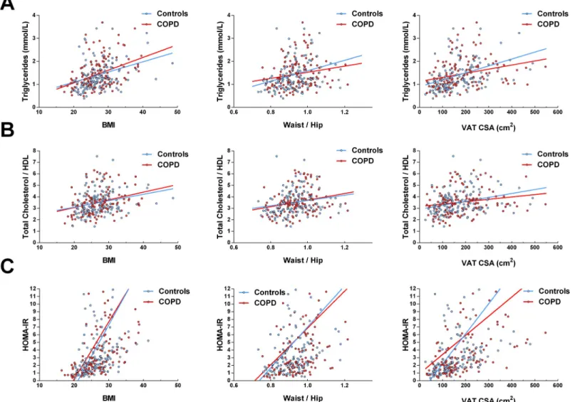
Triglycerides, total/HDL cholesterol ratio and HOMA-IR were positively associated with the three indices of adiposity (BMI, waist-to-hip ratio and VAT area) in individuals with COPD and controls. (Fig 2, all regression lines with a p<0.05). However, the slopes of the regression lines were similar for both groups (p>0.05 for all comparisons) suggesting that the relationships between metabolic markers and adiposity were not modified in the presence of COPD.
In linear multivariate analyses, the COPD status was not significantly associated with tri-glyceride levels, total/HDL cholesterol, insulin resistance (HOMA-IR) or VAT area (Table 2). A higher BMI was associated with an increase in triglycerides, total/HDL cholesterol, and HOMA-IR levels, and current smokers with one third of additional VAT. No significant inter-action was observed between the COPD status and any characteristic tested.
Lastly, a triglyceride level above 1.5 mmol/L, a total/HDL cholesterol ratio above 4 or a HOMA-IR above 3 were respectively observed in 54 (37.5%), 36 (25%) and 58 (40.3%) of COPD patients and in 37 (31.1%), 33 (27.7%) and 54 (45.4%) of control subjects. In multivari-ate analysis, the COPD status (or COPD 2+) was not associmultivari-ated with triglyceride >1.5 mmol/ L, total/HDL cholesterol ratio >4, HOMA-IR>3, or VAT area>75thpercentile. Only the COPD 2+ status was associated with a VAT area>75thpercentile (OR = 2.27, CI95% 1.00; 5.15, p = 0.05). Complete regression models are available in supplementaryS1–S8Tables.
Discussion
In a population-based cohort consisting of individuals with mild to moderate COPD and con-trol subjects, we found metabolic profiles (lipid profile, HOMA-IR, and VAT accumulation)
Table 1. Baseline characteristics by group.
Control subjects (n = 119) COPD (n = 144) P value
Age, years 65 [59–71] 65 [59–71] 0.88
Male, n (%) 73 (61.3) 93 (64.6) 0.61
BMI, kg/m2 26.5 [23.5–29.7] 26.6 [23.7–29.4] 0.96
Waist-to-hip ratio, mean (95% CI) 0.93 (0.92–0.94) 0.94 (0.93–0.95) 0.14
Waist circumference, cm 96 [87.8–103] 98 [89–106] 0.20 Current smokers, n (%) 12 (10.1) 39 (27.1) <0.001 Former smokers, n (%) 70 (58.8) 74 (51.4) 0.26 Never smokers, n (%) 37 (31.1) 31 (21.5) 0.09 Pack/year 11 [0–28] 27 [0–50] <0.001 Comorbidities Hypertension, n (%) 32 (26.9) 53 (36.8) 0.11 Diabetes, n (%) 10 (8.4) 14 (9.7) 0.83 Dyslipidemia, n (%) 31 (26.1) 41 (28.5) 0.68
Coronary artery disease, n (%) 6 (5.0) 14 (9.7) 0.17
Stroke, n (%) 1 (0.8) 9 (6.3) 0.02
Sleep apnea, n (%) 4 (3.4) 9 (6.3) 0.39
Pulmonary Function, post BD
FEV1, L 2.88 [2.37–3.48] 2.14 [1.55–2.99] <0.001
FEV1, % predicted 101 [92–110] 79 [65–93] <0.001
FVC, L 3.79 [3.09–4.52] 3.63 [2.75–4.69] 0.48
FVC, % predicted 120 [112–132] 118 [103–135] 0.21
FEV1/FVC, % 76.8 [73.4–79.9] 62.3 [55.7–66.5] <0.001
PEF, mean L/sec (95% CI) 7.42 (6.98–7.86) 7.42 (7.02–7.82) <0.001
FEF 25–75, L/sec 1.60 [0.96–2.41] 1.58 [0.95–2.41] <0.001 GOLD 1, n (%) - 70 (48.6) -GOLD 2, n (%) - 61 (42.4) -GOLD 3–4, n (%) - 13 (9.0) -GOLD A, n (%) - 91 (63.2) -GOLD B, n (%) - 40 (27.8) -GOLD C, n (%) - 3 (2.1) -GOLD D, n (%) - 10 (6.9) -Medications at baseline Short-acting BD, n (%) 2 (1.7) 24 (16.6) <0.001 Long-acting BD, n (%) 1 (0.8) 1 (0.7) 1 Inhaled CS, n (%) 3 (0.3) 33 (22.9) <0.001 Statins, n (%) 28 (23.5) 38 (26.4) 0.67
Other hypolipidemic drugs, n (%) 4 (3.3) 3 (2.1) 0.70
Insulin, n (%) 1 (0.8) 0 (0) 0.45
Oral hypoglycemic agents, n (%) 9 (7.6) 10 (6.9) 1
Values are median [IQR] if not stated otherwise. COPD: chronic obstructive pulmonary disease; BMI: body mass index; CI: confidence interval; BD: bronchodilator; FEV1: forced expiratory volume in 1 second; FVC: forced vital capacity; BD: bronchodilator; CS: corticosteroids; GOLD: global Initiative for obstructive lung disease
classification.
that were not influenced by the presence of COPD. The well-established relationships between triglycerides, total/HDL cholesterol ratio, and HOMA-IR to indices of adiposity [36,37], which were confirmed here, were not modified in the presence of COPD. Univariate and multivariate analyses showed an absence of association between COPD and metabolic disorders or visceral adiposity. Therefore, based on this thorough statistical approach, we conclude that COPD does not emerge as an independent risk factor for metabolic disorders and visceral adiposity in a cohort that can be considered representative of the entire population.
Numerous studies have explored possible physiopathological links between COPD, asthma or sleep apnea and cardiometabolic components [38]. In those respiratory diseases, several bidirectional mechanisms have been proposed to enhance the risk of hypertriglyceridemia, adipose tissue accumulation and insulin resistance, including hypoxia [8–12] and hypercapnia [39]. Activation of lipolysis in adipose tissue in the presence of hypoxia led to the "adipose tis-sue hypoxia" concept [11]. Adipose tissue would then appear to play a central role in the devel-opment of chronic inflammation, macrophage infiltration, and would also be responsible for increasing circulating free fatty acids [8,10,11]. In addition, fat-induced systemic inflammation involving adipokines [38,40–42], insulin and its receptor, has been implicated in lung injury and airway responsiveness [38,43,44], causing a deleterious pathophysiological loop.
Fig 1. Metabolic parameters according COPD status. COPD: chronic obstructive pulmonary disease; HDL: high density lipoprotein; HOMA-IR:
homeostasis model assessment of insulin resistance; VAT CSA: visceral adipose tissue cross-sectional area on L4-L5. p>0.05 for all between-group comparisons.
In light of the above potential pathophysiological links between chronic respiratory diseases and cardiometabolic risk factors, it was deemed legitimate to propose that COPD may contrib-ute to the development of metabolic abnormalities. In one of the most large-scale studies in the field, Leone et al. [13] found an association between lung function impairment and “classi-cal” components of the metabolic syndrome. This result was obtained in a heterogeneous pop-ulation (obstructive and restrictive ventilatory defects), and a sub-analysis restricted to individuals with an obstructive ventilatory defect failed to find an association between glucose or lipid levels and lung function impairment, in line with our present results as well as previous ones [45,46].
The phenotypic heterogeneity of COPD patients and many confounding factors must be considered when comparing the interaction between COPD and metabolic variables across studies. The prevalence of obesity in COPD is highly variable between studies and countries [47]. Some populations showed higher prevalence of obesity [48] with an over-representation
Fig 2. Relationships between metabolic parameters and BMI, waist-to-hip ratio and VAT CSA in individuals with COPD and controls. COPD: chronic obstructive
pulmonary disease; BMI: body mass index; HDL: high density lipoprotein; HOMA-IR: homeostasis model assessment of insulin resistance; VAT CSA: visceral adipose tissue cross-sectionnal area on L4-L5. All coefficients of determination (R2) are<0.3; All regression line slopes were significantly different from 0 (p<0.05); however,
none of the regression lines couples (COPD vs. controls) were significantly different (p>0.05 for all comparisons).
in patients with moderate airflow limitation [49,50], whereas in the worldwide population-based BOLD study [47], obesity was less frequent in COPD than in non-COPD. The impor-tance of BMI as a confounding factor in the observed link between COPD and metabolic parameters is clearly illustrated in our data (S1 Fig,Table 2). In multivariate analyses, BMI was the factor with the strongest association with the metabolic parameters studied. In the same way, treatment with inhaled corticosteroids (present in only 23% of our COPD subjects) could also confound the relationship between COPD, metabolism and adipose tissue accumu-lation. Inhaled corticosteroids have been related to a 3-fold increase in the likelihood of having a VAT > 75thpercentile (S7 Table). Based on these considerations, it becomes obvious that differences in population phenotypes across studies could at least partially account for incon-sistent conclusions about COPD being a risk factor for altered metabolic status [5]. In this regard, data obtained from clinical cohorts are unlikely to be generalizable to the populational level where the majority of patients has only mild to moderate COPD.
Our study has some limitations. First, given the relatively small sample size, a lack of statisti-cal power could be proposed to explain the absence of differences in endpoints between COPD subjects and controls. However, the similitude in the distribution of metabolic variables and obesity in the two groups studied makes this explanation unlikely. Second, the relatively small size of our otherwise well phenotyped sample could have led to a lesser representative image of the population than did the entire CanCOLD cohort. Despite this, the distribution of study participants’ characteristics in this sub-study was very similar to that of the entire cohort [51], with a majority of subjects with GOLD 1 and few GOLD 3 and 4 COPD. Furthermore,
Table 2. Multivariate linear regression models.
Effect (%)# p-value Triglycerides COPD +5.5 0.262 Age (years) -0.6 0.029 BMI Kg/m2 +3.8 <0.001 Total/HDL cholesterol COPD +1.2 0.721 Sex (men) +10.3 0.005 BMI Kg/m2 +2.3 <0.001 Hypolipidemic (yes) -18.0 <0.001 HOMA-IR COPD -1.8 0.858 Sex (men) +27.0 0.020 BMI Kg/m2 +12.0 <0.001 VAT CSA COPD +1.1 0.866 Age (years) +1.0 0.007 Sex (men) +14.7 0.047
Current smoker (yes) -32.6 <0.001
Pack-years (n) +0.7 <0.001
Significant p-values are shown in bold.
#: effect on variable in %, per increase in variable. COPD: chronic obstructive pulmonary disease; BMI: body mass index; HDL: High Density Lipoprotein; HOMA-IR: Homeostasis Model Assessment of Insulin Resistance; VAT CSA: Visceral Adipose Tissue Cross-sectionnal Area. Only significant factors and COPD are kept in the model by a backward selection.
only 30% of individuals with COPD in this sub-study were previously diagnosed with the dis-ease, another similitude with other population-based cohorts [52], providing further reassur-ance regarding how representative the present cohort is of the general population. That said, despite all the care devoted to building a cohort of individuals representative of the general population, some biases may still be present. For example, the most fragile or diseased subjects would probably be less inclined to participate in a clinical study. Third, focusing on a represen-tative and occidental population of COPD, our findings do not necessarily apply to individuals with severe COPD or to those exhibiting particular phenotypes (inflammatory, underweight or obese, with preponderant vascular comorbidities). Finally, physical activity, an important confounder for cardiovascular risk, was not included in the analysis; also, sleep apnea, another potential contributor, was underdiagnosed by far in this cohort when considering the reported prevalence.
Conclusions
In our cohort randomly drawn from the general population in which individuals with COPD mostly had mild-to-moderate airflow limitation, no difference in the distribution of metabolic parameters appeared compared to control subjects. As such, COPD did not emerge as a spe-cific risk factor for metabolic disorders or visceral adiposity. Although a strong mechanistic rationale can be developed for the existence of physiopathological links between chronic respi-ratory diseases and dyslipidemia, insulin resistance or visceral adiposity, their existence is likely restricted to specific phenotypes or to the most severely affected patients who are not widely represented in the general population.
Supporting information
S1 Table. Multivariate logistic regression on triglycerides > 1.5 mmol/L.
(DOCX)
S2 Table. Multivariate logistic regression on triglycerides > 1.5 mmol/L, COPD 2+ only.
(DOCX)
S3 Table. Multivariate logistic regression on TC/HDL > 4.
(DOCX)
S4 Table. Multivariate logistic regression on TC/HDL > 4, COPD 2+ only.
(DOCX)
S5 Table. Multivariate logistic regression on HOMA-IR > 3.
(DOCX)
S6 Table. Multivariate logistic regression on HOMA-IR > 3, COPD 2+ only.
(DOCX)
S7 Table. Multivariate logistic regression on visceral adipose tissue cross-sectional area (VAT CSA) > 75thpercentile by sex of the total population.
(DOCX)
S8 Table. Multivariate logistic regression on visceral adipose tissue cross-sectional area (VAT CSA) > 75thpercentile by sex of the total population, COPD 2+ only.
(DOCX)
S1 Fig. Univariate analysis stratified by BMI for all metabolic parameters. COPD: chronic
homeostasis model assessment of insulin resistance; VAT CSA: visceral adipose tissue cross-sec-tionnal Area on L4-L5. p>0.05 for all between-group (COPD vs. controls) comparisons. (TIF)
S1 Dataset. Anonymised data.
(XLSX)
Acknowledgments
We wish to thank the participants of this cohort and support staff who made the study possi-ble. We also thank Gae´tan Daigle for his statistical advice, and Dr Yves Deshaies for proofread-ing English.
CanCOLD Collaborative research Group:
Executive Committee: Jean Bourbeau, lead author (McGill University, Montreal, QC, Can-ada, jean.bourbeau@mcgill.ca); Wan C. Tan, J. Mark FitzGerald, D. D. Sin (UBC, Vancouver, BC, Canada); D. D. Marciniuk (University of Saskatoon, Saskatoon, SASK, Canada) D. E. O’Donnell (Queen’s University, Kingston, ON, Canada); Paul Hernandez (University of Hali-fax, HaliHali-fax, NS, Canada); Kenneth R. Chapman (University of Toronto, Toronto, ON, Can-ada); Robert Cowie (University of Calgary, Calgary, AB, CanCan-ada); Shawn Aaron (University of Ottawa, Ottawa, ON, Canada); F. Maltais (University of Laval, Quebec City, QC, Canada); International Advisory Board: Jonathon Samet (the Keck School of Medicine of USC, CA, USA); Milo Puhan (John Hopkins School of Public Health, Baltimore, USA); Qutayba Hamid (McGill University, Montreal, QC, Canada); James C. Hogg (UBC James Hogg Research Cen-ter, Vancouver, BC, Canada). Operations Center: Jean Bourbeau (Principal Investigator), Car-ole BaglCar-ole, CarCar-ole Jabet, Palmina Mancino, Yvan Fortier (University of McGill, Montreal, QC, Canada); Wan C. Tan (co-PI), Don Sin, Sheena Tam, Jeremy Road, Joe Comeau, Adrian Png, Harvey Coxson, Miranda Kirby, Jonathon Leipsic, Cameron Hague (University of British Columbia James Hogg Research Center, Vancouver, BC, Canada). Economic Core: Mohsen Sadatsafavi (University of British Columbia, Vancouver, BC). Public Health Core: Teresa To, Andrea Gershon (University of Toronto). Data Management and Quality Control: Wan C. Tan, Harvey Coxson (UBC, Vancouver, BC, Canada); Jean Bourbeau, Pei-Zhi Li, Jean-Fran-cois Duquette, Yvan Fortier, Andrea Benedetti, Denis Jensen (McGill University, Montreal, QC, Canada), Denis O’Donnell (Queen’s University, Kingston, ON, Canada). Field Centres: Wan C. Tan (PI), Christine Lo, Sarah Cheng, Cindy Fung, Nancy Ferguson, Nancy Haynes, Junior Chuang, Licong Li, Selva Bayat, Amanda Wong, Zoe Alavi, Catherine Peng, Bin Zhao, Nathalie Scott-Hsiung, Tasha Nadirshaw (UBC James Hogg Research Center, Vancouver, BC, Canada); Jean Bourbeau (PI), Palmina Mancino, David Latreille, Jacinthe Baril, Laura Labonte (McGill University, Montreal, QC, Canada); Kenneth Chapman (PI), Patricia McClean, Nad-een Audisho (University of Toronto, Toronto, ON, Canada); Brandie Walker, Robert Cowie (PI), Ann Cowie, Curtis Dumonceaux, Lisette Machado(University of Calgary, Calgary, AB, Canada); Paul Hernandez (PI), Scott Fulton, Kristen Osterling (University of Halifax, Halifax, NS, Canada); Shawn Aaron (PI), Kathy Vandemheen, Gay Pratt, Amanda Bergeron (Univer-sity of Ottawa, Ottawa, ON, Canada); Denis O’Donnell (PI), Matthew McNeil, Kate Whelan (Queen’s University, Kingston, ON, Canada); Francois Maltais (PI), Cynthia Brouillard (Uni-versity of Laval, Quebec City, QC, Canada); Darcy Marciniuk (PI), Ron Clemens, Janet Baran (University of Saskatoon, Saskatoon, SK, Canada).
Author Contributions
Data curation: Damien Viglino, Mickae¨l Martin, Franc¸ois Maltais. Formal analysis: Damien Viglino, Franc¸ois Maltais.
Funding acquisition: Franc¸ois Maltais.
Investigation: Mickae¨l Martin, Cynthia Brouillard, Jean-Pierre Despre´s, Natalie Alme´ras,
Wan C. Tan, Vale´rie Coats, Jean Bourbeau, Franc¸ois Maltais.
Methodology: Damien Viglino, Marie-Eve Piche´, Franc¸ois Maltais. Resources: Jean Bourbeau, Franc¸ois Maltais.
Supervision: Franc¸ois Maltais.
Validation: Marie-Eve Piche´, Jean-Pierre Despre´s. Visualization: Damien Viglino.
Writing – original draft: Damien Viglino.
Writing – review & editing: Mickae¨l Martin, Marie-Eve Piche´, Jean-Pierre Despre´s, Natalie
Alme´ras, Wan C. Tan, Vale´rie Coats, Jean Bourbeau, Jean-Louis Pe´pin, Franc¸ois Maltais.
References
1. Divo MJ, Casanova C, Marin JM, Pinto-Plata VM, de-Torres JP, Zulueta JJ, et al. COPD comorbidities network. Eur Respir J. 2015; 46(3):640–50.https://doi.org/10.1183/09031936.00171614PMID:
26160874
2. Chen W, Thomas J, Sadatsafavi M, FitzGerald JM. Risk of cardiovascular comorbidity in patients with chronic obstructive pulmonary disease: a systematic review and meta-analysis. Lancet Respir Med. 2015; 3(8):631–9.https://doi.org/10.1016/S2213-2600(15)00241-6PMID:26208998
3. Mu¨llerova H, Agusti A, Erqou S, Mapel DW. Cardiovascular comorbidity in COPD: systematic literature review. Chest. 2013; 144(4):1163–78.
4. Mirrakhimov AE. Chronic obstructive pulmonary disease and glucose metabolism: a bitter sweet sym-phony. Cardiovasc Diabetol. 2012; 11:132.https://doi.org/10.1186/1475-2840-11-132PMID:23101436
5. Wouters EFM. Obesity and Metabolic Abnormalities in Chronic Obstructive Pulmonary Disease. Ann Am Thorac Soc. 2017; 14(Supplement_5):S389–94.
https://doi.org/10.1513/AnnalsATS.201705-371AWPMID:29161076
6. Cebron Lipovec N, Beijers RJHCG, van den Borst B, Doehner W, Lainscak M, Schols AMWJ. The Prev-alence of Metabolic Syndrome In Chronic Obstructive Pulmonary Disease: A Systematic Review. COPD. 2016; 13(3):399–406.https://doi.org/10.3109/15412555.2016.1140732PMID:26914392
7. Breyer M-K, Spruit MA, Hanson CK, Franssen FME, Vanfleteren LEGW, Groenen MTJ, et al. Preva-lence of metabolic syndrome in COPD patients and its consequences. PLoS ONE. 2014; 9(6):e98013.
https://doi.org/10.1371/journal.pone.0098013PMID:24950070
8. Jun JC, Shin M-K, Yao Q, Bevans-Fonti S, Poole J, Drager LF, et al. Acute hypoxia induces hypertrigly-ceridemia by decreasing plasma triglyceride clearance in mice. Am J Physiol Endocrinol Metab. 2012; 303(3):E377–388.https://doi.org/10.1152/ajpendo.00641.2011PMID:22621867
9. Xuan L, Han F, Gong L, Lv Y, Wan Z, Liu H, et al. Association between chronic obstructive pulmonary disease and serum lipid levels: a meta-analysis. Lipids Health Dis. 2018; 17(1):263.https://doi.org/10. 1186/s12944-018-0904-4PMID:30463568
10. Plihalova A, Bartakova H, Vasakova M, Gulati S, deGlisezinski I, Stich V, et al. The effect of hypoxia and re-oxygenation on adipose tissue lipolysis in COPD patients. Eur Respir J. 2016; 48(4):1218–20.
https://doi.org/10.1183/13993003.00602-2016PMID:27587548
11. Ye J. Emerging role of adipose tissue hypoxia in obesity and insulin resistance. Int J Obes (Lond). 2009; 33(1):54–66.
12. van den Borst B, Schols AMWJ, de Theije C, Boots AW, Ko¨hler SE, Goossens GH, et al. Characteriza-tion of the inflammatory and metabolic profile of adipose tissue in a mouse model of chronic hypoxia. J Appl Physiol. 2013; 114(11):1619–28.https://doi.org/10.1152/japplphysiol.00460.2012PMID:
13. Leone N, Courbon D, Thomas F, Bean K, Je´go B, Leynaert B, et al. Lung function impairment and meta-bolic syndrome: the critical role of abdominal obesity. Am J Respir Crit Care Med. 2009; 179(6):509–16.
https://doi.org/10.1164/rccm.200807-1195OCPMID:19136371
14. Vanfleteren LEGW, van Meerendonk AMG, Franssen FM, Wouters EFM, Mottaghy FM, van Kroonen-burgh MJ, et al. A possible link between increased metabolic activity of fat tissue and aortic wall inflam-mation in subjects with COPD. A retrospective 18F-FDG-PET/CT pilot study. Respir Med. 2014; 108 (6):883–90.https://doi.org/10.1016/j.rmed.2014.04.001PMID:24785152
15. Vanfleteren LEGW Spruit MA, Groenen MTJ Bruijnzeel PLB, Taib Z Rutten EPA, et al. Arterial stiffness in patients with COPD: the role of systemic inflammation and the effects of pulmonary rehabilitation. Eur Respir J. 2014; 43(5):1306–15.https://doi.org/10.1183/09031936.00169313PMID:24311762
16. Kahn R, Buse J, Ferrannini E, Stern M, American Diabetes Association, European Association for the Study of Diabetes. The metabolic syndrome: time for a critical appraisal: joint statement from the Ameri-can Diabetes Association and the European Association for the Study of Diabetes. Diabetes Care. 2005; 28(9):2289–304.https://doi.org/10.2337/diacare.28.9.2289PMID:16123508
17. Burgel P-R, Paillasseur J-L, Peene B, Dusser D, Roche N, Coolen J, et al. Two distinct chronic obstruc-tive pulmonary disease (COPD) phenotypes are associated with high risk of mortality. PLoS ONE. 2012; 7(12):e51048.https://doi.org/10.1371/journal.pone.0051048PMID:23236428
18. Rennard SI, Locantore N, Delafont B, Tal-Singer R, Silverman EK, Vestbo J, et al. Identification of Five Chronic Obstructive Pulmonary Disease Subgroups with Different Prognoses in the ECLIPSE Cohort Using Cluster Analysis. Ann Am Thorac Soc. 2015; 12(3):303–12.https://doi.org/10.1513/AnnalsATS.
201403-125OCPMID:25642832
19. Garcia-Aymerich J, Go´mez FP, Benet M, Farrero E, Basagaña X, GayeteÀ, et al. Identification and pro-spective validation of clinically relevant chronic obstructive pulmonary disease (COPD) subtypes. Tho-rax. 2011; 66(5):430–7.https://doi.org/10.1136/thx.2010.154484PMID:21177668
20. Vanfleteren LEGW Spruit MA, Groenen M, Gaffron S, van Empel VPM, Bruijnzeel PLB, et al. Clusters of comorbidities based on validated objective measurements and systemic inflammation in patients with chronic obstructive pulmonary disease. Am J Respir Crit Care Med. 2013; 187(7):728–35.https://doi. org/10.1164/rccm.201209-1665OCPMID:23392440
21. Coats V, Despre´ s J-P, Alme´ras N, Martin M, Sin DD, Rabasa-Lhoret R, et al. Ectopic adiposity and car-diometabolic health in COPD. Int J Chron Obstruct Pulmon Dis. 2018; 13:3331–40.https://doi.org/10.
2147/COPD.S168963PMID:30410322
22. Gayoso-Diz P, Otero-Gonza´lez A, Rodriguez-Alvarez MX, Gude F, Garcı´a F, De Francisco A, et al. Insulin resistance (HOMA-IR) cut-off values and the metabolic syndrome in a general adult population: effect of gender and age: EPIRCE cross-sectional study. BMC Endocr Disord. 2013; 13:47.https://doi. org/10.1186/1472-6823-13-47PMID:24131857
23. Radikova Z, Koska J, Huckova M, Ksinantova L, Imrich R, Vigas M, et al. Insulin sensitivity indices: a proposal of cut-off points for simple identification of insulin-resistant subjects. Exp Clin Endocrinol Dia-betes. 2006; 114(5):249–56.https://doi.org/10.1055/s-2006-924233PMID:16804799
24. Qu H-Q, Li Q, Rentfro AR, Fisher-Hoch SP, McCormick JB. The Definition of Insulin Resistance Using HOMA-IR for Americans of Mexican Descent Using Machine Learning. PLoS One. 2011; 6(6):e21041.
https://doi.org/10.1371/journal.pone.0021041PMID:21695082
25. Yamada C, Mitsuhashi T, Hiratsuka N, Inabe F, Araida N, Takahashi E. Optimal reference interval for homeostasis model assessment of insulin resistance in a Japanese population. J Diabetes Investig. 2011; 2(5):373–6.https://doi.org/10.1111/j.2040-1124.2011.00113.xPMID:24843516
26. Rutter MK, Wilson PWF, Sullivan LM, Fox CS, D’Agostino RB, Meigs JB. Use of alternative thresholds defining insulin resistance to predict incident type 2 diabetes mellitus and cardiovascular disease. Circu-lation. 2008; 117(8):1003–9.https://doi.org/10.1161/CIRCULATIONAHA.107.727727PMID:18250267
27. Elshazly MB, Nicholls SJ, Nissen SE, St John J, Martin SS, Jones SR, et al. Implications of Total to High-Density Lipoprotein Cholesterol Ratio Discordance With Alternative Lipid Parameters for Coronary Atheroma Progression and Cardiovascular Events. Am J Cardiol. 2016; 118(5):647–55.https://doi.org/ 10.1016/j.amjcard.2016.06.021PMID:27392507
28. Despre´s J-P, Lemieux I. Abdominal obesity and metabolic syndrome. Nature. 2006; 444(7121):881–7.
https://doi.org/10.1038/nature05488PMID:17167477
29. Despre´s J-P, Lemieux I, Bergeron J, Pibarot P, Mathieu P, Larose E, et al. Abdominal obesity and the metabolic syndrome: contribution to global cardiometabolic risk. Arterioscler Thromb Vasc Biol. 2008; 28(6):1039–49.https://doi.org/10.1161/ATVBAHA.107.159228PMID:18356555
30. Bourbeau J, Tan WC, Benedetti A, Aaron SD, Chapman KR, Coxson HO, et al. Canadian Cohort Obstructive Lung Disease (CanCOLD): Fulfilling the need for longitudinal observational studies in COPD. COPD. 2014; 11(2):125–32.https://doi.org/10.3109/15412555.2012.665520PMID:22433011
31. Shorofsky M, Bourbeau J, Kimoff J, Jen R, Malhotra A, Ayas N, et al. Impaired Sleep Quality in COPD Is Associated With Exacerbations: The CanCOLD Cohort Study. Chest. nov 2019; 156(5):852–63.
https://doi.org/10.1016/j.chest.2019.04.132PMID:31150638
32. Martin M, Almeras N, Despre´ s J-P, Coxson HO, Washko GR, Vivodtzev I, et al. Ectopic fat accumula-tion in patients with COPD: an ECLIPSE substudy. Int J Chron Obstruct Pulmon Dis. 2017; 12:451–60.
https://doi.org/10.2147/COPD.S124750PMID:28203068
33. Bastien M, Poirier P, Brassard P, Arsenault BJ, Bertrand OF, Despre´s J-P, et al. Effect of PPARγ ago-nist on aerobic exercise capacity in relation to body fat distribution in men with type 2 diabetes mellitus and coronary artery disease: a 1-yr randomized study. Am J Physiol Endocrinol Metab. 01 2019; 317 (1):E65–73.https://doi.org/10.1152/ajpendo.00505.2018PMID:30964707
34. Antuna-Puente B, Disse E, Rabasa-Lhoret R, Laville M, Capeau J, Bastard J-P. How can we measure insulin sensitivity/resistance? Diabetes Metab. 2011; 37(3):179–88.https://doi.org/10.1016/j.diabet. 2011.01.002PMID:21435930
35. Catapano AL, Graham I, De Backer G, Wiklund O, Chapman MJ, Drexel H, et al. 2016 ESC/EAS Guide-lines for the Management of Dyslipidaemias. Eur Heart J. 2016; 37(39):2999–3058.https://doi.org/10. 1093/eurheartj/ehw272PMID:27567407
36. Mannino DM, Gagnon RC, Petty TL, Lydick E. Obstructive lung disease and low lung function in adults in the United States: data from the National Health and Nutrition Examination Survey, 1988–1994. Arch Intern Med. 2000; 160(11):1683–9.https://doi.org/10.1001/archinte.160.11.1683PMID:10847262
37. Tan WC, Hague CJ, Leipsic J, Bourbeau J, Zheng L, Li PZ, et al. Findings on Thoracic Computed Tomography Scans and Respiratory Outcomes in Persons with and without Chronic Obstructive Pulmo-nary Disease: A Population-Based Cohort Study. PLoS One. 2016; 11(11):e0166745.https://doi.org/ 10.1371/journal.pone.0166745PMID:27861566
38. Baffi CW, Wood L, Winnica D, Strollo PJ, Gladwin MT, Que LG, et al. Metabolic Syndrome and the Lung. Chest. 2016; 149(6):1525–34.https://doi.org/10.1016/j.chest.2015.12.034PMID:26836925
39. Dimoulis A, Pastaka C, Tsolaki V, Tsilioni I, Pournaras S, Liakos N, et al. Non-Invasive Ventilation (NIV) and Homeostatic Model Assessment (HOMA) Index in Stable Chronic Obstructive Pulmonary Disease (COPD) Patients with Chronic Hypercapnic Respiratory Failure: A Pilot Study. COPD. 2015; 12(4):427– 34.https://doi.org/10.3109/15412555.2014.974738PMID:25415619
40. Bolton CE, Evans M, Ionescu AA, Edwards SM, Morris RHK, Dunseath G, et al. Insulin resistance and inflammation—A further systemic complication of COPD. COPD. 2007; 4(2):121–6.https://doi.org/10.
1080/15412550701341053PMID:17530505
41. Machado FVC, Pitta F, Hernandes NA, Bertolini GL. Physiopathological relationship between chronic obstructive pulmonary disease and insulin resistance. Endocrine. 2018; 61(1):17–22.https://doi.org/10. 1007/s12020-018-1554-zPMID:29512058
42. Sideleva O, Suratt BT, Black KE, Tharp WG, Pratley RE, Forgione P, et al. Obesity and asthma: an inflammatory disease of adipose tissue not the airway. Am J Respir Crit Care Med. 2012; 186(7):598– 605.https://doi.org/10.1164/rccm.201203-0573OCPMID:22837379
43. Forno E, Han Y-Y, Muzumdar RH, Celedo´ n JC. Insulin resistance, metabolic syndrome, and lung func-tion in US adolescents with and without asthma. J Allergy Clin Immunol. 2015; 136(2):304–311.e8.
https://doi.org/10.1016/j.jaci.2015.01.010PMID:25748066
44. Nie Z, Jacoby DB, Fryer AD. Hyperinsulinemia potentiates airway responsiveness to parasympathetic nerve stimulation in obese rats. Am J Respir Cell Mol Biol. 2014; 51(2):251–61.https://doi.org/10.1165/
rcmb.2013-0452OCPMID:24605871
45. Lin W-Y, Yao C-A, Wang H-C, Huang K-C. Impaired lung function is associated with obesity and meta-bolic syndrome in adults. Obesity (Silver Spring). 2006; 14(9):1654–61.https://doi.org/10.1038/oby. 2006.190PMID:17030977
46. Nakajima K, Kubouchi Y, Muneyuki T, Ebata M, Eguchi S, Munakata H. A possible association between suspected restrictive pattern as assessed by ordinary pulmonary function test and the metabolic syn-drome. Chest. 2008; 134(4):712–8.https://doi.org/10.1378/chest.07-3003PMID:18625672
47. Vanfleteren LE, Lamprecht B, Studnicka M, Kaiser B, Gnatiuc L, Burney P, et al. Body mass index and chronic airflow limitation in a worldwide population-based study. Chron Respir Dis. 2016; 13(2):90–101.
https://doi.org/10.1177/1479972315626012PMID:26768010
48. Vozoris NT O’Donnell DE. Prevalence, risk factors, activity limitation and health care utilization of an obese, population-based sample with chronic obstructive pulmonary disease. Can Respir J. 2012; 19 (3):e18–24.https://doi.org/10.1155/2012/732618PMID:22679617
49. Steuten LMG, Creutzberg EC, Vrijhoef HJM, Wouters EF. COPD as a multicomponent disease: inven-tory of dyspnoea, underweight, obesity and fat free mass depletion in primary care. Prim Care Respir J. 2006; 15(2):84–91.https://doi.org/10.1016/j.pcrj.2005.09.001PMID:16701766
50. Rutten EPA, Calverley PMA, Casaburi R, Agusti A, Bakke P, Celli B, et al. Changes in body composition in patients with chronic obstructive pulmonary disease: do they influence patient-related outcomes? Ann Nutr Metab. 2013; 63(3):239–47.https://doi.org/10.1159/000353211PMID:24216978
51. Labonte´ LE, Tan WC, Li PZ, Mancino P, Aaron SD, Benedetti A, et al. Undiagnosed Chronic Obstruc-tive Pulmonary Disease Contributes to the Burden of Health Care Use. Data from the CanCOLD Study. Am J Respir Crit Care Med. 01 2016; 194(3):285–98.https://doi.org/10.1164/rccm.201509-1795OC
PMID:26836958
52. Martinez CH, Mannino DM, Jaimes FA, Curtis JL, Han MK, Hansel NN, et al. Undiagnosed Obstructive Lung Disease in the United States. Associated Factors and Long-term Mortality. Ann Am Thorac Soc. de´c 2015; 12(12):1788–95.https://doi.org/10.1513/AnnalsATS.201506-388OCPMID:26524488