Dopaminergic modulation of prefrontal cortex subpopulations
By
Caitlin Miya Vander Weele
B.S. PsychologyUniversity of Michigan, 2011
Submitted to the Department of Brain and Cognitive Sciences In partial fulfillment of the requirements for the degree of
DOCTOR OF PHILOSOPHY IN NEUROSCIENCE at the
MASSACHUSETTS INSTITUTE OF TECHNOLOGY
Ap-298 ne2.0o&3
Massachusetts Institute of Technology 2018. All rights reserved.
Signature of Author:
Signature redacted
V
Department of Brain and Cognitive Sciences April 13, 2018
/-Certified by:
Signature redacted
Kay M. Tye,
P(<1
Associate Professor, Picower Institute epartment rain and Cognitive Sciences Thesis Supervisor
Acceptedby:(Signature
redacted,
Matthew A. W on, P
Sherman Fairchild Professor of Neuroscience
MASSACHUSETTS INSTITUTE Director of Graduate Education for Brain and Cognitive Sciences OF TECHNOLOGY
OCT 11
2018
LIBRARIES
Dopaminergic modulation of prefrontal cortex subpopulations
ByCaitlin M. Vander Weele
Submitted to the Department of Brain and Cognitive Sciences on March 30, 2018 in partial fulfillment of the requirements for the degree of
DOCTOR OF PHILOSOPHY IN NEUROSCEINCE
Abstract
Despite abundant evidence that dopamine modulates medial prefrontal cortex (mPFC) activity to mediate diverse behavioral functions, the precise circuit computations remain elusive. One potentially unifying theoretical model by which dopamine can modulate functions from working memory to schizophrenia is that dopamine serves to increase the signal-to-noise ratio in mPFC neurons, where neuronal activity conveying sensory information (signal) are amplified relative to spontaneous firing (noise). To connect theory to biology, we lack direct evidence for dopaminergic modulation of signal-to-noise in neuronal firing patterns in vivo and a mechanistic explanation of how such computations would be transmitted downstream to instruct specific behavioral functions. Here, we demonstrate that dopamine increases signal-to-noise ratio in mPFC neurons projecting to the dorsal periaqueductal gray (dPAG) during the processing of an aversive stimulus. First, using electrochemical approaches, we reveal the precise time course of tail pinch-evoked dopamine release in the mPFC. Second, we show that dopamine signaling in the mPFC biases behavioral responses to punishment-predictive stimuli, rather than reward-predictive cues. Third, in contrast to the well-characterized mPFC-NAc projection, we show that activation of mPFC-dPAG neurons is sufficient to drive place avoidance and defensive behaviors. Fourth, to determine the natural dynamics of individual mPFC neurons, we performed single-cell projection-defined microendoscopic calcium imaging to reveal a robust preferential excitation of mPFC-dPAG, but not mPFC-NAc, neurons to aversive stimuli. Finally, photostimulation of VTA dopamine terminals in the mPFC revealed an increase in signal-to-noise ratio in mPFC-dPAG neuronal activity during the processing of aversive, but not rewarding stimuli. Together, these data unveil the utility of dopamine in the mPFC to effectively filter sensory information in a valence-specific manner.
Thesis Supervisor: Kay M. Tye
Title: Associate Professor of Neuroscience Picower Institute of Learning and Memory Department of Brain and Cognitive Sciences
To my mom, sister, and brother You are my world
Acknowledgements
"We love ourselves in the things we love. We find ourselves there, too" - Kristin Martz
To my teachers and mentors.
My Ph.D. mentor, Dr. Kay M. Tye, thank you for your relentless belief in my potential over the past 7 years. I never could have imagined the things I would accomplish under your supervision and support. You have grown a stronger, smarter, and more confident woman. To my
committee members, Drs. Li-Huei Tsai, Matt Wilson, and Ki Goosens, for leaving me
encouraged and determined to continue this path after every meeting. You've made me the best scientist I can be. My undergraduate mentors, Drs. Brandon Aragona and Shanna Harkey-Resendez, for nourishing my mind and soul at all stages over the past decade.
To my team, for sticking with me.
To my partner in crime, Cody Siciliano. From hooking up imaging animals to evenings over Bookers, you've inspired, comforted, and encouraged me during some of my most difficult times. Thank you for dragging me over the finish line- while making me laugh the entire way. Gillian Matthews, you are such a rock star. You've taught me tenacity and aesthetic. Thanks for patching an insane number of cells over an equally insane number of conditions. To all of my other co-authors, Edward Nieh, Praneeth Namburi, Isabella Espinel, Ehsan Izadmehr, Anna Beyeler, Romy Wichmann, Eyal Kimchi, Nancy Padilla, Tony Burgos-Robles, Evelien Schut, and Kay Tye, I couldn't be prouder of the things we've accomplished together. Thank you for the deepest parts of my heart.
To my friends, for believing in, supporting, and inspiring me.
All members of the Tye lab, past & present. In particular, Cody Siciliano, Craig Wildes, Anna Beyeler, Gwendolyn Calhoon, Edward Nieh, Praneeth Namburi, and Gillian Matthews - for my being my Boston family. Craig Wildes, someday you will miss me and appreciate the value of the beanbag. Special thanks to favorite bartenders, Tony Yohe and Curt Hancock, and the entire staff at Catalyst, for feeding me and making me smile after long lab days. My Ph.D. role models and confidants, Steve Ramirez, Emily Doucette, and Pedro Tsividis. My digital support system, my science Twitter community and Interstellate contributors and sponsors, for
helping me find and cultivate a passion for science communication.
To my family, for everything. Always.
My parents, Kitty Flaherty and Rick Vander Weele for raising me to follow my dreams and supporting my decisions at every step. I am incomplete without my siblings Meagan and Eric Vander Weele, who give me identity and strength. To others in my extended family, Ken Smith and Patrick Metzger. Finally, to Pasquale D'Silva for teaching me to dream bigger, love
deeper, and laugh harder. I love you.
I would also like to thank all of the BCS and animal care staff for providing support in various ways, both big and small.
Table of Contents
Chapter 1: Background
Functions of the prefrontal cortex ... 10
P re fro nta l a nato m y ... . 12
The mesolimbic dopamine system ... 13
Dopaminergic mechanisms of action in the prefrontal cortex ... 16
Dopaminergic modulation of prefrontal function and physiology ... 18
S u m m a ry ... 2 0 Chapter 2: Aversive stimuli increase dopamine in the prefrontal cortex and activation of dopamine terminals in the prefrontal cortex biases behavior towards aversion In tro d u ctio n ... . . 2 3 R e s u lts ... 2 4 Experim ental procedures ... 42
S u m m a ry ... . . 5 4 A utho r co ntributio ns ... . 54
Chapter 3: Prefrontal projections to the striatum and periaqueductal gray are distinct and differentially encode and promote reward and aversion In tro d u c tio n ... . . 5 5 R e s u lts ... . . 5 7 E xpe rim e nta l pro ced u re s ... 8 5 S u m m a ry ... . . 9 9 A utho r co ntributio ns ... . . 99
Chapter 4: Dopamine enhances neuronal responses to aversive stimuli in periaqueductal gray-projecting prefrontal neurons and inhibits striatal-projecting neurons In tro d u c tio n ... 1 0 1 R e s u lts ... ... 1 0 2 E xpe rim e nta l pro ced ure s ... 13 1 Summary ... 149
A u th o r c o ntrib utio n s ... 15 0 Chapter 5: Conclusions - Dopamine in the prefrontal cortex enhances signal-to-noise for aversive stimuli via projections to the periaqueductal gray S u m m a ry ... 1 5 1 D is c u s s io n ... 1 5 1 C a v e a ts ... 1 5 6 Im plicatio ns fo r m e nta l hea lth ... 158 C h a p te r 6 : R efe re n c es ... 15 9
List of Figures
Figure 1. Peer-reviewed studies of dopamine in appetitive and aversive motivation ... 22
Figure 2. Dopamine terminals are densest in deep layers of the prefrontal cortex ... .. . ... ... ... ... ... ... ... ... ... ... ... ... ... ... ... ... .. . ... ... ... ... ... ... ... ... ... ... ... ... ... ... ... ... ... ... ... ... ... ... .2 5 Figure 3. LC inactivation does not alter catecholamine release in the mPFC in response to ta il p in c h ... 2 8 Figure 4. VTA DA inhibition attenuates catecholamine release in response to tail pinch ... ... ... ... ... ... ... ... ... ... ... ... ... ... ... ... ... ... . . . 3 2 Figure 5. Activation of dopamine terminals in the mPFC does not support real-time or conditioned place preference / aversion ... 35
Figure 6. Activation of dopamine terminals in the mPFC biases behavior towards aversion during competitive stimulus presentations ... 38
Figure 7. Putative connection strength of mPFC projections to downstream targets ... 58
Figure 8. mPFC-dPAG and mPFC-NAc projector populations are distinct ... 61
Figure 9. Collateralization of mPFC-NAc and mPFC-dPAG projector subpopulations ... 64
Figure 10. Activation of mPFC-NAc neurons does not support real-time or conditioned placed preference / aversion ... 66
Figure 11. Activation of mPFC-dPAG neurons supports real-time or conditioned placed a v e rs io n ... . . .. 6 9 Figure 12. Activation of mPFC-dPAG neurons increases marble burying ... 73
Figure 13. Activation of mPFC terminals in the dPAG evokes marble burying ... 76
Figure 14. mPFC-NAc neurons exhibit heterogeneous responses to rewarding and aversive stimuli, while mPFC-dPAG neurons preferentially respond to aversive stimuli ... ... ... ... ... ... ... ... ... ... ... ... ... ... ... ... ... ... ... ... ... ... ... ... ... ... ... ... ... ... ... ... ... ... ... ... ... ... ... . . 8 0 Figure 15. In vivo calcium imaging re-analyzed with a background subtracted ROI a p p ro a c h ... 8 3 Figure 16. mPFC-dPAG and mPFC-NAc projectors have different electrophysiological p ro p e rtie s ... 1 0 3 Figure 17. Dopamine inhibits NAc-projectors via D2-receptors ... 107
Figure 19. Investigation of VTA projections to the dPAG for simultaneous epifluorescent imaging in mPFC-dPAG neurons and excitation of VTADA terminals ... 113 Figure 20. Stimulation of dopamine terminals in the mPFC increase calcium transient amplitude and decreases frequency in dPAG-projectors ... 115 Figure 21. Ex vivo latency validation for head-fixed electrophysiological recordings with p h o to ta g g in g ... 1 1 8 Figure 22. m PFC-dPAG neurons are excited to airpuff ... 121 Figure 23. Representative m PFC-dPAG units ... 125 Figure 24. Dopamine selectively enhances mPFC-dPAG responses to airpuff ... 127 Figure 23. Dopamine increase baseline firing rate, but not stimulus-evoked responses in photo inhibited m P FC neuro ns ... 129
Chapter 1
Background
Functions of the prefrontal cortex
The medial prefrontal cortex (mPFC) is a heterogeneous brain region implicated in a diverse range of cognitive and behavioral functions- including attention, decision-making, working memory, long-term memory, emotional control, inhibitory control, motivation, among many others (Arnsten, 2009; Bechara et al., 2000; Miller and Cohen, 2001; Ridderinkhof et al., 2004). While these functions may appear disparate, they all are related to cognitive control, or the ability to coordinate emotions and actions to support internally held goals (Miller and Cohen, 2001). Lesions of the mPFC result in robust deficits in performance in tasks that require one to adapt behavior to contexts or tasks with dynamic or unexpected rules (Dias et al., 1996a, 1996b, 1997; Gregoriou et al., 2014; Milner, 1963; Rossi et al., 2007; Wilkinson et al., 1997). While many brain regions, neurotransmitter systems, and neural circuits support goal-directed behavior, the mPFC is well positioned to act as an anatomical hub coordinating adaptive behavioral output.
At a basic level, successfully seeking rewards and avoiding punishments is critical for survival, and a defining characteristic of adaptive behavior. To mediate these behaviors, neural systems must integrate internal motivational states with external information to orchestrate approach and avoidance of rewarding and aversive stimuli, respectively. For example, an animal engaging in foraging behavior for food must constantly evaluate its environment for potential threats, which necessitate a transition from reward-seeking to threat avoidance. While the mPFC is not critical for basic stimulus-driven unconditioned behaviors, such as those found in subcortical structures like the periaqueductal gray (Assareh et al., 2016; Bandler and Carrive,
1988; Bandler et al., 1985a, 1985b; Carrive et al., 1987; Deng et al., 2016; Meyer et al., 2017; Tovote et al., 2016; Zhang et al., 1990) and hypothalamus (Anand and Brobeck, 1951; Betley et al., 2015; Burton et al., 1976; Jennings et al., 2015; Nieh et al., 2015, 2016), the mPFC plays a substantial role in deciphering ambiguous situations. In the example above, the foraging animal must not only perceive the environmental threat, but also evaluate its importance in relation to its ongoing efforts (e.g., How far away is the threat? How severe is the threat? How hungry am I? When will I be able to forage again?).
There is substantial evidence supporting a crucial role of the mPFC in decision-making in the context of risk and reward, particularly in situations involving competition (i.e., when both rewarding and aversive stimuli are simultaneously present) (Bechara et al., 1994, 2005;
Botvinick et al., 2004; Burgos-Robles et al., 2017; Mansouri et al., 2009; Milham et al., 2001). In rodents, pharmacological inactivation of the mPFC produces deficits in the coordination of reward-seeking and threat avoidance (Sangha et al., 2014; Sierra-Mercado et al., 2011). In humans, mPFC activity is modulated by the proximity of threat (Mobbs et al., 2007) suggesting a role in coordinating behavioral coping strategies. Not surprisingly, mPFC neuronal activity is
robustly modulated by motivationally-relevant stimuli- including delivery, reward-expectation, omission of reward, pain, and pain-predictive cues (for review, see: Euston et al., 2012)- and mPFC activity is highly correlated with aspects of reward- and fear-motivated
behavioral output (Burgos-Robles et al., 2009, 2013). In competitive situations, neurons encoding different information compete for representation and subsequent expression of behavior. However, it is still unknown if these neural responses carrying competing valence-defined information can be mapped onto specific mPFC circuits, what modulates their competitive interactions, and how the competition results are translated into behavior.
Prefrontal anatomy
Consistent with the idea that the mPFC is crucial for cognitive control, this neocortical region is most developed in primates and animals that have elaborate behavioral repertoires (Adolphs, 2009; Dunbar, 2009; Kerney et al., 2017; Noonan et al., 2018). Understanding the mPFC and its functional relevance relies on an understanding of its complex macro- and micro-anatomical organization. The mPFC is often subdivided into four distinct subregions - medial precentral / second frontal area (PrCM / Fr2), the anterior cingulate cortex (ACC), the prelimbic cortex (PL), and infralimbic cortex (IL) - based on afferent / efferent connections and
cytoarchitectural differences (Heidbreder and Groenewegen, 2003). However based on afferent and efferent connectivity the ventral portion of the PL and dorsal part of the IL are often
combined and referred to as the ventral medial PFC (vmPFC). For simplicity, the remainder of this manuscript will use vmPFC synonymously with mPFC, as manipulations and recordings in this region often contained the ventral aspect of the PL and dorsal aspect of the IL.
The local mPFC network consists of excitatory, glutamatergic pyramidal cells and inhibitory GABAergic interneurons, representing -80-90% and 10-20% of the population, respectively (Gabbott et al., 1997, 2005). Traditionally it has been thought that pyramidal
neurons comprise the majority of the long-range projection neurons in the mPFC while
GABAergic interneurons powerfully regulate their activity locally. However, recently long-range GABAergic projections have also been characterized (Lee et al., 2014a). Both of these
populations can be further dissected based on functional, laminar, morphological, and molecular properties (Gabbott et al., 1997, 2005).
Similar to other cortical structures, the mPFC is laminarly organized (Douglas and Martin, 2004; Gabbott et al., 2005; Kritzer and Goldman-Rakic, 1995). The mPFC receives diverse sensory and limbic afferent connections (Hoover and Vertes, 2007) arriving
1999a, 1999b). mPFC afferent connections are equally robust projecting to cortical and subcortical brain regions (Gabbott et al., 2005; Groenewegen et al., 1997; Heidbreder and Groenewegen, 2003; Sesack et al., 1989; Vertes, 2004). Long-range efferent projections originate mainly from the deep layers (Layers 5 and 6), while projections to other cortical areas are located in superficial Layer 2/3 (DeFelipe and Farihas, 1992; Douglas and Martin, 2004; Gabbott et al., 2005). Notably, downstream connections appear to be densest to brain regions involved in emotional and autonomic control- including the amygdala, ventral striatum, insula, hypothalamus, periaqueductal gray, habenula, and midline thalamic structures (Gabbott et al., 2005; Heidbreder and Groenewegen, 2003; Vertes, 2004). Through this dense interconnectivity, the mPFC is thought to exert top-down processing of rewarding and aversive stimuli and
subsequent emotional regulation and control of actions. However precise, causal investigations of these mPFC outputs are still lacking. It is unclear which specific mPFC connections convey valence-defined information and under what conditions.
The mesolimbic dopamine system
The mPFC also maintains reciprocal connectivity with a wide range of neuromodulatory systems known to be involved in adaptive responses to rewarding and aversive events-including the midbrain dopamine system (Gabbott et al., 2005; Heidbreder and Groenewegen, 2003; Vertes, 2004). Dopamine neurons originate in both the ventral tegmental area (VTA, also known as Al0) and substantia nigra pars compacta (SNc, also known as A9) (for review, see: BjOrklund and Dunnett, 2007), which are both anatomically and functionally distinct populations. While VTA dopamine neurons regulate motivation and encode reward expectation, substantia nigra neurons appear to be more involved in motor functions via connections to the dorsal striatum / caudate putamen (Berridge, 2007; Bjorklund and Dunnett, 2007; Michel et al., 2016; Schultz et al., 1997; York, 1973). VTA dopamine neurons send dense projections to the ventral
striatum (including the nucleus accumbens [NAc]) and sparser projections to the mPFC, amygdala, and hippocampus (for review, see: Haber and Fudge, 1997).
VTA dopamine neurons transmit signals in two modes: 1) "tonic" characterized by a consistent, pacemaker-like 2-5 Hz firing rate, and 2) "phasic" characterized by transient bursts of high frequency 10-20 Hz firing lasting 100-500 ms (Bunney et al., 1991; Grace and Bunney, 1984, 1984; Grace et al., 2007). Tonic firing maintains a consistent basal, extracellular level of dopamine in downstream targets. On the other hand, bursting rapidly and transiently increases dopamine levels, estimated to be -100 pM in the synapse (Garris and Wightman, 1994; Grace et al., 2007), termed 'phasic dopamine release' or dopamine 'transients' (for review, see:
Robinson et al., 2003). Dopamine release in target regions modulates the activity of specific populations of neurons expressing postsynaptic dopamine receptors (see below for more detailed discussion) (Dreyer et al., 2010, 2016). However, it is important to note that bursting (i.e., dopamine neuron action potentials) may not evoke dopamine neurotransmission
considering dopamine terminals are influenced by local inputs and autoreceptors (Cachope and Cheer, 2014; Cachope et al., 2012; Cragg and Rice, 2004; Dreyer and Hounsgaard, 2013; Rice and Cragg, 2004; Zhang and Sulzer, 2004). As such, it is important to use multidisciplinary approaches to appreciate the complex and dynamic interactions between somatic activity, dopamine release, and reuptake.
Phasic dopamine neuron activity and phasic dopamine release have been studied extensively in the context of reward (Berridge, 2007; Berridge and Robinson, 1998; Fields et al., 2007; Grace et al., 2007; Ikemoto, 2007; Robinson et al., 2003; Schultz, 2013; Schultz et al., 1997; Volkow et al., 2017; Wise, 2006, 2008). In a seminal study by Schultz and colleagues (1997), dopamine neurons were found to change their activity based on reward expectation. Here, dopamine neurons increased their firing to unexpected reward delivery, but shifted this response to cues that predict reward delivery after pairing. Further, VTA dopamine neurons
exhibited a decrease in firing if an expected reward (i.e., predicted by a conditioned stimulus) was omitted. Together, these neurons have been described as encoding "reward prediction error" (RPE) by signaling discrepancies between expected and received reward. As such, RPEs provide a mechanism for learning the outcomes of predictive stimuli and updating their internal representations when the magnitude or valence of their predicted outcomes are altered.
Considering this, and dopamine's role in other appetitive motivational functions, VTA dopamine has been extensively studied in the context of reward - particularly through its connections with the striatum (Berridge, 2007; Berridge and Robinson, 1998; Hamid et al., 2016; Howe et al., 2013; Niv, 2007; Phillips et al., 2003; Salamone and Correa, 2012).
There is considerable debate over dopamine's role in the processing of aversive stimuli. As previously mentioned, several theories have suggested that phasic dopamine responses
primarily encode reward-related events- including food reward, water reward, social targets, sex, and drugs of abuse (for review, see: Berridge, 2007; Berridge and Robinson, 1998; Fields et al., 2007; Robinson et al., 2003; Salamone and Correa, 2012; Schultz et al., 1997). Further, consistent with the reward prediction hypothesis, some studies report inhibition of dopamine activity in response to aversive events (Cohen et al., 2012; Roitman et al., 2008; Schultz et al.,
1997; Ungless et al., 2004). However, dopamine responses have also been reported to various salient and aversive stimuli- including surprising events, stress, pain, and fear-predictive cues (Badrinarayan et al., 2012; Brischoux et al., 2009; Budygin et al., 2012; Horvitz, 2000; Mantz et al., 1989; Matsumoto and Hikosaka, 2009; McCutcheon et al., 2012; Salamone and Correa, 2012). As a result, new theories of dopamine function have suggested roles in alerting (Bromberg-Martin et al., 2010), invigoration of ongoing behaviors (Niv, 2007; Salamone et al., 2007; da Silva et al., 2018), or encoding the value of work (Hamid et al., 2016) (for review, see: Salamone and Correa, 2018; Schultz et al., 2017).
It remains unclear whether reward-encoding dopaminergic signals recorded in denser terminal regions, like the striatum, are also transmitted to regions like the mPFC. Heterogeneity observed within the VTA might result from distinct dopamine neuron subpopulations defined by projection target. Indeed, growing evidence supports distinct behavioral roles of VTA dopamine subpopulations depending on their ascending forebrain projection (e.g., "projection-defined") (Gunaydin et al., 2014; Kim et al., 2016; Lammel et al., 2011, 2012; Roeper, 2013). This notion is anatomically supported by the finding that the majority of VTA dopamine neurons do not collateralize and send independent, non-overlapping ascending projections to target regions (Albanese and Minciacchi, 1983; Fallon, 1981). These projection-defined VTA dopamine subpopulations maintain unique molecular signatures (Bannon and Roth, 1983; Bannon et al., 1981; Lammel et al., 2008), electrophysiological properties (Chiodo et al., 1984; Lammel et al., 2008), anatomical locations (Lammel et al., 2012), afferent connectivity (Lammel et al., 2012), and responses to valence-defined stimuli (see below for discussion) (Abercrombie et al., 1989; Badrinarayan et al., 2012; Bassareo et al., 2002; Kim et al., 2016; Lammel et al., 2011; Mantz et al., 1989).
Dopaminergic mechanisms of action
In the mPFC, dopaminergic terminals contact both pyramidal neurons (Goldman-Rakic et al., 1989; Seguela et al., 1988; Zhang et al., 2010) and GABAergic interneurons (Verney et al., 1990). Postsynaptically, dopamine exerts its effects on neuronal activity via two main types of receptors- D1-type (including D1 and D5) and D2-type (including D2, D3, and D4) (de Almeida et al., 2008; Lidow et al., 1991; Santana and Artigas, 2017; Seamans and Yang, 2004; Sesack and Bunney, 1989). Both D1 and D2 sub-types are G-protein-coupled receptors, exerting slow, metabotropic modulation of postsynaptic cells (for review, see: Seamans and Yang et al., 2004). Activation of D1 receptors in the mPFC exerts excitatory effects through
sodium (Na+), potassium (K+), and calcium (Ca2+) currents (Gonzelez-Burgos et al., 2002; Gorelova and Yang, 2000; Henze et al., 2000; Yang and Seamans, 1996; Yang et al., 1996). In contrast, activation of mPFC D2 receptors exerts an inhibitory effect through the modulation of glutamaterigic receptors and Na+ conductances (Gorelova and Yang, 2000; Gulledge and Jaffe, 1998, 2001). In both cases, activation of receptors can produce long-lasting (-30 min) changes in excitability (Gorelova and Yang, 2000; Gulledge and Jaffe, 1998, 2001; Seamans et al., 2001a, 2001b).
In the mouse mPFC, D1 and D2 receptors are expressed in both pyramidal neurons and GABAergic interneurons (Gaspar et al., 1995; Lee et al., 2014b; Vincent et al., 1993). D1
receptors are primarily located on the dendritic spines and shafts of pyramidal neurons and on the axon terminals of putative GABAergic interneurons (Bergson et al., 1995a, 1995b; Muly et al., 1998). Further, D1 immunoreactivity is present on axon terminals that form asymmetric (i.e.,
putative glutamatergic) synapses with mPFC dendrites (Paspalas and Goldman-Rakic, 2005), suggesting that dopamine may influence the excitability of mPFC neurons both directly and
indirectly. In pyramidal neurons, D1 and D2 receptors are expressed in Layer 5 (Gaspar et al., 1995; Gee et al., 2012; Santana et al., 2009; Seong and Carter, 2012) - one of the major output layers of the mPFC. D1 receptors are expressed in 20% Layer 5 neurons, whereas 25% express D2 (Santana et al., 2009). Dopamine has been reported by some investigators to
increase (Henze et al., 2000; Penit-Soria et al., 1987; Yang and Seamans, 1996) and by others to decrease (Bunney and Aghajanian, 1976; Gulledge and Jaffe, 1998; Sesack and Bunney,
1989) the excitability mPFC neurons - suggesting differential modulation by dopamine
depending on mPFC cell-type or projection target. However, the relatively low expression level of dopamine receptors in mPFC projection neurons suggest that a larger portion of Layer 5 pyramidal neurons are not directly modulated by dopamine, but may be subject to dopaminergic modulation of presynaptic inputs (Seamans et al., 2001b; Tritsch and Sabatini, 2012).
Dopaminergic modulation of prefrontal functions and physiology
Although dopaminergic terminals in the mPFC are sparse (Descarries et al., 1987) and correspondingly, dopamine release is lower compared to other forebrain regions, such as the
NAc (Garris and Wightman, 1994), dopamine has a profound impact on mPFC-dependent cognition and behavior. The role of dopamine in the medial prefrontal cortex (mPFC) has been linked to basic functions such as working memory (Brozoski et al., 1979; Sawaguchi and Goldman-Rakic, 1991; Zahrt et al., 1997) and attention (Chudasama and Robbins, 2004; Granon et al., 2000), and is also associated with disease states such as schizophrenia (Creese et al., 1976; Nolan et al., 2004; Okubo et al., 1997a, 1997b; Rolls et al., 2008; Weinberger et al.,
1988a, 1988b), depression (Di Chiara et al., 1999; Tanda et al., 1994), and stress/anxiety (Abercrombie et al., 1989; Arnsten, 2009; Finlay et al., 1995; Gunaydin et al., 2014; Mizoguchi et al., 2000; Thierry et al., 1976). However, effects of mPFC dopamine on top-down regulation of rewarding and aversive stimuli remain controversial given several lines of paradoxical reports.
In an important series of studies by Lammel and colleagues, they demonstrated that projection-defined VTA dopamine neurons differentially respond to rewarding and aversive stimuli. More specifically, these studies have suggested that VTA dopamine neurons projecting to the NAc encode aspects of reward or reinforpement, while VTA dopamine neurons projecting to the mPFC may encode aspects of aversion (Lammel et al., 2011, 2012). First, Lammel and colleagues (2011) show that enhanced synaptic plasticity onto VTA dopamine neurons following
rewarding and aversive events depends on projection-defined subpopulations. In vitro AMPA/NMDA ratios, as a measure of synaptic strength, are enhanced in VTA dopamine
neurons projecting to the NAc medial shell after cocaine exposure - a highly rewarding and reinforcing experience (Calipari et al., 2015, 2016; Deroche et al., 1999; Nomikos and Spyraki, 1988; Roberts et al., 1977). In contrast, aversive stimulus experience - formalin injection into the paw (Dubuisson and Dennis, 1977) - did not change AMPA/NMDA ratios in NAc medial
shell-projecting VTA dopamine neurons. The opposite pattern of results was observed in VTA dopamine neurons projecting to the mPFC: synaptic plasticity was enhanced following aversive experience but not after rewarding experience (Lammel et al., 2011).
Measurements of neurotransmitter release and neuronal activity show the mPFC dopamine pathway is activated by both rewarding (Ahn and Phillips, 1999; Bassareo et al., 2002; Ellwood et al., 2017; St Onge et al., 2012) and aversive stimuli (Abercrombie et al., 1989; Kim et al., 2016; Mantz et al., 1989; Thierry et al., 1976). However, several studies have suggested that the mPFC dopamine system is preferentially sensitive to aversive stimuli. For example, in addition to the aforementioned Lammel studies (2011, 2012), dopamine
neurotransmission measured by in vivo microdialysis is robustly elevated following tail pinch, compared to release in the dorsal and ventral portions of the striatum, despite significantly denser striatal innervation (Abercrombie et al., 1989). Further, bulk calcium signals from dopamine axon terminals recorded simultaneously in the mPFC and NAc with in vivo fiber photometry show divergent responsivity to rewarding and aversive stimuli (Kim et al., 2016). Here, VTA dopamine terminals in the NAc were activated by water reward and inhibited by tail shock, whereas terminals in the mPFC were activated by tail shock and not robustly responsive to water reward (Kim et al., 2016). Finally, in vivo electrophysiological recordings of putative mPFC- and NAc-projecting VTA dopamine neurons revealed preferential responsivity of the mesocortical pathway to tail pinch - with 65% exhibiting excitation and 25% inhibition - whereas NAc-projecting units were largely non-responsive (Mantz et al., 1989).
The impact of dopamine neurotransmission on reward- and aversion-motivated behaviors also suggests functional differences, although this is still debated and likely dependent on many factors including stress-levels, context, and stimulus characteristics. In a second study by Lammel and colleagues (2012) they show that activation of inputs onto VTA
aversion, respectively. Further, direct activation of VTA dopamine terminals in the mPFC has been shown to support conditioned place avoidance and promote anxiety-related behavior (Gunaydin et al., 2014) and destruction of dopaminergic innervation of the mPFC disrupts escape behavior (Sokolowski et al., 1994). Yet the role of mesocortical dopamine in aversive motivation remains controversial. For example, Ellwood and colleagues (2017) recently reported that neither phasic nor tonic optical activation of VTA dopamine axon terminals supports
conditioned place preference / aversion (Ellwood et al., 2017), in contrast to Gunaydin and colleagues (2014). Popescu and colleagues (2016), show that activation of VTA DA terminals promotes stimulus-discrimination for predictive cues, suggesting a role in
reward-learning (Popescu et al., 2016). These seemingly contradictory reports fail to provide a cohesive understanding of the functional role of dopamine in the mPFC. Importantly, these studies do not explore dopaminergic modulation of competitive situations where both rewarding and aversive stimuli are simultaneously presented - which may profoundly impact the functional role of dopamine in the mPFC considering its importance to higher-order cognition and
decision-making.
Summary
Dopamine neurotransmission from midbrain VTA dopamine neurons are critically involved in modulating neural circuits responsible for appetitive and aversive motivation. While the impact of dopamine release within the striatum has been extensively studied, particularly in the context of reward-related behaviors, the mesocortical pathway's role in motivation has been vastly understudied (Figure 1). Mounting evidence suggests that under certain conditions, the
sensitivity of the mesocortical system to aversive stimuli may promote avoidance or escape behaviors. Further, considering mPFC neurons are responsive to both rewarding and aversive stimuli (Burgos-Robles et al., 2009, 2013; Euston et al., 2012), it is possible that mPFC neuronal
circuits encoding aversive or rewarding events are differentially modulated by dopamine, resulting in a vast array of computations and behavioral outcomes. Here we seek to identify the neural circuit mechanisms and environmental conditions in which dopamine modulates valence-encoding in the mPFC and subsequent behavioral output.
dopamine + motivation
reward+ dopamine + motivation 250
aversion + dopamine + motivation 200 0 00 a100r 50-0on r- r- 00 CO M~ M 0 0CD
~
0) 0) 0) 0) 0)0) 0 0D 0 C0 T- '-C - ' - N q CN Nq YearFigure 1. Peer-reviewed studies of dopamine in appetitive and aversive motivation. Results of PubMed search, April 2015.
-Chapter 2
Aversive stimuli increase dopamine in the prefrontal cortex and activation of dopamine terminals in the prefrontal cortex biases behavior towards aversion
Introduction
Aversive and stressful stimuli robustly increase dopamine release in the mPFC (Abercrombie et al., 1989; Thierry et al., 1976). However, the techniques used in these cardinal studies, lack temporal resolution and it remains unclear how noxious stimuli affect rapid dopamine release in the mPFC. As such, this slow time course does not allow us to determine whether dopamine is released in a time-locked manner (sub-second) or whether it is released in response to the slower stress response (minutes) following the aversive stimulus. Considering behaviorally-relevant stimuli are often discrete, aversive stimuli must rapidly evoke dopamine release in order to influence real-time behavioral responses. Recently, dopaminergic terminals in the mPFC were shown to respond to shock, while no change in activity was observed after receipt of a water reward using fiber photometry to measure calcium signals (Kim et al., 2016). However, increased axonal calcium is an indirect measure of neurotransmitter release and cannot dissociate dopamine release from other neurotransmitters that are co-released (Stuber et al., 2011; Tritsch and Sabatini, 2012) or assess rates of dopamine uptake. As such, methodological constraints have made it has been impossible to selectively study how dopamine neurotransmission within the mPFC may elicit aversive behavior.
Fast-scan cyclic voltammetry (FSCV) is an electrochemical technique that is capable of measuring "real-time" (i.e., 10 Hz) catecholamine release in vivo. While FSCV offers a direct measurement of catecholamine neurotransmission with precise temporal resolution, it is rarely used outside the NAc (for review, see: Robinson et al., 2003) (but see: Garris and Wightman,
1994; Garris et al., 1993; Matthews et al., 2016) due to difficulty in discriminating between norepinephrine and dopamine (Heien et al., 2003) - which has limited its application to brain regions like the mPFC which receive both dopaminergic and noradrenergic innervation. Thus, the time course and source of dopamine release in the mPFC upon aversive stimuli presentation is unknown. In this line of research, we will use electrochemical, optogenetic, pharmacological, and behavioral approaches to: 1) characterize the topography of catecholaminergic innervation in the mPFC, 2) record rapid dopamine signaling in the mPFC during aversive stimulation, 3) causally manipulate dopamine release in the mPFC to assess its impact on a variety of behaviors.
Results
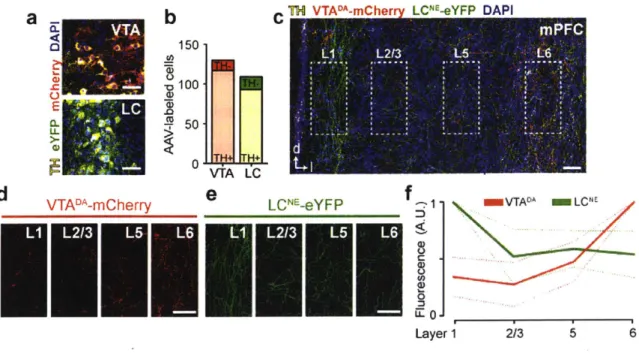
To assess noradrenergic and dopaminergic terminal density in the mPFC, we injected a Cre-dependent virus carrying a fluorescent protein (eYFP or mCherry, counter balanced) into the VTA and locus coeruleus (LC) of transgenic DAT-Cre mice to selectively express two distinct fluorescent tags in VTA dopamine (DA) neurons and LC norepinephrine (NE) neurons (Figure 2a-b). After 8 weeks of incubation, terminal fluorescence was observed in the mPFC (Figure 2c) and quantified across cortical layers (Figure 2d-e). VTA-DA terminals were densest in deep layers (5 and 6), consistent with previous reports (Berger et al., 1976; Sesack et al., 1998; Zhang et al., 2010) while LC-NE terminals were densest in the superficial layers (Figure 2f). These data suggest that dopamine is acting in the deep layers of the mPFC.
M
150 _1 0 50d
VTADA-mCherry'Eli
VTA LCe
L CNE -eYFP mVTAO^ mLCI
U.
0-Layer 1 2/3 5 6
Figure 2. Dopamine terminals are densest in deep layers of the prefrontal cortex.
(a) Injection of viral constructs enabling Cre-dependent expression into the LC and VTA of TH::Cre mice (n = 3).
(b) Fluorescent labeling of TH positive (TH+) noradrenergic (NE) neurons in the LC and
dopaminergic neurons in the VTA.
(c) Examination of VTADA and LCNE fluorescent terminal labeling in the mPFC.
(d) VTADA terminals (from inset) across cortical layers in the prelimbic subregion of the mPFC. (e) LCNE terminals (from inset) across cortical layers in the prelimbic subregion of the mPFC. (f) VTADA terminals were densest in the deep (5 and 6) layers of the mPFC, while LCNE terminals were denser in superficial (1 and 2/3) layers.
To investigate the time course of dopamine release, we injected a viral vector enabling Cre-dependent expression of channelrhodopsin-2 (ChR2) in the VTA of transgenic tyrosine hydroxylase (TH)::Cre rats (Witten et al., 2011) and inserted carbon-fiber electrodes into the prelimbic subregion of the mPFC for in vivo FSCV measurements (Figure 3a-c). To convert recorded changes in current to changes in catecholamine concentration and to eliminate contributions from transient pH fluctuations, chemometric and principal component analyses were performed (Figure 3d-f) (Keithley and Wightman, 2011; Keithley et al., 2009; Vander Weele et al., 2014). Electrodes were aimed at the deep layers (5-6) where VTA dopaminergic terminals in the mPFC (VTADA-mPFC) were densest, compared to norepinephrine (NE) locus coeruleus (LC) terminals (Figure 3g-h) and secured in locations, which supported optically-evoked dopamine release upon stimulation of VTADA neurons (Figure 3i-j). To investigate how an aversive stimulus impacts rapid catecholamine release, we performed a 10 s tail pinch and observed a reliable catecholaminergic signal, which peaked just after tail pinch offset and
lingered for -40 s (Figure 31-n). Given the relatively slow decay kinetics, our data are consistent with other reports suggesting that dopamine reuptake is slower in the mPFC than in the striatum
(Cass and Gerhardt, 1995; Garris and Wightman, 1994; Garris et al., 1993; Kaenmaki et al., 2010; Sesack et al., 1998) (Figure 4c). This is consistent with reports indicating that
mesocortical dopamine neurons lack D2-type autoreceptors, which provide negative feedback and autoinhibit the activity of dopamine neurons (Bannon et al., 1981; Chiodo et al., 1984). In addition, dopamine transporter (DAT) expression is low (Sesack et al., 1998), resulting in slower dopamine reuptake in the mPFC.
Since norepinephrine release in the mPFC has also been reported using microdialysis in the minutes following an aversive stimulus (Finlay et al., 1995) we sought to eliminate LC norepinephrine contribution. To do this, we repeated this experiment immediately after infusing the sodium-channel blocker, tetrodotoxin (TTX) into the LC (Figure 3k-o) and observed
catecholamine release before and after LC inactivation were indistinguishable (Figure 3p-r). TTX microinfusions were co-injected with fast green for cover and spread visualization, which were inspected and noted during tissue sectioning. We speculate that the previously reported LC signal measured in the minutes following an aversive stimulus (Finlay et al., 1995) may have a slower onset and may be related to the subsequent stress response rather than the detection of the noxious stimulus itself.
FSCV AAV-DIO- TTX
a
ChR2-mCherry MPFC VTA LC TH :Cre ra'd
-0.6 -0.6 _b
Ce
pH Training Set DA Training Seth
VTADA-mPFC::ChR2I
j
mPFC mPFC -0p 0. AP: +3.72 mm AP: +3.24 0 " 3s pre postm
n
pinch 3 - .2 Current (nA) 0.5 * sbase pinchq
P-7 N L 5 pinch pinch _r
0.5 * 01 base pinch ChR2 THg
C 0 ic ok
DAPI TH C 'U -J .0 0 AR -9.84 mm -0.4-+1.3 -0.4 Ss +1 -043M
55 -0.4 5SFigure 3. LC inactivation does not alter catecholamine release in the mPFC in response to tail pinch.
(a) Schematic of strategy for differentiating dopamine and NE neurotransmission in the mPFC using fast-scan cyclic voltammetry (FSCV). VTADA neurons were selectively transduced with ChR2 in TH::Cre rats (n = 5). After incubation, rats were prepared for anesthetized FSCV recordings where an optical fiber was implanted over the VTA and a guide cannula was positioned over the LC for tetrodotoxin (TTX)-mediated pharmacological inhibition. A glass-encased carbon-fiber recording electrode was lowered into the mPFC for FSCV neurochemical measurements.
(b) 20x representative confocal image of ChR2-mCherry expression (red) in VTADA cell bodies (yellow = TH immunostaining). The optical fiber track is indicated by the white rectangle.
(c) 40x representative confocal image of ChR2-mCherry expression (left, red) in VTADA cell bodies (right, yellow = TH immunostaining).
(d) Overview of chemometric and principal component analyses of FSCV recordings to isolate catecholamine signals from pH shifts. Representative false color plot showing the presence of both basic pH shifts (left inset, pH cyclic voltammogram) and catecholamine release (right inset, catecholamine cyclic voltammogram).
(e) pH training set composed of acidic and basic pH voltammograms derived from in vivo and in
vitro (flow cell) preparations. Dopamine training set composed of in vivo (optically- and
electrically- evoked) and in vitro (flow cell) preparations.
(f) pH and dopamine traces derived from the representative recording using principal component regression
(h) Schematic representation of all recording electrode locations for ChR2 FSCV experiments.
(i) Evoked dopamine release in the mPFC following 20 Hz (60, 30, and 10 pulses) optical activation of VTADA::ChR2-mCherry cell bodies in the VTA.
U)
Optical activation (473 nm, 20 Hz, 60 pulses, 5 ms pulse-duration, 20 mW) of VTADA::ChR2 neurons evoked dopamine release in the mPFC (paired t-test, t(3) = 3.72, p = 0.034), ensuring FSCV recording electrodes were positioned in dopamine-rich locations.(k) Representative image of guide cannula track positioned over LCNE cell bodies (yellow = TH).
(I-n) When VTADA and LCNE neurons were intact, tail pinch (10 s in duration) rapidly increased extracellular catecholamine concentration ([CAT]) release, as shown in (I) a representative false color plot, (m) average CAT trace, and (n) concentration quantification (paired t-test, t(4) =
3.402, p = 0.027). Color plot insets: representative cyclic voltammograms.
(o) Tetrodotoxin (TTX) + fast-green injection locations were viewed and recorded while tissue sectioning on a freezing microtome, prior to staining and mounting. The center of each injection was recorded. Rats that had injections but did not have spread covering the majority of the LC were excluded. Cannula implant locations were subsequently verified on a confocal microscope.
(p-q) After LC inactivation via intra-LC infusion of TTX, tail pinch evoked responses were maintained as shown in (p) a representative false color plot, (q) average CAT trace, and (r) concentration quantification (paired t-test, t(4) = 5.249, p = 0.006). Color plot insets: representative cyclic voltammograms.
Error bars indicate SEM. Scale bars (histology) = 50 um. "pre" = 5 s time period prior to stimulation onset; "post" = 5 s time period after stimulation onset.
Since both dopamine and norepinephrine can be released from multiple sources (Bjorklund & Dunnet et al., 2007), and it is possible that local modulation of LCNE-mPFC terminals could occur, we sought to determine the primary source for the pinch-induced dopaminergic signal in the mPFC. To do this, we performed halorhodopsin (NpHR)-mediated photoinhibition of VTADA neurons during tail pinch (Figure 4a-b). We found that photoinhibition of VTADA neurons significantly attenuated the pinch-induced dopaminergic signal in the mPFC (Figure 4d-f). These data suggest that the majority of the pinch-evoked time-locked signal in the mPFC was mediated by dopamine release from VTA terminals.
d
b VTAI- r-nPFC..NpHR C~ ~
a
FSCV AAV-DIO- eSim+ 0 T NAc NpHR-eYFP 593 nm mPFC mPFC -mPFCI
mPFC VTA LC 3 s 52 Hz0p TH::Cre rat OFF ON-4
6 - 4 Current (nA) 6m -4 Current(nA) 1.6+1.3
-0p4
g-3
pinch 5 pinch 15s 593 nm ON*
OFF ON
Figure 4. VTA DA inhibition attenuates catecholamine release in response to tail pinch. (a) Schematic of strategy to verify dependence of pinch-evoked increases in CAT
neurotransmission on VTADA neurons.
(b) Histologically verified FSCV recording electrode locations for NpHR experiments.
(c) Electrical stimulation (60 Hz, 60 pulses, 200 uA) of the dorsal VTA evoked distinct patterns
of dopamine release in the NAc and mPFC.
(d) Representative false color plots showing tail pinch evoked CAT responses before (left) VTADA inhibition and during (right) transient VTADA inhibition surrounding the tail pinch (593 nm
laser light, 20 s in duration) (n = 5) Color plot insets: representative cyclic voltammograms. (e) Average CAT traces with (orange) and without (gray) optical inhibition of VTADA neurons.
(f) Optical inhibition of NpHR-expressing VTADA neurons attenuated tail pinch evoked CAT release in the mPFC (concentration quantification (paired t-test, t(4) = 5.884, p = 0.004)).
Error bars indicate SEM. Scale bars (histology) = 50 um. "pre" = 5 s time period prior to stimulation onset; "post" = 5 s time period after stimulation onset.
Next, given that dopamine is released in the mPFC following aversive stimuli, we wanted to explore the causal relationship between VTADA-mPFC and valence processing. To address this, we tested whether photostimulation of dopaminergic VTA terminals in the mPFC was sufficient to drive aversion. In TH::Cre rats, we injected a viral vector enabling Cre-dependent expression of ChR2 in the VTA and implanted optical fibers over the mPFC (Figure 5a-b). We did not find any detectable differences between ChR2- and eYFP-expressing rats in closed-loop real-time place avoidance (RTPA) (Figure 5c-d) or conditioned place aversion (CPA) assays (Figure 5e-f),
a
TH :Cre rat
AAV-DIO-ChR2-eYFP
mPFC VTA
C Real-time place d =ChR2 MeYFPj preference / aversion 14
OFF
ON_
F -60
'
60 min sessions ChR2 eYFP
Figure 5. Activation of dopamine terminals conditioned place preference I aversion.
b
X VTADkmPFC:.ChR2 X VTAr-mPFC:.eYFPmPFC
mPFC
PLL
AP:+3.72 mm AP +324 mm * Conditioned place f MChR2 MeYFP
preference I aversion 300 Habitua0on
k
OFF
N
ON 0
Test -100
15 min sessions ChR2 OYFP
in the mPFC does not support real-time or
(a) Schematic strategy for manipulating dopamine release in the mPFC. VTADA neurons were selectively transduced with ChR2 in TH::Cre rats.
(b) Guide cannulae were implanted over the mPFC for the insertion of an optical fiber for light
delivery.
(c) Schematic of experimental design for real-time place preference/avoidance assays (RTPP/A). When rats entered the ON zone, laser light stimulation was activated for the duration of the time spent in the ON zone (20 Hz, 60 p, every 30 s, 20 mW of 473 nm for VTADA-mPFC experiments). When rats entered the OFF zone, light stimulation was terminated for the duration of time spent in the OFF zone.
(d) Optogenetic stimulation of VTADA neurons did not evoke real-time place avoidance or
measured by difference score [minutes spent in the ON zone - OFF zone] (unpaired t-test, t(8) = 0.9337, p = 0.3778).
(e) Schematic of experimental design for all conditioned place preference/aversion assays (CPP/A). Day 1 consisted of a habituation period where time spent on each compartment of the arena was recorded. On days 2 and 3, a divider was placed in the middle of the chamber to separate the two compartments and rats received either no stimulation (OFF) or stimulation (ON) (20 Hz, 60 p, every 30 s, 20 mW of 473 nm for VTADA-mPFC experiments), counter-balanced across days. On day 4, the divider was removed and time spent in each compartment was recorded in the absence of stimulation (i.e., test day).
(f) Optogenetic stimulation of VTADA neurons did not support conditioned place aversion or preference in VTADA-mPFC::ChR2 animals, compared to VTADA-mPFC::eYFP controls, measured by difference score [minutes spent in the ON zone - OFF zone] (unpaired t-test, t(9) = 0.3192, p = 0.7569).
However, in RTPA and CPA there are no discrete stimuli presented, and in light of the model for dopaminergic involvement in enhancing signal-to-noise, we considered the potential importance of dopamine in enhancing neural activity in response to discrete, predictive cues. To test whether dopamine would bias behavioral responses to discrete cues predicting either punishment or reward, we used a recently described behavioral paradigm (Burgos-Robles et al., 2017). Here, rats expressing ChR2 in VTADA neurons (Figure 6a-b) and were trained on a stimulus competition behavioral paradigm (Figure 6c-d). First trained to associate auditory or visual cues (counter-balanced across subjects) with sucrose delivery to a nearby port (Figure 6e) and ChR2 and control eYFP subjects learned that the cue predicted reward delivery at the same rate - as measured by time spent in the sucrose port during the conditioned stimulus (Figure 6f). Next rats were trained to associate the alternative auditory or visual cues (counter-balanced across subjects) with footshock (Figure 6g). During discrimination sessions, and ChR2 and control eYFP subjects discriminated sucrose- and shock-predictive cues similarly, as measured by time spent in the sucrose port (Figure 6h-i) and time spent freezing (Figure 6j-k), a species-specific defensive reaction. Once rats learned to discriminate the cues predicting shock and sucrose by freezing or approaching the delivery port, respectively, we tested their behavioral responses to the competition of simultaneously-presented cues driving conflicting motivational outputs (Figure 6d). We found that photostimulation of VTADA-mPFC terminals (using empiricallydetermined optical parameters, Figure 2ij) during the "competition" cues -wherein both shock-predictive and reward-predictive stimuli were co-presented - caused ChR2-expressing rats to spend significantly less time in the sucrose delivery port and more time freezing than eYFP controls (Figure 61-0). There were no differences between groups during sucrose and shock only trials during competitions sessions (Figure 6p-s).
a
H Cfirb -- -er mPFC ChR2-eYFPmPFC - VTA t
co dio ON OFF 2CHLM00 pe 5 s
c
discrimination coptiton competion vauumDandt - 4 -N OFF I 'I lShock
Days 1-3 4- 8-10 11-13 'Tm s 6 )2O0
o
sucrose conditioningBilll~~l
VsOW=I
M ChR2 M YFP C o 40-0 Session 2 3g
discrimination-
rshock 6 Time(s) 2 6 Ti" (s) 20 discrimination sessionsh
-Cs-M
CS" MChR2 MeYFP - CS_= Cs"
ChR2 MYFP 100 80 -h00Rv
rh
w
1
80 60 80. .S 80. 60 '1 *)4 40- 1 V.0. 0 400 . . . 40 -10 Time (s) 30 Cs CSf -10 ThiMe (s) 30 CS" CS" competition sessionsm
n
CS-"w trials 0 CSQ"'" trials -CS- -CS -CS- - - " - CSM fYFP --aYFP 100 s0 80 MChR2 0 MChR20.0
.I2.I
0
,
II0
I
-10 Time (s) 30 -10 Time (8) 30 ON OFF ON OFF CS-C trials
q
CS" trialsr
CS* trials*
CS*" trials80 A 80 -ChR2 s0
m
ChR2 60.r-3eYFP r3 """eYFp
0 . 0 .1 I MIL "O"L rn11111 *0 1 .1
ON OFF ON OFF ON OFF ON OFF
Figure 6. Activation of dopamine terminals in the mPFC biases behavior towards aversion during competitive stimulus presentations.
(a) Schematic of strategy for manipulating dopamine release in the mPFC.
(b) VTADA neurons were selectively transduced with ChR2 in TH::Cre rats and guide cannulae were implanted over the mPFC for insertion of an optical fiber for light delivery.
(c) Schematic of task used to examine dopamine modulation of reward and fear-motivated behaviors during competition.
(d) During competition trials (CScomP), VTADA terminals were activated (473 nm, 20 Hz, 60 pulses, 5 ms pulse-duration, every 5 s, 18 mW) to assess the impact of dopamine signaling in the mPFC on behavior during competition. During "competition OFF", light was not delivered for the entire session to assess recovery.
(e) During sucrose training, a CS (light or tone, counterbalanced) predicted sucrose delivery (C S"c). Sucrose was removed from the delivery port by vacuum if not collected.
(f) VTADA-mPFC::ChR2 rats and VTADA-mPFC::eYFP controls acquired sucrose conditioning similarly (two-way repeated measures ANOVA, F2,2 2 = 0.7, p = 0.5090).
(g) During discrimination, the alternative CS (light or tone, counterbalanced) was introduced and predicted foot shock (CSh"k)
(h) Average traces showing time spent in the sucrose port before, during, and after each CS presentation.
(i) Time spent in the sucrose port did not differ between VTADA-mPFC::ChR2 rats and VTADA_ mPFC::eYFP controls during CSsuc or CSShk (repeated measures two-way ANOVA, Fm1 = 0.54, p = 0.4789, Bonferroni multiple comparisons tests p > 0.05) presentation.
(j) Average traces showing time spent freezing before, during, and after each CS presentation. (k) Time spent in the freezing did not differ between VTADA-mPFC::ChR2 rats and VTADA_
mPFC::eYFP controls during CSUC or CS"hk (repeated measures two-way ANOVA, F1,11 = 0.01, p = 0.9281, Bonferroni multiple comparisons tests p > 0.05) presentation.
(I) Percent time spent in the reward port during the three trial types in the last competition ON session.
(m) Percent time spent freezing during the three trial types in the last competition ON session. (n) During competition sessions, the average time spent in the reward port during CScomp ON trials was lower in ChR2 rats (closed bars) compared with eYFP controls (open bars, repeated measures two-way ANOVA, F1,11 = 8.13, p = 0.0157, Bonferroni multiple comparisons tests p < 0.05).
(o) During competition sessions, the average time spent freezing during CScomp ON trials was greater in ChR2 rats (closed bars) compared with eYFP controls (open bars, repeated measures two-way ANOVA, F1,11 = 13.29, p = 0.0039, Bonferroni multiple comparisons tests p < 0.05).
(p) During competition sessions, the average time spent in the reward port for CS" trials during ON sessions and CSuc trials during OFF sessions did not differ between ChR2 rats (closed bars) and eYFP controls (open bars, repeated measures two-way ANOVA, F1,11 = 0.82, p = 0.3845). Note that during ON sessions, stimulation was only delivered during the CScomP trials. (q) Average time spent freezing for CS'"" trials during ON sessions and CSs"c trials during OFF sessions did not differ between ChR2 rats (closed bars) and eYFP controls (repeated measures two-way ANOVA, F1,11 = 1.35, p = 1.35).
(r) During competition sessions, the average time spent in the reward port for CSShk trials during ON sessions and CSshk trials during OFF sessions was not different between ChR2 (closed bars) and eYFP controls (open bars, repeated measures two-way ANOVA, F1,11 = 0.94, p = 0.354).
(s) During competition sessions, the average time spent freezing for CSShk trials during ON sessions and CSShk trials during OFF sessions was not different between ChR2 rats (closed bars) and eYFP controls (open bars, repeated measures two-way ANOVA, F1,11 = 0.16, p = 0.6998).
Experimental Procedures General virus surgery
Specific subject/surgery details for each experiment are detailed below. For all subjects, surgeries were performed under aseptic conditions and body temperature was maintained with a heating pad. Rodents were anesthetized with isoflurane mixed with oxygen (5% for induction, 2.5-2% for maintenance, 1L/min oxygen flow rate) and placed in a digital small animal stereotax (David Kopf Instruments, Tujunga, CA, USA). Following initial induction, hair was removed from the dorsal surface of the head with hair clippers, ophthalmic ointment was applied to the eyes, the incision area was scrubbed with alcohol pads and betadine (x3 each), and 2% lidocaine was injected just under the skin surface above the skull for topical anesthesia. All measurements were made relative to bregma (unless noted otherwise) for virus/implant surgeries. Viral injections were performed using a beveled microinjection needle (26 gauge for rat; 33 gauge for mice) with a 10 pL microsyringe (Nanofil; WPI, Sarasota FL, USA) delivering virus at a rate of 0.05-0.01 pL/min using a microsyringe pump (UMP3; WPI, Sarasota, FL, USA) and controller (Micro4; WPI, Sarasota, FL, USA). For injections at multiple locations on the dorsal-ventral axis, the most ventral location was completed first and the injection needle was immediately relocated to the more dorsal location and initiated. After injection completion, 15 mins were allowed to pass before the needle was slowly withdrawn. After viral infusions were completed, craniotomies were filled with bone wax and the incision closed with nylon sutures. Subjects were maintained under a heat lamp and provided 0.05 mg/kg (rat) / 0.10 mg/kg (mouse) buprenophine (s.c., diluted in warm Ringers solution) until fully recovered from anesthesia.
All experiments involving the use of animals were in accordance with NIH guidelines and approved by the MIT Institutional Animal Care and Use Committee. For all experiments involving viral or tracer injections, animals containing mistargeted injection(s) were excluded after histological verification.