HAL Id: inserm-00868819
https://www.hal.inserm.fr/inserm-00868819
Submitted on 2 Oct 2013
HAL is a multi-disciplinary open access
archive for the deposit and dissemination of
sci-entific research documents, whether they are
pub-lished or not. The documents may come from
teaching and research institutions in France or
abroad, or from public or private research centers.
L’archive ouverte pluridisciplinaire HAL, est
destinée au dépôt et à la diffusion de documents
scientifiques de niveau recherche, publiés ou non,
émanant des établissements d’enseignement et de
recherche français ou étrangers, des laboratoires
publics ou privés.
Alteration of skin perfusion in mottling area during
septic shock.
Hafid Ait-Oufella, Simon Bourcier, Mikael Alves, Arnaud Galbois, Jean-Luc
Baudel, Dimitri Margetis, Naike Bige, Georges Offenstadt, Eric Maury,
Bertrand Guidet
To cite this version:
Hafid Ait-Oufella, Simon Bourcier, Mikael Alves, Arnaud Galbois, Jean-Luc Baudel, et al.. Alteration
of skin perfusion in mottling area during septic shock.. Annals of Intensive Care, SpringerOpen, 2013,
3 (1), pp.31. �10.1186/2110-5820-3-31�. �inserm-00868819�
R E S E A R C H
Open Access
Alteration of skin perfusion in mottling area
during septic shock
Hafid Ait-Oufella
1,2,3*, Simon Bourcier
1, Mikael Alves
1,3, Arnaud Galbois
1, Jean-Luc Baudel
1, Dimitri Margetis
1,
Naike Bige
1,3, Georges Offenstadt
1,3,4, Eric Maury
1,3,4and Bertrand Guidet
1,3,4Abstract
Background: Mottling score has been reported to be a strong predictive factor during septic shock. However, the pathophysiology of mottling remains unclear.
Methods: In patients admitted in ICU for septic shock, we measured on the same area the mean skin perfusion by laser Doppler, the mottling score, and variations of both indices between T1 (6 hours after vasopressors were started) and T2 (24 hours later).
Results: Fourteen patients were included, SAPS II was 56 [37–71] and SOFA score at T1 was 10 [7–12]. The mean skin surface area analyzed was 4108 ± 740 mm2; 1184 ± 141 measurements were performed over each defined skin surface area. Skin perfusion was significantly different according to mottling score and decreased from 37 [31–42] perfusion units (PUs) for a mottling score of [0–1] to 22 [20–32] PUs for a mottling score of [2–3] and 23 [16–28] for a score of [4–5] (Kruskal-Wallis test, P = 0.05). We analyzed skin perfusion changes during resuscitation in each patient and together with mottling score variations between T1 and T2 using a Wilcoxon signed-rank test. Among the 14 patients included, mottling score increased (worsened) in 5 patients, decreased (improved) in 5 patients, and remained stable in 4 patients. Baseline skin perfusion at T1 was arbitrarily scored 100%. Mean skin perfusion
significantly decreased in all the patients whose mottling score worsened from 100% baseline to 63.2 ± 10.7% (P = 0.001), mean skin perfusion significantly increased in all patients whose mottling score improved from 100% baseline to 172.6 ± 46.8% (P = 0.001), and remained stable in patients whose mottling score did not change (100.5 ± 6.8%, P = 0.95).
Conclusions: We have shown that mottling score variations and skin perfusion changes during septic shock resuscitation were correlated, providing additional evidence that mottling reflects skin hypoperfusion. Keywords: Septic shock; Microcirculation; Mottling; Intensive care medicine
Background
The identification of endothelial dysfunction and abnor-mal microcirculation as the main cause of organ damage and death during septic shock [1,2] prompts intensivists to develop tools to assess microcirculation and organ perfusion. Sublingual videomicroscopy using Sidestream Dark Field (SDF) imaging provided interesting informa-tions on microcirculatory status during septic shock at the admission and during resuscitation with a direct
visualisation of microvessels [3]. Noninvasive measure-ments of tissue oxygenation using near-infrared spec-troscopy (NIRS) technology informs indirectly about microcirculation and mostly muscle perfusion [4]. In our unit, we focused on skin perfusion through the explor-ation of mottling. We have developed a clinical score based on the extension of mottling around the knee and showed in a prospective, observational study that mot-tling score was a strong predictor of 14-day mortality for patients admitted for septic shock. Mottling score was related to other parameters that reflected organ perfu-sion, such as arterial lactate level or diuresis [5]. How-ever, the physiopathology of mottling remains unknown, and the link between mottling and skin perfusion was * Correspondence:hafid.aitoufella@sat.aphp.fr
1
Service de réanimation médicale, Hôpital Saint-Antoine, Assistance Publique-Hôpitaux de Paris, 184 rue du Faubourg Saint-Antoine, Cedex 12, Paris 75571, France
2Paris Research Cardiovascular Center, Inserm U970, Paris, France
Full list of author information is available at the end of the article
© 2013 Ait-Oufella et al.; licensee Springer. This is an Open Access article distributed under the terms of the Creative Commons Attribution License (http://creativecommons.org/licenses/by/2.0), which permits unrestricted use, distribution, and reproduction in any medium, provided the original work is properly cited.
speculative and based on indirect evidences. To explore in a more accurate way the physiopathology of mottling, we conducted a pilot observational study using a laser Doppler imager to analyze skin perfusion according to mottling extension during septic shock management.
Methods
Septic shock inclusion and management
We conducted a prospective, observational study in a 16-bed ICU in a tertiary teaching hospital during 4 months. All consecutive patients, older than 18 years, admitted for septic shock were included. Septic shock, within 24 hours after ICU admission, was defined according to the 2001 SCCM/ESICM/ACCP/ATS/SIS International Sepsis Definitions conference [6]. Pa-tients were included (H0) when vasopressor infusion was started within 24 hours of admission. Circulatory support was guided by our local protocol, adapted from international guidelines [7]. Intravenous volume expansion and intravenous norepinephrine were used in a stepwise manner to achieve predefined endpoints of resuscitation from invasive hemodynamic monitor-ing: mean arterial pressure (MAP) >65 mmHg, central venous pressure (CVP) between 8 and 12 mmHg, urin-ary output >0.5 ml/kg/h. General characteristics of the patients were recorded: demographic data, diagnoses, severity of illness evaluated by the Sequential Organ Failure Assessment (SOFA) score (within 6 hours of admission) [8] and Simplified Acute Physiology Score II (SAPS II) [9]. Hemodynamic variables were recorded 6 hours (T1) and 24 hours (T2) after start of vasopres-sor therapy. We measured MAP, CVP, diuresis, cardiac index using echocardiography, arterial lactate level, and mottling score. Mottling score, recently described by our group, provided a semiquantitative evaluation of mottling based on skin area extension on legs. Score 0 no mottling, score 1 small mottling area (coin size) localized to the center of the knee, score 2 mottling area that does not exceed the superior edge of the knee cap, score 3 mottling area that does not exceed the middle thigh, score 4 mottling area that does not ex-ceed the fold of the groin and score 5 otherwise [5]. Fi-nally, we measured mean skin perfusion using Laser Doppler Imager on the legs and compare its changes to mottling score variations between T1 and T2.
Scanning laser Doppler
The PeriScan PIM 3 System is based on the established laser Doppler technique [10,11]. When laser light pene-trates the tissue, it is scattered and partly absorbed. Some of the scattered light returns to the tissue surface, where it is registered by a photo detector inside the instrument. This signal is then processed to extract information about the microcirculatory blood flow.
According to the Doppler principle, light particles which hit moving blood cells undergo a change in wavelength/frequency (a Doppler shift), while light particles which encounter static structures return un-changed. The perfusion can be calculated, because the magnitude and frequency distribution of the Doppler shifted light are directly related to the number and velocity of blood cells. Blood perfusion is measured in perfusion units (PU) and there is a documented lin-earity between PU and the true blood perfusion in the tissue being imaged. The penetration depth is be-tween 0.5 and 1 mm. The PeriScan PIM 3 System automatically scans a defined skin surface area (4 mil-liseconds per pixel, resolution of 255 × 255 pixels). The data are computed and visualized as a two-dimensional colour-coded image, mapping skin perfu-sion. The mean blood flow is computed to yield an average of pixels in a region of interest within the scanned area with the software provided by the manu-facturer (LDPIwin software). The total time required for a complete image at these settings is approxi-mately 5 minutes. The scanning Laser Doppler pro-vides a skin perfusion mapping and the LDPIWin software led us to measure skin perfusion, expressed in Perfusion Units (PU), in the same area that we quantified the mottling score.
Study protocol
Experiments were performed at bedside. The first ex-ploration (T1) was done 6 hours after vasopressor in-fusion has been started and the second 24 hours later (T2). The laser Doppler imaging was performed with the patient in a reclined position, the thigh at heart level. The distance between the skin and the lowest part of the scanner head during image capturing was fixed to 17 cm with the system resolution set on medium. Studied skin area was standardized and was focused on the anterior face of the thigh between the middle of the knee cap and the fold of the groin (Figure 1). When capturing images, the ambient light level was kept at the minimum to avoid any influence on the laser light and the quality of the recorded perfusion signal. We measured mean skin perfusion at T1 and T2, and we also compared skin perfusion changes with mot-tling score variations between T1 and T2. The investigator that analyzed the data provided by the laser Doppler imager was blinded to the mottling score.
The observational protocol was approved by the ethical committee of the Société de Réanimation de Langue Française (SRLF). This is an observational study without any specific intervention. All patients and families were informed through the admission leaflet that anonymous data could be used for aca-demic research and gave their consent.
Ait-Oufella et al. Annals of Intensive Care 2013, 3:31 Page 2 of 6 http://www.annalsofintensivecare.com/content/3/1/31
Statistical analysis
Data were summarized as median (25th-75thpercentiles) for skewed distributions and percentages as appropriate. Skin perfusion expressed on PUs according to mottling score was tested using the Kruskal-Wallis rank-sum test. Hemodynamic variables and skin perfusion changes be-tween T1 and T2 were tested using a Wilcoxon signed-rank test. All tests were computed with the R software. Significance was defined as a two-sided P value < 0.05.
Results
Studied population
During the 4-month study, fourteen patients were in-cluded. General characteristic of the studied population were summarized in Table 1. The median SAPS II was
56 [37–71], the median SOFA at T1 was 10 [7-12], and the 14-day mortality was 36% (5/14 patients). All the patients initially received intravenous norepinephrine; median dose was 0.20 μg/kg/min [0.10-0.37] at T1 and 0.11 μg/kg/min [0.01-0.72] at T2. Hemodynamic para-meters at both T1 and T2 were summarized in Table 2.
Laser Doppler analysis
The mean skin surface area analyzed was 4108 ± 740 mm2; 1184 ± 141 measurements were performed over each defined skin surface area. First of all, we compared the mean skin perfusion according to mottling score at T1. Interestingly, mean skin perfusion was significantly
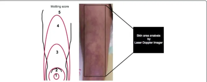
Figure 1 Mottling score is based on mottling area extension on legs. Score 0, no mottling; score 1, modest mottling area (coin size) localized to the center of the knee; score 2, moderate mottling area that does not exceed the superior edge of the kneecap; score 3, mild mottling area that does not exceed the middle thigh; score 4, severe mottling area that does not exceed the fold of the groin; and score 5, extremely severe mottling area that exceeds the fold of the groin. The area of laser Doppler scanning was superposed to the area of mottling score classification.
Table 1 Baseline characteristics of included population
Patients (n) 14
Age, yr 65 [53–68]
Gender, female (n) 5/14 Primary site of infection [n]
Lung 3 Abdomen 6 Urinary tractus 1 Endocarditis 1 Primary bactaeremia 3 SAPS II 56 [37–71] 14-day mortality (n) 5/14
SAPS II (Simplified Acute Physiology Score) was calculated within 24 hours of admission.
Values are given as median [25th-75thpercentiles] and numbers (n).
Table 2 Hemodynamic parameters recorded at T1 (6 hours after vasopressor start) and T2 (24 hours after vasopressor start) T1 T2 P SOFA score 10 [7–12] 11 [3–16] NS Norepinephrine N 14 10 Doses μg/kg/min 0.20 [0.10-0.37] 0.25 [0.01-0.72] NS Mean arterial pressure (mmHg) 77 [66–88] 84 [72–91] NS Central venous pressure (mmHg) 13 [10–15] 12 [11–16] NS Cardiac index (L/min/m2) 2.4 [2.2-2.9] 2.3 [2.1-3] NS Arterial lactate level (mmol/L) 3.3 [1.8-7.2] 1.8 [1.2-6] NS ScVO2(%) 70 [67–75] 71 [67–77] NS
SOFA Sequential Organ Failure Assessment, ScVO2, central venous saturation in oxygen, NS not significant.
Values are given as median [25th-75thpercentiles]. Cardiac index was
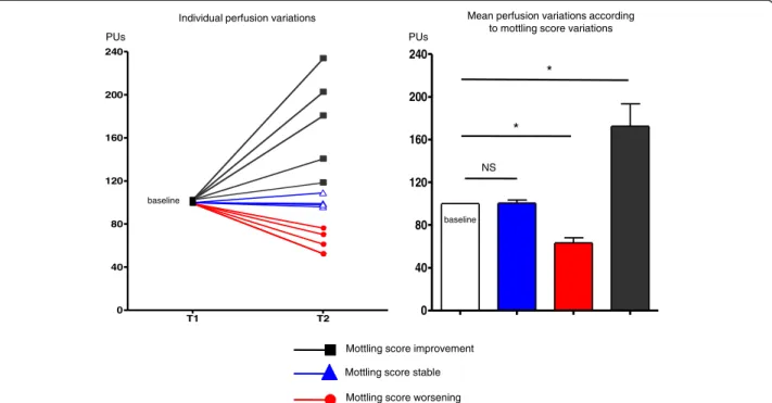
different according to mottling score and decreased from 37 [31–42] PUs for a mottling score of [0–1] (n = 5) to 22 [20–32] PUs (n = 5) for a mottling score of [2,3] and 23 [16–28] for a score of [4,5] (n = 4) (P = 0.05) (Figure 2). However, value variability was important be-cause PU depends on several individual parameters such as microvessel density or skin thickness. For this reason, we subsequently analyzed the skin perfusion changes during resuscitation in each patient and compared the results to mottling score variations between T1 and T2. Among the 14 patients included, mottling score increased (worsened) in 5 patients, decreased (improved) in 5 pa-tients and remained stable in 4 papa-tients. Baseline value at T1 was noted 100%. Interestingly, we observed that mean skin perfusion significantly decreased in all the patients whose mottling score worsened from 100% baseline to 63.2 ± 10.7% (P = 0.001), mean skin perfusion significantly increased in all the patients whose mottling score im-proved from 100% baseline to 172.6 ± 46.8% (P = 0.02), and remained stable in patients whose mottling score did not change (100.5 ± 6.8%, P = 0.91; Figure 3).
Discussion
The identification and the quantification of microcircu-lation dysfunction during severe infection is a major challenge in ICU [1,12]. We focused on a very old clin-ical sign of shock, mottling [13,14], and hypothesized that mottling was the consequence of abnormal skin perfusion. To quantify mottling, we developed a clinical score based on its skin extension over the leg. This score, easy to learn and to use at the bedside, is very re-producible (kappa 0.87, 95% confidence interval (0.72-0.97)) [5]. In a prospective, observational study, we have
shown that mottling score and its variations during sep-tic shock resuscitation provides powerful predictive in-formation. Moreover, mottling score was strongly related to parameters (lactate level or urinary output) that re-flect tissue perfusion [5].
However, the physiopathology of mottling remains un-known. Several indirect evidences led us to associate mottling and abnormal skin perfusion. First, mottling areas are colder than normally colored skin (personal observations). Moreover, we have recently reported, using NIRS Technology, that tissue oxygen saturation on knee area was inversely related to mottling score, the higher the mottling score the lower the knee StO2[15].
In healthy volunteers, Lima et al. also reported that per-ipheral vasoconstriction induced by body surface cooling reduced StO2[16]. As StO2measures tissue oxygen
sat-uration within 15 mm of depth that included skin and muscle, it could be an argument for abnormal skin per-fusion in mottling area but the respective part of both tissues (skin and muscle) in the final result of stO2is
un-known. In this pilot study, we evaluated the link between mottling extension and skin perfusion. The skin perfu-sion was measured using a scanning laser Doppler (PIM3 Perimed System) on the same area that we quan-tified the mottling score (Figure 1). We measured mean skin perfusion with more than 1,000 measurements, and we observed that mean skin perfusion was inversely re-lated to mottling score. Distribution of the mean perfusion values between patients was large, because skin perfusion depends on several individual parameters, such as micro-vessels density, skin thickness, hemoglobin level, and skin temperature. Between T1 and T2, we did not observe any difference regarding the central temperature or the hemoglobin level. Between T1 and T2, we observed three different profiles, a diminution (improvement) of mottling score in five patients, an augmentation (worsening) in five patients, and no variation in four patients. Mottling score changes during resuscitation are very inform-ative, because we have previously shown in a prospect-ive study on septic shock that patients whose mottling score improved had a better prognosis that patients whose mottling score did not (14-day mortality 23% vs. 88%, P = 0.004) [5]. We measured mean skin perfusion on the area on which we have previously computed the mottling score (Figure 1). The area of interest, the dis-tance between the camera and the skin, was stan-dardized. Interestingly, we found a close relationship between changes of mean skin perfusion and changes of mottling score. In all the patients whose mottling score increased, the mean skin perfusion decreased, in patients whose mottling score decreased, the mean skin perfusion decreased, and finally in patients whose mottling score did not change, the mean perfusion also remained stable. It is the first direct evidence that
Figure 2 Mean skin perfusion expressed as mean perfusion units (PUs) according to mottling score. Boxes show 1st and 3rd quartiles, with the median as a thick line. Whiskers extend to 1.5 interquartile ranges (Q75-Q25). *P = 0.05.
Ait-Oufella et al. Annals of Intensive Care 2013, 3:31 Page 4 of 6 http://www.annalsofintensivecare.com/content/3/1/31
mottling extension was related to skin perfusion and cutaneous microcirculation impairment.
During septic shock, microvascular dysfunction is het-erogeneous, because it does not alter all of the organs or all of the territories of every organ [17,18]. Mottling preferentially develops around the knee like area of het-erogeneous discoloration. Interestingly, laser Doppler imager confirmed heterogeneity of skin perfusion on the thigh (Figure 4). Furthermore, laser Doppler mapping showed that color-encoded signal was more important on the knee surface area compared with other leg surfaces
(Figure 4), suggesting that vascular density or blood flow are more important in this area.
Our study has several limitations. It is a monocentric study and results need to be confirmed in a larger popu-lation. Nevertheless, although the size of this preliminary study was rather limited, it was sufficient to highlight significant results. Moreover, perfusion indices were cal-culated by the laser Doppler imager software and the physician who analyzed the data was blind to the mot-tling score and therefore was not influenced by the result of the clinical observation. Skin temperature was
T1 T2 0 40 80 120 160 200 240 0 40 80 120 160 200 240
Mottling score worsening Mottling score stable Mottling score improvement
*
*
baseline baseline
Individual perfusion variations Mean perfusion variations according to mottling score variations
PUs PUs
NS
Figure 3 Left: individual skin perfusion changes between T1 and T2. Baseline value was 100%. Right: mean skin perfusion changes between T1 and T2 according to mottling score variations. Baseline value was 100%. *P < 0.05, **P < 0.01.
Score 5 Score 3 Score 0 Score 2 Patient A Patient B
T1 T2 T1 T2
not measured in the study, and we cannot exclude varia-tions of skin temperature between T1 and T2 that could affect microvascular skin blood flow. Finally, we ex-plored global microvascular blood flow, but we did not analyze the precise mechanisms that lead to skin perfu-sion changes. Several intricate mechanisms, such as intravascular coagulation, increased leucocyte adhesion, or vascular tone modifications [18], potentially partici-pated to the changes of microvascular perfusion.
Conclusions
Using a laser Doppler imager, we have shown in patients admitted for septic shock that mottling score was related to skin perfusion. Moreover, we have shown that mot-tling score variations during resuscitation were related to skin perfusion changes providing additional evidence that mottling is the clinical expression of skin hypoperfusion.
Competing interests
The authors had no competing of interest. Authors’ contribution
Conception and design: HAO, SB, BG, EM ; Analysis and interpretation: HAO, MA, AG, JLB, EM, BG ; Drafting the manuscript for important intellectual content: HAO, MA, AG, JLB, DM, NB, GO, EM, BG. All authors read and approved the final manuscript.
Author details
1Service de réanimation médicale, Hôpital Saint-Antoine, Assistance
Publique-Hôpitaux de Paris, 184 rue du Faubourg Saint-Antoine, Cedex 12, Paris 75571, France.2Paris Research Cardiovascular Center, Inserm U970, Paris,
France.3Université Pierre et Marie Curie-Paris 6, Paris, France.4Inserm U707, Paris F-75012, France.
Received: 15 March 2013 Accepted: 21 August 2013 Published: 16 September 2013
References
1. Vincent JL, De Backer D: Microvascular dysfunction as a cause of organ dysfunction in severe sepsis. Crit Care 2005, 9(Suppl 4):S9–S12. 2. Ince C: The microcirculation is the motor of sepsis. Crit Care 2005,
9(Suppl 4):S13–S19.
3. De Backer D, Creteur J, Preiser JC, Dubois MJ, Vincent JL: Microvascular blood flow is altered in patients with sepsis. Am J Respir Crit Care Med 2002, 166:98–104.
4. Creteur J, Carollo T, Soldati G, Buchele G, De Backer D, Vincent JL: The prognostic value of muscle StO2 in septic patients. Intensive Care Med 2007, 33:1549–1556.
5. Ait-Oufella H, Lemoinne S, Boelle PY, Galbois A, Baudel JL, Lemant J, Joffre J, Margetis D, Guidet B, Maury E, Offenstadt G: Mottling score predicts survival in septic shock. Intensive Care Med 2011, 37:801–807.
6. Levy MM, Fink MP, Marshall JC, Abraham E, Angus D, Cook D, Cohen J, Opal SM, Vincent JL, Ramsay G: SCCM/ESICM/ACCP/ATS/SIS International Sepsis Definitions Conference. Crit Care Med 2001, 2003(31):1250–1256. 7. Dellinger RP, Levy MM, Carlet JM, Bion J, Parker MM, Jaeschke R, Reinhart K,
Angus DC, Brun-Buisson C, Beale R, et al: Surviving Sepsis Campaign: international guidelines for management of severe sepsis and septic shock: 2008. Crit Care Med 2008, 36:296–327.
8. Moreno R, Vincent JL, Matos R, Mendonca A, Cantraine F, Thijs L, Takala J, Sprung C, Antonelli M, Bruining H, Willatts S: The use of maximum SOFA score to quantify organ dysfunction/failure in intensive care. Results of a prospective, multicentre study. Working Group on Sepsis related Problems of the ESICM. Intensive Care Med 1999, 25:686–696.
9. Le Gall JR, Lemeshow S, Saulnier F: A new Simplified Acute Physiology Score (SAPS II) based on a European/North American multicenter study. Jama 1993, 270:2957–2963.
10. Opazo Saez AM, Mosel F, Nurnberger J, Rushentsova U, Gossl M, Mitchell A, Schafers RF, Philipp T, Wenzel RR: Laser Doppler imager (LDI) scanner and intradermal injection for in vivo pharmacology in human skin microcirculation: responses to acetylcholine, endothelin-1 and their repeatability. Br J Clin Pharmacol 2005, 59:511–519.
11. Terada K, Miyai N, Maejima Y, Sakaguchi S, Tomura T, Yoshimasu K, Morioka I, Miyashita K: Laser Doppler imaging of skin blood flow for assessing peripheral vascular impairment in hand-arm vibration syndrome. Ind Health 2007, 45:309–317.
12. Boerma EC: The microcirculation as a clinical concept: work in progress. Curr Opin Crit Care 2009, 15:261–265.
13. Altemeier WA, Cole W: Septic shock. Ann Surg 1956, 143:600–607. 14. Vachon F, Emile J, Bazin C: Conception actuelle du traitement du choc
infectieux. Paris: Editions Arnette; 1968.
15. Ait-Oufella H, Joffre J, Boelle PY, Galbois A, Bourcier S, Baudel JL, Margetis D, Alves M, Offenstadt G, Guidet B, Maury E: Knee area tissue oxygen saturation is predictive of 14-day mortality in septic shock. Intensive Care Med 2012, 38:976–983.
16. Lima A, van Genderen ME, Klijn E, Bakker J, van Bommel J: Peripheral vasoconstriction influences thenar oxygen saturation as measured by near-infrared spectroscopy. Intensive Care Med 2012, 38:606–611. 17. Aird WC: Endothelial cell heterogeneity. Crit Care Med 2003, 31:S221–S230. 18. Ait-Oufella H, Maury E, Lehoux S, Guidet B, Offenstadt G: The endothelium: physiological functions and role in microcirculatory failure during severe sepsis. Intensive Care Med 2010, 36:1286–1298.
doi:10.1186/2110-5820-3-31
Cite this article as: Ait-Oufella et al.: Alteration of skin perfusion in mottling area during septic shock. Annals of Intensive Care 2013 3:31.
Submit your manuscript to a
journal and benefi t from:
7 Convenient online submission
7 Rigorous peer review
7 Immediate publication on acceptance
7 Open access: articles freely available online
7 High visibility within the fi eld
7 Retaining the copyright to your article
Submit your next manuscript at 7 springeropen.com
Ait-Oufella et al. Annals of Intensive Care 2013, 3:31 Page 6 of 6 http://www.annalsofintensivecare.com/content/3/1/31