HAL Id: hal-03166082
https://hal.sorbonne-universite.fr/hal-03166082
Submitted on 11 Mar 2021
HAL is a multi-disciplinary open access
archive for the deposit and dissemination of
sci-entific research documents, whether they are
pub-lished or not. The documents may come from
teaching and research institutions in France or
abroad, or from public or private research centers.
L’archive ouverte pluridisciplinaire HAL, est
destinée au dépôt et à la diffusion de documents
scientifiques de niveau recherche, publiés ou non,
émanant des établissements d’enseignement et de
recherche français ou étrangers, des laboratoires
publics ou privés.
Cilia, ciliopathies and hedgehog-related forebrain
developmental disorders
Abraham Andreu-Cervera, Martin Catala, Sylvie Schneider-Maunoury
To cite this version:
Abraham Andreu-Cervera, Martin Catala, Sylvie Schneider-Maunoury.
Cilia, ciliopathies and
hedgehog-related forebrain developmental disorders. Neurobiology of Disease, Elsevier, 2020, 150,
pp.105236. �10.1016/j.nbd.2020.105236�. �hal-03166082�
Neurobiology of Disease 150 (2021) 105236
Available online 28 December 2020
0969-9961/© 2020 The Authors. Published by Elsevier Inc. This is an open access article under the CC BY-NC-ND license
(http://creativecommons.org/licenses/by-nc-nd/4.0/).
Review
Cilia, ciliopathies and hedgehog-related forebrain developmental disorders
Abraham Andreu-Cervera
a,b, Martin Catala
a,*, Sylvie Schneider-Maunoury
a,*aSorbonne Universit´e, Centre National de la Recherche Scientifique (CNRS) UMR7622, Institut national pour la Sant´e et la Recherche M´edicale (Inserm) U1156, Institut de Biologie Paris Seine – Laboratoire de Biologie du D´eveloppement (IBPS-LBD), 9 Quai Saint-Bernard, 75005 Paris, France
bInstituto de Neurociencias, Universidad Miguel Hern´andez – CSIC, Campus de San Juan; Avda. Ram´on y Cajal s/n, 03550 Alicante, Spain
A R T I C L E I N F O Keywords: Forebrain Cerebral cortex Brain development Primary cilia Sonic hedgehog Gli transcription factor Ciliopathy
Holoprosencephaly Microcephaly RPGRIP1L
A B S T R A C T
Development of the forebrain critically depends on the Sonic Hedgehog (Shh) signaling pathway, as illustrated in humans by the frequent perturbation of this pathway in holoprosencephaly, a condition defined as a defect in the formation of midline structures of the forebrain and face. The Shh pathway requires functional primary cilia, microtubule-based organelles present on virtually every cell and acting as cellular antennae to receive and transduce diverse chemical, mechanical or light signals. The dysfunction of cilia in humans leads to inherited diseases called ciliopathies, which often affect many organs and show diverse manifestations including forebrain malformations for the most severe forms.
The purpose of this review is to provide the reader with a framework to understand the developmental origin of the forebrain defects observed in severe ciliopathies with respect to perturbations of the Shh pathway. We propose that many of these defects can be interpreted as an imbalance in the ratio of activator to repressor forms of the Gli transcription factors, which are effectors of the Shh pathway.
We also discuss the complexity of ciliopathies and their relationships with forebrain disorders such as hol-oprosencephaly or malformations of cortical development, and emphasize the need for a closer examination of forebrain defects in ciliopathies, not only through the lens of animal models but also taking advantage of the increasing potential of the research on human tissues and organoids.
1. Introduction
Malformations affecting the forebrain are now very well documented by the extensive use of cerebral magnetic resonance imaging (MRI). Exome sequencing has greatly accelerated the identification of causal genes, and cellular and animal models are instrumental in identifying the molecular, cellular and developmental origin of these diseases. Classifications of these syndromes are proposed according to the mo-lecular mechanisms likely generating the associated phenotypes. How-ever, genetic and phenotypic heterogeneity and overlaps make the classification of these diseases sometimes difficult to establish.
In the last 20 years, a novel category of pleiotropic diseases, the “ciliopathies”, has been defined as a group of diseases with overlapping phenotypes resulting from dysfunctions of cilia, cellular organelles with motile, sensory and/or signaling functions (Baker and Beales 2009;
Mitchison and Valente 2017). A wide range of brain abnormalities, from intellectual disability to anencephaly, is observed in syndromic cil-iopathies. Mice with mutations in cilia- or ciliopathy-associated genes also display brain malformations (Han and Alvarez-Buylla, 2010). Accordingly, cilia regulate many aspects of brain development, from progenitor fate specification and neurogenesis to neuronal migration and axonal pathfinding (Han and Alvarez-Buylla, 2010; Baudoin et al., 2012; Guo et al., 2019). Moreover, functions of the cilium as diverse as fluid movement, sensory functions and transduction of signaling path-ways are involved in brain development. The study of cilia function in brain development is therefore complex and of extreme importance for the understanding of the developmental causes of brain defects in ciliopathies.
Among the signaling pathways transduced at the cilium, we focus on the Hedgehog (Hh) pathway, essential for early regionalization and
Abbreviations: ACLS, Acrocallosal syndrome; ANR, Anterior neural ridge; BBS, Bardet-Biedl syndrome; BMP, Bone morphogenetic protein; CC, Corpus callosum;
FGF, Fibroblast growth factor; HH, Hedgehog; HLS, Hydrolethalus syndrome; HPE, Holoprosencephaly; IFT, Intraflagellar transport; JBTS, Joubert syndrome; MKS, Meckel-Grüber syndrome; MRI, Magnetic resonance imaging; OB, Olfactory bulb; OFD, Oro-facial-digital syndrome; PKD, Polycystic kidney disease; SHH, Sonic Hedgehog; SRTD, Short-rib thoracic dystrophy; ZLI, Zona Limitans Intrathalamica.
* Corresponding authors.
E-mail addresses: [email protected] (M. Catala), [email protected] (S. Schneider-Maunoury).
Contents lists available at ScienceDirect
Neurobiology of Disease
journal homepage: www.elsevier.com/locate/ynbdi
https://doi.org/10.1016/j.nbd.2020.105236
Neurobiology of Disease 150 (2021) 105236
morphogenesis of the forebrain. The other functions of cilia in signaling, although likely contributing to brain anomalies in ciliopathies, are beyond the scope of this review. The Hh pathway is an evolutionarily conserved signaling cascade essential for many developmental processes and perturbed in genetic diseases and cancer. The importance of the Hh pathway in human forebrain development is illustrated by its frequent perturbation in holoprosencephaly (HPE), a human condition defined as a defect in the formation of midline structures of the forebrain and face (Muenke and Beachy, 2000; Fernandes and H´ebert, 2008), as well as in other brain malformations.
Here we review data from the literature involving primary (non- motile) cilia in forebrain development and disease and its relationship to perturbations of the Hh pathway. The main Hh ligand in the mammalian brain being Sonic Hedgehog (Shh), the pathway will be hereafter referred to as the Shh pathway. We explain how the topology of the anterior neural tube and the localization of the Shh-expressing orga-nizing centers during development may explain the defects found in severe ciliopathies. Our aim is to provide the reader with a develop-mental and molecular framework for a global understanding of the
causes of forebrain abnormalities associated with ciliopathies. We also discuss the phenotypic and genetic overlap of ciliopathies with other human conditions involving forebrain developmental defects.
2. Cilia and the Hh pathway
2.1. Primary cilia and ciliopathies
The cilium is a microtubule-based organelle that projects from the cell surface (Fig. 1). It consists of a specialized plasma membrane sup-ported by a specific microtubule skeleton, the axoneme, anchored to the centriole-derived basal body. The proximal-most region of the axoneme, the transition zone, has a distinct structure and protein composition (Fig. 1). The basal body and transition zone form the “ciliary gate” that controls entry and exit of cytosolic components and ciliary membrane composition, making the cilium a true cellular compartment specialized for signal reception and transduction (Reiter et al., 2012; Reiter and Leroux, 2017). Ciliogenesis and ciliary function require intraflagellar transport (IFT) (Fig. 1). Anterograde transport (to the tip of cilia) is
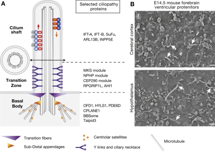
Fig. 1. Primary cilia and ciliopathy proteins.
A) Structure and composition of the primary cilium. The basal body (BB), made of nine microtubule triplets, matures from the mother centriole of the cell and anchors to the cell membrane via transition fibres (TF). The axoneme consists of nine microtubule doublets (see cross-section). Intraflagellar transport (IFT) com-plexes transport cargoes to the tip of cilia and back with their molecular motors, Kinesin II (anterograde transport, IFT-B complex) and cytoplasmic dynein (retrograde transport, IFT-A complex). At the proximal side of the axoneme, the transition zone is characterized by Y-shaped links that link the axoneme with the ciliary membrane at the level of the ciliary necklace. The distal basal body and the transition zone together form the ciliary gate, which controls the entry and exit of ciliary components such as cytoplasmic and membranes proteins as well as lipids via the ciliary localization of INPP5E (Garcia-Gonzalo et al. 2011; Reiter et al. 2012; Gonçalves and Pelletier 2017). The mature basal body is also decorated by subdistal appendages, which link microtubules along which centriolar satellites transport proteins toward the basal body. The MKS modules includes MKS1, CC2D2A, B9D1–2, TCTN1-3, TMEM67-107-216-231–237, the NPHP module includes NPHP1 and NPHP4, and the CEP290 module is made of CEP290 and NPHP5. These complexes interact with other proteins such as RPGRIP1L, AHI1, IFT proteins and the BBSome (Gonçalves and Pelletier 2017). The BBSome is a protein complex involved in trafficking toward and within cilia, whose components are mutated in BBS. B) Scanning electron microscopy of cilia on E14.5 forebrain ventricular progenitor cells. Cilia length is typically 1 μm in the cortex and 2 μm in the hypothalamus.
essential for cilium assembly while defects in retrograde transport (back to the cell body) lead to longer and/or bloated cilia with impaired function (Pazour et al., 2000; Follit et al., 2006; Tsao and Gorovsky, 2008; Iomini et al., 2009; Ishikawa and Marshall, 2017). Cilia are typically classified into motile and primary (non-motile) types. Motile cilia and related ciliopathies have been reviewed elsewhere (Horani et al., 2016; Mitchison and Valente, 2017; Spassky and Meunier, 2017) and will not be covered here. Primary cilia are present in most verte-brate cells where they act as cellular antennae that receive and trans-duce a variety of chemical, mechanical and light signals. In vertebrates, primary cilia are implicated in the transduction of several signaling pathways essential during development and perturbed in genetic disease and/or cancer, such as the Hh (see below), Wnt (Gerdes and Katsanis, 2008; Wallingford and Mitchell, 2011), PDGFa (Schneider et al., 2005), Notch (Ezratty et al., 2011), Hippo (Habbig et al., 2011) and mTOR (Boehlke et al., 2010; Foerster et al., 2017) pathways. In cultured cells, ciliogenesis is usually associated with quiescence, but in embryos, pri-mary cilia are also present on cycling progenitor cells, and cilium as-sembly and disasas-sembly are tightly coupled to the cell cycle (Plotnikova et al., 2009; Izawa et al., 2015). Moreover, cilia control proliferation and cell cycle length through their modulation of signaling pathways such as Wnt and Hh (Chizhikov et al., 2007; Spassky et al., 2008; Han et al., 2008; Lancaster et al., 2011; Tong et al., 2014).
Primary cilia in the developing central nervous system are present on progenitor cells as well as differentiated neurons. On ventricular pro-genitor cells (Fig. 1), they are typically 1–3 μm-long depending on the
region (Besse et al., 2011; Andreu-Cervera et al., 2019), are anchored to the apical membrane and point into the cerebrospinal fluid-filled ven-tricles, rich in morphogens and growth factors such as Shh, Fgf2 and IGF (Sawamoto et al., 2006; Huang et al., 2010; Lehtinen and Walsh 2011;
Lehtinen et al., 2011). During asymmetric division in the mouse cerebral cortex, cilia regrow on the lateral membrane of progenitor cells committed to delamination (Wilsch-Braüninger et al., 2012), which removes them from the ventricular surface and allows them to receive and transduce signals from neighbouring cells. In the neuroepithelium of the chick and mouse spinal cord, new-born neurons undergo abscission of their apical membrane, leading to abrupt loss of the cilium and of canonical (Gli-mediated) Hh signaling (Das and Storey 2014; Toro-Tapia and Das, 2020). Thus, the dynamics of cilia in neural progenitor cells is tightly linked to their division mode and fate and appears essential for signal reception and behavior. Cilia on differentiated neurons have been shown to regulate neuronal migration (Baudoin et al., 2012; Higginbo-tham et al., 2012; Stoufflet et al., 2020) and axon tract formation (Guo et al., 2019), processes that will not be reviewed here.
20 years after the seminal discovery that mutations in the same IFT gene, Polaris/IFT88, lead to loss of flagella in the green algae Chlamy-domonas reinhardtii and to polycystic kidney disease (PKD) in mice (Pazour et al., 2000), the involvement of cilia dysfunctions in a variety of human inherited diseases is now widely recognized. Defects found in these diseases, the ciliopathies, include renal disease (PKD or neph-ronophthisis), retinal degeneration, hearing loss, anosmia, laterality defects, intellectual disability, obesity, polydactyly, skeletal abnormal-ities and brain malformations (Mitchison and Valente 2017; Reiter and Leroux 2017). Brain developmental anomalies are found in severe cil-iopathies, such as Joubert syndrome (JBTS) and/or Meckel-Grüber syndrome (also called Meckel syndrome, MKS). JBTS is defined by a characteristic cerebellar and brainstem malformation including cere-bellar vermis hypoplasia, thick and horizontally oriented superior cerebellar peduncles and a deep interpeduncular fossa, recognizable on axial brain MRI as the “Molar Tooth Sign”. JBTS patients present as infants with hypotonia, abnormal eye movements, respiratory control disturbances, and as children or adults, ataxia and/or cognitive impairment (Bachmann-Gagescu et al., 2020). MKS is a lethal devel-opmental syndrome characterized by occipital encephalocele, cystic kidneys, hepatic ductal plate malformation with fibrosis and cysts. Postaxial polydactyly is commonly found, as well as brain
malformations (Hartill et al., 2017). More than 35 genes have been involved in JBTS, with 6 major genes, AHI1, CC2D2A, CEP290, CPLANE1, KIAA0586 and TMEM67 and more that 15 in MKS, with extensive genetic overlap between these two syndromes (Mitchison and Valente 2017). Our laboratory is particularly implicated in the study of one of these severe ciliopathy genes, RPGRIP1L (also called MKS5, NPHP8, JBTS7 and FTM). Mutations in RPGRIP1L produce MKS (OMIM 611561) and JBTS type B (OMIM 213300) (Arts et al., 2007; Delous et al., 2007; Brancati et al., 2008). The Rpgrip1l protein is part of the ciliary gate and has additional roles in proteasome activity at the cilium base and in autophagy (Mahuzier et al., 2012; Reiter et al., 2012; Ger-hardt et al., 2015; Shi et al., 2017; Struchtrup et al., 2018; Wiegering et al., 2018).
Ciliopathies can be associated with impairment of ciliogenesis, ciliary trafficking, ciliary signaling or sensory reception. Interestingly, the transition zone seems to be a hotspot of ciliopathy proteins. Tran-sition zone proteins form three main modules, called the MKS, NPHP and CEP290 modules (Fig. 1), grouped according to their physical and genetic interactions in different model systems. They are composed of soluble and membrane-associated proteins that collaborate for the as-sembly and gating function of the transition zone (Gonçalves and Pel-letier 2017). For instance, the transition zone protein RPGRIP1L is essential for the localization of many other transition zone proteins (Reiter et al., 2012; Shi et al., 2017; Wiegering et al., 2018). Other severe ciliopathy proteins are localized at the axoneme and involved in ciliary signaling, such as ARL13B, INPP5E or PDE6D (Reiter and Leroux 2017) (Fig. 1). Mutations affecting basal body proteins also lead to ciliopathies. For instance, Oro-Facial-Digital syndrome type 1 (OFD1, OMIM 311200), a X-linked dominant ciliopathy with frequent brain malfor-mations, results from mutations in the OFD1 gene, encoding a basal body and centriolar satellite protein involved in the regulation of ciliogenesis (Ferrante et al., 2006; Macca and Franco 2009; Lopes et al., 2011; Singla et al., 2010) (Fig. 1). Mutations in components of the distal region of the basal body, such as HYLS1, produce hydrocephaly and brain defects (Finnish hydrolethalus syndrome, HLS) or JBTS (Mee et al., 2005; Oka et al., 2016). Of note, most of these ciliopathy proteins can be found at several locations depending on the cell type or cell state, such as the transition zone, basal body, cilium shaft, centriolar satellites, or cell junctions (Tang et al., 2013; Gonçalves and Pelletier, 2017; Choi et al., 2019), which may in part explain the diversity and complexity of their functions and the broad phenotypic spectrum linked to their dysfunctions.
2.2. Cilia and Hh/Gli signaling
The role of primary cilia in Hh signaling was first discovered thanks to a genetic screen for mutant mice with defects in neural tube patterning performed in Kathryn Anderson’s laboratory (Huangfu et al., 2003). Several mutations in IFT genes identified in this screen led to a reduction of ventral neural progenitor subtypes in the spinal cord, as well as reduced expression levels of Hh target genes. Following this seminal discovery, further studies have shown that Hh pathway proteins traffic in the cilium and that Hh signal transduction in the receiving cell requires cilium integrity (Ribes and Briscoe 2009; Bangs and Anderson 2017) (Fig. 2). In the receiving cell, the Hh pathway comprises its membrane receptor Patched (Ptch) of which Ptch1 is the main member in vertebrates, and the G protein coupled receptor family protein Smoothened (Smo) that initiates intracellular signaling. The effectors of Hh signaling are a family of zinc finger proteins, the Gli transcription factors (Gli1-3 in mammals). In the absence of ligand, Ptch1 is at the base of cilia and inhibits the activity of Smo. The binding of the Shh ligand to Ptch1 relieves the inhibition of Smo, leads to its translocation into the cilium and induces the activation of the pathway. In the absence of Shh ligand, Gli2 and Gli3 are subject to a cascade of post-translational modifications initiated by phosphorylation by Protein Kinase A (PKA), which leads to their targeting to the proteasome. Gli2 is mainly
Neurobiology of Disease 150 (2021) 105236
degraded by the proteasome, while Gli3 is cleaved into a short form with strong transcriptional repressor activity (Gli3R). In the presence of Hh ligand, Gli2 is transformed through several post-translational modifi-cations into a strong transcriptional activator (Gli2A), which activates the expression of target genes, among which Ptch1 and Gli1. Gli1 is a constitutive activator, whose expression is enhanced by the Hh pathway. Many components of the Hh pathway traffic in and out of the cilium in a Hh-dependent manner (Fig. 2A). In the absence of ligand, the Ptch1 receptor localizes in and around of the cilium and prevents translocation of Smo into the cilium. The Gli transcription factors (GliFL (full length)) traffic in the cilium in a complex with SuFu, a chaperone that stabilizes them and maintains them in an inactive complex. The GPR161 mem-brane protein is in the cilium and activates adenylyl cyclases, which in turn increases cAMP concentration in the cilium and activates Protein kinase A (PKA) (Mukhopadhyay et al., 2013). PKA initiates a cascade of post-translational modifications (phosphorylation and ubiquitination)
that promotes the proteasome-dependent processing of the Gli tran-scription factors Gli2 and Gli3 at the cilium base. The truncated forms of these proteins (GliR) translocate to the nucleus and repress the tran-scription of Hh target genes. In the presence of Shh ligand, Shh binding to its Ptch1 receptor removes Ptc1 from the cilium and allows Smo to translocate into the cilium. The activation of Smo leads to the removal of GPR161 (Pal et al., 2016) and to an accumulation of full length Gli (GliFL) at the tip of cilia, allows their dissociation from SuFu, inhibits their proteolytic processing and initiates a poorly understood cascade of events to produce activated forms (GliA), which activate target gene expression in the nucleus. Kif7 is a kinesin that regulates microtubule dynamics at the cilia tip, thus restricting the length of the axoneme (He et al., 2014, 2017). As a consequence, it accumulates signaling at the cilia tip, including the Gli-SuFu complex. Interestingly, in addition to being negative regulators of the Hh pathway, SuFu and Kif7 are ciliop-athy proteins, further stressing the intricate relationship between cilia
Fig. 2. Cilia, Hh signaling and neural tube patterning.
A simplified scheme of Hh pathway transduction in ciliated cells, in the absence (a) or in the presence (b) of Hh ligand. Adapted from Ribes and Briscoe (2009). B) Hh in neural tube patterning. Shh (purple dots) is first produced by the notochord (NC, purple), a mesodermal structure underlying the neural plate. Hh response in the neural tissue leads to the activation of Shh expression in the floor plate. Graded Shh signaling then specifies five ventral progenitor domains ventro-dorsally, p3, pMN, p2, p1 and p0 (Stamataki et al. 2005). Low levels of Shh also repress the expression of genes that specify dorsal progenitor domains (dp) (Dessaud et al. 2010; Sasai and Briscoe 2012). C) Schematics of cell specification defects in Hh pathway and ciliary/ciliopathy gene mutant mice. The progenitor types are color-coded as shown in C. Colors are the same for B and C.
and Hh signaling (see Kopinke et al., 2020 for a comprehensive review of the roles of cilia in Hh signaling).
In sum, cilia are essential for two aspects of Hh/Gli signaling that normally occur at very different levels of pathway activity (Fig. 2A). They are required for the formation of activator forms of Gli transcrip-tion factors, GliA (Gli2A and Gli3A) normally achieved in the presence of high levels of Shh ligand. They are also required for the proteasomal processing of Gli into a repressor form, GliR (mainly Gli3R), which is normally present in the absence of Hh ligand. These dual and opposite roles of primary cilia in Hh/Gli signal transduction are key to under-stand the central nervous system patterning defects in ciliary mutant mice, as well as the forebrain defects found in ciliopathies, as discussed below.
In the mouse, the role of Shh as a morphogen to specify cell fate has been extensively studied in the spinal cord (Ribes and Briscoe 2009;
Sasai and Briscoe 2012). Shh is expressed first in the underlying noto-chord and is then activated in the floor plate of the neural tube in a Hh pathway-dependent manner. In the ventral neural tube, Shh forms a ventral-to-dorsal decreasing gradient, which eventually determines the expression domains along the dorso-ventral axis of a series a transcrip-tion factor genes. In additranscrip-tion to this spatial gradient, a temporal gradient of Hh signaling is also involved, more ventral neuronal progenitor cells receiving longer exposure to the ligand, which leads to higher pathway activity (Dessaud et al., 2007). A single progenitor may thus undergo temporal fate changes in response to levels and duration of Hh signals it receives. In receiving progenitor cells, the Shh gradient is transformed into opposite gradients of GliA (high ventrally) and GliR (high dorsally). Gli transcription factors bind the cis-regulatory domains of their target genes and control their expression. Thus, the spatial expression pattern of a target gene depends on the GliA/GliR ratio and on the affinity of its cis-regulatory domains for Gli factors. The final neural progenitor do-mains are established through multiple cross-repressive interactions between the transcription factors, whose expression controls progenitor cell fate (Balaskas et al., 2012; Briscoe and Small 2015 for a compre-hensive review). In the ventral spinal cord, graded response to Hh signaling induces different ventral progenitor fates, from p3 ventrally to p0, and represses dorsal progenitor fates (Fig. 2B). In the absence of Shh or Smo, the ventral fates are not specified and dorsal fates are expanded ventrally. In contrast, in the absence of functional Ptch1 or SuFu, the pathway is constitutively activated leading to a dorsal expansion of the floor plate, P3 and/or pMN domains (Jeong and McMahon 2005; Hwang and Mukhopadhyay 2015) (Fig. 2C).
Mutant mice that lack cilia show defects of intermediate severity, with a variable loss of ventral spinal cord domains (Fig. 2C) (Sasai and Briscoe 2012; Hwang and Mukhopadhyay 2015). In mutant mice with impaired anterograde transport, such as Kif3a, Ift172 and Ift88, which lack cilia in spinal cord progenitor cells, the p3 domain that requires high levels of GliA activity is lost, while the pMN (motoneuron pro-genitor cells) domain is reduced and ventrally displaced (Goetz et al., 2009). The phenotype of these IFT mutant embryos is similar to that of embryos lacking both Gli2 and Gli3. A similar phenotype is found in mice mutant for genes encoding components of the basal body or tran-sition zone (such as Talpid 3 and Rpgrip1l, respectively). (Davey et al., 2006; Vierkotten et al., 2007; Bangs et al., 2011). In contrast, other mutant mice with abnormal morphology of primary cilia present a partial overactivation of Shh signal transduction. For example, mouse mutants for the gene encoding the GTPase Arl13b lack p3 cells but show a strong dorsal expansion of pMN cells (Caspary et al., 2007; Sasai and Briscoe 2012). Interestingly, mouse mutants of genes involved in retrograde IFT (IFT139, IFT122, Dync2h1) fall in either of the two cat-egories, illustrating the complexity of the relationship between cilia and Shh signaling and the multiple functions of individual ciliary proteins (Goetz et al., 2009; Sasai and Briscoe 2012) (Fig. 2C). In the brain, cilia also pattern ventral neural structures via Hh signaling (Gazea et al., 2016 and see below for the forebrain).
2.3. Early development and patterning of the forebrain
The brain arises from a sheet of epithelial cells, the neural plate, which then folds to form the neural tube, a process called neurulation. During neurulation, the anterior central nervous system undergoes drastic changes, generating, by differential proliferation, the three pri-mary vesicles of the brain: in caudo-rostral order, the hindbrain (or rhombencephalon) midbrain (mesencephalon) and forebrain (prosen-cephalon). The forebrain is further subdivided into the diencephalon, the hypothalamus, the eye primordium and the telencephalon (Marin and Rubenstein, 2002). The early embryonic forebrain organization is not reflected in an obvious manner in the anatomy and histology of the adult brain, particularly in humans with the spectacular expansion of the cerebral cortex derived from the telencephalon. Data accumulated in the last 30 years or so, from anatomical observations, fate mapping, gene expression and functional studies in many vertebrate species, led to a reconsideration of the antero-posterior and dorso-ventral axes of the forebrain and of the topological relationships between its different do-mains. The prosomeric model, initially developed by Puelles and Rubenstein, offers a coherent theory to describe forebrain development (Inoue et al., 2000; Puelles 2001; Puelles and Rubenstein, 2003; Puelles et al., 2013). Primary postulates of this theory are that 1) the topological organization of the early forebrain primordium is circular and not bilateral like more caudal brain regions (Fig. 3A). This is essential to understand how different domains of the forebrain primordium are influenced by mediolateral (and later dorsoventral) external signals. 2) during development the differential growth of different brain regions leads to bending of the antero-posterior axis (Fig. 3B); and 3) the fore-brain can be subdivided into its antero-posterior axis into distinct developmental units called “prosomeres” (Fig. 3A, B). Thus, in terms of embryonic origin, if not in adult position, the rostral extremity of the forebrain lies in the hypothalamus. The thalamus (formerly dorsal thalamus), prethalamus (formerly ventral thalamus) and hypothalamus are not aligned dorso-ventrally as suggested by their former names but caudo-rostrally (Fig. 3C). Moreover, the telencephalon is not anterior to the hypothalamus but is dorsal to it (Fig. 3B, C). The telencephalon is itself subdivided into the pallium (dorso-caudally) and the subpallium (ventro-rostrally). While continuously refining, the prosomeric model in its outline is widely corroborated by the studies mentioned above and is a key paradigm to understand forebrain development and congenital malformations.
During development, forebrain patterning relies on a series of signaling centers called secondary organizers. They secrete diverse signaling molecules or morphogens that act at a distance and in a concentration-dependent manner to organize cell fate and cell prolif-eration in adjacent territories. The earliest organizing centers along the antero-posterior axis of the brain are the anterior neural ridge (ANR) at the rostral end of the anterior neural tube and the isthmic organizer at the midbrain-hindbrain boundary (Fig. 3A, B) (Vieira et al., 2010). The ANR is a source of Fibroblast growth factors (Fgf), principally Fgf8. It is essential for the regionalization of the telencephalon (Shimamura and Rubenstein 1997; Ye et al., 1998). Fgf8 is required for the expression of FoxG1, encoding a transcription factor important for cell proliferation in the telencephalon (Xuan et al., 1995; Shimamura and Rubenstein 1997;
Ye et al., 1998). In the forebrain as in more caudal regions, dorso-ventral patterning relies on a ventral Shh-positive organizing center and a dorsal center expressing BMPs and Wnts. According to the circular organiza-tion of the forebrain, antero-posterior and dorso-ventral organizing centers abut and closely interact with each other. In close proximity to the ANR, Shh is required for the specification of the subpallium, mainly through the inhibition of the formation of Gli3R (Rallu et al., 2002). The telencephalic dorsal midline (including the roof plate and the cortical hem) expresses Wnt3a and BMPs and is essential for patterning the medial pallium and the cortex. All these signaling centers interact with one another to set up cell proliferation, neurogenesis and cell fate in the telencephalon (H´ebert and Fishell, 2008).
Neurobiology of Disease 150 (2021) 105236
2.4. The Shh pathway in forebrain development and disease
The use of mouse models has been instrumental in dissecting the roles of the Hh pathway in different regions and at different stages of forebrain development. Null mouse mutants for Shh display a severe HPE phenotype including cyclopia and proboscis (Chiang et al., 1996) and studies involving gene inactivation in mouse, lineage tracing, and loss- and gain-of-function approaches in chick identified multiple,
successive functions of the Hh pathway in the diencephalon, hypothal-amus and eyes (Furimsky and Wallace 2006; Szab´o et al., 2009a, 2009b;
Vue et al., 2009; Jeong et al., 2011; Alvarez-Bolado et al., 2012; Haddad- T´ovolli et al., 2012, 2015; Blaess et al. 2015; Zhang and Alvarez-Bolado, 2016). In mouse embryos, Shh is initially expressed from E7.5 in axial tissues underlying the neural plate, notochord posteriorly and pre-chordal plate anteriorly (Fig. 3A), where it signals to the overlying neural plate to induce ventral structures. Shh signaling induces Shh
Fig. 3. The developing mouse forebrain.
A) The neural plate at E8-E8.5 indicating the fate map of the forebrain (Inoue et al. 2000) and the position of the organizing centers: the FGF8-expressing ANR and the SHH-expressing medial signaling center. B) E9.5-E10.5. Major forebrain subdivisions. Shh source (in red), Wnts/BMPs (in green) and FGF (in blue) sources in the forebrain. Shh is expressed in the ZLI, basal plate and/or floor plate of the diencephalon and hypothalamus. Wnts and FGFs are expressed dorsal to the ZLI and in the roof plate. C) Representation of E13.5 parasagittal forebrain section showing its different subdivisions: the telencephalon, hypothalamus and diencephalon. The telencephalon can be divided along the dorso-ventral axis in two main regions, the pallium and the subpallium. The pallium is located in the most dorsal part and is separated by the pallio-subpallial boundary from the subpallium. Along the dorso-ventral axis, the brain is divided into four main regions: the floor plate (FP), the basal plate (BP), the alar plate (AP) and the roof plate (RP). ANR: anterior neural ridge; ANT: anterior hypothalamus; CX: cerebral cortex; DI: diencephalon; HYP: hypothalamus; LGE: lateral ganglionic eminence; MAM: mammillary area; MES: mesencephalon; MGE: median ganglionic eminence; MHB: midbrain-hindbrain boundary; NT: notochord; OB: olfactory bulb; OV: optic vesicle; P1: prosomere 1 (future pretectum); P2: Prosomere 2 (future thalamus); P3: Prosomere 3 (future prethalamus); PAL: pallium; POA: pre-optic area; PP: prechordal plate; PSB: pallial-subpallial boundary; PT, pretectum; PTH, prethalamus; RH: rhombencephalon (hindbrain); TA: tegmental areas; TUB: tuberal hypothalamus. TEL: telencephalon; TH, thalamus; ZLI: zona limitans intrathalamica. Adapted from Marin and Rubenstein, 2002.
expression in the ventral forebrain from E8.0 onward (Dale et al., 1997;
Alvarez-Bolado et al., 2012). At E9.0-E9.5, Shh is expressed in the floor plate of the diencephalon. In the hypothalamus, Shh is down-regulated in the most ventral region and activated in two lateral stripes in the basal plate (Fig. 3B, C) (Szab´o et al., 2009a; Alvarez-Bolado et al., 2012;
Blaess et al., 2015). From E10 onward, Shh expression is progressively activated in a dorso-ventral stripe in the diencephalon, between P2 and P3 (Fig. 3B, C, D). This stripe of Shh-expressing cells, called the Zona Limitans Intrathalamica (ZLI), is fully formed at E12.5 and organizes cell fate in the thalamus and prethalamus (Epstein 2012; Zhang and Alvarez- Bolado 2016) (Figs. 3A-D). Shh is also activated in the region of the preoptic area - telencephalic septum, where it counteracts Gli3 function in the telencephalon (Rallu et al., 2002) (Fig. 3C, D), The dynamic and complex expression pattern of Shh explains its stage and region-specific functions in forebrain development (Alvarez-Bolado et al., 2012, Blaess et al., 2015).
In humans, HPE is the main developmental disease associated to SHH pathway perturbation. It is a complex malformation characterized by a lack or a partial defect of cleavage of the forebrain along its ventral and dorsal midlines. In addition to impaired brain septation, defects include cleft lip and palate, single maxillary incisor, and eye separation phe-notypes ranging from hypotelorism to cyclopia. It is observed in 1/ 10,000 to 1/20,000 live births (Mercier et al., 2011) and 1/250 termi-nated pregnancies (Cohen Jr., 1989). Several anatomical forms have been described according to the extent and severity of the malforma-tions. Classically recognized forms are alobar HPE in which the telen-cephalon is a totally noncleaved holosphere, semi-lobar forms with the presence of an inter-hemispheric fissure only at the posterior pole, and lobar forms in which cerebral hemispheres are completely separated but a bridge of cerebral cortex connecting the two sides and passing over the corpus callosum is present. Other forms have been described more recently (Barkovich and Quint 1993; Lewis et al., 2002; Simon et al., 2002).
HPE presents different transmission patterns and is in a proportion of the cases associated with chromosomal abnormalities (Mercier et al., 2011; Dubourg et al., 2018). SHH was the first causal gene identified in HPE (Roessler et al., 1996). Heterozygous mutations in more than 17 genes have now been identified as causing HPE, four of which are considered as major causal genes: SHH, ZIC2, SIX3 and TGIF, which in total amount to 25% of the known HPE cases (Mercier et al., 2011). SHH mutations are the most frequent (10% of the cases in a European cohort) (Mercier et al., 2011). Other members of the Hh pathway are also mutated in HPE: GLI2, PTCH1 (probably dominant active mutations), DISP1 (regulation of Hh ligand secretion and internalization), GAS1 and CDON (membrane proteins that act with Ptch1 and positively regulate Hh signaling). Mouse studies point to a prime involvement of defects in Shh expression and signaling in the forebrain. According to these studies, the pathogenicity of the mutations in the three major HPE genes, outside Shh, are explained by the role of these genes in Shh expression or pathway activity in the forebrain. Zic2 is essential for the formation of the Shh-expressing prechordal plate (Warr et al., 2008); Six3 directly regulates Shh expression in the forebrain (Geng et al., 2008; Jeong et al., 2008); Tgif1 has an indirect action on Shh signaling by regulating Gli3 and Nodal independently (Taniguchi et al., 2012, 2017). Thus, the comparative study of HPE in humans and in mouse models shows that the importance of Hh signaling in forebrain morphogenesis is conserved in mammals. Of note, only heterozygous mutations are found in human HPE, suggesting that homozygous mutations would lead to very early death of the embryo.
Genetic evidence also shows a major role of Gli3 in forebrain patterning, both in mice and humans. In humans, two main syndromes with brain anomalies are linked to heterozygous mutations in GLI3: Greig cephalopolysyndactyly syndrome (OMIM 175700) and Pallister- Hall syndrome (OMIM 146510). Greig cephalopolysyndactyly includes macrocephaly with frontal bossing, hypertelorism associated with pol-ysyndactyly and may be associated to different mutations throughout
the GLI3 gene including whole gene deletions (Biesecker 2008). Pallister-Hall syndrome comprises hypothalamic/diencenphalic hamartomas, pituitary dysfunction and central polydactyly. Most of the mutations responsible for Pallister-Hall syndrome occur near the middle of the GLI3 gene and lead to a truncated Gli protein with constitutive repressor activity (Johnston et al., 2005; Naruse et al., 2010). Of note, somatic mutations in GLI3 as well as in the PRKACA gene encoding a subunit of PKA involved in production of Gli3R, were found in sporadic cases of hypothalamic hamartomas with gelastic epilepsy (Hildebrand et al., 2016). In mice, Gli3 is a central player in telencephalic patterning. The mouse extra-toes mutant (XtJ), considered as a null Gli3 mutant, has a severely reduced dorsomedial telencephalon, lacks olfactory bulbs (OBs) and presents an expanded subpallium, in particular in the most anterior region (Hui and Joyner 1993; Theil et al., 1999; Tole et al., 2000; Aoto et al., 2002; Kuschel et al., 2003). In addition, Gli3 controls the switch from symmetric proliferative to asymmetric neurogenic cell division of cortical radial glial cells (Hasenpusch-Theil et al., 2018). The dorsal telencephalon predominantly forms Gli3R and double [Shh−/−,
Gli3−/−] embryos show partial rescue of telencephalic patterning,
strongly suggesting that Gli3 acts mainly as a repressor in this structure (Rallu et al., 2002; Fotaki et al., 2006). Gli3 is also involved in hypo-thalamus, diencephalon and eye development, acting mostly as a repressor (Haddad-T´ovolli et al., 2012, 2015; Wiegering et al., 2019). While mouse Gli3 mutants have phenotypes similar to Greig cepha-lopolysyndactyly, a mouse mutant with a deletion of the Gli3 C-termi-nus, Gli3∆699, displays constitutive repressor activity and mimics PSH
(Hill et al., 2007).
3. Forebrain defects in ciliopathies and in mouse models of cilia dysfunction
Forebrain malformations are described in severe forms of cil-iopathies such as MKS and, to a lesser extent, in JBTS. Other cilcil-iopathies with forebrain anomalies include OFD1 syndrome, acrocallosal syn-drome (ACLS) and Finnish HLS, and more rarely Bardet-Biedl synsyn-drome (BBS) and short rib thoracic dysplasia (SRTD) syndrome. These fore-brain malformations include agenesis or hypoplasia of the olfactory bulbs (OB) (Fig. 4A) and corpus callosum (CC) (Fig. 4B), micro-phthalmia or coloboma, malformations of cortical development (Fig. 4C-F), hypothalamic or diencephalic hamartomas (Table 1). Hy-drocephalus and ventriculomegaly, which impact forebrain develop-ment, will also be reviewed, while malformations of other brain regions such as cerebellar hypoplasia and occipital encephalocele will not be discussed here. Old series of lethal ciliopathies usually involved cases observed late in gestation, allowing to perform a very detailed neuro-pathological examination. In more recent series, often focused on the identification of causal genes, severe cases led to early termination of pregnancy. In these cases, the central nervous system was either poorly preserved or very immature (with brain structures such as the cerebral cortex not yet developed), preventing precise description of malformations.
The observation of forebrain malformations in severe ciliopathies stresses the importance of studying their developmental origin in mouse mutants for ciliary and ciliopathy genes. Many mouse knock-out mu-tants for IFT genes die early, but forebrain defects are observed in conditional or hypomorphic mouse mutants, which display malformed cilia, such as the Ift88/cobblestone, Ift172/wimple and Ift139/Ttctb1 mutant mice (Willaredt et al., 2008; Gorivodsky et al., 2009; Stottmann et al., 2009). Forebrain defects are also observed in mouse mutants for the transcription factor Rfx3 that controls ciliary gene expression (Benadiba et al., 2012) and for Ofd1, a ciliogenesis regulator (D’Angelo et al., 2012). A thorough analysis of forebrain defects has been per-formed by our laboratory in the mouse Ftm mutant for the ciliopathy gene Rpgrip1l. Most Ftm−/− fetuses survive until birth, which allows the
precise analysis of forebrain development. Moreover, they display strongly reduced numbers of cilia in the neuroepithelium as soon as E8.5
Neurobiology of Disease 150 (2021) 105236
(Besse et al., 2011; Andreu-Cervera et al., 2019), validating this mutant for studying the role of cilia in forebrain morphogenesis. At the end of gestation, Ftm−/− fetuses show a severe MKS-like phenotype (Delous et al., 2007; Vierkotten et al., 2007). In the brain, they display agenesis of the corpus callosum (CC), OB agenesis, anophthalmia, a loss of the anterior pituitary, a reduction of the ventral hypothalamus and a disorganization of diencephalic nuclei and axonal tracts. Exencephaly is partially penetrant and dependent on the genetic background (Besse et al., 2011; Andreu-Cervera et al., 2019). In the next paragraphs we will detail these different anomalies and discuss their developmental and molecular origin. As we will see, most of them can be in a large part explained by perturbations in the GliA/GliR balance.
3.1. Malformations of the olfactory structures
Anomalies of the olfactory structures in ciliopathies can be separated into two main categories of different severities, olfaction disorders (anosmia, hyposmia) and agenesis of the olfactory bulbs (OB) (Fig. 4A). OBs are the first relay center for olfaction in the brain. They are composed of projection neurons (mitral and tufted cells) born in the OB primordium and of interneurons born in the subpallium and migrating into the OB. OBs receive the axons of olfactory sensory neurons located in the nasal olfactory epithelium, which contact mitral cell dendrites in glomeruli. Odorant receptors being localized on the cilia of olfactory sensory neuron dendrites, defects in cilia sensory function are expected to impair olfaction. Accordingly, functional olfaction disorders are constant in BBS patients (Kulaga et al., 2004; Iannaccone et al., 2005;
Brinckman et al., 2013; Braun et al., 2014) and Bbs mutant mice show a
decrease in behavioral and/or electrophysiological responses to olfac-tion (Kulaga et al., 2004; Nishimura et al., 2004; Tadenev et al., 2011). In these mice, cilia on olfactory sensory neurons are reduced in number and olfactory axons toward the OB display fasciculation defects, some-times leading to OB hypoplasia (Tadenev et al., 2011; Braun et al., 2016). In contrast, OB agenesis is only encountered in severe cil-iopathies (Table 1). It is a constant feature in the Finnish HLS, a genetically homogenous condition due to the mutation of the HYSL1 gene (Paetau et al., 2008). OB agenesis is also very frequent on several classical MKS series (Paetau et al., 1985; Ahdab-Barmada and Claassen, 1990). In these cases, OB agenesis is part of a more global malformation of the entire forebrain, including HPE and CC agenesis. OB agenesis has been also described in SRTD syndrome (Sharma et al., 1992; Alby et al., 2015). Individual associations between gene mutations and OB agenesis in ciliopathies are indicated in Table 1.
Understanding the causes of OB agenesis in severe ciliopathies re-quires a knowledge of OB developmental mechanisms. In mice, the OB primordia originate in the anterior part of the pallium, just above the subpallium (Cobos et al., 2001; Marin and Rubenstein, 2002) (Figs. 3 and 5). From about E12 they are morphologically distinguishable as anterior buddings of the neuroepithelium in each telencephalic hemi-sphere (Fig. 5). OB outgrowth is concomitant with the initiation of mitral cell differentiation and is thought to require a decrease in pro-genitor proliferation that occurs at this stage in the anterior telenceph-alon (Gong and Shipley 1995; Hebert et al., 2003). Axons of olfactory sensory neurons enter the telencephalon and make contacts with mitral cell dendrites in glomeruli (Fig. 5) (Whitesides and LaMantia, 1996;
Blanchart et al., 2006), and OB lamination and full outgrowth depend on
Fig. 4. Illustrations of forebrain defects in cases of severe ciliopathies.
A) Basal view of the brain of a fetus affected with MKS. Termination of pregnancy was performed at 22 weeks of gestation. Note bilateral absence of both olfactory tracts and bulbs underneath the frontal lobe (Front). The causal gene is not known for this case. Photography: courtesy of Dr. Catherine Fallet-Bianco (Montreal, Canada). B, C) Brain MRI of an adult female patient affected with OFD1 due to heterozygous mutation in exon 7 of the OFD1 gene (c.613_615delAGAinsTATAA). B shows a medio-sagittal section (T1-weighted sequence). The corpus callosum and the hippocampal commissure are lacking whereas the posterior (white arrow) and the anterior (white arrowhead) commissures are present. C is an axial section (T1-weighted sequence). Probst’s bundle (white arrow) is present on the right hemisphere and absent on the left hemisphere. The latter appears hypoplastic with ventricular dilation (asterisk) and curvilinear frontal heterotopia (yellow arrows). Martin Catala, unpublished observation. D) Brain MRI of an adult female patient affected with JBTS (COACH syndrome). The patient presents a compound het-erozygous mutation of KIAA0856 gene (c.428del, p.Arg143Lysfs*4 and c.1412 + 2 T > C). Axial section (T1-weighetd sequence). A small heterotopia lies in the hemispheric white matter (black arrow). Martin Catala, unpublished observation. E, F) Neuropathological examination of a fetus (25 weeks of gestation) affected with JBTS due to homozygous mutation of the RPGRIP1L gene (c.1864C > T, p.Gln622X). In E, an external view of the right hemisphere shows polymicrogyria predominating on the perisylvian region (white bracket). F presents the histological aspect of the polymicrogyric region showing the infolded cortex with fusion of the molecular layers (ML). Martin Catala and Martine Bucourt, unpublished observation.
this contact (Gong and Shipley 1995; Hirata et al., 2006). In terms of molecular mechanisms, several genes involved in dorso-ventral patterning of the telencephalon such as Gli3, Emx1/2 and Pax6 are required for OB formation and outgrowth (Dellovade et al., 1998;
LaMantia 1999; Bishop et al., 2003; Balmer and LaMantia 2004). Moreover, the OB forms in an anterior region close to the ANR source of FGF signals and to the frontonasal mesenchymal source of retinoic acid (RA), which are both required for OB formation (Anchan et al., 1997;
Garel et al., 2003; Hebert et al., 2003; Balmer and LaMantia 2005) (Fig. 5).
The developmental causes of OB agenesis in ciliopathies have been first elucidated in the Ftm/Rpgrip1l mouse mutant (Besse et al., 2011). In the anterior telencephalon of Ftm−/− fetuses at the end of gestation, OB
projection neurons and interneurons are present but show abnormal localization and disrupted layering, and the characteristic OB outgrowth is never detected (Fig. 5A). At E12.5, the first OB projection neurons are specified more dorsally and OB outgrowth does not occur. Olfactory sensory neurons differentiate normally and their axons reach, but do not enter, the telencephalon (Fig. 5). In these mutant embryos, the sub-pallium is expanded dorsally, whereas pallial structures are reduced, and the boundary between these two domains is fuzzier than in controls, with a mixing of subpallial and pallial cell fates at the level of the boundary (Fig. 5). Similar to the FgfR1 conditional mutant (Hebert et al., 2003), E12 Ftm−/− embryos do not show the decrease in progenitor
proliferation that normally occurs in the anterior telencephalon (Besse et al., 2011). The defect in OB outgrowth may have several non- exclusive causes: i) a position of the OB primordium further away from the FGF8 source in the ANR required for its outgrowth, ii) a dispersion of the OB cells due to cell mixing at the subpallial-pallial boundary, and iii) the absence of contact with the olfactory sensory axons (Fig. 5B, C). In conclusion, OB agenesis of Ftm mutant mice does not result from a cell fate specification defect but from a morphogenesis defect (Besse et al., 2011).
The phenotype of the Ftm−/− mouse is very reminiscent of that of
Gli3 mutant mice, which display OB agenesis and expansion of the subpallial telencephalon at the expense of the pallium (Theil et al., 1999;
Aoto et al., 2002; Kuschel et al., 2003; Besse et al., 2011). In addition, Gli3R amount is severely reduced in Ftm−/− embryos, suggesting that
this reduction could be the cause of OB agenesis. To test this hypothesis, we crossed the Ftm mutant with the Gli3∆699 mutant (Besse et al., 2011).
Table 1
Most frequent forebrain malformations in human ciliopathies.
Forebrain malformation Ciliopathy Selected genes References OB agenesis /
arhinencephaly MKS MKS1, CEP290, CC2D2A/MKS6 Paetau et al. (1985); Ahdab- Barmada and Claassen (1990); Paetau et al. (2008); Khaddour et al. (2007); Baala et al. (2007); Mougou-Zerelli et al. (2009). HLS/
SRTD HYLS1, KIAA0586 Paetau et al. (2008), Sharma
et al. (1992), Alby
et al. (2015),
CC agenesis ACLS KIF7 Putoux et al. (2011)
HLS HYLS1 Paetau et al. (2008)
OFD OFD1, TCTN3, CPLANE1, KIAA0753 Bisschoff et al. (2013); Del Giudice et al. (2014); Thomas et al. (2012)a; Lopez et al. (2014); Chevrier et al. (2016) MKS MKS1, TMEM67, CC2D2A Ahdab-Barmada and Claassen (1990); Khaddour et al. (2007); Szymanska et al. (2012); Mougou- Zerelli et al. (2009)
JBTS CC2D2A, KIF7 Gorden et al.
(2008); Dafinger
et al. (2011)
SRTD KIAA0586 Alby et al. (2015)
Holoprosencephaly MKS MKS1 Fried et al. (1974);
Paetau et al. (1985); Ahdab- Barmada and Claassen (1990); Auber et al. (2007). Microcephaly and malformations of cortical development (heterotopia, polymicrogyria, lissencephaly,) MKS Paetau et al. (1985); Ahdab- Barmada and Claassen (1990).
SRTD KIAA0586 Alby et al. (2015)
OFD OFD1, CPLANE1, C2CD3, TMEM107 Poretti et al. 2011; Darmency- Stamboul et al. 2013; Del Giudice et al. 2014; Lopez et al. (2014); Lambacher et al. (2016); Boczek et al. (2018).
HLS HYLS1 Paetau et al. (2008)
JSRD TMEM216 Valente et al. (2010)
JBTS AHI1, ARMC9 Dixon-Salazar et al.
(2004); Giordano et al., 2009; Poretti et al., 2011; Van De Weghe et al. (2017). Anophthalmia, Microphthalmia, coloboma MKS nd RPGRIP1L, TMEM216, TCTN2, TMEM67, CEP290, Ahdab-Barmada and Claassen (1990) Delous et al. (2007); Valente et al. (2010); Baala Table 1 (continued)
Forebrain malformation Ciliopathy Selected genes References
et al. (2007);
Shaheen et al.
(2011); Szymanska
et al. (2012)
SRTD KIAA0586,
CEP120, INTU Alby et al. (2015)Roosing et al. ;
(2016); Toriyama
et al. (2016)
HLS nd Kivel¨a et al. (1996)
JBTS CSPP1, PDE6D,
TOGARAM1 Shaheen et al. (2014); Thomas
et al. (2014);
Latour et al. (2020)
OFD C2CD3,
CPLANE1 Lopez et al. (2014)Boczek et al. (2018) ;
Hypothalamic/ diencephalic hamartomas MKS CC2D2A Ahdab-Barmada and Claassen (1990); Roume et al. (1998); Mougou-Zerelli et al. (2009).
HLS HYLS1 Paetau et al. (2008)
JBTS nd Poretti et al. (2011)
OFD OFD1,
CPLANE1 Del Giudice et al. (2014); Lopez et al.
Neurobiology of Disease 150 (2021) 105236
Gli3∆699 is an allele of Gli3 in which only the N-terminal repressor form
is present and thus acts as a constitutive Gli3R (Hill et al., 2007). [Ftm−/ −, Gli3∆699+] fetuses had a spectacular restoration of OBs, and this despite the absence of cilia (Besse et al., 2011), demonstrating that the OB agenesis found in Ftm−/− fetuses is mainly due to the reduction in
Gli3 repressor activity. This experiment provided the first in vivo demonstration that the role of cilia in forebrain patterning is mainly mediated by the production of Gli3R. Interestingly, OB agenesis and ventralization of the telencephalon are also found in the Alien (aln) mutant, a mouse mutant in the Tct21b/IFT139 gene encoding an IFT A
protein involved in retrograde transport (Stottmann et al., 2009), and in a conditional mutant in the telencephalon of Sufu, a negative regulator of the Hedgehog pathway (Yabut et al., 2015). Like Ftm, Tct21baln shows an increase Gli3FL/GLI3R ratio. However, unlike Ftm, it shows increased Shh/Gli signaling and an upregulation of Shh target genes in the spinal cord and brain. Interestingly, OB agenesis is rescued in [Tct21baln/aln, Shh+/−] embryos, confirming the altered GliA/R balance as a prime
cause of OB agenesis in ciliary mutant mice.
In conclusion, the study of mouse ciliopathy models indicates that OB agenesis is caused by a defect in Gli3R production. Interestingly, the observation of OB defects in human syndromes resulting from GLI3 mutations, Greig cephalopolysyndactyly and Pallister-Hall syndrome (Subramanian et al., 2019), suggests that mechanisms of OB develop-ment are conserved between mice and humans. Thus, one may propose a reduction in Gli3R levels as a pathogenic mechanism of OB agenesis observed in severe ciliopathies.
3.2. Agenesis of the Corpus callosum
Complete or partial CC agenesis is frequently reported in ciliopathies (Table 1; Fig. 4B and C). By definition, callosal anomalies are present in ACLS. Patients with a mutation in KIF7, the first gene associated with this syndrome, have either complete CC agenesis or hypoplasia (Putoux et al., 2011). CC agenesis is almost always observed in the HLS (Paetau et al., 2008). It is found in more than 80% of the patients suffering from OFD1 and presenting neurological features (Bisschoff et al., 2013; Del Giudice et al., 2014). In MKS, CC agenesis is less frequent (Table 1). However, the analysis of the CC is not always possible in fetuses with either anencephaly or large occipital encephalocele, particularly if the latter is broken during abortion (Delous et al., 2007). CC agenesis has also been reported in JBTS and in OFD6 (due to mutations in the CPLANE1 gene) (Table 1).
Mouse models have been instrumental in determining the critical steps of CC development. These include the specification of callosal neurons in the cortex, the guidance of their axons across the midline to their final target in the contralateral hemisphere and the early patterning of the commissural plate, the portion of the telencephalic midline that provides guideposts for the guidance of commissural axons (Fig. 6). Guideposts are made of populations of neurons and glia, including the indusium griseum (IG), the glial wedge (GW), the sub-callosal sling (SCS) and the midline zipper glia (MZ), strategically located above and below the CC to channel the growing callosal axons across the midline (Lindwall et al., 2007; Donahoo and Richards, 2009;
Edwards et al., 2014; Su´arez et al., 2014) (Fig. 6). Disruption of any of these processes can result in CC agenesis or dysgenesis.
The origin of CC agenesis has been investigated in the Rfx3 mouse mutant, which exhibits several hallmarks of ciliopathies, consistent with the functions of RFX transcription factors in ciliogenesis (Thomas et al., 2012b). Rfx3-deficient mice harbour aberrant distribution of guidepost cells, associated with a mild ectopic expansion of Fgf8 expression and an increased Gli3FL/Gli3R ratio (Benadiba et al., 2012). Interestingly, FGF and Gli3 have both been shown to be essential for CC formation (Amaniti et al., 2013; Magnani et al., 2014). The cause of CC agenesis was also analysed in the FtmRpgrip1l mutant (Laclef et al., 2015). Ftm−/−
mutant callosal neurons form normally and their axons reach the medial pallium where, instead of crossing the midline, they form dense ipsi-lateral “Probst” bundles or aberrant ventral projections (Fig. 6). Grafts of forebrain slice explant tissues show that the defect lies in the midline: axons of Ftm−/− callosal neurons can reach and cross the wild type
midline, while axons of wild type callosal neurons are unable to cross a Ftm−/− midline (Laclef et al., 2015). In Ftm−/− fetuses, midline
guide-post cells show abnormal localization and this correlates with impaired dorso-ventral patterning of the commissural plate. In [Ftm−/−, Gli3∆699/
+] fetuses, CC formation is totally restored. Thus, as for the OB, it
ap-pears that the role of cilia in CC formation is in a large part mediated by Gli3 processing. A similar rescue of CC agenesis by crossing with Gli3∆699
Fig. 5. Developmental origin of olfactory bulb agenesis in the Ftm/Rgprip1l
mouse mutant.
Top: Schematics of the mouse brain at the end of gestation in dorsal view, showing the olfactory bulbs (OBs) in the anterior telencephalon in wt, and the position of the non-evaginated OB-like structures in Ftm−/− fetuses. Middle:
schematics of the E12.5 mouse brain showing telencephalic patterning and the position of the first differentiating mitral cells at (black dots) relative to the FGF-expressing ANR (blue). In Ftm−/− embryos, the anterior subpallium is
expanded at the expense of the pallium, and mitral cells differentiate more dorsally and laterally, further away from the ANR. Bottom: schematics of par-asagittal sagittal sections of the telencephalon at E13.5. The axons of the ol-factory sensory neurons reach the OB in wt, but cannot enter the telencephalon in Ftm−/− embryos.(For interpretation of the references to color in this figure
legend, the reader is referred to the web version of this article.)
was observed in the Rfx3 mutant in the same study (Laclef et al., 2015) and, more recently, in the Kif7 mouse mutant (Putoux et al., 2018), thus strengthening the conclusion that cilia defects cause CC agenesis through the impairment of Gli3R production.
Recently, mild CC defects were reported in conditional mutant mice with neuron-specific knock-out of Arl13b or Inpp5e, two JBTS genes (Guo et al., 2019). In this work CC defects have been linked to a role of cilia in axonal fasciculation and guidance. Although a role of cilia in neurons can explain mild fasciculation defects found in ciliopathies, we think it unlikely that it is the basis of total CC agenesis in severe forms. 3.3. Holoprosencephaly, hypothalamic and diencephalic defects
HPE is very rarely encountered in ciliopathies and only in MKS cases. The neuropathological description is poor, but a few cases of semi-lobar forms have been reported (Fried et al., 1974; Paetau et al., 1985; Ahdab- Barmada and Claassen 1990; Auber et al., 2007). In addition, benign tumors called diencephalic or hypothalamic hamartomas have been reported in MKS and other ciliopathies (Ahdab-Barmada and Claassen 1990; Roume et al., 1998; Paetau et al., 2008; Poretti et al. 2011, 2017;
Del Giudice et al., 2014). Interestingly, hypothalamic/diencephalic hamartomas have been linked to mutations in GLI3, SMO and other SHH pathway genes (Shin et al., 1999; Hildebrand et al., 2016; Le et al., 2020), suggesting that those observed in ciliopathies could also be caused by defects in SHH/GLI signaling.
A mild HPE phenotype characterized by an incomplete or delayed separation of the two telencephalic hemispheres at early stages of development has been reported in mouse models of ciliary/ciliopathy genes (Stottmann et al., 2009; Gorivodsky et al., 2009; Besse et al., 2011). However, a detailed analysis of the ventral forebrain defects in relation to the Hh pathway has only been performed in the Ftm/Rpgrip1l mutant (Andreu-Cervera et al., 2019). At the end of gestation, a highly dysmorphic ventral hypothalamus, a perturbed position and shape of the 3rd ventricle, and a disorganization of the diencephalic nuclei and tracts, in particular the thalamo-cortical and cortico-thalamic axons, are observed. At earlier developmental stages, ventral forebrain structures (derived from the floor and basal plates of the hypothalamus and diencephalon) are severely reduced. This is accompanied by a ventral
expansion of alar diencephalic domains and a loss of the rostral thal-amus (Fig. 7). The telencephalic-diencephalic boundary is also per-turbed. The disorganization of the telencephalic-diencephalic boundary and of TCA tracts has also been observed in other ciliary mutant mice and in Gli3 mutant mice (Willaredt et al., 2008, 2013; Magnani et al., 2015). The ventral diencephalon and hypothalamus, as well as the rostral thalamus, are located close to forebrain Shh sources and are known to require high Shh/Gli signaling for their formation. Accord-ingly, Gli target genes are strongly downregulated in the ventral hypo-thalamus and diencephalon of Ftm−/− embryos, as well as around the
ZLI. Moreover, the use of a Gli reporter transgenic line confirms the severe downregulation of Gli activator activity in these sites (Fig. 7A). Shh expression in the ventral forebrain, known to require Gli activity, is virtually absent as soon as E8.5, while the gene remains expressed in the ZLI (Andreu-Cervera et al., 2019, Fig. 7A). Thus, in ventral hypothala-mus and diencephalon, as well as in the thalahypothala-mus close to the ZLI, cilia defects lead to severely reduced Hh-mediated Gli activator activity, a defect opposite to that observed in the telencephalon.
The profound diencephalic and hypothalamic defects found in Ftm mouse mutants are not clearly reflected in human ciliopathies. Hypo-thalamic hamartomas are reported in ciliopathies and in diseases linked to HH pathway mutations, but the link between these benign tumors and the severe dorso-ventral patterning defects found in Ftm mutant mice is unclear. More careful studies of the brain of severe ciliopathies such as MKS are required to understand whether this reflects differences in cilia biology or a lack of careful examination of this brain region in severe ciliopathies.
Despite these differences, several data suggest that ciliopathy genes could play a role as modifier genes for HPE. Incomplete penetrance and variable expressivity in single HPE families suggests that integration of multiple defects is involved (Roessler and Muenke 2010; Mouden et al., 2016). In a recent study, rare variants in six ciliary and/or ciliopathy- associated genes were identified: STK36, IFT172, B9D1, MKS1, TCTN3 and TULP3, arguing in favour of an involvement of ciliopathy genes as modifiers in HPE (Kim et al., 2019). In other studies, homozygous mu-tations in the STIL gene encoding a pericentriolar and centrosomal protein have been found in patients with HPE and microcephaly (Mouden et al., 2015; Kakar et al., 2015). Mouse Stil−/− embryos display Fig. 6. Developmental origin of corpus callosum
agenesis in the Ftm/Rgprip1l mouse mutant. A) Schematics of a brain section at the level of the forming CC. Callosal neurons (red) have their cell bodies in the neocortex. Pioneer cal-losal neurons (green) have their cell bodies in the cingulate cortex, The axons of both callosal neuron populations successively extend toward the telencephalic midline, cross it and reach their neuronal targets in the contralateral hemisphere. The guidance of their axons across the midline to their final target in the contra-lateral hemisphere and the early patterning of the commissural plate. Populations of guidepost neurons and glia located in the commissural plate of the telencephalic midline include the indusium griseum (IG), the glial wedge (GW), the subcallosal sling (SCS) and the midline zipper glia (MZ). B) Schematics of the guidepost and CC axon position in wt fetal brain and of their defects in Ftm−/− fetuses. For clarity, in A- C only one neuron of each type is drawn, and on one side of the brain only, while in reality both neuronal populations are bilateral.
Neurobiology of Disease 150 (2021) 105236
severe forebrain midline defects (Izraeli et al., 1999) and ciliogenesis, centriole duplication and Shh signaling are defective in the absence of STIL (Mouden et al., 2015), further suggesting an involvement of cilia defects in HPE. These studies pave the way for a deeper analysis of ciliopathy genes as modifiers in HPE.
3.4. Microphthalmia and other eye defects
Anophthalmia/microphthalmia (more rarely coloboma) is often ported is MKS and HLS. In contrast, micro/anophthalmia is rarely re-ported in JBTS (see Table 1 for references). The origin of these severe
eye defects had remained unexplained until the recent analysis in the Ftm/Rpgrip1l mutant, which displays anophthalmia (Andreu-Cervera et al., 2019). In Ftm−/− embryos, optic vesicles form but optic cup
morphogenesis does not occur. Eye development starts with the subdi-vision of an initial eye field in the anterior forebrain by the prechordal mesoderm (Li et al., 1997). This leads to the formation of two bilateral optic vesicles that then separate in medial (optic stalk) and lateral (retina) structures. Inductive interactions between the optic vesicle and the bilateral ectodermal lens placodes give rise to retinal invagination. Hh signals have temporally successive functions in eye development: initial separation of the optic field (cyclopia is found in HH knock-out
Fig. 7. Summary of eye and forebrain patterning defects in the Ftm/Rpgrip1l mouse mutant and their relationship to GliA/GliR balance.
A) Combined immunofluorescence for Shh and GFP in the Ftm/Rpgrip1L mutant crossed with the Gli reporter Tg[GBS::GFP] transgenic line, in which GFP expression is driven by a concatemer of Gli-binding sites (Balaskas et al. 2012). The images illustrate Shh expression in the ZLI and Gli activity (revealed by GFP immuno-staining) in the neighbouring rostral thalamus (TH-R). White arrowheads point to GFP-positive blood cells. The genotypes are indicated on the Figure. Note that GBS:: GFP expression is severely reduced in Ftm homozygotes, despite the presence of the Shh ligand. B) Coronal sections of wt, Ftm−/− and [Ftm−/−, Gli3∆699/+] mutant fetuses stained by in situ hybridization for Pax6, which labels the retina and the lens. In Ftm−/− fetuses, the retina do not form. In the compound [Ftm−/−, Gli3∆699/+]
mutant, formation of the retina is restored, but the two eyes are brought closer under the forebrain due to a shortening of the optic stalk. C) Schematics of forebrain defects in the Ftm−/− mouse forebrain, adapted from Andreu-Cervera et al. 2019. Black arrowheads in wt point to the OB, the ventral forebrain, the rostral thalamus and the retina. All these structures are absent in the Ftm−/− forebrain (right, empty arrowheads). OB: olfactory bulb; os: optic stalk; R: retina; VF: ventral forebrain;
TH-R: rostral thalamus. Bottom: interpretative representation of GliA/GliR balance in the control and Ftm−/− forebrain.