ORIGINAL PAPER
Clinical, laboratory and pathological findings in dogs
experimentally infected with Angiostrongylus vasorum
Manuela Schnyder&Anna Fahrion&Barbara Riond&Pete Ossent&Pia Webster&Asja Kranjc&Tony Glaus&
Peter Deplazes
Received: 25 June 2010 / Accepted: 27 July 2010 / Published online: 11 August 2010 # Springer-Verlag 2010
Abstract The aim of this comparative study was to investigate the development of clinical signs and accompa-nying haematological, coproscopic and pathological find-ings as a basis for the monitoring of health condition of Angiostrongylus vasorum infected dogs. Six beagles were orally inoculated with 50 (n=3) or 500 (n=3) A. vasorum third stage larvae (L3) obtained from experimentally infected Biomphalaria glabrata snails. Two dogs were treated with moxidectin/imidacloprid spot-on solution and two further dogs with an oral experimental compound
92 days post infection (dpi), and were necropsied 166 dpi. Two untreated control dogs were necropsied 97 dpi. Prepatency was 47–49 days. Dogs inoculated with 500 L3 exhibited earlier (from 42 dpi) and more severe respiratory signs. Clinical signs resolved 12 days after treatment and larval excretion stopped within 20 days in all four treated dogs. Upon necropsy, 10 and 170 adult worms were recovered from the untreated dogs inoculated with 50 and 500 L3, respectively. Adult worms were also found in two treated dogs, in the absence of L1 or eggs. Despite heavy A. vasorum infection load and severe pulmonary changes including vascular thrombosis, only mild haematological changes were observed. Eosinophilia was absent but the presence of plasma cells was observed. Neutrophilic leucocytes showed a transient increase but only after treatment. Signs for coagulopathies were slight; neverthe-less coagulation parameters were inoculation dose depen-dent. Ten weeks after treatment pulmonary fibrosis was still present. Infections starting from 50 L3 of A. vasorum had a massive impact on lung tissues and therefore on the health of affected dogs, particularly after prepatency, although only mild haematological abnormalities were evident.
Introduction
Angiostrongylus vasorum is a metastrongylid nematode that resides as adult stage (13–21 mm) in the pulmonary arteries and the right ventricle, atrium and auricle of the heart in dogs and other carnivores. Snails and slugs are obligatory intermediate hosts (Guilhon and Bressou1960; Eckert and Lämmler 1972). The distribution of A. vasorum encom-passes Europe, South and North America and Africa. Isolated endemic foci are generally observed throughout these regions although increasingly, reports of infections
P. Ossent - Deceased in February this year
M. Schnyder (*)
:
A. Fahrion:
P. DeplazesInstitute of Parasitology, Vetsuisse Faculty, University of Zürich, Winterthurerstrasse 266a,
8057 Zürich, Switzerland
e-mail: manuela.schnyder@access.uzh.ch B. Riond
Clinical Labor, Vetsuisse Faculty, University of Zürich, Winterthurerstrasse 260,
8057 Zürich, Switzerland P. Ossent
Institute of Veterinary Pathology, Vetsuisse Faculty, University of Zurich,
Winterthurerstr 268, 8057 Zurich, Switzerland P. Webster
Danish Centre for Experimental Parasitology, Department of Veterinary Pathobiology, University of Copenhagen, Copenhagen, DK, Denmark
A. Kranjc
:
T. GlausClinic for Small Internal Medicine, University of Zürich, Winterthurerstrasse 260,
outside of these focal areas have been documented (reviewed in Koch and Willesen2009).
Clinical manifestations of A. vasorum infection in dogs are highly variable. Respiratory signs induced by vermin-ous pneumonia are very frequent. Other signs may include coagulopathies with bleeding disorders, neurological, gastrointestinal or non-specific signs such as anorexia and exercise intolerance. Sudden death may also occur due to the obstruction of the pulmonary or other important arteries or heart failure (Mason1987; Patteson et al.1993; Cury and Lima1996; Ramsey et al.1996; Chapman et al. 2004; Garosi et al.2005; Staebler et al. 2005; Wessmann et al. 2006). Naturally infected asymptomatic dogs have also been reported (Martin et al. 1993; Patteson et al. 1993; van Doorn et al.2009). In a study with 20 naturally infected dogs, 10% of the dogs showed anaemia, 40% had neutrophilic leukocytosis, 45% eosinophilia, 30% mono-cytosis, 15% lymphocytosis and a fourth of the dogs had basophilia (Chapman et al. 2004). Also hypercalcaemia (Boag et al.2005), low serum fructosamine concentrations (Willesen et al. 2006) and other haematological factors, reviewed by Koch and Willesen (2009), have been associated with angiostrongylosis. Since also coagulopa-thies have been repeatedly reported in naturally infected dogs (Chapman et al.2004; Garosi et al.2005; Wessmann et al. 2006), testing of the coagulation system is recommended when a progressed case of angiostrongylo-sis is suspected (Koch and Willesen 2009). The main parameters for the detection of coagulation dysfunctions include one-stage prothrombin time (PT), the activated partial thromboplastin time (aPTT), platelet counts and occasionally, thrombin time (TT) and fibrin degradation products. However, haematological and chemical changes have been shown to exhibit high variation between different studies with naturally infected dogs (Bolt et al. 1994). The infection time(s) and worm burden are important unknown factors occurring during natural infections. They are possibly responsible for these varia-tions and can be further influenced by an individual’s immunological state and concomitant infections and diseases.
Experimental studies allowed following up the course of haematological and coagulation parameters during A. vaso-rum infection, however contradictory results were described in such studies. Additionally, these parameters were not correlated with onset of clinical signs, coproscopic findings and pathological manifestations (Prestwood et al. 1981; Schelling et al. 1986; Cury et al. 2002). The aim of this comparative study was to investigate the development of clinical signs and accompanying haematological, copro-scopic and pathological findings as a basis for the monitoring of health condition of A. vasorum infected dogs in upcoming treatment studies.
Materials and methods
Study design, experimental inoculation
Six healthy facility-born beagle dogs, with a body weight of 10.8–16.2 kg were experimentally inoculated with L3 of A. vasorum on study day 0. Dogs were both males (n=2) and females (n=4) aged 2 (n=5) or 6 years (n=1). Larvae were obtained from experimentally infected snails (Biomphalaria glabrata) and isolated as previously described (Schnyder et al. 2009). Dogs were randomly divided into two groups. Three dogs received 50 L3 (group A, dog A1, A2, A3) and three dogs received 500 L3 (group B, dog B1, B2, B3), orally by mixing with wet cat food. Two dogs (A1 and B1) were euthanized 97 days post inoculation (dpi). Two dogs (A2, B2) were treated 92 dpi with imidacloprid 10 mg/kg body weight (BW)/moxidectin 2.5 mg/kg BW spot-on (Advocate®, Bayer Animal Health) applied at the minimum recommended dosage of 0.1 ml per kg body weight. Two dogs (A3, B3) were also treated 92 dpi with an experimental compound (EC) in oral tablet form. Treated dogs were euthanized 166 dpi. Clinical follow-up
All dogs underwent physical examination by a veterinarian before inoculation and then weekly starting from 4 days post inoculation (dpi) until 84 dpi (all dogs), then 104, 111, 119 and 126 dpi (four dogs). Reference values for rectal temperature, heart rate and respiratory rate were previously obtained from a set of 96 values from healthy dogs of the same breed housed at the same institution. Weight of the dogs was determined before and at 63, 70, 77 dpi.
Additionally, starting on 42 dpi, mucous membranes, heart rate, rectal temperature, lymph nodes and respiration were assessed weekly in all dogs. The respiration assess-ment included the intensity of inspiratory and expiratory sounds on a scale from 0 to 3 (0=no sound; 1=slight sound; 2=moderate sound; 3=severe sound), the quality of the respiratory sound (normal/deepened normal sound/ stertor/stridor/rhonchus/wheeze/crackle), abdominal in-volvement and cough/retch (yes or no).
Coproscopic examination
A faecal examination from each dog was performed before inoculation using combined sedimentation/flotation and the Baerman–Wetzel larval emigration technique (Eckert et al. 2008) in order to exclude intestinal and lung parasite infestations. Starting from 42 to 89 dpi, faeces were collected daily from each dog and examined for shedding of L1 of A. vasorum by Baerman–Wetzel technique. From 90 to 92 dpi (before chemotherapy) and from 92 to 110 dpi (after chemotherapy) the number of L1 per gramme of faeces
(LPG) was determined by the same method in 10 g of faeces. Afterwards, faeces were analysed every second day until 140 dpi, then weekly until the study end (166 dpi).
Haematology and coagulation analysis
Venous blood samples were collected from all dogs during the study in intervals of 3–7 days until 77 dpi, later on 91, 104 and 111 dpi. Samples were immediately forwarded to the clinical laboratory of the Vetsuisse Faculty, University of Zurich, for haematological and coagulation analysis. A complete blood cell count (CBC) was performed using EDTA-anticoagulated blood in a haematology analyzer (CellDyn 3500, Abbott, Baar, Switzerland) and a manual white blood cell differential. Citrated plasma was used to determine prothrombin time (PT), thrombin time (TT), and activated partial thromboplastin time (aPTT) by automated analysis (Start 4, Roche Diagnostics, Rotkreuz, Switzerland). Post mortem examination
Adult worm burdens were determined by reverse lung perfusion and dissection applying a method described previ-ously (Schnyder et al. 2009). The extent and severity of pneumonic change was assessed semi-quantitatively using a scale from “+” to “+++” (“+”=slight; “++”=moderate; “+++”=severe). Tissue samples from affected areas were removed from each lung and fixed in formalin for subsequent histological examination. The sections were graded according to the extent of pneumonic alteration, frequency of thrombi and the presence of parasites using the same scale.
Statistical analysis
Statistical analysis was performed using GraphPad Prism version 4.00 for Windows, GraphPad Software, San Diego, CA, USA,www.graphpad.com. To give an overview about trends, comparison of means of group A and B concerning haematological and coagulation results and LPG before treatment was made using a non-parametric Mann–Whitney test. The level of significance was set at 0.05.
Results
Clinical follow-up
The heart rate of the dogs did not significantly change during the study and ranged from 72 to 144/min. Intermittent elevated rectal temperature up to 40.5°C in five of six dogs was observed.
Before inoculation the mean respiratory rate in the 6 dogs was 25 (range 10–32). The respiratory rate significantly
increased during the study to a maximum of 58 (32–92) at 63 dpi. Forced respiration and increased respiratory sounds were first detected in dog B3 at 42 dpi and in all dogs from group B at 49 dpi. In group A, these signs were observed starting between 49 and 56 dpi. Signs tended to be stronger in dogs of group B and were still present at 92 dpi, although respiratory signs tended to improve. Faeces containing blood were observed during the study, particularly between 77 and 84 dpi in dogs of group B. Loss of appetite was common. Five out of six dogs showed weight loss. Two dogs in group A had a weight loss of 1% and 7%, while all dogs in group B had presented weight loss (7%, 12% and 16%, respectively). The difference between groups was significant. In the treated dogs, no more clinical manifestations were observed after 104 dpi, i.e. 12 days after treatment.
Coproscopic examination
Faecal examination before experimental inoculation was negative. Larval shedding started from 47 dpi (dog B2), 48 dpi (dogs A1, A2, B3) and 49 dpi (dogs A3, B1) and was constantly positive until necropsy or treatment. LPG before chemotherapy or necropsy on 90–92 dpi was 30– 1,608 (mean, standard deviation (SD): 317, 469) and 306– 16,992 (mean, SD: 2,764; 5,171) in dogs of group A and B, respectively. This difference between groups was however, not significant (p=0.20). After chemotherapy, LPG in dogs treated with imidacloprid/moxidectin decreased from 678 to 0 (dog A2) and from 2976 to 0 (dog B2), both within 15 days. In dogs treated with EC, the LPG decreased from 30 to 0 in 20 days (dog A3), and from 7,278 to 0 in 15 days in dog B3. No larvae were found from 21 days after treatment (113 dpi) until the study end (day 166).
Haematology and coagulation analysis
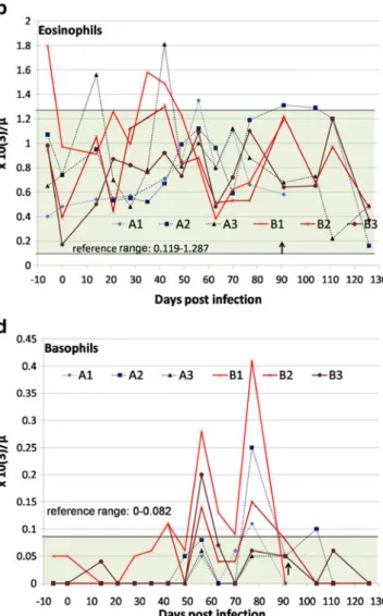
Over the course of the experimental infection, only mild changes in the CBCs were observed for both groups. The packed cell volume (PCV) showed a mild decrease after patency (63 dpi, data not shown). Dogs of group B tended to show lower PCV values than group A. Most dogs showed an occasionally mild neutrophilic leucocytosis starting around 21 dpi. Instead, 12 days after treatment (104 dpi) all four dogs developed a significant (p<0.001) transient and mild to moderate neutrophilic leucocytosis (Fig. 1a) compared to values of the six dogs before inoculation. The eosinophil count did not show a clear increase but rather fluctuated over time (Fig.1b). Notably one dog of group B (B1) had eosinophil counts mildly above the reference range already before inoculation and on two occasions at 42 and 35 dpi. There was a mild increase in the monocyte count in four out of six dogs (A1, A2, B1, B3) on one to three occasions starting from 56 dpi (Fig.1c).
The lymphocyte count was essentially unchanged through-out the study, except after treatment, where there was a mild increase, comparable to the neutrophil count (data not shown). In four dogs (A1, A2, B1, B3), an occasional mild basophilia at 1–5 time points between 42 and 104 dpi was observed, but never above 0.28×10(3)/μl (Fig. 1d). Baso-phils were more often (6 of 46 CBCs) above the reference range in dogs in group B as compared to values of dogs in group A (3 of 46 CBCs). In the blood smear of dog A1, plasma cells were found 70 dpi (60/μl).
The coagulation times PT, aPTT and TT were essentially within reference ranges throughout the experiment. In dogs B1 and B2, PT and aPTT were once minimally above the upper reference range, respectively. Nevertheless, aPTT (Fig. 2a) was significantly higher in group B on 63 and 70 dpi (p=0.024 and 0.02, respectively). Platelets were often clustered, preventing accurate counts at some time
points. The platelet counts (Fig. 2b) remained virtually in the normal range during the study, even though there was a decline starting around patency in all dogs. A mild to moderate thrombocytopenia was observed in three individ-ual dogs (A2, B1, B2) between 49 and 70 dpi and the number of thrombocytes was significantly lower in group B on 77 and 91 dpi (p=0.017 and 0.015, respectively). Post mortem examination
The number of adult specimens of A. vasorum recovered by reverse lung perfusion and the extent of the gross and histological lesions for each dog are summarised in Table1. Approximately 80% of the lungs of the two untreated animals necropsied 97 dpi (dogs A1 and B1) were consolidated (Fig. 3a) and their appearance was macro-scopically similar. All the lobes contained large, confluent,
Fig. 1 Haematological values of six dogs experimentally inoculated with 50 (n=3, group A, dotted lines) or 500 (n=3, group B, continuous
lines) third stage larvae of Angiostrongylus vasorum from −6 to
126 days post inoculation (dpi). The arrows (↑) indicate the point of
treatment of four dogs (A2, A3, B2 and B3) 92 dpi. Two dogs (A1, B1) were necropsied 97 dpi. a Segmented neutrophils, b eosinophils, c monocytes d basophils
firm and raised nodules that were irregularly discoloured from pale beige to brownish yellow and frequently associated with dark red hemorrhagic patches. The pleural surface of some nodules had a grey translucent centre. The cut surface in the less affected areas appeared granular (Fig.3b). In contrast, lung tissue of the treated dogs was far less affected, usually restricted to disseminated pale beige slightly raised foci (∼1 cm diameter) and areas of yellowish discoloration in the aerated tissues (Fig. 4). An exception was dog B2, whose lungs still manifested hemorrhagic patches, foci with grey translucent centres and remnants of fibrous adhesions. The lung lymph nodes were consistently enlarged and moist in both treated and untreated dogs.
Histological sections confirmed that the consolidated lung tissues of untreated dogs were totally solidified and consisted mainly of masses of plasma cells and macro-phages (Fig. 5a). When thrombi were present they were
often associated with incorporated larvae and eggs (Fig. 5b). Often, the arterial walls were thickened. The granulomas consisted mainly of macrophages, multinucle-ated giant cells and lymphocytes that had accumulmultinucle-ated around larvae and eggs and were associated with pneumo-cyte proliferation, haemorrhage and intracellular hemosid-erin (Fig.5c). Eosinophilic granulocytes were present only very sporadically. In the dogs treated 92 dpi and euthanized 166 dpi, pneumonia and fibrosis were still present (Fig.5d), but to a lesser extent. No parasitic stages were histologi-cally present in three out of the four treated dogs. One treated dog (dog A3) had only adult worms in a larger vessel. Differences between dogs of group A and B concerning gross and histological lesions were not obvious, with the exception of fibrosis which was more pronounced in group B.
Discussion
This study correlates the successful experimental infection of six dogs with two different A. vasorum L3 inoculation doses of A. vasorum, leading to severe clinical and pathological manifestations on one hand, but in contrast with mild haematological and coagulation changes. Animal numbers were small, due to animal welfare reasons and the study’s pilot character intended for further experimental studies, therefore significant differences were considered cautiously and are mentioned to highlight trends in several parameters.
Pre-treatment
The prepatency of 47–49 days in this study was in the range (38–57 days) of previous observations (reviewed in Bolt et al.1994). During patency, the number of detected larvae in the faeces was variable and therefore did not allow an estimate of the present worm burden, as also shown in previous studies (Oliveira-Junior et al.2006).
All dogs in this trial developed respiratory signs and the progressive worsening of these clinical signs was typical for infections with A. vasorum (Koch and Willesen 2009). Weight loss was higher and respiratory signs were more pronounced and started earlier (1 week before onset of patency) in dogs of group B, followed 1 week later by respiratory signs in dogs of group A. This is in contrast with previous experimental inoculations with similar inoculation doses (Prestwood et al. 1981; Schelling et al. 1986) and other experimental infections (Neff1971) where no clinical abnormalities suggestive for A. vasorum were noted before patency. In a study performed under similar conditions as described for this study, with the exception that 200 L3 were administered, slight to moderate
respira-Fig. 2 Coagulation profiles of six dogs experimentally inoculated with 50 (n=3, group A, dotted lines) or 500 (n= 3, group B, continuous lines) third stage larvae of Angiostrongylus vasorum
from−6 to 92 days post inoculation. a Activated partial
tory sounds were observed starting from 41 dpi, confirming that an attentive physical examination including lung auscultation can furnish indications for A. vasorum infec-tion before the onset of potential fatal coagulopathies and notably before patency and therefore aetiological diagnosis by Baerman–Wetzel technique (Schnyder et al. 2009). Indeed pathological (Neff 1971; Prestwood et al. 1981) and radiological (Mahaffey et al. 1981) examinations of experimentally infected animals (always with similar inoculation doses) confirmed lesions in lungs starting from 13 to 30 days post inoculation, however these changes were not correlated with clinical signs. Clinical outcome and larval excretion during A. vasorum infection may be influenced by the age of infected dogs. In studies using naturally infected dogs, young animals are regularly overrepresented (Chapman et al.2004; Koch and Willesen 2009) and more often exhibited severe clinical signs with fatal outcome than older ones (Staebler et al. 2005). Animals of the presented experimental study were adult dogs and all, including the oldest of 6 years of age, presented clinical signs and at least temporarily high larval excretion. In an experimental study with dogs of 1 year of age, larval excretion was very irregular (Oliveira-Junior et al. 2006). We therefore conclude that the age of affected animals is not a factor helping to predict the course of an experimental A. vasorum infection and most probably also not in naturally infected dogs.
The anaemia found in the dogs was mild and not detected clinically by observation of a pale mucosa. Similarly, in another experimental study, young dogs aged 6–8 months receiving inoculation doses of 50–100 L3/kg BW (Cury et al. 2002) showed PCV values mildly under the reference range starting from 20 dpi. Our inoculation doses were lower (3.2–4.6 and 31–36 L3/kg BW for group A and B, respectively) and thus we conclude that even though differences between groups A and B were observed,
anaemia does not clearly develop with inoculation doses of approximately 500 L3 per dog and can therefore not serve as an indicator for canine angiostrongylosis.
A high degree of eosinophilia caused by endoparasitism is often reported in literature. Migration within the tissue and prolonged host-tissue contact lead to the production of interleukin (IL) 5. Also a subsequent exposure to the same parasite elicits a quicker and more drastic eosinophilia (Schultze 2000). Surprisingly then, eosinophilia was often absent or mild for the dogs in this study. In addition, no explanation can be provided for one dog presenting with eosinophilia before inoculation. In previous studies, dogs with angiostrongylosis presented with or without eosino-philia (Cury et al.2002; Chapman et al.2004; Willesen et al.2009). In our study, histology of lung tissues confirmed the very sporadic presence of eosinophilic granulocytes. Therefore, it has to be considered that the absence of eosinophilia does not exclude an A. vasorum infection.
Mild monocytosis started from 56 dpi equally in both groups. This result is in agreement with the observed leucocytosis and neutrophilia, as each are indicative of chronic inflammation. Monocytosis due to A. vasorum infection had not been described in experimental infections before however has been reported in up to 30% of clinical cases (Chapman et al. 2004) and does not represent a characteristic sign for canine angiostrongylosis.
Basophilia is rare in dogs and is reported with hypersensitivity or inflammatory conditions (Schultze 2000). The mild basophilia seen in this study may be a reflection of the high imprecision when measuring baso-philes by blood smear evaluation as this leukocyte subpopulation is often underrepresented in peripheral blood. However basophilia occurred also in 25% of dogs naturally infected with A. vasorum (Chapman et al. 2004) and a frequent reported cause of basophilia is the infection with another heart worm, Dirofilaria immitis (Rawlings
Table 1 Post-mortem findings of treated and untreated dogs after experimental inoculation with 50 (group A) or 500 (group B) third stage larvae of Angiostrongylus vasorum: gross and histological
lesions in the lungs and adult worms recovered by reverse lung perfusion and dissection of heart and lungs
Dog ID Treatment/ necropsy (dpi) Inoculation dose (L3) Post-mortem findings Number of adult A. vasorum Gross findings Histological findings
Pneumonia Eggs/L1 Thrombi Fibrosis
A1 None/97 50 10 +++ +++ +++ + ++
A2 92, I/M/166 50 0 + + − − +
A3 92, EC/166 50 13 + ++ Adults (no larvae) + +
B1 None/97 500 170 +++ +++ ++ + +
B2 92, I/M/166 500 1 ++ ++ − + ++
B3 92, EC/166 500 0 + + − + +++
I/M imidacloprid/moxidectin (Advocate®), EC experimental compound, + slight, ++ moderate, +++ severe Table 1 Post-mortem findings of treated and untreated dogs after
experimental inoculation with 50 (group A) or 500 (group B) third stage larvae of Angiostrongylus vasorum: gross and histological
lesions in the lungs and adult worms recovered by reverse lung perfusion and dissection of heart and lungs
1982). Basophils are reported to respond quickly to inflammatory signals that are induced by helminth infec-tions and can be recruited into tissues such as lung where they contribute to initiation of protective immunity by local release of cytokines IL-4, IL5 and IL-13 (Voehringer2009). Plasma cells, indicating generally a humoral B-cell response after antigen-stimulation (Schultze2000), are very rarely observed in the peripheral blood circulation in dogs but were detected in the blood of one dog 70 dpi. The presence of plasma cells associated with A. vasorum infection is, to our knowledge, described for the first time. Particularly leishma-niosis and ehrlichiosis are reported to be associated with intensive B-cell activation and massive presence of plasma cells in concerned tissues in dogs (Kuehn and Gaunt1985;
Martínez-Moreno et al. 1993) but this did not implicit the presence of plasma cells in the peripheral blood circulation. However, it can be expected that plasma cells could be detectable since A. vasorum infection induced the production of circulating antibodies (Cury et al. 1996; De Oliveira Vasconcelos et al.2008; Verzberger-Epshtein et al.2008).
In contrast to previous cited experimental studies (Schelling et al. 1986; Cury et al.2002) and several case reports (Dodd1973; Ramsey et al.1996; Garosi et al.2005; Wessmann et al.2006; Schmitz and Moritz2009), we only found very limited coagulation abnormalities, coinciding with the absence of haemorrhages. The only potential signs of coagulopathy observed for the current experimental infections included occasional blood in faeces and a higher aPTT between 63 and 70 dpi in highly inoculated dogs versus dogs inoculated with 50 L3.
The decrease of platelet counts observed from 42 to 70 dpi coincides with the onset of patency but was not clinically relevant. Temporal patterns were very similar to those observed during other experimental studies with comparable (Schelling et al. 1986) or higher (Cury et al. 2002) inoculation doses. Our significantly lower values in highly inoculated dogs on 77 and 91 dpi indicates that thrombocy-topenia due to A. vasorum infections may be dose dependent, a result that was not detected in a previous experimental infection (Schelling et al. 1986). It is during this time that adult A. vasorum are established within the pulmonary arteries and are producing eggs and L1 (Guilhon and Cens 1973). Such infections may directly cause a reduction in platelet number; however, generalised inflammation may also produce an increased depletion of platelets as a consequence of consumption coagulopathy (Breitschwerdt 1988; Grindem et al. 1991). In contrast to experimental studies, more than half of naturally infected dogs (12/20) exhibited thrombocytopenia (Chapman et al. 2004). The
Fig. 4 Lungs of dog B3 inoculated with 500 third stage larvae (L3) of Angiostrongylus vasorum, treated with an anthelmintic experimental compound 92 days post inoculation (dpi) and necropsied 166 dpi. The lungs exhibit hemorrhagic patches, foci with grey translucent centres and remnants of fibrin adhesions within the pleura
Fig. 3 Lungs of dog B1, necropsied 97 days after inoculation with 500 third stage larvae of Angiostrongylus vasorum. a Large areas were consolidated and presented firm, raised nodules with dark red hemorrhagic patches or discoloration to brownish yellow. b The cut surface appeared granular
mechanism of the onset of thrombocytopenia and generally of bleeding disorders during A. vasorum infections have been repeatedly discussed but are still not fully understood (Morgan et al. 2005; Koch and Willesen 2009). Chronic disseminated intravascular coagulation (DIC) as a conse-quence of consumptive coagulopathy (Ramsey et al.1996), as well as an immune-mediated thrombocytopenia (Gould and McInnes 1999), von Willebrand factor deficiency (Whitley et al.2005) or accumulation of immune complexes in the lung vessels stimulating the intrinsic pathway in addition to the secretion of antigens by adult worms and larvae (Caruso and Prestwood1988) have all been suggested as potential influencing factors.
Additional techniques for the diagnosis of coagulopa-thies and particularly DIC have been previously considered. In particular, thromboelastography, a method introduced in 1948 (Hartert 1948), has been presented as the assay allowing the identification of hypo-, normo- or hypercoag-ulable patients (Wiinberg et al.2009) and is suggested to be
a helpful tool to identify also dogs with coagulopathies due to A. vasorum infections, but devices for this assay are not yet widely distributed in veterinary laboratories.
Post-treatment
Imidacloprid/moxidectin and the EC stopped larval shed-ding in all four treated dogs within 20 days until the study end. Potentially sterile, adult parasites were however found in one dog for each anthelmintic treatment. With imidaclo-prid/moxidectin, a single worm was found in a dog that had received 500 L3, showing high efficacy. In contrast, one dog inoculated with 50 L3 and treated with the EC still presented with 13 adults at necropsy. The worms of this dog were apparently not producing eggs. Particularly, larvae and eggs were shown to cause granulomatous pneumonia (Prestwood et al. 1981; Schnyder et al. 2009). Possibly some anthelmintic treatments may not completely eliminate adult worms but are able to sterilise them,
Fig. 5 Histological sections (hematoxylin and eosin stain) of lung tissue from dogs inoculated with third stage larvae (L3) of Angiostrongylus vasorum. a Dog B1, necropsied 97 days after inoculation (dpi) with 500 L3: the alveoli are invaded with masses of plasma cells, macrophages, multinucleated giant cells and lympho-cytes. b Dog B1: larval stages are surrounded by a granulomatous
inflammation. Thrombi and thickened arterial walls were regularly present. c Dog B1: granulomas around larvae and eggs were associated with pneumocyte proliferation, haemorrhage and intracel-lular hemosiderin. d Dog A3, inoculated with 50 L3, treated with an anthelmintic experimental compound 92 dpi and necropsied 166 dpi. Pneumonia and fibrosis were still present, but to a lesser extent
limiting therefore the pathogenicity of the infection, as demonstrated by necropsy and the absence of clinical signs 12 days after treatment in dogs of this study.
Imidacloprid/moxidectin has been previously tested for the treatment of A. vasorum in naturally and experimentally infected dogs in a single application (Willesen et al.2007; Schnyder et al. 2009) as well as milbemycin oxime (0.5 mg/kg PO) weekly for 4 weeks (Conboy 2004), supporting the results obtained for two dogs in this study. Macrocyclic lactones are therefore suggested to replace the classical long-lasting fenbendazole therapy. Pathological observations of the lungs revealed considerable improve-ments in gross morphology for three of four dogs when compared with untreated dogs. Tissues for histological analysis were only sampled from affected areas and as such, could not be used to assess quantitative differences between each of the dog groupings. However, interestingly, eggs and larvae were only present in untreated dogs. Thrombi and particularly fibrosis were also still present in the treated dog lungs. Fibrosis represents a reparative process in response to damage of an organ or a tissue; the substitutive tissue is usually not as functional as the original tissue, but apparently this did not impact on clinical performances for these dogs. However, as they were always kept in kennels, the demand for high level exertion was limited.
An interesting outcome is the distinct increase of leucocytes and segmented neutrophils 12 days after treatment. It may indicate that the elimination of parasitic stages due to treatment enhanced the inflammatory response in these dogs. Such immune responses were comparable in dogs of both groups, resolving to baseline values within 34 days after treatment, and, to our knowledge, have not been described before. Similar patterns were observed for the lymphocytes, indicating a cellular immune reaction to dead parasites. This is sustained by a study with 42 naturally infected dogs in which leucocytes were within the reference range on the day of diagnosing A. vasorum, but significantly decreased 42 days after treatment (Willesen et al.2009).
The results from this study showed that respiratory sounds detected by an attentive auscultation of the lungs may indicate an early, prepatent infection of A. vasorum in dogs. Clinical signs tended to be associated with the inoculation doses but were not correlated with the age of the dogs. Despite heavy A. vasorum infection load and severe pulmonary changes including vascular thrombosis, only mild haematological changes were observed and without serial measurements they may have to be consid-ered carefully. The absence of eosinophilia did not exclude an A. vasorum infection and rarely plasma cells were observed in the peripheral blood circulation. Signs for coagulopathies in form of a prolonged aPTT and thrombo-cytopenia were slight and could also be missed without
serial measurements. Nevertheless, in this study changes concerning coagulation parameters showed inoculation dose dependent.
A further important point the practitioners have to consider is that larval excretion may continue for over 3 weeks, even if anthelmintic treatment was successful. Leucocytosis shortly after treatment returning to baseline within 5 weeks may indicate an enhanced immune response that has not been previously described. Ten weeks after treatment fibrosis was still present, however this did not have a clinical impact on the dogs. Possibly some anthelmintic treatments may not completely eliminate adult worms but be able to sterilise them leading to a reduced pathogenicity of A. vasorum infection.
Ethical standards
The study was carried out at the experimental units of the Vetsuisse Faculty at the University of Zurich after approval by the Cantonal Veterinary Office of Zurich (permission number 26/2007).
Acknowledgements We kindly thank the keepers, Armin Ruedemann
and Esther Merz, for their great participation and support throughout the study, Lucia Kohler for intense laboratory tasks, the veterinarian Sandra Staebler for her help during the trial and Ryan Jefferies for revising the English and for precious comments and suggestions on the manuscript. We also acknowledge Bayer Animal Health GmbH, Germany, for the financial contribution.
References
Boag AK, Murphy KF, Connolly DJ, Boag AK, Murphy KF, Connolly DJ (2005) Hypercalcaemia associated with
Angios-trongylus vasorum in three dogs. J Small Anim Pract 46:79–84
Bolt G, Monrad J, Koch J, Jensen AL (1994) Canine
angiostrongy-losis: a review. Vet Rec 135:447–452
Breitschwerdt EB (1988) Infectious thrombocytopenia in dogs. Comp
Cont Ed Pract Vet 10:1177–1190
Chapman PS, Boag AK, Guitian J, Boswood A (2004)
Angiostrongy-lus vasorum infection in 23 dogs (1999–2002). J Small Anim
Pract 45:435–440
Caruso JP, Prestwood AK (1988) Immunopathogenesis of canine angiostrongylosis: pulmonary effects of infection. Comp Immun Microbiol Infect Dis 11:85–92
Conboy G (2004) Natural infections of Crenosoma vulpis and Angiostrongylus vasorum in dogs in Atlantic Canada and their
treatment with milbemycin oxime. Vet Rec 155:6–18
Cury MC, Lima WS (1996) Rupture of femoral artery in a dog infected
with Angiostrongylus vasorum. Vet Parasitol 65:313–315
Cury MC, Lima WS, Vitor RWA (1996) Enzyme-linked immunosor-bent assay (ELISA) for the diagnosis of Angiostrongylus
vaso-rum (Baillet, 1866) infection in dogs. Rev Méd Vét 147:525–530
Cury MC, Lima WS, Guimaraes MP, Carvalho MG (2002) Hemato-logical and coagulation profiles in dogs experimentally infected with Angiostrongylus vasorum (Baillet, 1866). Vet Parasitol
De Oliveira Vasconcelos VV, Wagner De Almeida VR, Dos Santos LW (2008) Identification of stage-specific proteins of Angiostrongylus
vasorum (Baillet, 1866) Kamensky. Parasitol Res 102:389–395
Dodd K (1973) Angiostrongylus vasorum (Baillet, 1866) infestation in a greyhound kennels. Vet Rec 92:195–197
Eckert J, Lämmler G (1972) Angiostrongylose bei Mensch und Tier. Z Parasitenk 39:303–322
Eckert J, Friedhoff KT, Zahner H, Deplazes P (2008) Lehrbuch der Parasitologie für die Tiermedizin. Enke, Stuttgart
Garosi LS, Platt SR, McConnell JF, Wrayt JD, Smith KC (2005) Intracranial haemorrhage associated with Angiostrongylus
vaso-rum infection in three dogs. J Small Anim Pract 46:93–99
Gould SM, McInnes EL (1999) Immune-mediated thrombocytopenia associated with Angiostrongylus vasorum infection in a dog. J
Small Anim Pract 40:227–232
Grindem CB, Breitschwerdt EB, Corbett WT, Jans HE (1991) Epidemiologic survey of thrombocytopenia in dogs: a report on
987 cases. Vet Clin Pathol 20:38–43
Guilhon J, Bressou C (1960) Rôle des Limacidés dans le cycle
évolutif d’Angiostrongylus vasorum (Baillet, 1866). C R Acad Sc
251:2252–2253
Guilhon J, Cens B (1973) Angiostrongylus vasorum (Baillet, 1866): Étude biologique et morphologique. Ann Parasitol Hum Comp 48:567–596
Hartert H (1948) Blutgerinnungsstudien mit der Thromboelastographie, einem neuen Untersuchungsverfahren. Klin Wochenschr 26:577–583 Koch J, Willesen JL (2009) Canine pulmonary angiostrongylosis: an
update. Vet J 179:348–359
Kuehn NF, Gaunt SD (1985) Clinical and hematologic findings in
canine ehrlichiosis. J Am Vet Med Ass 186:355–358
Mahaffey MB, Losonsky JM, Prestwood AK, Mahaffey EA, Lewis RE (1981) Experimental canine angiostrongylosis: II.
Radio-graphic manifestations. J Am Anim Hosp Assoc 17:499–502
Martin MWS, Ashton G, Simpson VR, Neal C (1993) Angiostrongy-losis in cornwall: clinical presentation of eight cases. J Small
Anim Pract 34:20–25
Martínez-Moreno A, Martínez-Cruz MS, Blanco A, Hernández-Rodríguez S (1993) Immunological and histological study of T-and B-lymphocyte activity in canine visceral leishmaniosis. Vet Parasitol 51:49–59
Mason KV (1987) Canine neural angiostrongylosis: the clinical and therapeutic features of 55 natural cases. Austr Vet J 64:201–203 Morgan ER, Shaw SE, Brennan SF, De Waal TD, Jones BR, Mulcahy G (2005) Angiostrongylus vasorum: a real heartbreaker. Trends
Parasitol 21:49–51
Neff H (1971) Experimentelle infektionen von hunden mit Angios-trongylus vasorum (Nematoda). University of Zurich, Switzer-land, Dissertation
Oliveira-Junior SD, Barcante JM, Barcante TA, Dias SR, Lima WS (2006) Larval output of infected and re-infected dogs with Angiostrongylus vasorum (Baillet, 1866) Kamensky, 1905. Vet
Parasitol 141:101–106
Patteson MW, Gibbs C, Wotton PR, Day MJ (1993) Angiostrongylus
vasorum infection in seven dogs. Vet Rec 133:565–570
Prestwood AK, Green CE, Mahaffey EA, Burgess DE (1981) Experimental canine angiostrongylosis: I. Pathologic
manifesta-tions. J Am Anim Hosp Assoc 17:491–497
Ramsey IK, Littlewood JD, Dunn JK, Herrtage ME (1996) Role of chronic disseminated intravascular coagulation in a case of
canine angiostrongylosis. Vet Rec 138:360–363
Rawlings CA (1982) Clinical laboratory evaluations of seven heartworm infected beagles: during disease development and following treatment. Cornell Vet 72:49–56
Schelling CG, Greene CE, Prestwood AK, Tsang VC (1986) Coagulation abnormalities associated with acute Angiostrongylus vasorum infection in dogs. Am J Vet Res 47:2669–2673 Schmitz S, Moritz A (2009) Chronic disseminated intravascular
coagulopathy in a dog with lung worm infection (in German).
Schweiz Arch Tierheilk 151:281–286
Schnyder M, Fahrion A, Ossent P, Kohler L, Webster P, Heine J, Deplazes P (2009) Larvicidal effect of imidacloprid/moxidectin spot-on solution in dogs experimentally inoculated with
Angios-trongylus vasorum. Vet Parasitol 166:326–332
Schultze AE (2000) Interpretation of canine leukocyte responses. In:
Feldman BF, Zinkl JG, Jain NC (eds) Schalm’s veterinary
hematology. Lippincott Williams & Wilkins, Philadelphia,
pp 1155–1160
Staebler S, Ochs H, Steffen F, Naegeli F, Borel N, Sieber-Ruckstuhl N, Deplazes P (2005) Autochthonous infections with Angios-trongylus vasorum in dogs in Switzerland and Germany (in German). Schweiz Arch Tierheilk 147:121–127
van Doorn DC, van de Sande AH, Nijsse ER, Eysker M, Ploeger HW (2009) Autochthonous Angiostrongylus vasorum infection in
dogs in The Netherlands. Vet Parasitol 162:163–166
Verzberger-Epshtein I, Markham RJ, Sheppard JA, Stryhn H, Whitney H, Conboy GA (2008) Serologic detection of Angiostrongylus
vasorum infection in dogs. Vet Parasitol 151:53–60
Voehringer D (2009) The role of basophils in helminth infection.
Trends Parasitol 25:551–556
Wessmann A, Lu D, Lamb CR, Smyth B, Mantis P, Chandler K, Boag A, Cherubini GB, Cappello R (2006) Brain and spinal cord haemorrhages associated with Angiostrongylus vasorum infection
in four dogs. Vet Rec 158:858–863
Whitley NT, Corzo-Menendez N, Carmichael NG, McGarry JW (2005) Cerebral and conjunctival haemorrhages associated with von Willebrand factor deficiency and canine angiostrongylosis. J Small Anim Pract 46:75–78
Wiinberg B, Jensen AL, Rozanski E, Johansson PI, Kjelgaard-Hansen M, Tranholm M, Kristensen AT (2009) Tissue factor activated thromboelastography correlates to clinical signs of bleeding in dogs. Vet J 179:121–129
Willesen JL, Jensen AL, Kristensen AT, Kjelgaard-Hansen M, Jessen R, Koch J (2006) Serum fructosamine concentrations in 59 dogs naturally infected with Angiostrongylus vasorum. J Vet Med A
53:266–269
Willesen JL, Kristensen AT, Jensen AL, Heine J, Koch J (2007) Efficacy and safety of imidacloprid/moxidectin spot-on solution and fenbendazole in the treatment of dogs naturally infected with Angiostrongylus vasorum (Baillet, 1866). Vet
Parasitol 147:258–264
Willesen JL, Jensen AL, Kristensen AT, Koch J (2009) Haemato-logical and biochemical changes in dogs naturally infected with Angiostrongylus vasorum before and after treatment. Vet J