Compartmentalization and Axon Guidance in the Drosophila Brain by
Timothy D. Tayler B.A., Biology Earlham College
Submitted to the Department of Biology
in Partial Fulfillment of the Requirements for the Degree of Doctor of Philosophy in Biology
at the
Massachusetts Institute of Technology February 2005
© 2005 Massachusetts Institute of Technology All rights reserved
Signature of
Author...
. ....
...
...
Department of Biology\1)"~~"?.'....
tDecember
17,
2004
Certified ( by . ' Paul A. Garrity Assistant Professor of BiologyThesis Supervisor
Accepted
by a..., .... ... ... Stephen P. Bell Professor of Biology Co-Chair, Biology Graduate Committee
MASSACHUSETTS INSTITUTE OF TECHNOLOGY
Compartmentalization and Axon Guidance in the Drosophila Brain by
Timothy D. Tayler
Submitted to the Department of Biology on December 20, 2004 in partial fulfillment of the requirements for the Degree of Doctor of Philosophy in Biology
ABSTRACT
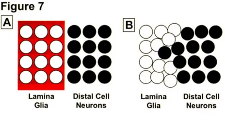
The Drosophila brain is composed of many morphologically and functionally distinct processing centers and brain morphogenesis depends on the creation and maintenance of distinct boundaries between adjacent regions to prevent cells from mixing. In the Drosophila visual system, I have found that Slit and Roundabout (Robo) proteins function to prevent cells from adjacent compartments from mixing. I have defined a boundary between two distinct compartments, the lamina and lobula, and find that the secreted ligand Slit is present in the lamina, while the Robo receptors (Robo, Robo2 and Robo3) are expressed on lobula neurons. I examined the function of theses proteins by identifying a tissue-specific allele of slit and
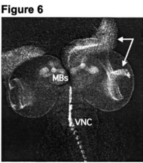
creating transgenic RNAi flies that inhibit the expression of the Robo proteins. Loss of Slit or all three Robo proteins in the visual system results in the invasion of lobula neurons into the lamina. Mixing of cells at the lamina/lobula boundary results in glial cell mispositioning and aberrant photoreceptor axon targeting. Thus, Slit and Robo proteins are required to restrict movement of cells across the lamina/lobula boundary. Additionally, I have characterized Ptpmeg, a highly conserved protein tyrosine phosphatase (PTP). In addition to the C-terminus PTP domain, Ptpmeg contains a central PDZ domain and an N-terminus FERM domain. The in vivo role of this family of proteins is unknown. To explore the function of Ptpmeg in flies, mutants were generated by targeted gene disruption. Examination of the adult nervous system of Ptpmeg mutants reveals a defect in the mushroom bodies (MB), brain structures required for olfactory learning and memory. In mutant animals, the MB lobes are disorganized and fail to elaborate their characteristic structure. I find that Ptpmeg is expressed on MB axons and targeted knockdown of Ptpmeg in the MB results in similar defects as seen in homozygous mutants. Thus, the MB neurons appear to require Ptpmeg for proper formation.
Thesis Supervisor: Paul Garrity Title: Assistant Professor of Biology
CHAPTER ONE
Introduction
The human brain contains more than 1012 neurons, and each neuron can make more than 1000 connections (Kandel, 2000). Each connection is precisely wired, with presynaptic and postsynaptic partners finding each other among the milieu of many other cells. How do axons navigate, often over long distances, to find their precise targets and how are
complex structures within the brain generated? The vertebrate brain contains many morphologically and functionally distinct compartments, but less complex brains, such as
those of insects, are similarly compartmentalized. In this thesis, I have used the model
system of Drosophila melanogaster, which contains _105 neurons, to understand how the
separation of distinct regions of the brain is maintained and how complex neuronal wiring is achieved.
This thesis is comprised of two parts. The first is concerned with the role of four
well characterized regulators of axon guidance and cell migration, Slit and the
Roundabout (Robo) family of proteins, in the developing visual system. The second part
of this thesis describes the first characterization of Drosophila Ptpmeg, a protein tyrosine phosphatase (PTP) that appears to regulate axon guidance in another important region of
the brain, the mushroom bodies. How neurons reach their appropriate targets within the brain, and how regions of the brain become compartmentalized are fundamental
questions in developmental neurobiology, and my work contributes towards an
understanding of these topics.
Here I will introduce the regions of the fly brain I have chosen to work on, the visual system and the mushroom bodies, the molecules I have found to be important in
the development of each of these structures, Slit, Robo, and Ptpmeg, and the processes of
neural development I have studied, axon guidance and compartmentalization.
Nervous System Development in Drosophila
The impressive architecture of the brain is a product of precisely followed genetic instructions and complex cellular interactions that begin in the earliest stages of
development. The Drosophila embryonic CNS develops from progenitor cells called neuroblasts (Lu et al., 2000). These neural precursors are singled out from a layer of undifferentiated epithelial cells through complex signaling mechanisms that result in the separation (delamination) of a neuroblast from the epithelial sheet into the embryo (Campos-Ortega, 1988). Neuroblasts divide asymmetrically to give rise to two different
daughter cells, a ganglion mother cell (GMC) and another neuroblast (Jan and Jan, 1998; Jan and Jan, 1999). The GMC will divide once more to produce two postmitotic neurons or a postmitotic neuron and a glial cell. Before the neuroblast divides, certain molecules
are unequally distributed in the cell, resulting in molecularly distinct daughter cells after
division (Hirata et al., 1995; Knoblich et al., 1995; Spana and Doe, 1995; Spana et al.,
1995). Asymmetric cell division helps drive the process of differentiation. Neuroblast
divisions are repeated over and over again to generate a great diversity of neuronal and glial cell types (Goodman, 1993). Some populations of neural progenitors are set aside
for later stages of development. They remain mitotically quiescent until reactivation
signals cause them to begin dividing again to generate mature neurons (Lu et al., 2000).
After the decision to become a neuron is ensured, axons and dendrites begin the process of nervous system wiring. Sensory structures at the tip of neurites, termed growth cones, guide axons and dendrites to specific targets (Goodman and Shatz, 1993).
Once the growth cone reaches the target cell or cells, connections are formed that give rise to functional synapses. Although the cell bodies of many neurons will remain near
their place of differentiation, a large number of neurons migrate to different locations using many of the same conserved molecules that are used by navigating axons and dendrites (Guan and Rao, 2003). Some of these molecules will be discussed later in this
chapter. My graduate work has focused primarily on a window of neural development that begins after cell fate has been specified and before synapses form.
Introduction to the Brain Regions
Wolff, 1997). Drosophila vision is mediated through two compound eyes that each consist of precisely arranged ommatidia, the individual facets of the eye (Wolff and Ready, 1993). The ommatidial lenses focus incoming light onto underlying
photoreceptors that contain light-absorbing visual pigment and through the
phototransduction process, the absorbed photons are transduced into membrane potentials
which are then converted into signals that are passed on to other neurons in the brain (Yarfitz and Hurley, 1994). The visual ganglia, which will be discussed below, are the image processing centers of the brain.
The Drosophila eye and brain provide exceptionally accessible systems to study
the mechanisms of axon guidance and compartmentalization. There are a large number
of molecular tools that allow disruptions in these highly ordered structures to be
identified. In addition, early cellular events that pattern the eye are well understood and the eye is amenable to genetic manipulation, as it is dispensable for the viability of the fly
(Dickson and Hafen, 1993; Pappu, 2002). The adult eye is composed of -800 repeated
ommatidial units (Wolff and Ready, 1993). Each ommatidium contains 8 uniquely
identifiable photoreceptor neurons (or R-cell for Retinula cell). There are three subtypes
of photoreceptors, R1-R6, R7 and R8. In the adult, each subtype expresses a different
photosensitive opsin and responds to different wavelengths of light (Hardie, 1985).
R-cells differentiate in the developing eye disc and send projections through the optic stalk
into distinct layers of the brain during larval and pupal development (Meinertzhagen,
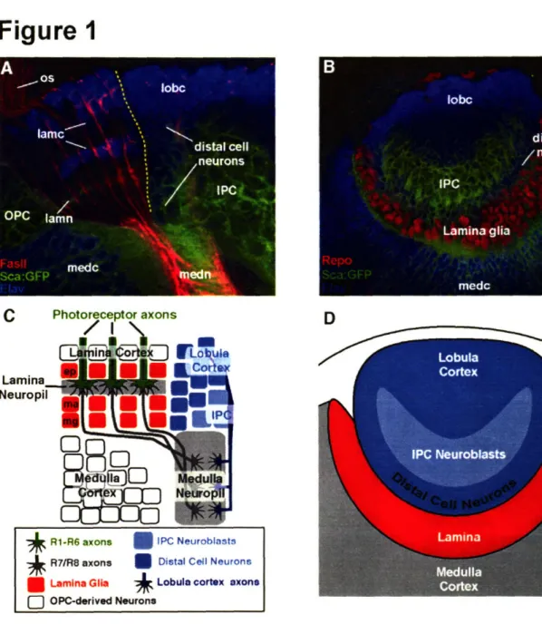
1993). R1-R6 project axons into the most superficial layer of the optic ganglion, the lamina, while R7 and R8 send axons past the lamina, into two deeper layers of the brain
in the medulla. The developing lamina and medulla are separated from another region of
the visual system, the lobula, by a distinct boundary (Tayler et al., 2004). The R-cell axonal projections and the developing target region can be visualized after dissection and
antibody staining. Therefore, mutations that disrupt R-cell targeting or target region
formation can be readily identified.
The Drosophila eye disc arises from a small group of cells (-20) during early embryogenesis (Garcia-Bellido and Merriam, 1969; Wieschaus and Gehring, 1976). The distinct morphology of the young eye disc is easily visible after hatching and the eye disc rapidly grows in size and cell number. From the end of the first larval instar to the
beginning of the third larval instar, cell numbers increase from approximately 130 to more than 1300 (Becker, 1957; Wolff and Ready, 1993). The adult eye begins to take shape at the beginning of the third larval instar. The differentiation of cell clusters that
will compose ommatidia begins at the posterior margin of the eye imaginal disc. The line of newly differentiated cells is marked by the morphogenetic furrow (MF), which sweeps across the eye disc toward the anterior margin (Wolff and Ready, 1993). The MF
represents the constriction and contraction of cells just prior to differentiation and appears
as an indentation in the eye disc (Tomlinson, 1985). Photoreceptor neurons differentiate in a sequential manner: R8, followed by R2 and R5, R3 and R4, R1 and R6, and finally R7. As the photoreceptors differentiate they secrete Hedgehog protein, a morphogen required in many developmental pathways, which triggers the differentiation of more anterior cells resulting in the progression of the MF (Heberlein et al., 1993; Lum and
Beachy, 2004; Ma et al., 1993). Photoreceptor differentiation begins in the posterior
margin of the eye disc at the dorsal-ventral midline. R8 is the founder cell of each
ommatidium and organizes the developmental events that follow (Wolff and Ready, 1993). Newly differentiated photoreceptor neurons begin sending out axons that travel
through the optic stalk into the brain. The axons converge on the optic stalk and then
spread out again as they enter the brain, maintaining the same position relative to their
neighbor in the eye disc and thus generating retinotopy in the brain. In the adult brain,
there are four visual processing centers: the lamina, medulla, lobula and lobula plate. The lamina and medulla receive input directly from the retina, while the lobula and lobula plate connect visual system neurons to higher processing centers of the brain
(Meinertzhagen, 1993).
A considerable amount of effort has been devoted to mapping the structure and
neuronal connections of the adult optic lobe (Bausenwein et al., 1992; Dittrich and
Fischbach, 1989; Meinertzhagen, 1993). However, the cellular and molecular
mechanisms that generate the precise organization of optic lobes are not well understood. The adult optic lobes are located beneath the compound eye and consist of the four
neuropil regions mentioned above. Each neuropil contains of an orderly array of columns that reflect the organization of the overlying eye (Meinertzhagen, 1993). The
fasciculated bundle. Hence, the number of columns formed in the lamina and medulla
optic lobe neuropil is nearly the same as the number of ommatdia in the eye (-800)
(Meinertzhagen, 1993). Stacks of neuronal projections running perpendicular to the
columns innervate specific layers within the optic lobes and have been postulated to have an integrative function. In addition, the visual centers of each brain hemisphere are connected by large tangential neurons and provide further integration of visual
information (Dittrich and Fischbach, 1989).
The adult optic lobes are derived from neuroblast proliferation centers (anlagen) that arise during embryonic development. Two crescent-shaped neuroblast proliferation
centers create most of the optic lobe, the inner optic anlagen (IOA) and the outer optic
anlagen (OOA) (Hofbauer and Campos-Ortega, 1990). The lamina (the R1-R6 target
region) and distal medulla (R7/R8 target region) are derived from cells of the OOA. The IOA generates neurons of the lobula, lobula plate and proximal medulla. As a result of cell division and cell migration, the spatial relationship of the two proliferation centers changes during the third larval instar stage, yet the cells formed remain separate and
distinct (Hofbauer and Campos-Ortega, 1990; Meinertzhagen, 1993).
Of the optic lobe structures, the lamina has received the most attention. The
differentiation of lamina precursors is triggered by photoreceptor innervation and therefore lamina development is completely dependent on ingrowth of photoreceptor
axons (Power, 1943; Selleck et al., 1992; Selleck and Steller, 1991). The medulla, lobula and lobula plate are less dependent on photoreceptor innervation, as the structures are still
present (although reduced in size) even when photoreceptors axons have been prevented from entering the brain (Fischbach and Heisenberg, 1981; Power, 1943).
Although it had long been observed that the development of the optic lobes
depended on photoreceptor innervation (Power, 1943), the mechanisms governing the
induction process were unknown. Shortly after photoreceptor differentiation begins, the
first photoreceptor axons travel through the optic stalk and enter the brain. Photoreceptor axon entry into the brain triggers a series of developmental events. Work from many groups has shown that the migrating photoreceptor axons are primarily engaged in two
concurrent tasks: establishing the patterning of their final targets and identifying their intermediate targets (Clandinin and Zipursky, 2002).
The organization of the lamina target field is accomplished, in part, by the
delivery of secrected cues to precursor cells in or near the target region. Elegant studies by Kunes and coworkers have shown that Hedgehog (Hh) protein is expressed in
photoreceptors and transported down their axons. Delivery of Hedgehog to the lamina induces the final cell division of lamina precursor cells (LPCs), the eventual R1-R6 synaptic targets (Huang and Kunes, 1996; Huang and Kunes, 1998). Later, they showed that Spitz, a member of the EGF family, was the factor that promoted the final
differentiation step of the lamina neurons (Huang et al., 1998).
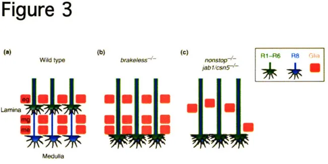
The observation that R1-R6 growth cones terminate precisely between rows of glia, suggested that glial cells provided the signal(s) for the R1-R6 growth cones to stop in lamina, effectively functioning as the intermediate target of the R1-R6 axons (Perez and Steller, 1996; Winberg et al., 1992). The lamina glia also depend on R-cell axons for migration and these anterograde signals are essential for proper visual system
development (Poeck et al., 2001; Suh et al., 2002; Winberg et al., 1992). It has recently
been shown that retrograde signals from these glia must also be required for the R-cells to make the correct target layer selection. In mutants in which glial cells do not migrate into
the lamina target region, R-cells fail to stop in the lamina and extend into the underlying
medulla (Poeck et al., 2001; Suh et al., 2002).
The Drosophila Mushroom Bodies
The mushroom bodies are lobed nervous system structures found in many marine annelids and all arthropod groups except crustaceans (Strausfeld et al., 1998). Although
the basic structure is the same, the size and complexity varies widely among species and
sometimes even within species. The mushroom bodies were first described in 1850 by the French biologist Felix Dujardin. He termed the structures "corps peloncules"
(stalk-like bodies), as their shape appeared similar to the fruiting bodies of lichens (Dujardin, 1850). Experiments that revealed a role for the mushroom bodies in learning and memory followed the early descriptive studies of Dujardin. Ants have the ability to navigate relatively complex mazes using specific olfactory cues and cockroaches have a keen ability to recall the location of food sources (place memory). These abilities are
Elegant genetic experiments in Drosophila later revealed a number of genes that were
required in the mushroom bodies for olfactory learning and memory (Dudai et al., 1976; Livingstone et al., 1984; Quinn et al., 1974; Quinn et al., 1979). These experiments importantly established that fruit flies could indeed learn and that single genes could
affect the process. Further anatomical, electrophysiological and genetic studies over
many years have contributed to the view that the mushroom bodies play a major role in olfactory processing and the integration of other sensory modalities, such as vision, touch and hearing (Strausfeld et al., 1998).
The Drosophila mushroom bodies (MBs) have been used as a model to study various aspects of neuronal morphogenesis. The development of MBs is well described and mutagenesis screens have uncovered genes that are important in regulating cell size,
neuroblast proliferation, axonal transport and axon and dendrite morphogenesis (Lee et
al., 1999; Ng et al., 2002; Reuter et al., 2003). A combination of Golgi staining and
genetic mosaic analysis has formed a comprehensive picture of mushroom body structure
and development (Kenyon, 1896a; Kenyon, 1896b; (Lee et al., 1999; Strausfeld, 1976).
There are three classes of mushroom body neurons (also called Kenyon cells), all of which originate from four neuroblasts in each hemisphere of the embryonic brain. The
neuroblasts begin dividing during mid-embryogenesis (stage 9) and continue into the pupal stages (Ito et al., 1997; Ito and Hotta, 1992). Densely packed Kenyon cell bodies (-2500 in each hemisphere) are located near the dorsal-anterior surface of the brain and are designated according to which lobes their axons innervate. y neurons are generated in the first instar larval stage, a' and ' neurons are generated between the third instar larval stage and puparian formation, and lastly, the a and f3 neurons are generated after puparian formation. As the newly generated mushroom body neurons extend axons to form the
a and 13 lobes, the y neurons partially retract their axons and then re-extend toward the midline, to form the y lobe (Lee et al., 1999). Sequential expression of transcription
factors appears to underlie the temporal development of the mushroom bodies (Isshiki et
al., 2001).
After Kenyon cells elaborate dendritic processes within the mushroom body
calyx, just beneath their cell bodies, the axons fasciculate, forming tightly bundled parallel axon fibers called a peduncle (Strausfeld et al., 1998). As the axons exit the
peduncle, they bifurcate and enter dorsal and medial lobes. An interesting feature of the mushroom bodies is the concentric development of the axons. Younger axons are found
in the interior, while the older axons are found on the exterior (Verkhusha et al., 2001).
Screens for defects in organization of the adult mushroom bodies have revealed a number of genes that regulate MB neuroblast proliferation: the histone acetyltransferase (enok), the p21-activated kinase-like protein serine/threonine kinase (mbt), and a
coiled-coil protein (mud) (Heisenberg, 1980; Melzig et al., 1998) (Guan et al., 2000; Scott et al.,
2001). Recent investigations have used mosaic analysis to study more specific aspects mushroom body development such as axon growth, guidance and branching (Ng et al., 2002; Reuter et al., 2003).
INTRODUCTION TO THE MOLECULES
In this thesis I have studied molecules required for the formation of the visual system and mushroom bodies, and will provide some background on these molecules below.
Slit and Roundabout
Mutations in slit were originally recovered by Nusslein-Volhard and Wiechaus in a screen for genes controlling pattern formation in Drosophila (Nusslein-Volhard et al., 1984). A few years later slit was cloned by Artavanis-Tsakonas and coworkers after
being identified in a homology based screen (Rothberg et al., 1988). The slit embryonic
phenotype was similar to a previously characterized gene called single-minded (sim)
(Crews et al., 1988; Nusslein-Volhard et al., 1984; Thomas et al., 1988). Sim protein is
expressed by glia and neurons of the embryonic CNS midline. Loss of Sim expression results in the collapse of the ladder-like structure of embryonic nerve cord, resulting from
the loss of midline cells. Slit protein was expressed in a similar pattern and the
loss-of-function phenotype was nearly identical to that of sim mutants. Therefore, it was concluded that the longitudinal axon collapse phenotype seen in slit mutants, likely resulted from loss or improper positioning of midline cells, in particular the midline glia
that had been shown to play an important role in the formation of axon commissures at the midline (Thomas et al., 1988). More than a decade after its initial characterization,
(Brose et al., 1999; Kidd et al., 1999; Li et al., 1999), axon branching (Wang et al., 1999),
and neuronal migration (Wu et al., 1999). Furthermore, these groups showed that Slit controlled these processes through a previously characterized guidance receptor called Roundabout (Robo).
Goodman and colleagues (Seeger et al., 1993) performed a screen for mutations
that disrupted the development of axon pathways in the developing CNS of Drosophila embryos. The first two mutant lines to be molecularly characterized were the previously unidentified genes roundabout (robo) and commissureless (comm). Mutations in robo
resulted in axons aberrantly crossing and recrossing the midline. Mutations in comm caused the absence of nearly all commissures, as axons rarely approached or crossed the midline. The robo gene was later shown to encode a highly conserved transmembrane protein, expressed on the growth cones of axons that never cross the midline and on axons that have recently crossed the midline (Kidd et al., 1998a; Seeger et al., 1993). It became the founding member of a novel family of axon guidance receptors. Ectopic expression of Robo resulted in a comm-like phenotype, where axons never entered the midline. Comm was shown to regulate Robo by preventing Robo protein from being expressed on the growth cone surface (Georgiou and Tear, 2003; Keleman et al., 2002). This suggested a model in which axons that never crossed the midline express high levels Robo protein on their growth cones, whereas axons destined to cross the midline express
little or no Robo. After crossing the midline, Robo expression is upregulated, preventing
axons from re-crossing (Kidd et al., 1998a; Kidd et al., 1998b).
The characterization of Robo as a transmembrane receptor led to the search for the unknown ligand. In Drosophila, the midline mutagenesis screen that had identified
robo and comm, apparently covered much of the genome. However, the corresponding
robo phenotype that would be predicted for the ligand, had not been found (Seeger et al., 1993). A closer look at other mutants recovered from the initial screen finally revealed
Slit as the missing ligand (Kidd et al., 1999). slit had initially been overlooked for two
reasons. First, the slit mutant phenotype did not fully resemble the robo mutant
phenotype. In robo mutants, too many axons cross and recross the midline, resulting in thicker commissures and thin or missing longitudinal axons. The phenotype in slit mutants is more dramatic: axons converge on the midline and do not reemerge. This
issue was resolved later when two more Robo-family members were identified. It was
shown that the Robo proteins function redundantly and loss of Robo and Robo2 proteins results in a slit-like phenotype (Simpson et al., 2000a; Simpson et al., 2000b). Second, the similarity of the slit and sim mutant phenotypes misled researchers to believe that the
CNS collapse was due to loss or mispositioning of midline cell rather than errors in axon
guidance. Better molecular markers revealed that the midline cells of slit mutants
appeared normal (Kidd et al., 1999).
While there are multiple forms of Slit in vertebrates, there is only a single slit gene in Drosophila. Slit is a secreted protein, containing four leucine rich repeats (LRRs), seven EGF repeats, a laminin G domain, an Agrin-Laminin-Perlecan-Slit
(ALPS) spacer domain, and a cysteine-rich repeat. It has been shown to be expressed in
both glia and neurons (Guan and Rao, 2003; Wong et al., 2002). The binding of Slit to Robo requires the LRRs but not the EGF repeats (Battye et al., 2001; Chen et al., 2001). Robo receptors belong to a novel subfamily of immunoglobulin (Ig) superfamily proteins. The extracellular portion of Robo receptors contains five Ig repeats and three fibronectin type III (FNIII) repeats. The intracellular region contains four conserved motifs, or CC domains, that share little homology to intracellular domains of other transmembrane receptors. Elimination of each CC domain reduces but does not abolish Robo function, suggesting built in redundancy of these motifs (Bashaw et al., 2000). The Robo family consists of three members, Robo, Robo2 and Robo3. In Drosophila, differences in the CC domain composition likely mediate differential responses to Slit (Simpson et al., 2000a; Simpson et al., 2000b). Both Slit and Robos can be proteolytically cleaved, but the significance of the cleavage is unclear (Wong et al., 2002).
A growing list of molecules has been identified to be important in Robo signal
transduction. Through biochemical studies, Enabled (a member of the Ena/Vasp family) and Abelson tyrosine kinase (Abl) have been identified as binding partners of Robo
(Bashaw et al., 2000). Such interactions may be responsible for the mechanism by
which the cellular signaling events downstream of Robo are induced. Bashaw et al
(2000) showed that both Abl and Ena are able to bind to one or more of the conserved
phosphorylation, acts to reduce Robo repulsion (Bashaw et al., 2000). Dreadlocks
(Dock), another established regulator of axon guidance events, also physically interacts
with Robo (Fan et al., 2003; Garrity et al., 1996). Previous studies had shown that Dock binds to p21activated kinase (Pak), an evolutionarily conserved regulator of the actin cytoskeleton (Bokoch et al., 1996; Hing et al., 1999; Leeuw et al., 1998; Sells et al.,
1997). Upon Slit stimulation, it appears that Dock and Pak are recruited to the Robo receptor, resulting in the apparent stimulation Rac GTPase activity (Fan et al., 2003).
Slit and Robo are also expressed outside of the nervous system, where they have been found to control the movements of cell types as diverse as trachea, muscle, and leukocytes (Englund et al., 2002; Kramer et al., 2001; Wong et al., 2002). Drosophila mesodermal and tracheal cell migration is directed by Slit through both repulsive and attractive mechanisms (Englund et al., 2002; Kramer et al., 2001). This suggests that there are conserved mechanisms that control movements in both immune system and nervous system.
PTPMEG
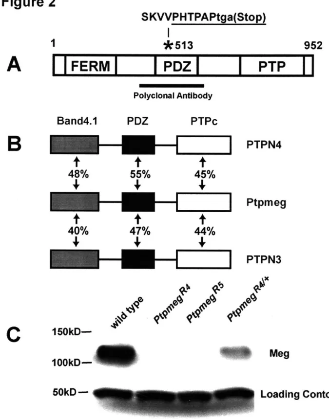
Ptpmeg belongs to a class of highly conserved proteins, that contain an N-terminal FERM domain, an internal PDZ-binding motif, and a C-terminal protein tyrosine phosphatase domain (PTP) (Edwards et al., 2001; Gu et al., 1991; Uchida et al., 2002; Yang and Tonks, 1991). Ptpmeg has been shown to be expressed in the nervous system of both C.
elegans and vertebrates (Hironaka et al., 2000; Uchida et al., 2002). The vertebrate
homologs have also been shown to be expressed in cancer cell lines, immune cells and testis (Gjorloff-Wingren et al., 2000; Sahin et al., 1995). However, there are no
published in vivo loss-of-function phenotypes in any species. In Chapter 3, I present the work that other members of the lab and I completed on Drosophila Ptpmeg. Here, I will briefly introduce two of the domains contained within Ptpmeg that likely regulate the protein localization and binding partners of Ptpmeg.
PDZ domains
PDZ domains were originally identified as conserved elements in the postsynaptic
the tight junction protein ZO-1 (Mitic and Anderson, 1998; Sheng and Sala, 2001; Woods and Bryant, 1991). PDZ domains commonly function as protein-protein interaction motifs and are found in a diverse set of proteins (Kim and Sheng, 2004). PDZ domains
are known to bind to the C-terminal of target proteins and several PDZ domains have been shown to bind the plasma membrane lipid PIP2 or short internal protein sequences
(Sheng and Sala, 2001; Zimmermann et al., 2002).
The wide range of PDZ domain targets include transmembrane receptors, ion channels and other PDZ domain proteins. These interactions suggest that PDZ domain-containing proteins are capable of functioning in a variety of cellular processes. Proteins
with multiple PDZ domains can serve as scaffolding proteins, by assembling
well-ordered, multi-protein complexes (Sheng and Sala, 2001; Tsunoda et al., 1998). However, Ptpmeg contains a single PDZ domain and is therefore unlikely to serve as a typical scaffolding protein, although it could serve a signaling function within a
scaffolding protein to which it is bound.
PDZ domains also exhibit significant sequence variation, which may underlie their ability to bind to a diverse set of ligands. Importantly, each PDZ domain appears to bind a small number of ligands and show a high degree of target sequence specificity (Sheng and Sala, 2001). Subcellular localization of PDZ-containing proteins suggests that they participate in the formation of cell junctions, receptor/channel clustering and intracellular signaling pathways (Kim and Sheng, 2004; Ponting et al., 1997).
FERM domains
Ptpmeg is a member of the FERM superfamily. FERM domains, named for prominent
family members 4.1 protein, Ezrin, Radixin, and Moesin, are found in a variety of
cytoplasmic proteins that are involved in the control of cell adhesion, cell motility, cell
shape and signal transduction (Bretscher et al., 2002). FERM domains participate in localizing proteins to the plasma membrane and have been shown to localize to the
cytoplasmic surface of the plasma membrane and bind to PIP2(phosphatidylinositol
4,5-bisphosphate) and phosphatidylserine (Chishti et al., 1998). A number of FERM
binding domain (Chishti et al., 1998). Unlike many other FERM domain-containing proteins, Ptpmeg does not appear to contain an actin-binding motif.
In addition to binding to the plasma membrane, FERM domains also associate with transmembrane proteins and PDZ domain-containing proteins (Chishti et al., 1998).
For example, the FERM domain of the protein Talin, interacts with the cytoplasmic tail of P integrin, a transmembrane protein, and is required for integrin function (Brown et al., 2002; Calderwood et al., 1999; Cram et al., 2003; Hynes, 2002). Although direct protein or plasma membrane interactions of Ptpmeg are unknown, the domains contained within Ptpmeg suggest several mechanisms by which Ptpmeg could be localized to the plasma
membrane to regulate phosphotyrosine signaling.
INTRODUCTION TO THE DEVELOPMENTAL PROCESSES
Axon Guidance
Precise neuronal wiring is fundamental to proper nervous system function. During development, axons navigate to their appropriate targets and establish synaptic
connections (Goodman and Shatz, 1993; Tessier-Lavigne and Goodman, 1996). Axons
must be able to correctly navigate through an extracellular environment of multiple
signals and cell types and distinguish their target from an array of potential targets. Growth cones at the tip of extending axons are largely responsible for the decisions made by navigating axons (Bentley and O'Connor, 1994). Growth cones use dynamic, actin-rich structures, filopodia and lamellipodia, to probe the environment. Filopodia and lamellipodia extend and retract in response to directional cues (O'Connor et al., 1990). The growth cone was initially described by Ramon y Cajal (1890), who observed
Golgi-stained preparations of embryonic chick spinal cord neurons (Ramon y Cajal, 1893). From these observations he proposed that axons could be attracted by diffusible signals
emanating from a target.
It is now clear that both attractive and repulsive forces guide axons to their destination (Guan and Rao, 2003; Mueller, 1999). For example, in the mouse embryonic spinal cord, commissural neurons differentiate near the roof plate in the dorsal region of the spinal cord and extend axons ventrally toward the floor plate where they cross the
midline (Augsburger et al., 1999; Colamarino and Tessier-Lavigne, 1995). The roof plate secretes diffusible factors that repel the commissural axons while the floor plate secretes
attractive proteins, such as Netrin, to guide the axons to the ventral midline (Kennedy et al., 1994). The Slit protein is also secreted by the floorplate and guides Robo-expressing axons (Long et al., 2004; Sabatier et al., 2004; Yuan et al., 1999).
Guidance cues can be diffusible, associated with the ECM, or cell-surface bound.
They often function as ligands for receptors located on the cell surface of growth cones.
It has been shown that an individual cue can be both an attractant and a repellent. For example, C. elegans UNC-6, a Netrin homolog, is expressed in ventral regions of the embryonic ectoderm and controls both ventral and dorsal migration of certain axons and cells, suggesting that it can function as both an attractant and a repellent (Culotti and Merz, 1998; Hedgecock et al., 1990; Wadsworth et al., 1996). Netrin-family proteins act
through two receptor families, the Deleted in Colorectal Cancer (DCC) and UNC-5 families. In C. elegans, the attractive response to UNC-6 requires the DCC homolog, UNC-40. The repulsive responses involve both UNC-40 and the UNC-5 receptor, suggesting that the combination of receptors determines the nature of the response (Kim et al., 1999). Additional work on the Drosophila homolog of DCC (Frazzled) and UNC-5 homolog suggests that Frazzled is required attractive as well as long-range repulsive effects and that UNC-5 is required for short-range and long-range repulsive effects (Keleman and Dickson, 2001; Kolodziej et al., 1996). These examples serve to illustrate
that the same cues and receptors can be used to achieve numerous effects.
The response of the growth cone to a given cue can also be affected by the
presence of other guidance cues. In Xenopus spinal neuron cultures, axons will migrate
toward a pipette dispensing Netrin (Ming et al., 1997; Song et al., 1997). However, when the ECM component laminin is added to the substrate, axons will steer away from the 1 source. cAMP levels in the growth cone normally increase upon
Netrin-exposure in this system. However, laminin alters signaling by lowering cAMP levels in the growth cone. If cAMP levels are artificially elevated in the presence of laminin, Netrin-l once again becomes attractive (Hopker et al., 1999).
Rao, 2003; Mueller, 1999). These forces, acting together, guide axons to their
appropriate targets. Dynamic remodeling of the cytoskeleton is the driving force behind growth cone motility. Actin filament assembly and disassembly along with retrograde
flow of F-actin appear to control the rapid protrusions and retractions of the highly motile growth cone (Lin and Forscher, 1995). Actin filaments predominate at the leading edge
of the growth cone in the lamellipodia and filopodia. Microtubules serve as the major
structural component of the axon body, but occasionally enter the leading edge (Dent et al., 1999).
The Rho-family of small GTPases, including Rho, Rac, Cdc42 and others, are important for axon guidance (Guan and Rao, 2003). GTPases cycle between an active
GTP-bound state and an inactive GDP-bound state. Transitions between the two states are influenced by guanine nucleotide exchange factors (GEFs), dissociation inhibitors
(GDIs), and activating proteins (GAPs) (Kozma et al., 1997). In fibroblast injection studies it was shown that activated Rho GTPase resulted in the assembly of stress fibers and focal adhesion complexes, while activated Rac and Cdc42 appeared to stimulate
formation of lamellipodia and filopodia, respectively (Hall, 1998). GTPase interactors
include proteins that are involved in regulating actin dynamics. Several of these proteins are characterized regulators of axon guidance, including UNC-73/Trio (a Rac and Rho GEF) (Awasaki et al., 2000; Debant et al., 1996), p21-activated serine/threonine kinase (Pak) (Hing et al., 1999) and the cell adhesion molecule N-WASP (Higgs and Pollard,
1999).
Compartments and Boundaries
The ability to keep discrete cell populations from mixing is critical for proper animal development. One commonly used strategy to maintain the separation of adjacent cell populations is compartmentalization (Herrup and Kuemerle, 1997; Irvine and Rauskolb, 2001). Compartments are groups of adjacent but non-intermingling cells that often arise from distinct progenitors (Garcia-Bellido et al., 1973; Irvine and Rauskolb, 2001). A
prevailing model is that cells from different compartments have distinct adhesive
properties that prevent them from intermingling across defined boundary regions and that
transcriptional regulators and signaling pathways (Irvine and Rauskolb, 2001; Lumsden, 2004).
The landmark studies of Garcia-Bellido in the early 1970s firmly established the concept of compartmentalization. Examination of mosaic animals revealed distinct compartments in the Drosophila wing that were not marked by any visible morphological boundary (Garcia-Bellido et al., 1973). Clonal analysis experiments revealed that cells on either side of the boundary were restricted by their lineage. For example, along the anterior-posterior (A/P) boundary, clonal populations would stay within the same anterior or posterior region and if they approached the adjacent region they would spread out, forming a smooth line, but never cross the other side. Regions on either side of the A/P
boundary were termed "compartments" (Garcia-Bellido et al., 1973; Garcia-Bellido et al., 1976). Distinct "cell affinities" were proposed to keep adjacent cell compartments
separate (Garcia-Bellido, 1975).
The A/P boundaries of the Drosophila wing are established by engrailed gene expression (Lawrence and Morata, 1976). Posterior cells express Engrailed which directs expression of Hedgehog (Hh) and prevents expression of Cubiutus interruptus (Ci), a component of Hedgehog signal transduction (Basler and Struhl, 1994). Anterior cells do not express engrailed, thus only anterior cells of the wing respond to the Hedgehog signal, permitting the expression of Hh target genes such as patched (a Hedgehog receptor) and dpp (a BMP homolog) (Chen and Struhl, 1996). As a result of regulation from both anterior and posterior cells, dpp expression is confined to the A/P boundary (Dahmann and Basler, 2000). It is hypothesized that these and other signaling pathways are then used to generate distinct compartments and confer differential cell adhesion properties to adjacent compartments by regulating the expression of cell adhesion molecules, ultimately through the regulation of transcription factors (Dahmann and
Basler, 1999; Irvine and Rauskolb, 2001).
As mentioned above, Hedgehog signaling plays an important role in A/P
boundary formation in the Drosophila wing. The Notch signaling pathway has also been shown to be important in this process. Notch signaling is involved in many
also important in the formation of compartment boundaries and specialized boundary
cells (Irvine, 1999; Tepass et al., 2002). The restriction of membrane-bound ligands Delta and Serrate and the ligand affinity factor Fringe to distinct cell populations increase
Notch signaling specificity (Bruckner et al., 2000; Moloney et al., 2000; Munro and Freeman, 2000; Panin et al., 1997). Consistent with previous studies that implicated Notch signaling as an important regulator of boundary formation, Lunatic fringe (L-fng) was found to be expressed at compartment borders and ectopic expression disrupted
border formation in the chick brain (Zeltser et al., 2001).
In some cases, specialized boundary cells are formed between adjacent
compartments. It has been postulated that these cells promote the refinement of borders and act as a specialized signaling center that affect further patterning events (Irvine and
Rauskolb, 2001). For example, border cells along the D/V compartments in the
Drosophila wing produce specialized bristle cells (de Celis and Garcia-Bellido, 1994;
Kim et al., 1995; Rulifson et al., 1996). The establishment of these specialized cells along the D/V border requires Notch signaling. In the vertebrate midbrain-hidbrain junction, cells along the border help direct further cell specification events (Liu and Joyner, 2001).
The most studied and best understood vertebrate compartments are the
rhombomeres of the developing hindbrain. As in the Drosophila wing, the partitioning of rhombomeres into compartments is hypothesized to be driven, in part, by differential
adhesion that arises from differential expression of regulatory genes. (Dahmann and
Basler, 1999; Fraser et al., 1990; Lumsden, 2004). In the vertebrate hindbrain the
neuroepitelium is subdivided along its A/P axis. After neural tube closure, visible bulges
in the presumptive hindbrain begin to form. As rhombomere boundaries begin to form,
the movement of cells within a delineated rhombomere is restricted (Fraser et al., 1990). Rhombomeres are arranged in a segmentally repeating pattern and distinct expression
domains are generated in an alternating pattern (Guthrie and Lumsden, 1991; Wizenmann and Lumsden, 1997b). For example, even-numbered rhombomers (r2, r4, r6) express the
transmembrane ligand ephrin-B. Odd-numbered rhombomers (r3 and r5) express three Eph receptors: EphA4, EphB2, EphB3. Eph receptor tyrosine kinases and their ligands,
repulsive cell migration events and cell adhesion (Poliakov et al., 2004). Interactions between this ligand-receptor pair appear to prevent cell mixing and sharpen compartment boundaries, implicating repulsive signaling in the process of compartmentalization
(Gilardi-Hebenstreit et al., 1992; Nieto et al., 1992; O'Leary and Wilkinson, 1999). A number of transcription factors have been shown to be crucial for hindbrain
compartment formation and their downstream control of genes such as Eph receptors and ephrins is assumed to be important for the establishment of compartments and
maintenance of compartment boundaries (Lumsden, 2004). The zinc-finger transcription factor Krox20 is expressed in two stripes of the neural plate before neurulation. These regions later become rhombomeres r3 and r5 (Wilkinson et al., 1989). In Krox20
mutants, r3 and r5 are absent, resulting in partial fusion of the even-numbered
rhombomeres r2, r4 and r6. Importantly, Krox20 was found to directly regulate EphA4
expression (Theil et al., 1998). These studies established Krox20 as one of the major
upstream regulators of rhombomere development. Another transcription factor
discovered to be important in compartment formation in the hindbrain is Kreisler, a leucine zipper containing protein. Kreisler is expressed in r5 and r6 (Manzanares et al.,
1999; Moens et al., 1998), both of which are missing in the mutant animal (McKay et al.,
1994). The Hox family of homeobox genes influence the early steps of brain patterning
in flies and vertebrates (Hirth et al., 1998). Hox gene expression occurs early in development, before the rhombomeres have begun to form. Rhombomere identity
appears to be controlled through the combined expression of a number of Hox genes (Keynes and Krumlauf, 1994; Krumlauf, 1994). It has been suggested that one role of compartments is to stably maintain homeotic gene expression within a given boundary (Struhl, 1984). Importantly, Krox-20 controls the expression of a number of Hox genes
(Nonchev et al., 1996).
As mentioned above, Eph receptors and their Ephrin ligands have been implicated
in the cell-cell signaling processes that restricts intermingling (Mellitzer et al., 1999; Xu et al., 1999). They have been shown to mediate repulsive and attractive interactions in axon guidance and cell migration (Wilkinson, 2001). In cell culture they were shown to
dominant-Although these studies are consistent with the idea that Eph/Ephrin signaling restricts cell
mixing, loss-of-function mutations in genes encoding Ephrins and Eph receptors have not revealed compartment boundary disruptions (Adams et al., 2001; Chen et al., 1996; Helmbacher et al., 2000).
Differential cell adhesion likely contributes to many of the complex
morphogenetic movements that underlie nervous system development. Classical cell
aggregation experiments showed that when cells of the ectoderm and neural tube were
dissociated from amphibian embryos and then mixed together, two populations of cells consequently separated over time, forming distinct aggregates (Townes, 1955). This implied that cells had adhesive properties located on their surfaces that permitted
associations with cells having the same adhesive properties and excluded associations
with cells that had different adhesive properties. Differential chemoaffinity properties of
the developing hindbrain have been tested by mixing cells of dissociated rhombomeres. Cell from even-numbered rhombomeres and odd-numbered rhombomeres do not mix and only form aggregates with cells from the same rhombomere. The segregation was
abolished when Ca2+was removed, suggesting that Ca2+-depedent cell adhesion molecules, such as the cadherins, would be required for rhombomere development
(Wizenmann and Lumsden, 1997a).
Cadherins are a class of cell surface glycoproteins that play an important role in tissue morphogenesis and cell-cell adhesion (Gumbiner et al., 1988; Takeichi, 1988). Cells expressing cadherin proteins are able to bind to other cells expressing the same cadherin. Homotypic binding is mediated through the extracellular domain and the presence of calcium is required for adhesion (Nakagawa et al., 1998; Redies, 2000).
There are multiple cadherin proteins, therefore combinatorial expression of different cadherin proteins can potentially generate numerous variations of adhesive specificity
(Redies, 2000). A family of cytoplasmic proteins, the catenins, interacts with the
intracellular domain of the cadherins. This association links the cadherins to the actin
cytoskeleton (Gooding et al., 2004). Cadherins are often restricted to discrete domains and segments of the developing nervous system (Redies, 2000). For example, in the
telencephalon (Inoue et al., 1997; Matsunami and Takeichi, 1995). However cadherin-6
knockout mice showed no telencephalic compartment phenotypes (Inoue et al., 2001). Although members of both the cadherin family and Eph/Ephrin family have been implicated as molecules that restrict cell mixing across compartment boundaries within the developing brain, loss-of-function analysis has not yet demonstrated the precise in
vivo role that these proteins play in compartmentalization (Cooke and Moens, 2002;
Inoue et al., 2001).
Concluding Remarks
In my work, I have sought to identify and characterize molecules that are required for nervous system development. Using the developing visual system of Drosophila as a
model, I uncovered a role for the secreted guidance cue Slit and the Robo-family
receptors in visual system morphogenesis. These molecules work together to promote
compartmentalization of the visual centers in the Drosophila brain. My work provides
insight into the process of compartmentalization by identifying Slit and Robo-family proteins as molecules that restrict cell mixing between compartments in the brain. It will be interesting to see if the signaling pathways that are used by Slit and Robo-family
proteins in the processes of cell migration and axon guidance are also used to control
compartmentalization. It will also be of interest to determine whether other molecules
that are known to guide migrating cells and neurites also participate in
compartmentalization within the brain and other developing tissues.
Precise neuronal wiring is fundamental to proper nervous system function. During development, axons and dendrites navigate to their appropriate targets and
establish synaptic connections. A growing list of cues and receptors has been identified
to be required for the formation of neuronal connections, although the intracellular signaling pathways that link cell surface detection of cues to directed rearrangements of
the cytoskeleton remain less clear. Unlike Slit and Robo-family proteins, very little is
known about the in vivo function of the protein tyrosine phosphatase Ptpmeg. The protein domains and protein localization patterns suggest that Ptpmeg could serve as a regulator of nervous system development and my initial characterization of Ptpmeg
bodies of the Drosophila brain. Ptpmeg is broadly expressed in neurons and it will be
interesting to determine whether Ptpmeg plays additional roles in nervous system
development. Additional work will be needed to identify Ptpmeg binding partners and substrates and further characterization of Ptpmeg could shed light on how
phosphotyrosine signaling is used to control aspects of neural development, such as axon guidance and branching.
References
Adams, R. H., Diella, F., Hennig, S., Helmbacher, F., Deutsch, U. and Klein, R.
(2001). The cytoplasmic domain of the ligand ephrinB2 is required for vascular morphogenesis but not cranial neural crest migration. Cell 104, 57-69.
Artavanis-Tsakonas, S., Rand, M. D. and Lake, R. J. (1999). Notch signaling: cell fate
control and signal integration in development. Science 284, 770-6.
Augsburger, A., Schuchardt, A., Hoskins, S., Dodd, J. and Butler, S. (1999). BMPs as
mediators of roof plate repulsion of commissural neurons. Neuron 24, 127-41.
Awasaki, T., Saito, M., Sone, M., Suzuki, E., Sakai, R., Ito, K. and Hama, C. (2000).
The Drosophila trio plays an essential role in patterning of axons by regulating their
directional extension. Neuron 26, 119-31.
Bashaw, G. J., Kidd, T., Murray, D., Pawson, T. and Goodman, C. S. (2000).
Repulsive axon guidance: Abelson and Enabled play opposing roles downstream of the
roundabout receptor. Cell 101, 703-15.
Basler, K. and Struhl, G. (1994). Compartment boundaries and the control of
Drosophila limb pattern by hedgehog protein. Nature 368, 208-14.
Battye, R., Stevens, A., Perry, R. L. and Jacobs, J. R. (2001). Repellent signaling by
Slit requires the leucine-rich repeats. J Neurosci 21, 4290-8.
Bausenwein, B., Dittrich, A. P. and Fischbach, K. F. (1992). The optic lobe of
Drosophila melanogaster. II. Sorting of retinotopic pathways in the medulla. Cell Tissue Res 267, 17-28.
Becker, H. J. (1957). [Roentgen mosaic spots & defective mutations at the eye of
Drosophila & the evolutive physiology of the eye.]. Z Indukt Abstamm Vererbungsl 88, 333-73.
Bentley, D. and O'Connor, T. P. (1994). Cytoskeletal events in growth cone steering. Curr Opin Neurobiol 4, 43-8.
Bokoch, G. M., Wang, Y., Bohl, B. P., Sells, M. A., Quilliam, L. A. and Knaus, U. G.
(1996). Interaction of the Nck adapter protein with p21-activated kinase (PAK1). J Biol
Chem 271, 25746-9.
Bretscher, A., Edwards, K. and Fehon, R. G. (2002). ERM proteins and merlin:
integrators at the cell cortex. Nat Rev Mol Cell Biol 3, 586-99.
Brown, N. H., Gregory, S. L., Rickoll, W. L., Fessler, L. I., Prout, M., White, R. A. and Fristrom, J. W. (2002). Talin is essential for integrin function in Drosophila. Dev Cell 3, 569-79.
Bruckner, K., Perez, L., Clausen, H. and Cohen, S. (2000). Glycosyltransferase
activity of Fringe modulates Notch-Delta interactions. Nature 406, 411-5.
Calderwood, D. A., Zent, R., Grant, R., Rees, D. J., Hynes, R. 0. and Ginsberg, M.
H. (1999). The Talin head domain binds to integrin beta subunit cytoplasmic tails and regulates integrin activation. J Biol Chem 274, 28071-4.
Campos-Ortega, J. A. (1988). Cellular interactions during early neurogenesis of
Drosophila melanogaster. Trends Neurosci 11, 400-5.
Chen, J., Nachabah, A., Scherer, C., Ganju, P., Reith, A., Bronson, R. and Ruley, H.
E. (1996). Germ-line inactivation of the murine Eck receptor tyrosine kinase by gene trap retroviral insertion. Oncogene 12, 979-88.
Chen, J. H., Wen, L., Dupuis, S., Wu, J. Y. and Rao, Y. (2001). The N-terminal
leucine-rich regions in Slit are sufficient to repel olfactory bulb axons and subventricular
zone neurons. J Neurosci 21, 1548-56.
Chen, Y. and Struhl, G. (1996). Dual roles for patched in sequestering and transducing
Hedgehog. Cell 87, 553-63.
Chishti, A. H., Kim, A. C., Marfatia, S. M., Lutchman, M., Hanspal, M., Jindal, H., Liu, S. C., Low, P. S., Rouleau, G. A., Mohandas, N. et al. (1998). The FERM domain:
a unique module involved in the linkage of cytoplasmic proteins to the membrane. Trends
Biochem Sci 23, 281-2.
Clandinin, T. R. and Zipursky, S. L. (2002). Making connections in the fly visual
system. Neuron 35, 827-41.
Colamarino, S. A. and Tessier-Lavigne, M. (1995). The role of the floor plate in axon
guidance. Annu Rev Neurosci 18, 497-529.
Cooke, J. E. and Moens, C. B. (2002). Boundary formation in the hindbrain: Eph only it were simple. Trends Neurosci 25, 260-7.
Cram, E. J., Clark, S. G. and Schwarzbauer, J. E. (2003). Talin loss-of-function
uncovers roles in cell contractility and migration in C. elegans. J Cell Sci 116, 3871-8.
Crews, S. T., Thomas, J. B. and Goodman, C. S. (1988). The Drosophila
single-minded gene encodes a nuclear protein with sequence similarity to the per gene product.
Cell 52, 143-51.
Culotti, J. G. and Merz, D. C. (1998). DCC and netrins. Curr Opin Cell Biol 10,
Dahmann, C. and Basler, K. (1999). Compartment boundaries: at the edge of
development. Trends Genet 15, 320-6.
Dahmann, C. and Basler, K. (2000). Opposing transcriptional outputs of Hedgehog
signaling and engrailed control compartmental cell sorting at the Drosophila A/P boundary. Cell 100, 411-22.
de Celis, J. F. and Garcia-Bellido, A. (1994). Roles of the Notch gene in Drosophila
wing morphogenesis. Mech Dev 46, 109-22.
Debant, A., Serra-Pages, C., Seipel, K., O'Brien, S., Tang, M., Park, S. H. and
Streuli, M. (1996). The multidomain protein Trio binds the LAR transmembrane tyrosine
phosphatase, contains a protein kinase domain, and has separate rac-specific and
rho-specific guanine nucleotide exchange factor domains. Proc Natl Acad Sci U S A 93, 5466-71.
Dent, E. W., Callaway, J. L., Szebenyi, G., Baas, P. W. and Kalil, K. (1999).
Reorganization and movement of microtubules in axonal growth cones and developing interstitial branches. J Neurosci 19, 8894-908.
Dickson, B. and Hafen, E. (1993). Genetic dissection of eye development in Drosophila.
Cold Spring Harbor: Cold Spring Harbor Laboratory Press.
Dittrich, A. P. and Fischbach, K. F. (1989). The optic lobe of Drosophila melanogaster.
Part I. A Golgi analysis of wild-type structure. Cell Tissue Res 258, 441-475.
Dudai, Y., Jan, Y. N., Byers, D., Quinn, W. G. and Benzer, S. (1976). dunce, a mutant
of Drosophila deficient in learning. Proc Natl Acad Sci U S A 73, 1684-8.
Dujardin, F. (1850). Memoire sur le systeme nerveux des insectes. Ann. Sci. Nat. Zool.
14, 195-206.
Edwards, K., Davis, T., Marcey, D., Kurihara, J. and Yamamoto, D. (2001).
Comparative analysis of the Band 4. 1/ezrin-related protein tyrosine phosphatase Pez from two Drosophila species: implications for structure and function. Gene 275, 195-205.
Englund, C., Steneberg, P., Falileeva, L., Xylourgidis, N. and Samakovlis, C. (2002).
Attractive and repulsive functions of Slit are mediated by different receptors in the Drosophila trachea. Development 129, 4941-51.
Fan, X., Labrador, J. P., Hing, H. and Bashaw, G. J. (2003). Slit stimulation recruits
Dock and Pak to the roundabout receptor and increases Rac activity to regulate axon
repulsion at the CNS midline. Neuron 40, 113-27.
Fischbach, K. F. and Heisenberg, M. (1981). Structural brain mutant of Drosophila
melanogaster with reduced cell number in the medulla cortex and with normal yaw optomoter response. Proc Natl Acad Sci U S A 78, 1105-1109.
Fraser, S., Keynes, R. and Lumsden, A. (1990). Segmentation in the chick embryo
hindbrain is defined by cell lineage restrictions. Nature 344, 431-5.
Garcia-Bellido, A. (1975). Genetic control of wing disc development in Drosophila. Ciba Found Symp 0, 161-82.
Garcia-Bellido, A. and Merriam, J. R. (1969). Cell lineage of the imaginal discs in
Drosophila gynandromorphs. J Exp Zool 170, 61-75.
Garcia-Bellido, A., Ripoll, P. and Morata, G. (1973). Developmental
compartmentalisation of the wing disk of Drosophila. Nat New Biol 245, 251-3.
Garcia-Bellido, A., Ripoll, P. and Morata, G. (1976). Developmental
compartmentalization in the dorsal mesothoracic disc of Drosophila. Dev Biol 48, 132-47.
Garrity, P. A., Rao, Y., Salecker, I., McGlade, J., Pawson, T. and Zipursky, S. L.
(1996). Drosophila photoreceptor axon guidance and targeting requires the dreadlocks SH2/SH3 adapter protein. Cell 85, 639-50.
Georgiou, M. and Tear, G. (2003). The N-terminal and transmembrane domains of
Commissureless are necessary for its function and trafficking within neurons. Mech Dev
120, 1009-19.
Gilardi-Hebenstreit, P., Nieto, M. A., Frain, M., Mattei, M. G., Chestier, A., Wilkinson, D. G. and Charnay, P. (1992). An Eph-related receptor protein tyrosine
kinase gene segmentally expressed in the developing mouse hindbrain. Oncogene 7,
2499-506.
Gjorloff-Wingren, A., Saxena, M., Han, S., Wang, X., Alonso, A., Renedo, M., Oh, P., Williams, S., Schnitzer, J. and Mustelin, T. (2000). Subcellular localization of
intracellular protein tyrosine phosphatases in T cells. Eur J Immunol 30, 2412-21.
Gooding, J. M., Yap, K. L. and Ikura, M. (2004). The cadherin-catenin complex as a
focal point of cell adhesion and signalling: new insights from three-dimensional
structures. Bioessays 26, 497-511.
Goodman, C. S. and Shatz, C. J. (1993). Developmental mechanisms that generate
precise patterns of neuronal connectivity. Cell 72 Suppl, 77-98.
Goodman, C. S. a. D., C. Q. (1993). Embryonic development of the Drosophila central
nervous system. Cold Spring Harbor: Cold Spring Harbor Laboratory Press.
Gu, M. X., York, J. D., Warshawsky, I. and Majerus, P. W. (1991). Identification, cloning, and expression of a cytosolic megakaryocyte protein-tyrosine-phosphatase with sequence homology to cytoskeletal protein 4.1. Proc Natl Acad Sci U S A 88, 5867-71.
Guan, K. L. and Rao, Y. (2003). Signalling mechanisms mediating neuronal responses
Guan, Z., Prado, A., Melzig, J., Heisenberg, M., Nash, H. A. and Raabe, T. (2000).
Mushroom body defect, a gene involved in the control of neuroblast proliferation in
Drosophila, encodes a coiled-coil protein. Proc Natl Acad Sci U S A 97, 8122-7.
Gumbiner, B., Stevenson, B. and Grimaldi, A. (1988). The role of the cell adhesion
molecule uvomorulin in the formation and maintenance of the epithelial junctional complex. J Cell Biol 107, 1575-87.
Guthrie, S. and Lumsden, A. (1991). Formation and regeneration of rhombomere
boundaries in the developing chick hindbrain. Development 112, 221-9.
Hall, A. (1998). Rho GTPases and the actin cytoskeleton. Science 279, 509-14. Hardie, R. C. (1985). Functional organization of the fly retina. Berlin: Springer.
Heberlein, U., Wolff, T. and Rubin, G. M. (1993). The TGF beta homolog dpp and the
segment polarity gene hedgehog are required for propagation of a morphogenetic wave in the Drosophila retina. Cell 75, 913-26.
Hedgecock, E. M., Culotti, J. G. and Hall, D. H. (1990). The unc-5, unc-6, and unc-40
genes guide circumferential migrations of pioneer axons and mesodermal cells on the
epidermis in C. elegans. Neuron 4, 61-85.
Heisenberg, M. (1980). Mutants of brain structure and function: what is the significance
of the mushroom bodies for behavior? Basic Life Sci 16, 373-90.
Helmbacher, F., Schneider-Maunoury, S., Topilko, P., Tiret, L. and Charnay, P.
(2000). Targeting of the EphA4 tyrosine kinase receptor affects dorsal/ventral pathfinding of limb motor axons. Development 127, 3313-24.
Herrup, K. and Kuemerle, B. (1997). The compartmentalization of the cerebellum. Annu Rev Neurosci 20, 61-90.
Higgs, H. N. and Pollard, T. D. (1999). Regulation of actin polymerization by Arp2/3
complex and WASp/Scar proteins. J Biol Chem 274, 32531-4.
Hing, H., Xiao, J., Harden, N., Lim, L. and Zipursky, S. L. (1999). Pak functions
downstream of Dock to regulate photoreceptor axon guidance in Drosophila. Cell 97,
853-63.
Hirata, J., Nakagoshi, H., Nabeshima, Y. and Matsuzaki, F. (1995). Asymmetric
segregation of the homeodomain protein Prospero during Drosophila development.
Nature 377, 627-30.
Hironaka, K., Umemori, H., Tezuka, T., Mishina, M. and Yamamoto, T. (2000). The
protein-tyrosine phosphatase PTPMEG interacts with glutamate receptor delta 2 and epsilon subunits. J Biol Chem 275, 16167-73.
Hirth, F., Hartmann, B. and Reichert, H. (1998). Homeotic gene action in embryonic
brain development of Drosophila. Development 125, 1579-89.
Hofbauer, A. and Campos-Ortega, J. A. (1990). Proliferation pattern and early
differentiation of the optic lobes in Drosophila melanogaster. Roux's Arch Dev Biol 198, 264-274.
Hopker, V. H., Shewan, D., Tessier-Lavigne, M., Poo, M. and Holt, C. (1999).
Growth-cone attraction to netrin-1 is converted to repulsion by laminin-1. Nature 401, 69-73.
Huang, Z. and Kunes, S. (1996). Hedgehog, transmitted along retinal axons, triggers
neurogenesis in the developing visual centers of the Drosophila brain. Cell 86, 411-22.
Huang, Z. and Kunes, S. (1998). Signals transmitted along retinal axons in Drosophila:
Hedgehog signal reception and the cell circuitry of lamina cartridge assembly.
Development 125, 3753-64.
Huang, Z., Shilo, B. Z. and Kunes, S. (1998). A retinal axon fascicle uses spitz, an EGF
receptor ligand, to construct a synaptic cartridge in the brain of Drosophila. Cell 95, 693-703.
Hynes, R. 0. (2002). Integrins: bidirectional, allosteric signaling machines. Cell 110,
673-87.
Inoue, T., Chisaka, O., Matsunami, H. and Takeichi, M. (1997). Cadherin-6
expression transiently delineates specific rhombomeres, other neural tube subdivisions,
and neural crest subpopulations in mouse embryos. Dev Biol 183, 183-94.
Inoue, T., Tanaka, T., Takeichi, M., Chisaka, O., Nakamura, S. and Osumi, N.
(2001). Role of cadherins in maintaining the compartment boundary between the cortex and striatum during development. Development 128, 561-9.
Irvine, K. D. (1999). Fringe, Notch, and making developmental boundaries. Curr Opin Genet Dev 9, 434-41.
Irvine, K. D. and Rauskolb, C. (2001). Boundaries in development: formation and
function. Annu Rev Cell Dev Biol 17, 189-214.
Isshiki, T., Pearson, B., Holbrook, S. and Doe, C. Q. (2001). Drosophila neuroblasts
sequentially express transcription factors which specify the temporal identity of their
neuronal progeny. Cell 106, 511-21.
Ito, K., Awano, W., Suzuki, K., Hiromi, Y. and Yamamoto, D. (1997). The
Drosophila mushroom body is a quadruple structure of clonal units each of which
Ito, K. and Hotta, Y. (1992). Proliferation pattern of postembryonic neuroblasts in the
brain of Drosophila melanogaster. Dev Biol 149, 134-48.
Jan, Y. N. and Jan, L. Y. (1998). Asymmetric cell division. Nature 392, 775-8. Jan, Y. N. and Jan, L. Y. (1999). Asymmetry across species. Nat Cell Biol 1, E42-4. Kandel, S. J., and Jessel TM. (2000). Principles of Neural Science. New York: McGraw
Hill.
Keleman, K. and Dickson, B. J. (2001). Short- and long-range repulsion by the
Drosophila Unc5 netrin receptor. Neuron 32, 605-17.
Keleman, K., Rajagopalan, S., Cleppien, D., Teis, D., Paiha, K., Huber, L. A., Technau, G. M. and Dickson, B. J. (2002). Comm sorts robo to control axon guidance
at the Drosophila midline. Cell 110, 415-27.
Kennedy, T. E., Serafini, T., de la Torre, J. R. and Tessier-Lavigne, M. (1994).
Netrins are diffusible chemotropic factors for commissural axons in the embryonic spinal cord. Cell 78, 425-35.
Keynes, R. and Krumlauf, R. (1994). Hox genes and regionalization of the nervous
system. Annu Rev Neurosci 17, 109-32.
Kidd, T., Bland, K. S. and Goodman, C. S. (1999). Slit is the midline repellent for the
robo receptor in Drosophila. Cell 96, 785-94.
Kidd, T., Brose, K., Mitchell, K. J., Fetter, R. D., Tessier-Lavigne, M., Goodman, C. S. and Tear, G. (1998a). Roundabout controls axon crossing of the CNS midline and
defines a novel subfamily of evolutionarily conserved guidance receptors. Cell 92, 205-15.
Kidd, T., Russell, C., Goodman, C. S. and Tear, G. (1998b). Dosage-sensitive and
complementary functions of roundabout and commissureless control axon crossing of the CNS midline. Neuron 20, 25-33.
Kim, E. and Sheng, M. (2004). PDZ domain proteins of synapses. Nat Rev Neurosci 5,
771-81.
Kim, J., Irvine, K. D. and Carroll, S. B. (1995). Cell recognition, signal induction, and
symmetrical gene activation at the dorsal-ventral boundary of the developing Drosophila wing. Cell 82, 795-802.
Kim, S., Ren, X. C., Fox, E. and Wadsworth, W. G. (1999). SDQR migrations in
Caenorhabditis elegans are controlled by multiple guidance cues and changing responses