HAL Id: inserm-00276757
https://www.hal.inserm.fr/inserm-00276757
Submitted on 19 Nov 2009
HAL is a multi-disciplinary open access archive for the deposit and dissemination of sci-entific research documents, whether they are pub-lished or not. The documents may come from teaching and research institutions in France or abroad, or from public or private research centers.
L’archive ouverte pluridisciplinaire HAL, est destinée au dépôt et à la diffusion de documents scientifiques de niveau recherche, publiés ou non, émanant des établissements d’enseignement et de recherche français ou étrangers, des laboratoires publics ou privés.
phospholipase D and enhances myogenic differentiation.
Saïda Mebarek, Hiba Komati, Fabio Naro, Caroline Zeiller, Monica Alvisi,
Michel Lagarde, Annie-France Prigent, Georges Némoz
To cite this version:
Saïda Mebarek, Hiba Komati, Fabio Naro, Caroline Zeiller, Monica Alvisi, et al.. Inhibition of de novo ceramide synthesis upregulates phospholipase D and enhances myogenic differentiation.. Journal of Cell Science, Company of Biologists, 2007, 120 (Pt 3), pp.407-16. �10.1242/jcs.03331�. �inserm-00276757�
407 Research Article
Introduction
Ceramide, the central molecule of the sphingolipid pathway, serves as a second messenger for cellular functions ranging from proliferation and differentiation to growth arrest and apoptosis. Ceramide generation may result from hydrolysis of sphingomyelin by various sphingomyelinases, or from synthesis involving ceramide synthases. These enzymes are regulated by physiological and environmental stimuli, and increasing evidence points to a role of this signaling pathway in response to stress and in pathogenesis (Mathias et al., 1998; Hannun and Obeid, 2002; Merrill, Jr, 2002). Thus, ceramide may be involved in insulin resistance associated with obesity and type-2 diabetes. In particular, defects in insulin signaling at the level of muscle cells have been attributed to ceramide action (Schmitz-Peiffer, 2000; Straczkowski et al., 2004). Besides, ceramide might be involved in muscle wasting closely associated with increased expression of proinflammatory cytokines, which occurs at the endpoint of several diseases such as cancer or chronic parasitic and bacterial infections (Meadows et al., 2000; Strle et al., 2004). Ceramide thus seems to play an important role in the pathophysiology of muscle tissue.
Myogenic differentiation takes place during embryonic development, and in the regenerating adult muscle after tissue
damage. Differentiation of proliferating myoblasts begins with expression of myogenesis-regulatory factors such as myogenin, withdrawal from the cell cycle, followed by expression of the proteins of contractile apparatus, and finally, by cell fusion to form the mature contracting multinucleated muscle fibers (Andres and Walsh, 1996). This highly ordered process can be regulated by hormones and growth factors, fibroblast growth factor and platelet-derived growth factor being repressors of myogenesis, whereas the insulin-like growth factors (IGFs) (Perry and Rudnick, 2000), the neurohypophyseal nonapeptide Arg8-vasopressin (AVP) (Nervi et al., 1995) and oxytocin (Breton et al., 2002) activate it. Proinflammatory cytokines such as tumor necrosis factor-␣ (TNF␣) and interleukin-1 (IL-1), which are negative effectors of myogenesis, are known to induce ceramide generation. Thus, TNF␣ might suppress murine myoblast differentiation and induce apoptosis in part through ceramide-mediated alteration of the IGF system (Meadows et al., 2000), and evidence suggests that ceramide is involved in the reduction of IGF-1-induced protein synthesis and expression of muscle-specific transcription factors caused by TNF␣ and IL-1 in C2C12 cells (Strle et al., 2004). However, the involvement of ceramide in the normal differentiation process is scarcely documented. To clarify this issue, we used a well-In L6 skeletal myoblasts induced to differentiate by
Arg8-vasopressin treatment, a short-lived lowering of ceramide levels was observed, followed by a long-lasting elevation that was prevented by inhibitors of the de novo synthesis pathway, fumonisin B1 and myriocin. Both inhibitors increased the expression of myogenic differentiation markers and cell fusion rate, whereas short-chain ceramides inhibited these responses. Similar drug effects were observed on primary mouse satellite cell differentiation. Furthermore, bacterial sphingomyelinase overexpression suppressed myogenin nuclear accumulation in L6 cells. These data suggested that endogenous ceramide mediates a negative feedback mechanism limiting myogenic differentiation, and that inhibitors of ceramide synthesis promoted myogenesis by removing this control. Phospholipase D (PLD), a recognized target of ceramide, is
required for myogenesis, as shown by the negative effects of PLD1 isoform depletion obtained by siRNA treatment. Fumonisin induced an increase in PLD activity of L6 cells, whereas C6-ceramide decreased it. The expression of PLD1 mRNA transcripts was selectively decreased by C6-ceramide, and increased by ceramide synthesis inhibitors. An early step of myogenic response is the PLD1-dependent formation of actin stress fiber-like structures. C6-ceramide addition or overexpression of sphingomyelinase impaired actin fiber formation. Ceramide might thus regulate myogenesis through downregulation of PLD1 expression and activity.
Key words: Myogenic differentiation, Satellite cells, Ceramide, Phospholipase D, Fumonisin, Cytoskeleton
Summary
Inhibition of de novo ceramide synthesis upregulates
phospholipase D and enhances myogenic
differentiation
Saïda Mebarek1,2,3, Hiba Komati1,2,3, Fabio Naro4, Caroline Zeiller1,2,3, Monica Alvisi4, Michel Lagarde1,2,3,
Annie-France Prigent1,2,3and Georges Némoz1,2,3,*
1INSERM, Unit 585, 2INSA-Lyon, Laboratoire de Physiopathologie des Lipides et Membranes and 3IMBL, Villeurbanne, F-69621 France 4Università di Roma-La Sapienza, Dipartimento di Istologia ed Embriologia Medica, Roma, Italy
*Author for correspondence (e-mail: georges.nemoz@insa-lyon.fr) Accepted 8 November 2006
Journal of Cell Science 120, 407-416 Published by The Company of Biologists 2007 doi:10.1242/jcs.03331
Jour
controlled model of in vitro myogenesis, the AVP-induced differentiation of L6 rat skeletal myoblasts in defined medium, in the absence of growth factors or serum components (Minotti et al., 1998). By using this model, we had previously shown that phospholipase D (PLD) activity, and its product, the phospholipid messenger phosphatidic acid, play an essential role in the differentiation of L6 myoblasts up to multinucleated myotube formation (Naro et al., 1997; Komati et al., 2005).
In the present study, we observed time-dependent variations of ceramide levels during the AVP-induced differentiation process, suggesting that this messenger could play a role in the control of myogenic response. We therefore investigated the effects on L6 cell myogenic differentiation of inhibitors targeted at different steps of the sphingolipid metabolism, of cell-permeant short-chain ceramides and of increased production of endogenous ceramide generated by ectopic expression of sphingomyelinase. The results of these experiments pointed to a role of ceramide as a regulator of myogenesis. Similar conclusions could be drawn from studies of the differentiation of primary mouse satellite cells. Since ceramide is considered a general regulator of PLD, we then examined the possibility that ceramide modulated differentiation through modifications of the PLD signaling pathway. Ceramide has been proposed to inhibit PLD activity by preventing its activation by protein kinases C (PKCs) and monomeric G-proteins (Venable and Obeid, 1999), by downregulating PLD gene transcription (Nakashima and Nozawa, 1999) or by a direct effect on the catalytic core of the enzyme (Singh et al., 2001). We observed here that ceramide levels can modulate PLD activity of L6 cells, at least in part by differentially regulating the expression of PLD isoforms. We then investigated whether exogenous or endogenously produced ceramide affected the PLD-dependent remodeling of actin cytoskeleton, which constitutes an early and essential step of myogenic differentiation. We conclude from the observed results that ceramide might modulate myogenesis through the regulation of PLD expression and PLD-dependent cytoskeletal changes, and that inhibition of ceramide synthesis constitutes a promising approach to promote myogenesis.
Results
Kinetics of ceramide level changes in differentiating L6 myoblasts
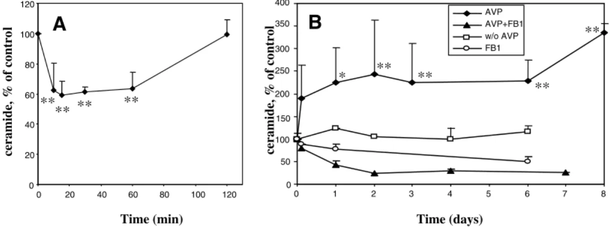
To investigate whether ceramide is involved in the differentiation of skeletal myoblasts, the levels of ceramide were measured in L6 cells induced to differentiate by treatment with AVP in serum-free medium. As shown in Fig. 1A, a transient decrease (–40%) rapidly occurred between 10-60 minutes of treatment. It was followed by a sustained increase (2 to 3-fold) starting from 3 hours and lasting for at least 8 days (Fig. 1B), i.e. throughout the differentiation process. Ceramide can be formed through different pathways, including the de novo synthesis pathway which can be selectively blocked by the fungal toxin fumonisin B1 (FB1), an inhibitor of ceramide synthase. Addition of FB1 totally prevented the delayed accumulation of ceramide induced by the differentiating treatment, even at the earliest time point of ceramide elevation (3 hours) (Fig. 1B), suggesting that ceramide accumulation principally occurred through the de novo pathway of synthesis in differentiating L6 myoblasts. FB1 addition to AVP-stimulated cells caused ceramide levels to drop under the values observed in cells treated by FB1 alone (Fig. 1B), suggesting that AVP amplified compensatory mechanisms, such as upregulation of glucosylceramide synthase, which take place in FB1-treated cells to ensure a minimal synthesis of essential glycosphingolipids at the expense of free ceramide (Meivar-Levy and Futerman, 1999). Another compound able to block the de novo pathway, the serine palmitoyltransferase selective inhibitor myriocin, showed effects similar to those of FB1 on ceramide levels measured at 3 and 24 hours (data not shown). By contrast, the inhibitor of acid sphingomyelinase desipramine did not significantly inhibit the increase in ceramide levels observed after 6 days of culture in the presence of AVP (control: 212±46% of initial level; 10 M desipramine-treated cells: 201±47%, n=3 and 5, respectively), consistent with the conclusion that ceramide was not formed through sphingomyelin hydrolysis in differentiating L6 myoblasts. A similar biphasic kinetics of ceramide level changes was observed in L6 cells induced to differentiate by switching to
ceramide, % of contro l ** ** ** **
A
120 20 40 60 80 100 ceramide, % of contro l 0B
** ** ** ** * w/o AVP FB1 AVP AVP+FB1 8 7 6 5 4 3 2 1 0 400 10 15 20 25 30 350 0 0 0 0 0 50 0 20 40 60 80 100 120 0Time (min) Time (days)
Fig. 1. Ceramide levels in L6 cells induced to differentiate by treatment with Arg8-vasopressin. L6 cells were treated for the time indicated with
10–7M AVP alone, 20 M FB1 alone, in the presence of both compounds or left untreated. Lipids were extracted, and the ceramide mass was
measured as described in the Materials and Methods using the DAG kinase method. The data are shown as percent of time 0 value (245±35 ng per mg protein, n=14). (A) Short-term (0-2 hours) AVP effects on ceramide levels. (B) Long-term (3 hours-8 days) AVP effects on ceramide levels. The data are the means ± s.e.m. of three to seven independent measurements. *Different from the time 0 value, P<0.05; **P<0.01.
Jour
409 Ceramide and phospholipase D in myogenesis
1% fetal bovine serum (FBS)-containing medium (data not shown).
Effects of drugs modulating cell ceramide content on myogenic differentiation
Since changes in ceramide content accompany myogenic differentiation of L6 cells, we asked whether drugs able to modify ceramide levels could influence the differentiative response of myoblasts. The effects of FB1 and myriocin were studied on the expression and nuclear accumulation of the muscle-specific transcription factor myogenin, which constitute one of the early crucial steps of the myogenic program (Perry and Rudnick, 2000). The synthesis of this transcription factor is maximal at day 2 after the induction of differentiation in several myogenic cell models (Nervi et al.,
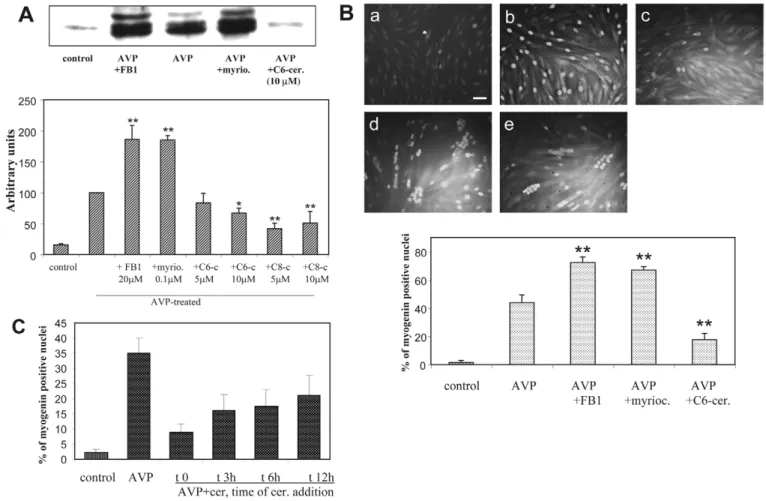
1995; Andres and Walsh, 1996). FB1 and myriocin both markedly increased the amount of myogenin expressed in cells induced to differentiate by a 48-hour AVP treatment, as evaluated by immunoblotting. Conversely, the cell-permeant short-chain C6-ceramide (N-hexanoylsphingosine) and C8-ceramide (N-octanoylsphingosine) significantly inhibited myogenin expression (Fig. 2A). FB1 and myriocin also potentiated the AVP-induced myogenin nuclear accumulation, as assessed by immunofluorescence, whereas C6-ceramide had an opposite effect (Fig. 2B). To evaluate whether ceramide effects vary in the course of myogenic response, C6-ceramide was added at different times after the onset of AVP stimulation, and nuclear myogenin was assessed by immunofluorescence (Fig. 2C). We observed that the exogenous ceramide inhibitory effect decreased when its addition was delayed, suggesting that
Fig. 2. Effects of ceramide on myogenin expression and nuclear accumulation. (A) Immunoblots of myogenin from L6 cells treated for 48
hours by AVP alone, or AVP in the presence of 20 M FB1, or 100 nM myriocin, or 5-10 M of short-chain ceramides or left untreated (control). The graph shows the videodensitometric quantitation of myogenin protein: the blots were reprobed for tubulin (data not shown), and myogenin amounts were normalized by tubulin. The means ± s.e.m. of four to nine determinations are shown. The absence of significant effects of FB1 or myriocin on myogenin expression in unstimulated cells was verified (data not shown). *Different from the + AVP value, P<0.05; **P<0.01. (B) Immunofluorescence analysis of myogenin expressed in L6 cells: the cells were left untreated (a, control), or were treated for 48 hours with AVP alone (b), or AVP in the presence of 20 M FB1 (d), 100 nM myriocin (e) or 10 M C6-ceramide (c). Nuclear myogenin was revealed by using a monoclonal anti-myogenin antibody and a fluorescein-conjugated secondary antibody. The total number of nuclei was evaluated on the phase contrast image. The mean percentage ± s.e.m. of myogenin-positive nuclei counted in four to six different fields (~100 cells per field) from one representative experiment of three performed is shown in the graph. The absence of significant effects of FB1 or myriocin on myogenin nuclear accumulation in unstimulated cells was verified (data not shown). **Different from the + AVP value, P<0.002. Magnification 32⫻; bar, 50 m. (C) Time-dependence of ceramide effects: 10 M C6-ceramide was added at different times after the onset of AVP stimulation. Nuclear myogenin was analyzed by immunofluorescence as in B. The data are the means ± s.e.m. of three independent experiments.
Jour
ceramide affects more markedly precocious events occurring in the first hours than later phases of the myogenic response. However, even when added 12 hours after AVP addition, ceramide still exerted some inhibitory effects (~40% inhibition of myogenin nuclear labeling versus 75% inhibition when added simultaneously with AVP) (Fig. 2C). Besides, microscopic examination showed that in cells simultaneously treated for 2 days by AVP and ceramide synthesis inhibitors, but not in cells treated by AVP alone, a substantial fraction of nuclei were already grouped inside highly multinucleated myotubes (Fig. 2B, Fig. 3A), suggesting that inhibiting ceramide synthesis accelerated the cell fusion process. This was confirmed by the evaluation of the cell fusion index (Fig. 3B). After 2 and 5 days of treatment by AVP in the presence of FB1, cell fusion was increased by twofold as compared with treatment by AVP alone. At later time points, the difference in the fusion index resulting from FB1 addition was proportionally less marked, and it was even null after 14 days, showing that FB1 affected the rate of the process rather than its maximal extent. Another marker of terminal myogenic differentiation, creatine kinase activity, was evaluated in L6 cells differentiated by a 6-day AVP treatment, in the absence or presence of ceramide level-modifying drugs. Addition of either FB1 or myriocin resulted in strongly increased creatine kinase activity of differentiated cells, whereas addition of C6-ceramide induced a 50% decrease in activity (Fig. 4). Since blocking the de novo pathway of ceramide synthesis can induce in 48 hours a marked general depletion in the cellular levels of complex sphingolipids such as glycosphingolipids (Stevens
and Tang, 1997), and because some biological effects of FB1 have been attributed to a depletion in glycosphingolipids rather than to ceramide decrease (Rentz et al., 2005), we assessed the effects of a glycosphingolipid-depleting agent, D-threo-1-phenyl-2-decanoylamino-3-morpholino-1-propanol (PDMP), on the induction of the late differentiation marker creatine kinase. Interestingly, PDMP, which inhibits glucosylceramide synthase, the first step in the synthesis of a majority of glycosphingolipids, is known to induce an increase in cellular ceramide, in contrast to FB1 (Shayman et al., 1991). We observed that at concentrations known to efficiently inhibit glucosylceramide synthase (10-20 M), PDMP markedly inhibited creatine kinase expression (Fig. 4). It is thus clear that the myogenesis-promoting effects of FB1 and myriocin did not result from a decrease in glycosphingolipid content of the cells. In mouse satellite cells, the effects on differentiation of drugs modifying ceramide levels were very similar to those observed with the L6 line: FB1 enhanced cell fusion, whereas C6-ceramide strongly inhibited it. Indeed, after 48 hours in differentiation medium, the fusion indexes were 6.6±1.5% for control cells, 14.0±2.5% for 20 M FB1-treated cells and 0% for 10 M C6-ceramide-treated cells (n=6 fields, ~100 cells per field, two experiments gave similar results).
In the main, these results strongly suggest that decreasing ceramide levels by blocking the de novo synthesis efficiently promoted myogenic differentiation, as evidenced by both an enhanced expression of muscle-specific proteins, such as myogenin and creatine kinase, and an acceleration of the myoblast fusion process.
Fig. 3. Effects of FB1 on the fusion of AVP-treated L6 cells. (A) L6 cells
were treated without (control) or with 10–7M AVP alone, or with the
concomitant addition of 20 M FB1 for 2 days. The cells were examined by phase contrast microscopy. Magnification, 32⫻; bar, 50 m. Myogenic differentiation was evidenced by the formation of multinucleated myotubes. (B) The cells treated as above were examined at different times, and the fusion index was evaluated as detailed in the Materials and Methods.
* * * * * * 350 300 250 200 150 100 50 0
control AVP AVP AVP AVP AVP AVP +FB1 +myrio +C6cer +PDMP +PDMP
10µM 20µM
CK activity, % of + AVP v
alue
Fig. 4. Effects of ceramide-modulating drugs on the
activity of the late differentiation marker creatine kinase. L6 myoblasts were cultured for 6 days in 1% BSA medium in the absence (control) or presence of 10–7M
AVP alone, or with the addition of 20 M fumonisin, or 100 nM myriocin, or 5 M C6-ceramide or 10-20 M PDMP. Creatine kinase-specific activity, expressed as the percentage of the + AVP value (1.7±0.25 OD units per minute perg protein), was measured in cell
homogenates. The mean ± s.e.m. of three measurements is shown. The absence of significant effects of FB1 or myriocin on creatine kinase activity of unstimulated cells was verified (data not shown). *Significantly different from the + AVP value, P<0.01. Three to five independent experiments gave similar results.
Jour
411 Ceramide and phospholipase D in myogenesis
Effect of sphingomyelinase overexpression on myogenic differentiation
To further substantiate the negative role of ceramide in the myogenic response of L6 cells, we transiently overexpressed a bacterial sphingomyelinase tagged with green fluorescent protein (GFP) and carrying a targeting sequence specific for endoplasmic reticulum (ER) localization. Expression of this construct induces both a marked increase in cell sphingomyelinase activity, and a substantial rise in ceramide levels at 24-72 hours, in transient transfection experiments (Birbes et al., 2001). The transfected L6 cells were treated by AVP for 24 hours and examined for myogenin immunofluorescence to assess differentiation. As shown in Fig. 5, a substantial fraction of AVP-stimulated cells expressing the fluorescent protein devoid of the sphingomyelinase sequence were positively stained for nuclear myogenin, and were thus undergoing differentiation (Fig. 5A), whereas almost none of the sphingomyelinase-expressing cells expressed nuclear myogenin (Fig. 5B), confirming that the enhanced production of endogenous ceramide correlated with an inhibition of the myogenic response.
Effects of ceramide-modulating drugs on PLD
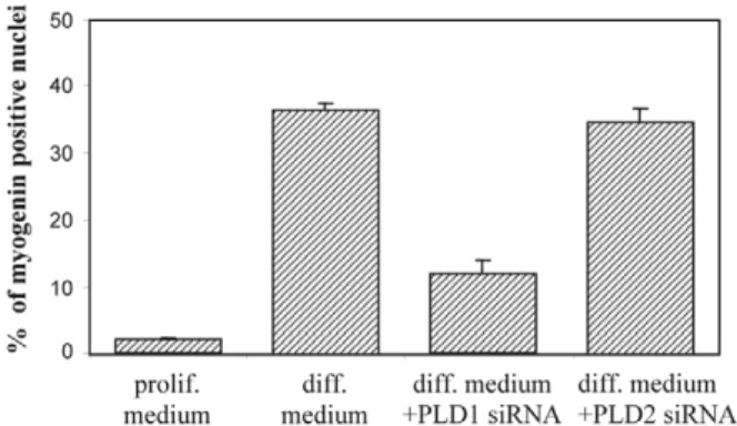
The two PLD genes identified in mammals, PLD1 and PLD2 (McDermott et al., 2004), are expressed in L6 myoblasts (Komati et al., 2005). We have previously shown that the PLD signaling pathway, and specifically the activity of the PLD1 isoform, is necessary for myogenic differentiation (Komati et al., 2004; Komati et al., 2005). To further support this conclusion, we assessed the effects of PLD1 and PLD2 knockdown by specific siRNAs on myogenin nuclear accumulation. Both PLD1- and PLD2-targeted siRNAs were able to efficiently deplete the corresponding protein (Fig. 6). Whereas PLD2 knockdown was without significant effects on L6 cell differentiation, PLD1 knockdown strongly inhibited it (Fig. 6), thus confirming that PLD1 activity is specifically required for myogenic differentiation.
Moreover, PLD has been identified as an intracellular target for ceramide action (Venable and Obeid, 1999). The effects of ceramide-modulating drugs on the PLD activity of L6 myoblasts stimulated by AVP were thus evaluated. In agreement with previous observations (Naro et al., 1997), AVP markedly increased PLD activity assayed in whole cells, after 30 minutes of stimulation (Fig. 7A). This response was significantly enhanced by a 3-hour pretreatment of the cells by FB1 (+30%). Conversely, pretreatment by C6-ceramide significantly decreased AVP-stimulated PLD activity (by 33% after 3 hours, by 50% after 24 hours) (Fig. 7A). These data show that ceramide effects on myogenesis can be linked to PLD activity modulation.
To determine whether the effects of ceramide on PLD activity can be ascribed to changes in PLD expression, reverse transcriptase-polymerase chain reaction (RT-PCR) experiments were performed to detect the various PLD transcripts after the treatment by ceramide-modulating drugs of L6 myoblasts stimulated by AVP and undergoing differentiation (Fig. 7B). A 3-hour incubation with either of the inhibitors of ceramide synthesis induced an increase in the amount of the two splicing variants transcribed from the PLD1 gene, namely PLD1a and PLD1b (+75% with myriocin, +120% with FB1). Conversely, a 3-hour incubation with C6-ceramide decreased the expression of the PLD1a and PLD1b transcripts. By contrast, the amount of PLD2 gene transcript was not affected by any of these treatments. The inhibitors of de novo ceramide synthesis, which did not induce differentiation on their own (Fig. 2A,B, Fig. 4), did not induce significant changes in PLD1 mRNA expression in unstimulated cells (data not shown). The above data suggest that ceramide accumulation selectively regulates PLD1 at the level of transcription in L6 myoblasts, and that the changes in PLD activity observed under ceramide-modulating drug treatments can, at least in part, be explained by modifications of PLD1 expression. Furthermore, it is suggested that PLD1 level of expression and the differentiative response of L6 myoblasts are positively correlated.
Ceramide impairs the PLD-dependent remodeling of cytoskeleton
We have previously shown that the differentiating agents 12-O-tetradecanylphorbol-13-acetate (TPA) (used at nanomolar concentrations) and AVP rapidly trigger the PLD-dependent formation of actin stress fiber-like structures (SFLS) in L6 cells. This response can also be induced by overexpressing the
Fig. 5. Bacterial sphingomyelinase overexpression inhibits
AVP-induced myogenic response. To induce the synthesis of endogenous ceramide, L6 cells were transfected with a vector allowing the expression of ER-targeted, GFP-tagged sphingomyelinase. Mock-transfection with a vector lacking the sphingomyelinase sequence was also performed. The transfected cells were cultured for 24 hours in 1% BSA medium in the absence (control) or presence of 10–7M
AVP, and nuclear myogenin immunofluorescence was assessed with a primary myogenin monoclonal antibody and a secondary anti-mouse IgG-rhodamine conjugate. (A) Mock-transfected cells + AVP. (B) Sphingomyelinase-transfected cells + AVP. Bar, 20 m. The graph shows the means ± s.e.m. of the percentages of myogenin-positive cells as calculated in the sole subsets of green fluorescent cells, in each condition (n=10 fields with ~10-15 cells per field).
Jour
PLD1 isoform (Komati et al., 2004; Komati et al., 2005). Owing to the major role of actin status in the regulation of myogenesis (Kuwahara et al., 2005), this step of cytoskeletal
reorganization seems to be essential to the initiation of the differentiation process. We evaluated the effects of exogenous ceramide on SFLS formation induced by AVP addition. As shown in Fig. 8, AVP induced the formation of a dense array of SFLS, with well-organized parallel fibers, in a large proportion of the cells. PLD1 overexpression also induced SFLS formation by itself, and potentiated AVP effects (Fig. 8A), in agreement with the PLD1-dependence of SFLS formation in response to AVP that we previously reported (Komati et al., 2005). A 3-hour pretreatment of the cells with C6-ceramide profoundly altered this response. In a majority of ceramide-treated, AVP-stimulated cells, the SFLS were thinner, incomplete and showed signs of actin disorganization, with non-parallel fibers and radial foci (Fig. 8B,C). Bacterial sphingomyelinase overexpression also disrupted SFLS formation induced by AVP, confirming the negative effect of endogenously formed ceramide on F-actin organization (data not shown). To substantiate the role of PLD in AVP induction of SFLS, we evaluated the effects of exogenous dioctanoyl-phosphatidic acid (DiC8-PA), a cell-permeant analog of the PLD product that can mimick the effects of PLD activity (Powner et al., 2005). We observed that addition of DiC8-PA induced by itself SFLS formation, confirming further the PLD-dependence of this process. Furthermore, DiC8-PA restored a normal SFLS response to AVP in the presence of ceramide (Fig. 8B,C), suggesting that ceramide disrupted the normal
Fig. 6. Effect of PLD isoform
selective depletion on myogenic differentiation. The cells were transfected with isoform-specific siRNAs. The cells were switched from proliferation medium (10% FBS) to differentiation medium (1% FBS + 10–7M AVP), and myogenin
immunofluorescence was assessed. The means ± s.e.m. of the
percentages of myogenin-positive cells are shown in the graph (n=10 fields). Western blotting analyses were performed with PLD1- and PLD2-specific antibodies to assess the efficacy of siRNA silencing.
Fig. 7. Effect of ceramide-modulating drugs on PLD of L6
myoblasts. (A) PLD activity was assayed by evaluating
phosphatidylbutanol formation in intact cells. The cells were labeled with [3H]palmitate, left untreated (control) or pretreated by 20 M FB1 or 10 M C6-ceramide for 3 hours. Butanol (1%) was added, with or without 10–7M AVP, for the last 30 minutes before lipid
extraction and analysis. Alternatively, the cells were pretreated by 10 M C6-ceramide for 24 hours before labeling. PLD activity is expressed as the percentage of total phospholipid radioactivity present in phosphatidylbutanol. The means ± s.e.m. of three to five independent experiments are shown. **Different from the + AVP value, P⭐0.001. (B) RT-PCR was performed with total RNA from myoblasts cultured for 3 hours in 1% BSA medium, in the absence (control) or presence of AVP, with or without different agents. Primers specific for either PLD1, PLD2 or -actin transcripts were used. The graphs show the amounts of PLD1 and PLD2
amplification products normalized by the amounts of -actin amplification product, as quantified by videodensitometry. The means ± s.e.m. of three experiments are shown for PLD1 (**different from + AVP, P⭐0.01), and the means of two determinations differing by less than 7% are shown for PLD2.
Jour
413 Ceramide and phospholipase D in myogenesis
SFLS formation through PLD inhibition and phosphatidic acid depletion.
Discussion
Triggering myogenic differentiation by AVP
treatment of rat L6 myoblasts cultured in defined medium induced changes in cell ceramide content. In a first short-lived phase ceramide levels were lowered, and then, after a few hours, they increased above the starting values, remaining elevated for at least 8 days, the period required for completion of in vitro differentiation. The early fall in ceramide levels might conceivably be attributed to activation of the ceramide-metabolizing enzyme ceramidase, because this enzyme has been shown to be involved in the regulation of ceramide levels by agonists: it is, for example, activated by growth factors in mesangial cells, and by interleukin-1 (IL-1) in hepatocytes, apparently through a tyrosine phosphorylation mechanism (Coroneos et al., 1995; Nikolova-Karakashian et al., 1997). This occurrence remains to be investigated. In terms of the later accumulation of ceramide, it can probably be attributed to activation of the de novo pathway of synthesis, because it was totally suppressed by inhibitors of two different enzymes of this pathway, FB1 and myriocin, and unaffected by the acid sphingomyelinase inhibitor desipramine. Many stimuli are known to activate the de novo pathway of synthesis. Serine palmitoyltransferase, which catalyzes the first step, is regulated at the transcriptional level by several agents, including cytokines and endotoxin (Merrill, Jr, 2002). Ceramide synthase, responsible for the acylation of sphinganine to dihydroceramide, can be activated by a range of cytotoxic agents (Merrill, Jr, 2002; Bose et al., 1995). The fact that this biphasic pattern of ceramide level changes was also observed in L6 myogenic cells differentiating in low-serum medium, in the absence of AVP, suggests that it is linked to the differentiation process itself, irrespective of its method of induction.
Activation of ceramide generation is generally considered to
be linked to cell cycle arrest, apoptosis and promotion of differentiation (Mathias et al., 1998; Hannun and Luberto, 2000). Increase in ceramide is reported to be involved in the differentiation of a range of cell types: myelocytic leukemia HL60 cells (Okazaki et al., 1990), EL4 thymoma cells (Mathias et al., 1993), cerebellar Purkinje cells (Furuya et al., 1998), U937 monoblastic leukemia cells (Ragg et al., 1998), etc. However, in view of the consequences of ceramide level manipulations, a negative, rather than positive, role of this messenger on myogenic differentiation can be inferred. Indeed, in L6 myoblasts, both FB1 and myriocin, which proved able to prevent the ceramide accumulation phase, strikingly enhanced nuclear myogenin accumulation, the rate of myotube formation, and their degree of differentiation as reflected by creatine kinase activity. It is noteworthy that myriocin is a selective inhibitor of the initial step of the de novo pathway of sphingolipid synthesis, the condensation of serine and palmitoyl-CoA, and that as such, unlike FB1, it does not induce an accumulation of bioactive sphingoid bases (Merrill, Jr, 2002). The observed positive effects of ceramide synthesis inhibition on myogenesis were thus not because of changes in sphingoid base levels. In addition, these effects did not result from a depletion of glycosphingolipids, because PDMP, an inhibitor of glucosylceramide synthase known to induce such a depletion together with ceramide accumulation, markedly inhibited myogenic differentiation. Conversely, increasing ceramide cellular levels at the onset of myogenic stimulation by using cell-permeant ceramides lowered the differentiative response. Similarly, overexpressing sphingomyelinase in ER, which is the compartment where de novo ceramide synthesis takes place (van Meer and Lisman, 2002), and thus mimicking a
Fig. 8. Ceramide impairs the PLD-dependent
remodelling of cytoskeleton. (A) L6 cells were transfected with either the empty vector or the PLD1-carrying vector, and stimulated or not by 10–7M AVP for 15 minutes. F-actin was stained
with rhodamine-phalloidin, and the percentage of cells forming stress fibers (SF) was assessed. The means ± s.e.m. of 10 fields are shown. **Indicates a significant difference to the empty vector + AVP value, P<0.001. (B) The cells were pretreated for 3 hours with 10 M C6-ceramide and/or 100 M DiC8-PA in 1% BSA medium. They were then stimulated or not with 10–7M AVP for 15 minutes,
and SF formation was assessed as above. Only the cells that formed thick parallel and continuous SFs were considered positive. (C) Fluorescence microscopy of F-actin in the above experiment. AVP stimulation induced the formation of regularly arranged, parallel fibers. When treated by ceramide, the AVP-stimulated cells displayed a fainter actin labeling because of a decrease in the number and thickness of the fibers and the presence of incomplete fibers. An irregular organization of actin, with some radial foci, was also noticed. Conversely, in the presence of PA the cells displayed a dense array of parallel fibers.
Jour
physiological activation of ceramide production, negatively affected differentiation. These results strongly suggest that ceramide conveys differentiation-limiting signals in myogenic cells. This regulation is not restricted to the myoblastic cell line L6 and seems to have a more general relevance, because similar effects of drugs modifying ceramide levels were observed on the differentiation of primary mouse satellite cells. This conclusion is in line with previous studies reporting that ceramide might participate in TNF␣- or IL-1-induced inhibition of the differentiation of murine C2 myoblasts promoted by low-serum medium or IGF-1 (Meadows et al., 2000; Strle et al., 2004). We observed here that exogenous ceramide had a stronger negative effect on myogenesis when added immediately at the onset of AVP stimulation. It can thus be hypothesized that the early lowering of endogenous ceramide levels that we observed in response to the differentiating agent AVP is essential for the initiation of myogenesis. When exogenous ceramide addition was delayed, it had a reduced but still noticeable negative effect on myogenesis, even 12 hours after AVP addition. The delayed phase of ceramide accumulation observed in response to AVP treatment might thus be viewed as a negative feedback mechanism moderating the rate of the process. Inhibitors of ceramide synthesis would then enhance differentiation by removing this negative control.
It has been reported that FB1, by disrupting sphingolipid metabolism and modifying the cellular levels of bioactive sphingolipid intermediates, is able to modulate cell proliferation, either in a positive or a negative way, depending on the considered cell model (Riley et al., 2001). In the case of L6 myoblasts, no significant effects of FB1 on [3H]thymidine incorporation measured in the presence of AVP could be found (results not shown). Thus, myogenesis-promoting effects of FB1 resulted from neither a facilitation of cell cycle exit nor from an increase in cell number favoring cell-cell contacts and myoblast fusion.
An identified cellular target of ceramide is PLD, and this signaling system plays an important role in the myogenic response. We previously showed that PLD activity is required for cytoskeleton rearrangements and myogenic differentiation induced by either AVP or the phorbol ester TPA, and that overexpressing the PLD1 isoform, but not the PLD2 isoform, can promote these responses (Komati et al., 2004; Komati et al., 2005). The conclusion that PLD1 is specifically required for myogenic differentiation is confirmed in the present study by PLD-depletion experiments using siRNAs. Our observations that (1) the cell-permeant C6-ceramide selectively downregulated the expression of PLD1 splicing variants, (2) ceramide synthesis inhibitors upregulated it and (3) none of these agents affected PLD2 expression, strongly suggest that ceramide can modulate myogenesis through the control of PLD1 gene expression. Such a control of PLD1 expression by ceramide has been observed in other cell systems such as C6 glial cells and RBL-2H3 mast cells undergoing apoptosis under C2-ceramide treatment (Nakashima and Nozawa, 1999). In addition, ceramide regulates PLD at the level of enzymatic activity, such a regulation being observed in cell-free systems (Singh et al., 2001; Venable et al., 1996; Abousalham et al., 1997; Mansfield et al., 2004). PLD inhibition might result from ceramide inhibition of the translocation of PLD-activating factors such as PKC␣ (Jones and Murray, 1995; Abousalham et al., 1997), PKC1, ADP-ribosylation factor 1 (ARF1), Cdc42 and RhoA (Abousalham et al., 1997), from
inhibition of PKC interaction with ARF-activated PLD (Venable et al., 1996) or from a direct interaction of ceramide with the catalytic core of PLDs (Singh et al., 2001). A direct regulation of PLD activity might participate, together with the regulation of PLD1 expression, in the changes in enzyme activity observed under the influence of treatments by FB1 or C6-ceramide (Fig. 7A). One way through which ceramide-induced downregulation of PLD1 activity might affect the myogenic response is impairment of cytoskeletal rearrangement, which involves the participation of PLD1 (Komati et al., 2005) (see also this study, Fig. 8). We observed that a short-term pretreatment (3 hours) by exogenous ceramide, or endogenous production of ceramide by forced expression of sphingomyelinase, hampered the formation of SFLS induced by AVP, and conversely that exogenous phosphatidic acid addition removed the disruption of fiber formation caused by ceramide. Actin cytoskeleton rearrangements play a prominent role in the induction of myogenesis, through the control by G-actin of the activity of serum response factor (SRF), which governs the expression of muscle-specific genes (Kuwahara et al., 2005). It can thus be postulated that disruption of the formation of dense regular actin fibers, resulting from the inhibition of phosphatidic acid accumulation, is the basis of the negative effects of ceramide on the myogenic process.
Thus, modulation of PLD activity by drugs acting on ceramide levels could explain their influence on the differentiation of myoblasts, and in particular, the myogenesis-promoting effects of FB1 and myriocin. Besides, it is suggested that the low ceramide levels observed during the first phase of AVP-induced differentiation are necessary in the onset of myogenic response because they allow the occurrence of a temporary elevation of PLD activity, whereas ceramide formed in the second phase would restrict phosphatidic acid synthesis and limit differentiation, possibly by preventing part of the cells from terminally differentiating.
The present study shows that blocking the de novo synthesis of ceramide leads to a strong enhancement of myogenesis, and sheds light on the signaling pathways involved in the control of muscle formation by revealing cross-talks between sphingolipid metabolism and phosphatidylcholine hydrolysis by PLD. Among the therapeutic strategies proposed for the treatment of muscular dystrophy or atrophy, increasing muscle mass by enhancing progenitor commitment seems a promising approach (Zammit and Partridge, 2002; Bogdanovich et al., 2004). This can be achieved either by stimulating positive regulators or by blocking negative regulators of muscle growth. Blockade of the ceramide signaling pathway might thus be taken into consideration as a possible target of pharmacological approaches aimed at favoring muscle regeneration or counteracting muscular atrophy.
Materials and Methods Materials
FB1 was obtained from Alexis Biochemicals (Illkirch, France); myriocin and DiPA were from Sigma-Aldrich (St-Quentin-Fallavier, France); C6-ceramide, C8-ceramide and PDMP were from Biomol Research Laboratories (Plymouth Meeting, PA). The compounds were dissolved in water (fumonisin), methanol (myriocin) or dimethyl sulfoxide (DMSO) (ceramides and PDMP), the final solvent concentration being less than 0.1%. Proteins were assayed by the Bradford method, using the protein assay kit from Bio-Rad Laboratories (Marnes-la-Coquette, France).
Cell culture
L6 cells of the C5 subclone had shown significant differentiation ability when cultured
Jour
415 Ceramide and phospholipase D in myogenesis
under appropriate conditions (Nervi et al., 1995; Minotti et al., 1998). They were maintained in Dulbecco’s modified Eagle’s medium (DMEM) supplemented with 10% heat-inactivated FBS, 100 U/ml penicillin and 100 g/ml streptomycin, in a humidified atmosphere containing 5% CO2, at 37°C. L6 cells were seeded at the
density of 10,000 per cm2. Twenty-four hours after plating, cells were rinsed twice
with PBS and shifted to serum-free medium supplemented with 1% bovine serum albumin (BSA) (Roche Diagnostics, Meylan, France; ref. 652237) with or without other additions. For differentiation studies, 10–7M AVP (Sigma-Aldrich; ref. V0377)
was added, and the cells were cultured for various periods of time, as stated. Mouse satellite cells were prepared from 3-day post-natal mice as previously described (Mezzogiorno et al., 1993). Briefly, muscles of the posterior limbs were finely minced, digested with 1 mg/ml collagenase/dispase (Roche Diagnostics) at 37°C for 30 minutes and filtered on a 70 m sterile filter. After a 4-hour pre-plating, the cells were plated on collagen-coated dishes and cultured in DMEM supplemented with 20% horse serum (Sigma-Aldrich) and 3% chick embryo extract for 3 days before the beginning of the experiment. Differentiation was induced by reducing horse serum to 5% and embryo extract to 0.75%. The concentrations of the studied drugs were chosen according to literature data: 5-10 M for short-chain ceramides (Hannun and Luberto, 2000), 20 M for FB1 (Hinkovska-Galcheva et al., 2003) and 100 nM for myriocin (Yamaji-Hasegawa et al., 2005). None of the different treatments significantly increased cell mortality as compared with controls, as evaluated by the Trypan Blue exclusion test (mortality was lower than 7% at days 2 and 6).
Ceramide measurement
After culture in the appropriate conditions, lipids were extracted according to Bligh and Dyer (Bligh and Dyer, 1959). Ceramide content was measured with a modified diacylglycerol (DAG) kinase assay (Preiss et al., 1986). This method measures the incorporation of 33P into ceramide-1-phosphate, obtained under the action of
diacylglycerol kinase (from Escherichia coli, Biomol Research Laboratories) on lipid extracts in the presence of [33P]ATP (Perkin Elmer Life Sciences, Courtaboeuf,
France). The phosphorylated lipids were separated by thin-layer chromatography (TLC) on a silica-gel plate using chloroform/methanol/acetic acid (65:15:5, v:v.) as a solvent. The spots corresponding to ceramide-phosphate were identified by comparison with standards. The intensity of spots was evaluated by autoradiography in a Storm phosphorimager, and quantification with ImageQuant software (Amersham Biosciences, Orsay, France). The linearity of a standard curve was assessed from 50 to 1000 ng of ceramide (non-hydroxy fatty acid ceramide, Sigma-Aldrich) treated in the same conditions as the samples. Given the amount of bacterial DAG kinase added per assay (3.6 g), and its indicated specific activity for ceramide phosphorylation, it was calculated that enzyme activity was in large excess with regard to the highest ceramide amounts in the samples (>250-fold), thus ensuring a total conversion of ceramide to ceramide-phosphate. Ceramide levels were normalized to proteins, measured according to the method of Bradford [no significant difference in the measured relative ceramide amounts were found when normalization was performed with respect to phosphorus content of lipid extracts, assessed as reported in Rouser et al. (Rouser et al., 1970)].
Myogenin immunoblotting and immunofluorescence
After 48 hours of culture in 1% BSA medium in the presence of appropriate agents, the cells were homogenized in ice-cold 20 mM Tris/HCl (pH 7.6) containing a protease inhibitor cocktail (Sigma-Aldrich; ref. P2714) diluted 1:4 in a Dounce homogenizer (30 strokes). The homogenates were analyzed on a 15% polyacrylamide gel, and immunoblotted with F5D anti-myogenin monoclonal antibody (Developmental Studies Hybridoma Bank, University of Iowa, Iowa City, IA) diluted 1:50. After stripping, the membranes were reprobed for normalization with an anti-␣-tubulin monoclonal antibody (Sigma-Aldrich). Immunoblots were revealed with the ECL detection system (Amersham Biosciences). Videodensitometric quantification of the bands was performed by using a CCD camera system (ImageMaster VSD-CL, Amersham Biosciences) and ImageQuant software. Alternatively, myogenin nuclear accumulation was detected by immunofluorescence. The cells were fixed by 3.7% formaldehyde for 10 minutes at 4°C, permeabilized with 0.1% Triton for 15 minutes at 4°C, and aspecific labeling was blocked in 1% BSA for 20 minutes. Anti-myogenin F5D monoclonal antibody was added undiluted and incubated overnight at room temperature. After extensive washing by 1% BSA, fluorescein- or rhodamine-conjugated anti-mouse immunoglobulin G (IgG) antibody (Jackson ImmunoResearch, West Grove, PA) was added, diluted 1:200 in 1% BSA, for 30 minutes. The cells were examined by fluorescence microscopy with an Axiovert 200 microscope, an objective LD A-plan, 20⫻/0.30 PHI ⬁/40, an Axiocam MRm camera and Axiovision 4.1 image acquisition software (Carl Zeiss, Göttingen, Germany). The total number of nuclei in the considered fields was assessed on phase contrast images.
Measurement of myoblast fusion
Cultures were evaluated for cell fusion by phase-contrast microscopy examination and nuclear staining with Hoechst 33342. Cells were considered fused only if cytoplasmic continuity and at least three nuclei were present in each myotube. The ratio between the number of nuclei in myotubes versus the total number of nuclei per microscopic field was expressed as the percentage of fusion.
Creatine kinase assay
Cells were homogenized in 20 mM Tris/HCl, 1 mM EDTA (pH 6.7) and the 20,000
g supernatant was used to measure creatine kinase activity by using the 47-10 kit
supplied by Sigma-Aldrich and assessing the rate of variation of 340 nm absorbance.
Sphingomyelinase overexpression
L6 cells were transfected with a pCMV/bSMase-GFP/ER vector carrying GFP-tagged sphingomyelinase of Bacillus cereus fused with an ER-targeting sequence from mouse Vh chain, or with the control vector devoid of the sphingomyelinase sequence (Birbes et al., 2001). These plasmids were a gift of Lina Obeid (Medical University of South Carolina, Charleston). The cells in suspension were treated with 3 g plasmidic DNA in Fugene (Roche) per 200,000 cells, and plated in 35 mm dishes, as detailed in Komati et al. (Komati et al., 2005). They were cultured for 48 hours in 10% FBS-containing medium, shifted to 1% BSA-containing medium, with or without 10–7M AVP, cultured for an additional 24 hours, and myogenin nuclear
accumulation was detected by immunofluorescence as described above. Alternatively, after 3 hours in BSA medium without AVP, the cells were treated or not for 15 minutes with 10–7M AVP, fixed and permeabilized as for myogenin
immunofluorescence. F-actin was stained with a rhodamine-phalloidin conjugate (Molecular Probes) diluted 1:200 for 5 minutes and examined by fluorescence microscopy to assess the formation of stress fibers.
SiRNA transfections
The siRNA used were targeted to PLD1 sequence 5⬘AAGTTAAGAG -GAAATTCAAGC-3⬘ and PLD2 sequence 5⬘-GACACAAAGTCTTGATGAG-3⬘, respectively. The oligonucleotide duplexes were synthesized by Sigma-Aldrich. The cells were plated the day before and transfected with 25 nM of siRNA complexed with 3.5 l of X-tremeGene reagent (Roche) per 3.5 cm dish. They were cultured for 24 hours in 10% FBS-containing medium, and then switched to differentiation medium consisting of 1% FBS medium supplemented with 10–7M AVP. The
presence of 1% serum in this set of experiments was required to prevent cell mortality induced by the treatment. After an additional 24 hours, the cells were either lyzed to perform western blotting of PLD1 and PLD2 as described in Komati et al. (Komati et al., 2005), with PLD1- and PLD2-specific antibodies kindly supplied by S. Bourgoin (Université Laval, Québec, Canada), or submitted to myogenin immunofluorescence analysis.
PLD activity assay
PLD activity was evaluated on the basis of its transphosphatidylation activity, by quantitating phosphatidylbutanol accumulated in intact cells. The cells cultured in 10% FBS-containing medium were shifted to serum-free-medium and incubated for 2 hours in the presence of 2 Ci/ml [3H]palmitic acid (Perkin Elmer Life Sciences).
After two washes, the labeled cells were shifted to 1% BSA-containing medium with or without 20 M FB1, or 10 M C6-ceramide, and incubated for three hours. AVP (10–7M) and 1% v/v 1-butanol were added during the last 30 minutes. When
required, the cells were shifted to BSA-medium and ceramide was added 24 hours before labeling. PLD assay incubations were terminated by washing with ice-cold PBS and adding 0.5 ml 0.1 N HCl in PBS. The cells were scraped and lipids were extracted according to Bligh and Dyer (Bligh and Dyer, 1959), in the presence of 50 M butylhydroxytoluene. Phosphatidylbutanol was separated by bidimensional TLC on silica-gel G60 plates (Merck, Darmstadt, Germany) by using chloroform/methanol/28% ammonia (65:35:5.5 by volume) as a solvent for the first migration, and ethyl acetate/isooctane/acetic acid (90:50:20 by volume) for migration in the second dimension. Spots stained by Coomassie Brilliant Blue R were scraped off and radioactivity was measured by liquid scintillation counting. The radioactivity associated with phosphatidylbutanol was expressed as the percentage of total phospholipid radioactivity.
RT-PCR
Total RNA was isolated from L6 cells using TriReagent (Sigma-Aldrich), as indicated by the manufacturer. RNA samples (5 g) were reverse transcribed using Moloney murine leukemia virus reverse transcriptase and oligo-dT (Promega, Charbonnières, France). Specific primers for the amplification of rat PLD1 transcripts were designed to discriminate between rPLD1a and rPLD1b splicing variants: 5⬘AGGACAGTCTCTGGGCTCTC3⬘ (sense) and 5⬘TGCCTTTCCGT -GAACCACAG-3⬘ (antisense). Primers designed for the amplification of rat PLD2 transcripts were: 5⬘TGAACAGGGGCAGTGTTTCC3⬘ (sense) and 5⬘AGG -TCTGGCCAGGTATTTGC-3⬘ (antisense). -actin transcripts were amplified using primers 5⬘TCATGAAGTGTGACGTTGACATCCGT3⬘ (sense) and 5⬘CCTAG -AA GCATTTGCGGTGCACGATG-3⬘ (antisense). PCRs were performed with two units per sample of Taq polymerase (Roche Diagnostics), by performing 40 cycles of 94°C for 45 seconds, 54°C for 45 seconds and 72°C for 45 seconds for PLD1 amplification; 35 cycles of 94°C for 45 seconds, 56°C for 45 seconds and 72°C for 30 seconds for PLD2 amplification; and 35 cycles of 94°C for 45 seconds, 65°C for 45 seconds and 72°C for 30 seconds for -actin amplification. The PCR products were analyzed on a 2% agarose gel by ethidium bromide staining, and quantified by videodensitometry.
Jour
PLD1 overexpression and fluorescence microscopy analysis of F-actin
L6 cells were transfected with either pCDNA3 empty vector or pCDNA3-PLD1b vector with Fugene reagent as detailed in Komati et al. (Komati et al., 2005). They were cultured for 18 hours in 10% FBS-containing medium, shifted to 1% BSA medium for 3 hours and treated or not for 15 minutes with 10–7M AVP. F-actin was
stained with a rhodamine-phalloidin conjugate and examined by fluorescence microscopy as detailed above.
We thank Lina Obeid (Medical University of South Carolina, Charleston) for kindly sharing the plasmids carrying B. cereus sphingomyelinase and Toshihide Kobayashi (Riken Institute, Tokyo) for helpful advice. The exchanges of researchers between the collaborating institutions were supported by the Franco-Italian Programme d’Action Intégrée Galilée and by an INSERM/CNR exchange program. F.N. was supported by grants from the University of Rome-La Sapienza and COFIN.
References
Abousalham, A., Liossis, C., O’Brien, L. and Brindley, D. N. (1997). Cell-permeable
ceramides prevent the activation of phospholipase D by ADP-ribosylation factor and RhoA. J. Biol. Chem. 272, 1069-1075.
Andres, V. and Walsh, K. (1996). Myogenin expression, cell cycle withdrawal, and
phenotypic differentiation are temporally separable events that precede cell fusion upon myogenesis. J. Cell Biol. 132, 657-666.
Birbes, H., El Bawab, S., Hannun, Y. A. and Obeid, L. M. (2001). Selective hydrolysis
of a mitochondrial pool of sphingomyelin induces apoptosis. FASEB J. 14, 2669-2679.
Bligh, E. G. and Dyer, W. J. (1959). A rapid method of total lipid extraction and
purification. Can. J. Biochem. Physiol. 37, 911-917.
Bogdanovich, S., Perkins, K. J., Krag, T. O. and Khurana, T. S. (2004). Therapeutics
for Duchenne muscular dystrophy: current approaches and future directions. J. Mol.
Med. 82, 102-115.
Bose, R., Verheij, M., Haimovitz-Friedman, A., Scotto, K., Fuks, Z. and Kolesnick, R. (1995). Ceramide synthase mediates daunorubicin-induced apoptosis: an alternative
mechanism for generating death signals. Cell 82, 405-414.
Breton, C., Haenggeli, C., Barberis, C., Heitz, F., Bader, C. R., Bernheim, L. and Tribollet, E. (2002). Presence of functional oxytocin receptors in cultured human
myoblasts. J. Clin. Endocrinol. Metab. 87, 1415-1418.
Coroneos, E., Martinez, M., McKenna, S. and Kester, M. (1995). Differential
regulation of sphingomyelinase and ceramidase activities by growth factors and cytokines. Implications for cellular proliferation and differentiation. J. Biol. Chem. 270, 23305-23309.
Furuya, S., Mitoma, J., Makino, A. and Hirabayashi, Y. (1998). Ceramide and its
interconvertible metabolite sphingosine function as indispensable lipid factors involved in survival and dendritic differentiation of cerebellar Purkinje cells. J. Neurochem. 71, 366-377.
Hannun, Y. A. and Luberto, C. (2000). Ceramide in the eukaryotic stress response.
Trends Cell Biol. 10, 73-80.
Hannun, Y. A. and Obeid, L. M. (2002). The ceramide-centric universe of
lipid-mediated cell regulation: stress encounters of the lipid kind. J. Biol. Chem. 277, 25847-25850.
Hinkovska-Galcheva, V., Boxer, L., Mansfield, P. J., Schreiber, A. D. and Shayman, J. A. (2003). Enhanced phagocytosis through inhibition of de novo ceramide synthesis.
J. Biol. Chem. 278, 974-982.
Jones, M. J. and Murray, A. W. (1995). Evidence that ceramide selectively inhibits
protein kinase C-alpha translocation and modulates bradykinin activation of phospholipase D. J. Biol. Chem. 270, 5007-5013.
Komati, H., Minasi, A., Naro, F., Lagarde, M., Prigent, A. F., Adamo, S. and Nemoz, G. (2004). Phorbol ester-induced differentiation of L6 myogenic cells involves
phospholipase D activation. FEBS Lett. 577, 409-414.
Komati, H., Naro, F., Mebarek, S., De Arcangelis, V., Adamo, S., Lagarde, M., Prigent, A. F. and Nemoz, G. (2005). Phospholipase D is involved in myogenic
differentiation through remodeling of actin cytoskeleton. Mol. Biol. Cell 16, 1232-1244.
Kuwahara, K., Barrientos, T., Teg Pipes, G. C., Shijie, S. and Olson, E. N. (2005).
Muscle-specific signaling mechanism that links actin dynamics to serum response factor. Mol. Cell. Biol. 25, 3173-3181.
Mansfield, P. J., Carey, S. S., Hinkovska-Galcheva, V., Shayman, J. A. and Boxer, L. A. (2004). Ceramide inhibition of phospholipase D and its relationship to RhoA and
ARF1 translocation in GTP gamma S-stimulated polymorphonuclear leukocytes. Blood
103, 2363-2368.
Mathias, S., Younes, A., Kan, C. C., Orlow, I., Joseph, C. and Kolesnick, R. N. (1993).
Activation of the sphingomyelin signaling pathway in intact EL4 cells and in a cell-free system by IL-1 beta. Science 259, 519-522.
Mathias, S., Pena, L. A. and Kolesnick, R. N. (1998). Signal transduction of stress via
ceramide. Biochem. J. 335, 465-480.
McDermott, M., Wakelam, M. J. and Morris, A. J. (2004). Phospholipase D. Biochem.
Cell Biol. 82, 225-253.
Meadows, K. A., Holly, J. M. and Stewart, C. E. (2000). Tumor necrosis
factor-alpha-induced apoptosis is associated with suppression of insulin-like growth factor binding protein-5 secretion in differentiating murine skeletal myoblasts. J. Cell Physiol. 183, 330-337.
Meivar-Levy, I. and Futerman, A. H. (1999). Up-regulation of neutral
glycosphingolipid synthesis upon long term inhibition of ceramide synthesis by fumonisin B1. J. Biol. Chem. 274, 4607-4612.
Merrill, A. H., Jr (2002). De novo sphingolipid biosynthesis: a necessary, but dangerous,
pathway. J. Biol. Chem. 277, 25843-25846.
Mezzogiorno, A., Coletta, M., Zani, B. M., Cossu, G. and Molinaro, M. (1993).
Paracrine stimulation of senescent satellite cell proliferation by factors released by muscle or myotubes from young mice. Mech. Ageing Dev. 70, 35-44.
Minotti, S., Scicchitano, B. M., Nervi, C., Scarpa, S., Lucarelli, M., Molinaro, M. and Adamo, S. (1998). Vasopressin and insulin-like growth factors synergistically induce
myogenesis in serum-free medium. Cell Growth Differ. 9, 155-163.
Nakashima, S. and Nozawa, Y. (1999). Possible role of phospholipase D in cellular
differentiation and apoptosis. Chem. Phys. Lipids 98, 153-164.
Naro, F., Donchenko, V., Minotti, S., Zolla, L., Molinaro, M. and Adamo, S. (1997).
Role of phospholipase C and D signalling pathways in vasopressin-dependent myogenic differentiation. J. Cell Physiol. 171, 34-42.
Nervi, C., Benedetti, L., Minasi, A., Molinaro, M. and Adamo, S. (1995).
Arginine-vasopressin induces differentiation of skeletal myogenic cells and up-regulation of myogenin and Myf-5. Cell Growth Differ. 6, 81-89.
Nikolova-Karakashian, M., Morgan, E. T., Alexander, C., Liotta, D. C. and Merrill, A. H., Jr (1997). Bimodal regulation of ceramidase by interleukin-1beta. Implications
for the regulation of cytochrome p450 2C11. J. Biol. Chem. 272, 18718-18724.
Okazaki, T., Bielawska, A., Bell, R. M. and Hannun, Y. A. (1990). Role of ceramide
as a lipid mediator of 1 alpha,25-dihydroxyvitamin D3-induced HL-60 cell differentiation. J. Biol. Chem. 265, 15823-15831.
Perry, R. L. and Rudnick, M. A. (2000). Molecular mechanisms regulating myogenic
determination and differentiation. Front. Biosci. 5, D750-D767.
Powner, D. J., Payne, R. M., Pettitt, T. R., Giudici, L., Irvine, R. F. and Wakelam, M. J. O. (2005). Phospholipase D2 stimulates integrin-mediated adhesion via
phosphatidylinositol 4-phosphate 5-kinase I␥b. J. Cell Sci. 118, 2975-2986.
Preiss, J., Loomis, C. R., Bishop, W. R., Stein, R., Niedel, J. E. and Bell, R. M. (1986).
Quantitative measurement of sn-1,2-diacylglycerols present in platelets, hepatocytes, and ras- and sis-transformed normal rat kidney cells. J. Biol. Chem. 261, 8597-8600.
Ragg, S. J., Kaga, S., Berg, K. A. and Ochi, A. (1998). The mitogen-activated protein
kinase pathway inhibits ceramide-induced terminal differentiation of a human monoblastic leukemia cell line, U937. J. Immunol. 161, 1390-1398.
Rentz, S. S., Showker, J. L., Meredith, F. I. and Riley, R. T. (2005). Inhibition of
sphingolipid biosynthesis decreases phosphorylated ERK2 in LLC-PK1 cells. Food
Chem. Toxicol. 43, 123-131.
Riley, R. T., Enongene, E., Voss, K. A., Norred, W. R., Meredith, F. I., Sharma, R. P., Spitsbergen, J., Williams, D. E., Carlson, D. B. and Merrill, A. H., Jr (2001).
Sphingolipid perturbations as mechanisms for fumonisin carcinogenesis. Environ.
Health Perspect. 109, 301-308.
Rouser, G., Fkeischer, S. and Yamamoto, A. (1970). Two dimensional thin layer
chromatographic separation of polar lipids and determination of phospholipids by phosphorus analysis of spots. Lipids 5, 494-496.
Schmitz-Peiffer, C. (2000). Signalling aspects of insulin resistance in skeletal muscle:
mechanisms induced by lipid oversupply. Cell. Signal. 12, 583-594.
Shayman, J. A., Deshmukh, G. D., Mahdiyoun, S., Thomas, T. P., Wu, D., Barcelon, F. S. and Radin, N. S. (1991). Modulation of renal epithelial cell growth by
glucosylceramide. Association with protein kinase C, sphingosine, and diacylglycerol.
J. Biol. Chem. 266, 22968-22974.
Singh, I. N., Stromberg, L. M., Bourgoin, S. G., Sciorra, V. A., Morris, A. J. and Brindley, D. N. (2001). Ceramide inhibition of mammalian phospholipase D1 and D2
activities is antagonized by phosphatidylinositol 4,5-bisphosphate. Biochemistry 40, 11227-11233.
Stevens, V. L. and Tang, J. (1997). Fumonisin B1-induced sphingolipid depletion inhibits
vitamin uptake via the glycosylphosphatidylinositol-anchored folate receptor. J. Biol.
Chem. 272, 18020-18025.
Straczkowski, M., Kowalska, I., Nikolajuk, A., Dzienis-Straczkowska, S., Kinalska, I., Baranowski, M., Zendzian-Piotrowska, M., Brzezinska, Z. and Gorski, J.
(2004). Relationship between insulin sensitivity and sphingomyelin signaling pathway in human skeletal muscle. Diabetes 53, 1215-1221.
Strle, K., Broussard, S. R., McCusker, R. H., Shen, W. H., Johnson, R. W., Freund, G. G., Dantzer, R. and Kelley, K. W. (2004). Proinflammatory cytokine impairment
of insulin-like growth factor I-induced protein synthesis in skeletal muscle myoblasts requires ceramide. Endocrinology 145, 4592-4602.
van Meer, G. and Lisman, Q. (2002). Sphingolipid transport: rafts and translocators. J.
Biol. Chem. 277, 25855-25858.
Venable, M. E. and Obeid, L. M. (1999). Phospholipase D in cellular senescence.
Biochim. Biophys. Acta 1439, 291-298.
Venable, M. E., Bielawska, A. and Obeid, L. M. (1996). Ceramide inhibits
phospholipase D in a cell-free system. J. Biol. Chem. 271, 24800-24805.
Yamaji-Hasegawa, A., Takahashi, A., Tetsuka, Y., Senoh, Y. and Kobayashi, T.
(2005). Fungal metabolite sulfamisterin suppresses sphingolipid synthesis through inhibition of serine palmitoyltransferase. Biochemistry 44, 268-277.
Zammit, P. S. and Partridge, T. A. (2002). Sizing up muscular dystrophy. Nat. Med. 8,
1355-1356.