HAL Id: hal-01418924
https://hal.archives-ouvertes.fr/hal-01418924
Submitted on 9 Nov 2018
HAL is a multi-disciplinary open access archive for the deposit and dissemination of sci-entific research documents, whether they are pub-lished or not. The documents may come from teaching and research institutions in France or abroad, or from public or private research centers.
L’archive ouverte pluridisciplinaire HAL, est destinée au dépôt et à la diffusion de documents scientifiques de niveau recherche, publiés ou non, émanant des établissements d’enseignement et de recherche français ou étrangers, des laboratoires publics ou privés.
Polymersome Popping by Light-Induced Osmotic Shock
under Temporal, Spatial, and Spectral Control
Ariane Peyret, Emmanuel Ibarboure, Arnaud Tron, Louis Beauté, Ruben
Rust, Olivier Sandre, Nathan Mcclenaghan, Sébastien Lecommandoux
To cite this version:
Ariane Peyret, Emmanuel Ibarboure, Arnaud Tron, Louis Beauté, Ruben Rust, et al.. Poly-mersome Popping by Light-Induced Osmotic Shock under Temporal, Spatial, and Spectral Con-trol. Angewandte Chemie International Edition, Wiley-VCH Verlag, 2017, 56 (6), pp.1566-1570. �10.1002/ange.201609231�. �hal-01418924�
COMMUNICATION
Author manuscript of Angew. Chem. Int. Ed. 2017, 56 1566-1570Polymersome popping by light-induced osmotic shock under
temporal, spatial and spectral control
Ariane Peyret[a], Emmanuel Ibarboure[a], Arnaud Tron[b], Louis Beauté[a], Ruben Rust[b], Olivier Sandre[a],
Nathan D. McClenaghan*[b], Sebastien Lecommandoux*[a]
Abstract: A high precision approach allowing light-triggered, programmed cell-sized vesicle rupture is described, with particular emphasis on self-assembled polymersome capsules. The mechanism involves a hypotonic osmotic imbalance created by accumulation of new photogenerated species inside the lumen, which cannot be compensated due to the low water permeability of the membrane. This simple and versatile mechanism can be adapted to a wealth of hydrosoluble molecules, which are either able to generate reactive oxygen species or undergo photocleavage. Ultimately, in a multi-compartmentalized and cell-like system, the possibility to selectively burst polymersomes with high specificity and temporal precision, and consequently deliver small encapsulated vesicles (both polymersomes and liposomes) is demonstrated.
Polymersomes are mechanically robust self-assembled vesicular structures that are widely studied and are proving central in increasing research and application areas ranging from nanomedicine to artificial cell design.[1] Control over their
membrane diffusion properties and structural integrity is crucial for the development of new complex systems, such as artificial cells. Compartmentalization is central in biological cells. Indeed, physical separation of biochemical species allows metabolic reactions to take place independently and simultaneously in a confined and crowded space.[2–10] For decades, different
methods have been proposed to construct elaborate structures that have been developed in the field of lipid and polymer chemistry.[11–14] Amongst others, double emulsion techniques[15– 17], phase transfer of emulsion droplets over an interface[18–20],
layer-by-layer assembly[21] or microfluidics[22] have proved
efficient in affording micron-sized vesicles, allowing the encapsulation of distinct biochemical species in different compartments and the ability to control simple biomimetic enzymatic reactions in a confined space.[13,23–25]
One additional major characteristic of natural cells is their ability to initiate metabolic reactions at a specific time and at a desired location, independently and repeatedly. In this regard, temporal control is crucial in artificial cell systems. However, most of the designed synthetic systems to date lack some control over the initiation of the reactions, which are generally induced by passive diffusion of species across semi-permeable
membranes, either as a result of intrinsic membrane permeability[23] or by the incorporation of channels or pores into
the membrane, resulting in a slow release of reactants.[26–28] As
a result, a remaining major challenge concerns the ability to trigger specific reactions by selectively and rapidly inducing the release of species from independent compartments, while controlling their concentration.[29,30]
Herein, we introduce a tunable protocol for light-driven specific polymersome rupture in time and space, which
combines the advantages of utilizing light as a trigger and the fast release of components from bursting vesicles. Our system is based on laser excitation of hydrophilic dyes encapsulated in the lumen of distinct giant poly(butadiene)-b-poly(ethylene oxide) (PBut-b-PEO) polymersomes, across the whole visible spectrum gamut. Upon excitation the dye is degraded, either through photofragmentation or reactive oxygen species (ROS)-mediated degradation, leading to an increase of the internal osmotic pressure until subsequent polymersome rupture. This process allows for a precise and fast release of entrapped species from different compartments. Additionally, such a selective mechanism allows discrimination between two types of polymersomes within a group of many and to successively trigger the release of their content without altering the remaining vesicles. This system offers great potential for the development of cell mimics where different species encapsulated in distinct organelle-like compartments have to be released independently, in a controlled manner, but also for the release of other (bio)active compounds.
[a] A. Peyret, E. Ibarboure, L. Beauté, Dr. O. Sandre, Prof. Dr. S. Lecommandoux
Laboratoire de Chimie des Polymères Organiques, LCPO Université de Bordeaux
CNRS, Bordeaux INP, UMR 5629, F-33600 Pessac, France. E-mail: [email protected]
[b] Dr. A. Tron, R. Rust, Dr. N. D. McClenaghan Institut des Sciences Moléculaires Université de Bordeaux,
CNRS UMR 5255, 33405 Talence, France. E-mail : [email protected]
Supporting information for this article is given via a link at the end of the document.
Scheme 1. a) Schematic representation of osmotic pressure increase in
polymersomes. The impermeable membrane prevents water from entering the vesicle or the internal solution to leak out and the osmotic pressure remains imbalanced until the vesicle ruptures. b) Schematic representation of
osmotic pressure increase in liposomes. The internal osmotic pressure increases transiently but it is rapidly compensated by water diffusion through the tenfold more permeable membrane of the liposome or through sub-critical resealing pores, resulting in vesicle swelling without irreversible rupture.
COMMUNICATION
Author manuscript of Angew. Chem. Int. Ed. 2017, 56 1566-1570Figure 2. a) Photocleavage of N-diethyl,
O-({7-[bis(carboxymethyl)-amino]coumarin-4-yl}methyl carbamate (coumarin derivative) under irradiation. b) Electronic absorption spectrum of a 80 μM coumarin
derivative in aqueous solution before and after (dashed line) 30 min irradiation at 365 nm with a 200 W Hg-Xe lamp. c) Confocal observation of
a 10 mM coumarin-loaded GUV (green channel, emission range of coumarin, 485 nm). The vesicle undergo fast (few milliseconds) rupture upon irradiation at 405 nm (50 mW, 25%). Scale bar = 10 μm.
Poly(butadiene)-b-poly(ethylene oxide) (PBut2.5-b-PEO1.3)
giant unilamellar vesicles (GUVs) were prepared by a previously reported emulsion-centrifugation method.[31] As suggested in
Scheme 1. a), due to the limited water permeability of the polymersome membrane, compared to liposomes, we initially hypothesized that a sudden increase in the internal osmotic pressure of the vesicles would lead to efficient rupture of the membrane. Indeed, water would be unable to diffuse into the cavity fast enough to compensate for the pressure difference between the lumen and the external medium. The outcome is that the membrane is exposed to a large lateral tension and ruptures irreversibly to release pressure. On the other hand, liposomes exhibit a tenfold larger permeability towards water compared to polymersomes (Supplementary Information (SI) p. 14,15), and whenever a pore opens up, the lateral stress on the membrane can be relaxed by hydrodynamic flow from inner to outer solutions through transient pores reported by many groups on large or giant liposomes irrespective of the means used to stress their bilayer: osmotic pressure[32], applied electric field[33],
lipid photo-oxidation[34] or membrane dye illumination[35]
(Scheme 1. b). Osmotic pressure was also shown to induce shell rupture of layer-by-layer coated gel beads releasing micro-capsules.[36] To test our hypothesis, a photodegradation
experiment was performed to confirm that fast in situ molecule fragmentation and subsequent osmotic pressure increase in the lumen of giant polymersomes could indeed cause vesicle
rupture. In this context, N-diethyl, O-({7-[bis(carboxymethyl)-amino]coumarin-4-yl}methyl carbamate (coumarin derivative (11), SI, p. 5-7) was synthesized, inspired by a previously described procedure.[37] It has been established that coumarin
derivatives undergo heterolytic C-O bond cleavage under UV irradiation. This cleavage results in the formation of a carbamate ion. After decarboxylation of the carbamate, carbon dioxide and diethylamine are released (Φreaction = 0.003 on irradiating at 405
nm) (figure 1. a).[38] Cleavage of the molecule was confirmed by
a decrease and a shift of the absorption band after 30 min UV irradiation (365 nm, 200 W Hg-Xe lamp) (figure 1. b). This photoinduced coumarin cleavage feature was used as a way to increase the osmotic pressure inside the polymersomes. The molecule was encapsulated inside the PBut2.5-b-PEO1.3 GUVs.
The vesicles were then irradiated under confocal observation (405 nm, 50 mW, 25%) resulting in a fast (few milliseconds) explosion (figure 1. c). As a control, dye-free (sucrose-loaded) polymersomes were irradiated at 405 nm and coumarin-loaded polymersomes were irradiated at 488 nm and 561 nm. In all cases, no rupture was observed, confirming that the explosion results from coumarin selective irradiation.
Figure 1. a) Chemical structure of calcein and confocal images of a 15 mM
calcein-loaded polymersome irradiated at 488 nm, with laser intensity 40 mW, 5% (green channel, emission range of calcein, 520 nm). b) Chemical
structure of methylene blue (MB) and confocal images of a 10 mM MB-loaded polymersome irradiated at 633 nm with laser intensity 10 mW, 90% (red channel, emission range of MB, 660 nm). c) Electronic absorption
spectrum of a 30 μM calcein photosensitizer in aqueous solution before and after (dashed line) 30 min irradiation in the 400 – 550 nm range with a 200 W Hg-Xe lamp equipped with a bypass filter, showing photoinduced degradation. d) Electronic absorption spectrum of a 30 μM methylene blue in
water solution before and after (dashed line) 30 min irradiation in the 240 – 550 nm range with a 200 W Hg-Xe lamp
COMMUNICATION
Author manuscript of Angew. Chem. Int. Ed. 2017, 56 1566-1570In order to broaden the scope and versatility of the release process, we reasoned that as increased osmolarity is a colligative process, any molecule able to degrade/cleave following illumination would potentially provide a complementary alternative release pathway. In this context, we chose two hydrophilic fluorescent dyes, calcein and methylene blue (MB), that are known to be effective photosensitizers.[39,40] Upon
irradiation in the visible region, they generate reactive oxygen species (ROS) including singlet oxygen (1O
2) via energy transfer
from the excited triplet state of the dye to the triplet ground state of molecular oxygen (3O
2).[41] We hypothesized that the fast
generation of reactive species upon irradiation would result in increasing the osmotic pressure inside the lumen of the polymersomes and would lead to vesicle bursting. As shown in the confocal images of Figure 2. a), and b), irradiation of GUVs loaded with either 15 mM calcein (λexc=488 nm,) or 10 mM MB
(λexc=633 nm,) led to a rapid (t <10 s) rupture of the membrane
and content release (SI, videos S1, S2). For both dye-loaded vesicles, no bursting occurred on illuminating at wavelengths outside the dye absorption bands, nor for dye-free vesicles (sucrose-loaded only) at any available wavelength (SI, video S3), confirming that the mechanism is not related to direct local heating or any other disturbance of the membrane due to the laser excitation. Further investigation on the exact role of ROS in
the overall process showed that ROS are responsible for dye degradation and osmotic pressure increase (SI, p. 8-12). This auto-degradation was evidenced by a decrease of the absorbance of both calcein and MB after 30 min irradiation (Figure 2. c, d)and is considered as the origin of polymersome bursting.
In order to demonstrate the role and versatility of compartmentalization and polymersome explosion in vesicle-based chemical factories or proto-cell design, further series of experiments were conducted. In a first series (Figure 4. a), calcein-loaded PBut2.5-b-PEO1.3 polymersomes were observed
with a confocal microscope in bright field (transmission) and in the green channel (emission range of calcein). Regions of interest (ROIs) in the form of circles were defined over several distinct polymersomes. At a defined time, these ROIs and consequently the chosen vesicles were subjected to intense irradiation (488 nm, 40 mW, 30%) corresponding to calcein absorption. The fluorescence intensity inside the ROIs could be followed during excitation. As shown in a series of snapshot images taken from the videos of the confocal observations, the chosen polymersomes rupture shortly (≈3 s) after being excited. The rupture is associated with the appearance of a peak (arrows) during the fluorescence intensity decay inside the ROIs. This decay corresponds to the bleaching of the dye. Because of Figure 3. Selective rupture of vesicles. a) Time and space. Confocal microscopy observation of 15 mM calcein-loaded PBut2.5-b-PEO1.3 vesicles. Regions of
interest (ROIs) are defined around chosen vesicles (circles) and allow specific irradiation of those vesicles. The fluorescence intensity inside the ROIs is monitored over time. At t=0 s, 5 vesicles (circled) are subjected to high irradiation intensity at 488 nm. Rapid vesicle rupture (t=3.1, 3.6, 4.3, 4.7 and 5.3 s) is indicated by a sudden burst in fluorescence intensity (arrows) in the 5 defined ROIs. The global decay of fluorescence intensity corresponds to the bleaching of the dye upon irradiation. b) Time and wavelength. Confocal observation of PBut2.5-b-PEO1.3 vesicles loaded with methylene blue (10 mM; red), calcein (15 mM;
green) or sucrose (0.35 M; arrow). The first column of pictures shows vesicles subjected to low laser intensity irradiation at 488 nm (40 mW, 2%, green channel) and 633 nm (10 mW, 10%, red channel). Then, vesicles are irradiated at 633 nm (high laser intensity), resulting in a rupture of red vesicles followed by an irradiation at 488 nm (high laser intensity), provoking rupture of green vesicles. Empty vesicles remain intact as can be seen in the transmission channel. (The movies corresponding to these two series of experiments are presented in SI, video S4 and video S5).
COMMUNICATION
Author manuscript of Angew. Chem. Int. Ed. 2017, 56 1566-1570its high concentration, calcein fluorescence that was initially quenched inside the polymersomes is rapidly recovered when diluted in the external medium due to vesicle rupture (see arrows on Figure 3. a). This method allows fast release of loaded species with high precision in space and time. In another series of experiments, calcein-, methylene blue -loaded and dye-free (sucrose-loaded) giant polymersomes were prepared, mixed together and visualized using confocal microscopy (Figure 3. b). MB-loaded vesicles were visualized in the red channel, calcein-loaded vesicles in the green channel and the native vesicles were only visible in bright field. First, the whole solution was illuminated in the absorption range of MB. As expected, only the red polymersome ruptured while the green and empty ones remained stable. Then, we excited calcein (λex=488 nm) and selectively-induced rupture of the green
polymersome. The sucrose-loaded vesicles remained stable through the whole process. This method of wavelength selectivity for explosion allows targeting of one or more types of vesicles within a group of many, and to specifically address them.
To illustrate and generalize this last example of controlled species release, an experiment was designed where nano-polymersomes and nano-liposomes (100 nm diameter) were encapsulated separately in PBut2.5-b-PEO1.3 GUVs (Figures S13,
S14). The PBut1.2-b-PEO0.6 nano-polymersomes tagged with
Alexa fluor 405 were encapsulated with calcein in PBut2.5
-b-PEO1.3 GUVs and the rhodamine tagged
1,2-dipalmitoyl-sn-glycero-3-phosphocholine (DPPC) nano-liposomes were loaded with MB in another batch of PBut2.5-b-PEO1.3 GUVs. Figure 4
shows confocal images of two neighboring GUVs. Visualization in the emission range of Alexa fluor and L-α-Phosphatidylethanolamine-N-(lissamine rhodamine B sulfonyl) (blue and red channel respectively) confirmed the effective encapsulation of the nano-vesicles (polymersomes and liposomes) in the cavity of the GUVs. A first irradiation (λex=488 nm) triggered selective rupture of calcein-loaded GUVs
and a consequent release of PBut1.2-b-PEO0.6 “blue”
nano-polymersomes, as schematically represented in Figure 4. Then, the sample was irradiated (λex=633 nm) to induce the DPPC “red”
liposomes release after MB-loaded GUV explosion. This is, to the best of our knowledge, the first reported example of selective triggered release of nano-vesicles (liposomes or polymersomes) loaded in giant polymersomes.
To summarize, photoirradation of vesicles (both polymersomes and liposomes) loaded with photofragmenting dyes (either via the intermediacy of ROS activity or direct photodecaging) is proposed as an efficient external trigger to modulate their membrane properties and structural integrity. Considering much more permeable phospholipid vesicles, the direct consequence of this dye photo-degradation process is vesicle swelling, which renders their membrane more taut (see SI). The ability to tune membrane surface tension of these micron-sized capsules can offer scope in studies of surface tension effect on motility in aqueous solution. Concerning polymersomes, which are much less capable of exchanging water with external bulk solution through passive permeability and cannot open up transiently (due to lower line energy, see SI), an increase in osmolarity accompanying photo-irradiation Figure 4. Controlled release of internalized cargo by selective vesicle rupture. Confocal observation of two neighbouring PBut2.5-b-PEO1.3 GUVs loaded
with 15 mM calcein (green) or 10 mM methylene blue (red). Nano-PBut1.2-b-PEO0.6 polymersomes tagged with Alexa Fluor 405 (blue) dye are loaded in the
green GUV and nano-DPPC liposomes doped with fluorescent L-α-Phosphatidylethanolamine-N-(lissamine rhodamine B sulfonyl) dye (red) are loaded in the red GUV. After a) irradiation at 488 nm (high laser intensity) the green vesicle ruptures and releases the nano-polymersomes. Then, b) irradiation at 633 nm (high laser intensity) caused rupture of the red vesicle and subsequent release of the nano-DPPC liposomes. (Movies corresponding to these two series of experiments are presented in SI, video S6).
COMMUNICATION
Author manuscript of Angew. Chem. Int. Ed. 2017, 56 1566-1570provokes bursting of their membrane, which is shown to release any cargo initially sequestered within the lumen (such as internalized molecules or nano-polymersomes and liposomes). We anticipate that such behavior can be generalized to any other kind of polymersomes or membranes with low water permeability. Additionally, this process is subject to spatial and temporal control based on the appropriate choice of the illumination source and of the irradiation wavelength, depending on the selected photosensitizer, making this phenomenon a universal and versatile approach. Envisioned applications range from directed delivery in nanomedicine, as well as controlled dosing of nutrients and cofactors locally, which may prove decisive in decrypting biochemical interplays in cascade reactions and enzyme function.
Acknowledgements
This work was funded by the French Ministry of Research and Education (fellow A.P. and A.T.) and the Agence Nationale de la Recherche (ANR program NO-SynthCell N° ANR-14-CE16-0015).
Keywords: polymersomes • photochemistry • photorelease • fluorescence • self-assembly
[1] D. E. Discher, Science 2002, 297, 967–973.
[2] A. H. Chen, P. A. Silver, Trends Cell Biol. 2012, 22, 662– 670.
[3] Y. Zhang, W. C. Ruder, P. R. LeDuc, Trends Biotechnol. 2008, 26, 14–20.
[4] N. P. Kamat, J. S. Katz, D. A. Hammer, J. Phys. Chem.
Lett. 2011, 2, 1612–1623.
[5] A. Pohorille, D. Deamer, Trends Biotechnol. 2002, 20, 123–128.
[6] P. Tanner, S. Egli, V. Balasubramanian, O. Onaca, C. G. Palivan, W. Meier, FEBS Lett. 2011, 585, 1699–1706. [7] S. Mann, Acc. Chem. Res. 2012, 45, 2131–2141. [8] J. W. Szostak, D. P. Bartel, P. L. Luisi, Nature 2001, 409,
387–390.
[9] J. Gaitzsch, X. Huang, B. Voit, Chem. Rev. 2016, 116, 1053–1093.
[10] L. Schoonen, J. C. M. van Hest, Adv. Mater. 2016, 28, 1109–1128.
[11] R. Chandrawati, F. Caruso, Langmuir 2012, 28, 13798– 13807.
[12] R. Chandrawati, M. P. van Koeverden, H. Lomas, F. Caruso, J. Phys. Chem. Lett. 2011, 2, 2639–2649. [13] M. Marguet, C. Bonduelle, S. Lecommandoux, Chem.
Soc. Rev. 2012, 42, 512–529.
[14] W. M. Aumiller Jr, C. D. Keating, Nat. Chem. 2016, 8, 129–137.
[15] A. Perro, C. Nicolet, J. Angly, S. Lecommandoux, J.-F. Le Meins, A. Colin, Langmuir 2011, 27, 9034–9042.
[16] H. C. Shum, Y. Zhao, S.-H. Kim, D. A. Weitz, Angew.
Chem. Int. Ed. 2011, 50, 1648–1651.
[17] H.-C. Chiu, Y.-W. Lin, Y.-F. Huang, C.-K. Chuang, C.-S. Chern, Angew. Chem. Int. Ed. 2008, 47, 1875–1878. [18] E. Mabrouk, D. Cuvelier, F. Brochard-Wyart, P. Nassoy,
M.-H. Li, Proc. Natl. Acad. Sci. 2009, 106, 7294–7298. [19] M. Marguet, L. Edembe, S. Lecommandoux, Angew.
Chem. Int. Ed. 2012, 51, 1173–1176.
[20] Y. Elani, A. Gee, R. V. Law, O. Ces, Chem. Sci. 2013, 4, 3332.
[21] L. Hosta-Rigau, S. F. Chung, A. Postma, R. Chandrawati, B. Städler, F. Caruso, Adv. Mater. 2011, 23, 4082–4087. [22] S.-H. Kim, H. C. Shum, J. W. Kim, J.-C. Cho, D. A. Weitz,
J. Am. Chem. Soc. 2011, 133, 15165–15171.
[23] R. J. R. W. Peters, M. Marguet, S. Marais, M. W. Fraaije, J. C. M. van Hest, S. Lecommandoux, Angew. Chem. Int.
Ed. 2014, 53, 146–150.
[24] R. J. R. W. Peters, I. Louzao, J. C. M. van Hest, Chem.
Sci. 2012, 3, 335–342.
[25] B. Städler, A. D. Price, R. Chandrawati, L. Hosta-Rigau, A. N. Zelikin, F. Caruso, Nanoscale 2009, 1, 68–73.
[26] B. Kim, T. Y. Lee, A. Abbaspourrad, S.-H. Kim, Chem.
Mater. 2014, DOI 10.1021/cm503831t.
[27] M. Lomora, F. Itel, I. A. Dinu, C. G. Palivan, Phys. Chem.
Chem. Phys. 2015, 17, 15538–15546.
[28] Y. Elani, R. V. Law, O. Ces, Nat. Commun. 2014, 5, DOI 10.1038/ncomms6305.
[29] J. Du, R. K. O’Reilly, Soft Matter 2009, 5, 3544–3561. [30] F. Meng, Z. Zhong, J. Feijen, Biomacromolecules 2009,
10, 197–209.
[31] S. Pautot, B. J. Frisken, D. A. Weitz, Langmuir 2003, 19, 2870–2879.
[32] C. Taupin, M. Dvolaitzky, C. Sauterey, Biochemistry 1975,
14, 4771–4775.
[33] K. A. Riske, R. Dimova, Biophys. J. 2005, 88, 1143–1155. [34] W. Caetano, P. S. Haddad, R. Itri, D. Severino, V. C.
Vieira, M. S. Baptista, A. P. Schröder, C. M. Marques,
Langmuir 2007, 23, 1307–1314.
[35] O. Sandre, L. Moreaux, F. Brochard-Wyart, Proc. Natl.
Acad. Sci. 1999, 96, 10591–10596.
[36] B. G. De Geest, S. De Koker, K. Immesoete, J.
Demeester, S. C. De Smedt, W. E. Hennink, Adv. Mater. 2008, 20, 3687–3691.
[37] W. A. Velema, J. P. van der Berg, W. Szymanski, A. J. M. Driessen, B. L. Feringa, ACS Chem. Biol. 2014, 9, 1969– 1974.
[38] P. Klán, T. Šolomek, C. G. Bochet, A. Blanc, R. Givens, M. Rubina, V. Popik, A. Kostikov, J. Wirz, Chem. Rev. 2013, 113, 119–191.
[39] H. Diehl, J. L. Ellingboe, Anal. Chem. 1956, 28, 882–884. [40] J. P. Tardivo, A. Del Giglio, C. S. de Oliveira, D. S.
Gabrielli, H. C. Junqueira, D. B. Tada, D. Severino, R. de Fátima Turchiello, M. S. Baptista, Photodiagnosis
Photodyn. Ther. 2005, 2, 175–191.
[41] C. Beghetto, C. Renken, O. Eriksson, G. Jori, P. Bernardi, F. Ricchelli, Eur. J. Biochem. 2000, 267, 5585–5592.
COMMUNICATION
Author manuscript of Angew. Chem. Int. Ed. 2017, 56 1566-1570COMMUNICATION
An original and versatile protocol is described for the controlled light-triggered rupture of giant polymersomes. This method allows precise and selective delivery of a range of loaded species with full control in time, space and excitation wavelength.
A. Peyret, E. Ibarboure, A. Tron, L. Beauté, R. Rust, O. Sandre, N. D. McClenaghan*, S. Lecommandoux*
Page No. – Page No.
Polymersome popping by light-induced osmotic shock under temporal, spatial and spectral control.
S1 Supporting Information
Polymersome popping by light-induced osmotic shock under temporal, spatial and
spectral control
Ariane Peyreta, Emmanuel Ibarbourea, Arnaud Tronb, Louis Beautéa,b, Ruben Rustb, Olivier Sandrea, Nathan D. McClenaghan*,b, Sébastien Lecommandoux*,a
a Univ. Bordeaux, LCPO, UMR 5629, F-33600, Pessac, France
CNRS, LCPO, UMR 5629, F-33600, Pessac, France Bordeaux INP, LCPO, UMR 5629, F-33600, Pessac, France
b Institut des Sciences Moléculaires, CNRS UMR 5255, Univ. Bordeaux, 33405 Talence, France.
Table of contents Page
Supplementary movies S1
Supplementary materials and methods S2-S4
Synthesis of the coumarin derivative (11) S5-S6
Supplementary discussion on the role of ROS S7-S12
Vesicle swelling and membrane tension rise upon irradiation S13-S14
Supplementary discussion on membrane pore opening dynamics S15-S19
Supplementary figures S20
References S21
List of Supplementary Movies:
Video S1: bursting of calcein-loaded PBut2.5-b-PEO1.3 polymersome (0.35 M sucrose, 30 mM calcein,
λex = 488 nm, 3% laser intensity).
Video S2: bursting of methylene blue-loaded PBut2.5-b-PEO1.3 polymersomes (0.37 M sucrose, 10 mM
methylene blue, λex = 633 nm, 90% laser intensity).
Video S3: selective explosion of 30 mM calcein-loaded PBut-b-PEO polymersomes in the presence of empty ones (λex = 488 nm, 3% laser intensity).
Video S4: spatio-temporal selective rupture of calcein loaded-vesicles.
Video S5: temporal and spectral selective rupture of calcein and methylene blue-loaded vesicles. Video S6: controlled release of internalized cargo by selective vesicle rupture in time and wavelength.
S2 Materials:
Poly(butadiene)-b-poly(ethylene oxide)PBut46-b-PEO30 and PBut23-b-PEO14 (Mn x 103 PBut-b-PEO:
2.5-b-1.3 and 1.2-b-0.6; Mw/Mn 1.04 and 1.09) were ordered from Polymer Source (P18422-BdEO and 10191-BdEO batches respectively, both comprising 89% 1,2-addition of butadiene). 1-Palmitoyl-2-oleoyl-sn-glycero-3-phosphocholine (POPC), 1,2-dipalmitoyl-sn-glycero-3-phosphocholine (DPPC) and L-α-phosphatidylethanolamine-N-(lissamine rhodamine B sulfonyl) (rhod-PE) were purchased from Avanti Polar Lipids (reference numbers: 850457, 850355 and 810146 respectively). Sodium azide (NaN3), sucrose, glucose, calcein, methylene blue and rhodamine were purchased from Sigma Aldrich
and used as received. Hydrosoluble caged coumarin and a dibenzofuran-based singlet oxygen trap were synthesized using standard protocols.
Methods:
NMR experiments were performed at 25 °C on a Bruker Advance 400 spectrometer operating at 400 MHz calibrated to the solvent peak in reference to the tetramethylsilane standard. Electronic absorption experiments were carried out on a Varian Cary 5000 UV-vis-NIR spectrophotometer. Mass spectrometry was performed by the CESAMO analytical centre (University of Bordeaux, France) on a QStar Elite mass spectrometer (Applied Biosystems). The instrument is equipped with an ESI source and spectra were recorded in the positive mode. The electrospray needle was maintained at 5000 V and operated at room temperature. Samples were introduced by injection through a 10 μL sample loop into a 200 μL/min flow of methanol from the LC pump. Dynamic Light Scattering (DLS) was used to determine the average size of extruded liposomes and polymersomes in solution. The measurements were performed on a Malvern ZetaSizer Nano ZS instrument with detection at 90°. Samples were analyzed at room temperature. Osmolarity measurements were performed with a freezing point Type-15M automatic osmometer (Löser, Berlin, Germany). Irradiation experiments were carried with a Mercury-Xenon 200 W lamp. A filter was used to cut UV light below 400 nm. The wavelength range was 400-550 nm. Samples (≈1-2 mL) were placed 1 cm from the light guide output end and irradiated in the dark under magnetic stirring for a defined time. Osmolarities were measured before and after irradiation. Viscosities were determined with a DMA generation M density meter equipped with a Lovis 2000 M/ME rolling ball micro-viscometer from Anton Paar at 20°C.
Vesicle preparation: Poly(butadiene)-b-poly(ethylene oxide) (PBut2.5-b-PEO1.3) giant polymersomes
were prepared by a previously reported emulsion-centrifugation method. Briefly, 5 µL of sucrose 0.38 M was poured into 3 mg/mL PBut2.5-b-PEO1.3 in 500 µL toluene. The solution was vigorously
hand-shaken for 30 seconds to create a water-in-oil emulsion. An interface was prepared by pouring 30 µL of PBut2.5-b-PEO1.3 (3 mg/mL) in toluene over 30 µL glucose 0.38 M and allowed to stabilize for 30
minutes. 60 µL of the above emulsion was slowly poured over the interface and the sample was immediately centrifuged (3 min, 500 g, ambient temperature). The resulting polymersomes were
S3 recovered in the lower phase. In the case of calcein or methylene blue (MB)-loaded polymersomes, the dye was dissolved in a sucrose solution to reach the desired concentration and 5 µL of this solution was used to form the emulsion. The concentration of glucose was adjusted to the same osmotic pressure as the dye in sucrose solution. For the ROS quenching experiment with sodium azide (NaN3), the desired
concentration of NaN3 was added to the calcein in sucrose solution and 5 µL of this solution was used to
form the emulsion. The concentration of glucose was adjusted to the same osmotic pressure (sucrose and dye-containing solution).
Confocal microcopy observations: Laser scanning confocal microscopy images were acquired on an inverted Leica TCS SP5 microscope equipped with an HCX PL APO 63×, NA 1.4 oil immersion objective in fluorescence mode. Samples (≈20 μL) were injected in a homemade chamber that was sealed to prevent evaporation. The laser outputs were controlled via the Acousto-Optical Tunable filter (AOTF) and the two collection windows using the Acousto-Optical Beam Splitter (AOBS) and photomultipliers (PMT) as follows: Alexa Fluor 405 was excited with a diode laser at 405 nm and measured with emission settings at 415-455 nm, calcein was excited with an argon laser at 488 nm (3-20 %) using a 418-423 nm window, rhodamine was excited at 561 nm using a 575-600 nm window and MB was excited at 633 nm with emission settings at 650–700 nm (90%). The Helium-Neon laser at 633 nm (10 %) was also used in transmission mode. Images were collected in simultaneous mode using a resonant scanner at 8000 Hz in bidirectional mode at a resolution of either 512×512, 1024×1024 or 1024×256 pixels. For the pore opening measurements, a lower resolution was used (128×128) to acquire 1frame/10ms. The FRAP wizard using the fly mode for faster time resolution was used to define regions of interest (ROIs) around chosen vesicles. Processing of fluorescence confocal acquisitions was performed with the ImageJ freeware.
Gel assisted giant unilamellar vesicle (GUV) formation: Giant liposomes were prepared by spin-coating and film rehydration on polyvinyl alcohol (PVA, Mw=143 000 g⋅mol-1, 100 % hydrolyzed, Merck
S6585 194 247) gel.[1] A few drops of 30 mM calcein in 5 % PVA solution in water (pH adjusted to 7.4)
were spin coated on a microscope coverslip (2500 rpm, 3 minutes). 1-Palmitoyl-2-oleoyl-sn-glycero-3-phosphocholine (POPC) lipids (1 mg/mL) tagged with rhod PE and dissolved in chloroform were spread out on the dried calcein-PVA film. After solvent evaporation, small chambers were formed and filled with 1 mL sucrose solution (0.35 M). GUV formation was followed under the confocal microscope (bright field). Glass micropipettes used to aspire the GUVs were prepared using a pipette puller (from Sutter Instrument: Model P97) followed by micro-forge treatment of the tip to adjust the inner diameter of the capillary around 5-10 µm. A micromanipulator (Eppendorf PatchMan NP2)
S4 connected to a water reservoir was used to move the micropipette. A micrometer allowed both to move the water reservoir with precision and to measure the low pressures applied to the aspired GUVs. Photocapture of singlet oxygen: Photocapture of singlet oxygen was determined upon excitation at 488 nm (calcein) or 633 nm (methylene blue) on an optical bench equipped with a 150 W Hg-Xe lamp and a monochromator. Samples (ratio fluorescent dye/oxygen trap : 10 µM / 40 µM in sucrose 0.35 M) were stirred during the irradiation and the amount of converted material was determined by UV-visible spectroscopy following the disappearance of the absorption band of the oxygen trap at 425 nm.
Small unilamellar liposome/polymersome preparation and incorporation in giant polymersomes: Liposomes were prepared by the thin film rehydration method, followed by extrusion. 1,2-Dipalmitoyl-sn-glycero-3-phosphocholine (DPPC) lipids (6 mg/mL) and L-α-phosphatidylethanolamine-N-(lissamine rhodamine B sulfonyl) (rhod PE) (0.1 mg/mL) were solubilized in chloroform. 1 mL of the above solution was evaporated on a rotary evaporator and rehydrated under magnetic stirring with 1.5 mL milliQ water (50 °C, overnight). The resulting suspension was extruded (Avanti Mini-Extruder, Avanti Polar Lipids) through a 400 nm poly-carbonate filter. For the encapsulation in the giant polymersomes, the vesicle preparation protocol was followed. 15 µL of the liposomes suspension was added to 15 µL of 30 mM calcein in sucrose 0.35 M and 5 µL of this solution was used to form the emulsion. The concentration of the glucose solution for the interface was adjusted to the same osmotic pressure as the liposomes in calcein-sucrose solution. The same procedure was applied to form PBut-b-PEO (Mn x 103: 1.2-b-0.6) polymersomes with ≈ 3 wt. % PBut (Mn=1700 g⋅mol-1) tagged with Alexa
S5 Synthesis of N-diethyl, O-({7-[bis(carboxymethyl)-amino]coumarin-4-yl}methyl carbamate (11)
Scheme 2. Synthesis reaction scheme of N-diethyl, O-({7-[bis(carboxymethyl)-amino]coumarin-4-yl}methyl carbamate
The synthesis protocol was adapted from literature procedures for a structurally-related photoactive conjugate.[3,4]
7-amino-4-methylcoumarin (6): Ethyl acetoacetate (45.8 mmol, 1eq) was added to a mixture of 3-aminophenol (45.8 mmol, 1eq) and Y(NO3)3.6H2O (4.58 mmol, 0.1 eq) in a 100 mL round bottom flask.
The reaction mixture was stirred at 90°C for 2 hours. The mixture was dispersed in water and the solid was filtered. The resulting crude product was then recrystallized from EtOH to give a yellow/green crystalline powder (50% yield).
1H NMR (DMSO, 300 MHz) δ (ppm): 7.41 (d, 1H), 6.57 (dd, 1H), 6.41 (d, 1H), 6.12 (s, 2H), 5.91 (s,
1H), 3.35 (s, 1H), 2.31 (s, 3H).
Di-tert-butyl 2,2'-((4-methyl-2-oxo-2H-chromen-7-yl)azanediyl)diacetate (7): Compound 6 (22.8 mmol, 1eq) was dissolved in MeCN (120 mL) in a round-bottom flask of 250 mL and then t-butylbromoacetate (114 mmol, 5 eq) , NaI (45.4 mmol, 2eq) and DIPEA (91 mmol, 4eq) were added. The mixture was heated at reflux for 3 days. Then the mixture was concentrated in vacuo and the residue was dissolved in EtOAc (100 mL) and washed with water (100 mL) and brine (100 mL) and dried over MgSO4. Following purification by column chromatography on silica gel
(cyclohexane/EtOAc, 5:1) and solvent removal, an orange solid was obtained (25% yield).
1H NMR (DMSO, 300 MHz) δ (ppm): 7.57 (d, 1H), 6.58 (dd, 1H), 6.43 (d, 1H), 6.05 (d, 1H), 4.19 (s,
4H), 2.36 (d, 3H), 1.43 (s, 18H).
Di-tert-butyl 2,2'-((4-formyl-2-oxo-2H-chromen-7-yl)azanediyl)diacetate (8): Selenium dioxide (10.9 mmol, 2eq) was added to a solution of compound 7 (5.45 mmol, 1eq) in p-xylene (55 mL). The resulting mixture was heated at reflux for 16 h and filtered while hot. The filtrate was concentrated in vacuo yielding a dark orange solid (94%).
1H NMR (DMSO, 300 MHz) δ (ppm): 10.10 (s, 1H), 8.25 (d, 1H), 6.78 (s, 1H), 6.66 (dd, 1H), 6.53 (d,
S6 Di-tert-butyl 2,2'-((4-(hydroxymethyl)-2-oxo-2H-chromen-7-yl)azanediyl)diacetate (9): Compound 8 (4.8 mmol, 1 eq) was dissolved in a mixture of methanol (50 mL) and THF (50 mL) and the solution was cooled on ice. NaBH4 (7.2 mmol, 1.5 eq) was added in small portions and the reaction mixture was
stirred for 3 h. After this time, the reaction was quenched by addition of 1 N aqueous HCl (20 mL) and the mixture was extracted twice with EtOAc. The combined organic layers were washed with brine and dried (MgSO4). The product was purified by column chromatography over silica gel
(cyclohexane:AcOEt; 1:1 v/v) and resulted in a green solid (40% yield).
1H NMR (DMSO, 300 MHz) δ (ppm): 7.50 (d, 1H), 6.55 (dd, 1H), 6.45 (d, 1H), 6.16 (s, 1H), 5.56 (t,
1H), 4.70 (d, 2H), 4.19 (s, 4H), 1.43 (s, 18H).
Di-tert-butyl 2,2'-((4-(((diethylcarbamoyl)oxy)methyl)-2-oxo-2H-chromen-7-yl)azanediyl)diacetate (10): Diethylcarbamoyl chloride (0.5 mL, excess) was added to a solution of compound 9 (0.24 mmol, 1 eq) in pyridine (3 mL) at ambient temperature. Then the mixture was heated to 90°C for 48 hours under a nitrogen atmosphere. After the reaction mixture was cooled to ambient temperature, 1N HCl was added and the mixture was extracted with EtOAc. The combined organic layers were washed with saturated NaHCO3 solution and concentrated. Then the crude product was
purified by silica column chromatography, eluting with cyclohexane:EtOAc (5:1, v/v) yielding the product (40% yield) upon solvent removal.
1H NMR (CDCl 3, 300 MHz) δ (ppm): 7.40 (d, 1H), 6.54 (m, 2H), 6.24 (s, 1H), 5.29 (d, 2H), 4.09 (s, 4H), 3.38 (q, 4H), 1.52 (s, 18H), 1.2 (t, 6H). 13C NMR (CDCl 3, 400 MHz) δ (ppm): 167.9, 160.7, 154.6, 153.8, 150.2, 149.5, 123.5, 108.1, 107.3, 106.8, 98.2, 81.5, 61.1, 53.3, 41.2, 40.4, 28.7, 27.1, 13.1, 13.4.
MS-TOF: m/z = 541.2520 calculated for C27H38N2O8Na; m/z = 541.2504 [M+Na]+
2,2'-((4-(((diethylcarbamoyl)oxy)methyl)-2-oxo-2H-chromen-7-yl)azanediyl)diacetic acid (11) : Compound 10 (0.058 mmol, 1eq) was dissolved in THF (0.5 mL). Then 0.13 mL of a water solution containing 100 mg of KOH in 1 mL of water was added (KOH = 4eq). The mixture was stirred at room temperature overnight. Water and THF were removed and the product was dissolved in water and precipitated on adding a small amount of HCl until precipitate formation was complete. The product, which was a red powder, was filtered off and dried in vacuo (50% yield).
1H NMR (DMSO, 600 MHz, 60°C) δ (ppm): 7.53 (d, 1H), 6.64 (dd, 1H), 6.5 (d, 1H), 6.01 (s, 1H),
5.28 (s, 2H), 4.22 (s, 4H), 3.31 (q, 4H), 1.12 (t, 6H).
13C NMR (MeOD, 400 MHz) δ (ppm): 175.8, 162.3, 155.6, 155.2, 152.0, 150.7, 125.0, 109.0, 107.7,
106.4, 98.0, 62.1, 56.3, 41.9, 41.3, 13.0, 12.2. TOF-MS [M-H]+: m/z=405.1303 calculated for C
S7 Supplementary discussion: ROS involvement in the photosensitizer degradation process
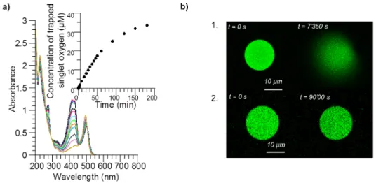
Figure S 1. a) UV-visible absorption spectra of a solution of calcein (λmax = 488 nm) and singlet oxygen scavenger (λmax = 425 nm) (ratio
1:4) during irradiation at 488 nm and singlet oxygen consumption over time. The decrease of the absorption of the scavenger indicates the consumption of singlet oxygen during irradiation. b) Confocal images of 5 mM calcein-loaded PBut2.5-b-PEO1.3 polymersomes during
irradiation (488 nm) 1. without and 2. with 3 eq. sodium azide (NaN3). NaN3 efficiently prevents vesicle rupture (verified on ~15 vesicles).
To further understand the mechanism of vesicle bursting at the molecular level, we investigated the role of ROS production in the overall phenomenon. We first evaluated the generation of ROS using a scavenger for singlet oxygen (1O
2), known to be predominantly generated by excited calcein in the
presence of oxygen.[5] The scavenger is a hydrophilic derivative of 1,3-diphenylisobenzofuran (DPBF),
used for the detection of 1O
2 in vitro.[6] A mixture of calcein in sucrose was prepared with a four-fold
excess of the scavenger. The evolution of the absorption band of the scavenger was followed spectrophotometrically during excitation of the solution at 488 nm on an optical bench (Figure S 1. a). The disappearance of the main emission band at 425 nm (arrow) as a function of illumination time, which is directly correlated with the consumption of 1O
2, confirmed that ROS were effectively
generated by calcein upon irradiation. If ROS generation is part of the process leading to membrane rupture, then the use of a scavenger should prevent vesicles from bursting, as confirmed in Figure S 1. b using sodium azide (NaN3) as a 1O2 quencher.[7] Under the same irradiation conditions, explosion was
inhibited by the presence of the oxygen scavenger, meaning that the scavenger efficiently trapped the generated singlet oxygen. In order to evaluate the consequence of singlet oxygen generation on the osmotic pressure change, a solution of calcein in sucrose (at varying concentration) was prepared at the same concentration as that used in polymersome formation (15 mM). The solution was irradiated at wavelengths ranging between 400 nm and 550 nm for 30 min using a mercury-xenon (Hg-Xe) lamp equipped with a broadband filter, and the osmolarity of the solutions was measured before and after the irradiation by the freezing point determination method.
S8 0.5 1 1.5 2 2.5 3 3.5 4 0 20 40 60 80 100 0 500 1000 1500 R el at ive vi sco si ty ( ν/ν Η 2Ο ) ∆ O smo lari ty ( m O sm/ kg ) Sucrose concentration (mM)
Figure S 2. Evolution of osmolarity increase upon irradiation correlated with the relative viscosity of 15 mM calcein in increasing sucrose
concentrations. Calcein solutions (15 mM) with different sucrose concentrations were irradiated for 30 min in the 400 - 550 nm range with a 200 W Hg-Xe lamp equipped with a filter. Osmolarites were measured before and after irradiation. The difference in osmolarity after irradiation increases with the sucrose concentration, which determines the relative viscosity of the solution (ratio compared to pure water).
Solution osmolarity was found to significantly increase (up to 80 mOsm/kg) with increasing sucrose concentration (Figure S 2). For a solution of 0.35 M sucrose and 15 mM calcein, there was an increase of 18 mOsm/kg after irradiation, whereas no increase was observed when irradiating pure sucrose solutions. Irradiation of calcein in the absence of sucrose led to the same increase of osmotic pressure, thus proving the non-interference of sucrose in the process. Then, we observed that increasing viscosity was correlated with a higher osmolarity difference after irradiation. Indeed, for 0.7 M sucrose, the osmolarity increase was about 40 mOsm/kg and it doubled for 1.4 M sucrose. We propose the interpretation that the viscosity of the solution has an impact on the diffusion of ROS and promotes degradation of the dye by lowering their diffusion away from the dye molecule.
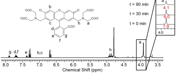
Figure S 3. 1H NMR in D
2O of calcein before and during irradiation (t=30 and 90 min). Integration of proton a resonance (-CH2-COOH,
δ=3.9 ppm) decreases with irradiation time indicating a probable cleavage of the –CH2-COOH arm mediated by ROS. Reference
S9
1H NMR analysis of calcein in D
2O during irradiation (Figure S 3) proved that the changes in
osmolarity were most certainly due to its degradation. Indeed, a decrease of the intensity of the resonance of the CH2 proton adjacent to the carboxylic moiety (a, δ=3.93 ppm, -CH2-COOH) was
observed as a function of irradiation time. Moreover, the apparition of a peak at 8.46 ppm after irradiation was consistent with the presence of formic acid, one of the probable degradation products.[8]
The control experiment, which consisted of irradiating solutions without calcein, did not show any change (Figure S 4 and Figure S 5). Additional control 1H NMR experiments were performed in order
to verify the non-interference of ROS with the membrane of the polymersomes (Figure S 6 and Figure S 7). Based on these results, as well as previous studies of ROS-mediated oxidation[8], the suggested
mechanism is based on a radical-mediated oxidation of calcein, leading to an increase of the internal osmotic pressure of the vesicle caused by the degradation products, arising through bond cleavage at the position labeled a on Figure S 3 (and also most likely at the two quasi-equivalent methylene groups adjacent to the carboxylate moiety).
The same experiments were performed with MB and showed that the dye underwent auto-degradation through the same process (Figure S 8 and Figure S 9).
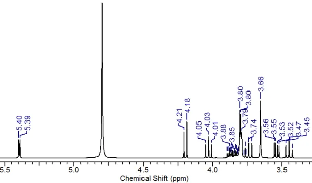
Figure S 4. 1H NMR of sucrose. (400 MHz, D
2O), δ (ppm): 5.42 (d, J=3.8 Hz, 1H), 4.22 (d, J=8.6 Hz, 1H), 4.06 (t, J=8.6 Hz, 1H), 3.91 –
3.86 (m, 1H), 3.84 (m, 1H), 3.81 – 3.78 (m, 4H), 3.77 (t, J=10.1 Hz, 1H,), 3.69 (s, 2H), 3.57 (dd, J=13.1 Hz, J=3.8 Hz, 1H), 3.48 (t, J=9.1 Hz, 1H).
S10 Figure S 5. 1H NMR of sucrose after 30 min irradiation between 400 and 550 nm. (400 MHz, D
2O), δ (ppm): 5.40 (d, J=4 Hz, 1H), 4.21
(d, J= 8.6 Hz, 1H), 4.03 (t, J=8.5 Hz, 1H), 3.89 – 3.86 (m, 1H), 3.84 (m, 1H), 3.81 – 3.78 (m, 4H), 3.74 (t, J=9.9 Hz, 1H), 3.66 (s, 2H), 3.55 (dd, J=9.9 Hz, J=3.8 Hz, 1H), 3.45 (t, J=9.3 Hz, 1H).
Figure S 6. 1H NMR spectrum of PBut
1.2-b-PEO0.6. (400 MHz, CDCl3), δ (ppm): 5.65 – 5.30 (m, 1H), 5.0 – 4.75 (m, 2H), 3.64 (s, 4H),
S11 Figure S 7. 1H NMR spectrum of PBut
1.2-b-PEO0.6 after irradiation at 488nm in presence of calcein. (400 MHz, CDCl3), δ (ppm): 5.65 –
5.30 (m, 1H), 5.0 – 4.75 (m, 2H), 3.64 (s, 4H), 1.25 (s, 2H).
PBut1.2-b-PEO0.6 polymer LUVs of 100 nm diameter were formed by the thin film rehydration method.
Briefly, a polymer film was dried in a round bottom flask and rehydrated overnight with 3 mL of a solution of 30 mM calcein in 0.35 M sucrose. The resulting suspension was extruded through a 100 nm polycarbonate filter (Avanti Polar Lipids). The calcein-loaded polymersomes were then irradiated at 488 nm overnight on an optical bench equipped with a 150 W Hg-Xe lamp and a monochromator. The polymer was recovered for NMR analyses by extraction with dichloromethane.
S12 Evidence of ROS generation by methylene blue
0 0.5 1 1.5 2 2.5 200 300 400 500 600 700 800 Abs or banc e Wavelength (nm) 0 8 16 24 32 40 0 100 200 300 400 C on cen tra tio n of tr ap pe d si ngl et o xy gen (µ M ) Time (min)
Figure S 8. Electronic absorption spectrum showing ROS photogeneration by methylene blue. UV-visible absorption spectrum of a
solution of methylene blue (λabs = 500 -700 nm) and singlet oxygen scavenger (λabs = 360-460 nm) (ratio 1:4) during irradiation at 633 nm
and singlet oxygen consumption over time (inset). The decrease of the absorption band of the scavenger indicates the consumption of singlet oxygen generated during irradiation.
Figure S 9. ESI mass spectrum of methylene blue before and after overnight irradiation. A solution of 5 mM methylene blue in D2O was
irradiated overnight at 633 nm and ESI analyses were performed a) before and b) after irradiation.
After irradiation of methylene blue the ESI spectrum shows an increase of the peak at 270.1 m/z that corresponds to dye degradation. The same peak is present in a smaller proportion in the spectrum before irradiation because the sample was left unprotected from light a few hours before the analysis.
S13 Vesicle swelling upon irradiation
Figure S 10. Confocal observations of POPC GUVs loaded with 30 mM calcein before and after irradiation at 488 nm. The membrane is
doped with L-α-Phosphatidylethanolamine-N-(lissamine rhodamine B sulfonyl) to allow observation in the red channel. Upon irradiation, the vesicle membrane tightens without rupturing.
If the limited diffusion of water across the membrane is responsible for the explosion, using a much more permeable membrane should prevent it. In order to test this hypothesis, we formed 1-palmitoyl-2-oleoyl-sn-glycero-3-phosphocholine (POPC) GUVs[1], as lipidic membranes are known to exhibit
tenfold higher permeability towards water than polymersomes.[9] Figure S 10 represents confocal
images of a POPC GUV loaded with 30 mM calcein before and after irradiation. For visualization in the red channel, the membrane was tagged with rhodamine. The images clearly show a floppy membrane before irradiation. After being excited (λexc= 488 nm; 5 % laser intensity; 20 s), the vesicle swelled
without rupturing, even if higher laser intensity and/or illumination times were applied. These observations confirmed that water indeed diffused into the interior of such permeable lipidic vesicles to compensate for the osmotic imbalance, inducing its swelling, but without a rupture of the membrane. This scenario was confirmed by an additional experiment where a POPC GUV was partially aspired through a micropipette to evaluate its internal volume increase and hence the initial osmotic pressure increase upon calcein irradiation (Figure S 11).
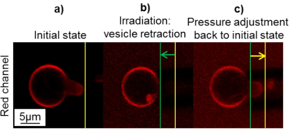
S14 Figure S 11. Controlled membrane deformation of a phospholipid GUV by pressure adjustments through aspiration in a capillary.
Confocal images of a 30 mM calcein-loaded POPC GUV aspired inside a micropipette. The membrane is tagged with rhodamine for visualization in the red channel. Image a) shows the vesicle before irradiation. In image b), the vesicle is irradiated at 488 nm (calcein) and
then (image c)) the pressure is adjusted (i.e. the level of the tank is decreased by a 55 mm height, corresponding to a negative pressure of
540 Pa) to aspire the vesicle portion within the capillary back to its initial position 𝑳P= 𝟓 µm.
In the initial state (a), the vesicle has not been irradiated at 488 nm and the floppy membrane is rendered tauter by aspiration inside the capillary. When the vesicle is irradiated at 488 nm (b), calcein generates ROS that degrade the dye and induce an increase of the osmotic pressure. As the lipid membrane is permeable, water is able to enter the GUV to compensate for the osmotic imbalance. This results in a swelling of the vesicle and a retraction of the membrane tongue from the capillary by a length ∆𝐿P ≈ −4 µm. Using an equation introduced by Olbrich et al. for osmotically-swollen giant
liposomes under micropipette aspiration,[10] we can estimate the volume increase of liposomes caused
by irradiation and ROS production: ∆𝑉 = −𝜋𝐷P(𝐷V− 𝐷P)∆𝐿P⁄ = 91 µm4 3 for a vesicle of diameter
𝐷V= 10.8 µm (initial volume 𝑉i= 660 µm3) aspired in a pipette of diameter 𝐷P = 5 µm. The ratio
∆𝑉 𝑉⁄ i≈ 0.14 can give an estimate of the osmotic pressure variation under illumination ∆𝐶 𝐶⁄ i≈ 0.14
where the initial osmolarity is 𝐶i = 380 mOsm ∙ kg−1. Therefore we can estimate the osmolarity
increase build up by light-induced ROS production and dye degradation at ∆𝐶 ≈ 52 mOsm ∙ kg−1 .
When the pressure inside the capillary (c) was adjusted to pull the vesicle tether end back to its initial position, the lateral tension was raised up to 𝜎 = 1.3 mN.m-1, a value still below the maximum lateral
S15 Supplementary discussion: membrane pore opening dynamics
The dynamics of pore opening were experimentally monitored and fitted with a theoretical model proposed by Mabrouk et al. in which they describe the curling of polymersome membranes induced by nucleation of a pore as a result of creation of an excess area on one of the two leaflets of the membrane through photo-isomerization of azobenzene moieties.[11] We show that our system exhibits the same
trends in pore opening dynamics although the initial constraint needed to induce membrane rupture is different in the two cases.
According to their model, the opening of a pore in the membrane can be compared with the curling rim instability of a bimetallic spring or an asymmetric elastic sheet. The dynamics of pore growth is governed by a transfer of the membrane surface elastic energy S into viscous dissipation of the curling rim within the solution (either the inside solution of the polymersome if the rim curls inward, or the outside solution if the rim curls outwards, depending on whether the photoisomerizable groups are located on the outer or inner leaflet of the asymmetric block copolymer bilayer, respectively). In our case, Fig. 2a clearly shows the calcein outward flow through the pore (the fluorescence bursts arising from a suddenly unquenched signal by dye dilution), whereas the still quenched solution pocket trapped inside the rim is moving into the external medium. Our experiments are thus consistent with their model: the vesicles might also experience a change of spontaneous curvature due to the photo-osmotic phenomenon described to occur in the inner compartment. The pore opening dynamics has two regimes:[11] constant velocity at the early stages, followed by a quasi-diffusive regime at longer times.
More precisely, the effective diffusion constant of the rim (governing the rate of pore opening at long times) is expected to have a bulk (external) solution viscosity dependence when the rim is directed outwards (provoked by azobenzene groups in the inner leaflet in the previous case or internal osmotic pressure in the current case). In their initial publication,[11] Mabrouk et al. gave an oversimplified form
𝑟2+ 2 𝑟 𝑟
𝑐 = 𝐷eff𝑡 that simply reduces to linear regime 𝑟~(𝐷eff⁄ ) 𝑡 at early stages and to pure 2𝑟𝑐
diffusive regime 𝑟~�𝐷eff𝑡 at long stages. Subsequently, they considered a slightly modified form of the
growth dynamics of a pore of radius r at time t as given by Equation (1) [12]:
2𝑟𝑐ṙ 𝐷⁄ eff = 2 (1 +𝑟𝑟 𝑐) 1 2 � − 1 (1 +𝑟𝑟 𝑐) � Eq. (1)
where Deff is the effective diffusion coefficient and rc is defined as the critical pore radius,
𝑟𝑐 = 𝜋𝜅 (𝑒𝑆⁄ 0) , κ being the bending modulus of the membrane, e the membrane thickness and S0 the
stored elastic energy per unit area at initial time t=0). As the line tension (or line energy) of a pore in the membrane is usually expressed as 𝜏 = 𝜅 2𝑒⁄ , we can also write 𝑟𝑐 = 2𝜋 𝜏 𝑆⁄ which represents well 0
the balance between the 2 driving forces that determine the nucleation pore radius: on one side the elastic energy that tends to open up the pore (𝜋𝑟𝑐2𝑆0) and on the opposite the line energy that
S16 The effective diffusion coefficient Deff is given by:
𝐷eff = 2𝑟𝑐𝑆0⁄(𝜂 𝑙𝑙)= 2𝜋𝜅 (𝑒𝜂𝑙𝑙)⁄ = 4π𝜏 (𝜂𝑙𝑙) ≈ 5𝜏 2𝜂⁄ ⁄ Eq. (2)
where η represents the viscosity of the solution in which the membrane curls (the outside glucose solution in our case) and ln is a logarithmic term arising from the drag coefficient of a cylinder that is considered as constant:[11] 𝑙𝑙 = ln(2𝜋 𝑟 𝐿⁄ ) + 1 2⁄ ≈ 5, and 4𝜋 𝑙𝑙⁄ ≈ 5 2⁄ (hence the simplified right
term on Eq. (2).)
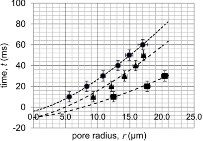
To compare our system with the aforementioned one, we prepared three different batches of giant vesicles with the emulsion-centrifugation technique (see experimental methods) with varying glucose concentrations. In each experiment, the concentration of the inner sucrose solution was adjusted to reach an osmotic equilibrium with the outer glucose solution. Calcein (15 mM) was encapsulated within the vesicles to enable bursting under laser irradiation (488 nm, 5-10 % laser intensity). Movies of pore bursting were obtained at the highest achievable speed of the Leica SP5 resonant scanner (100 frames/s). The extracted frames of the explosions were processed with ImageJ freeware to track and measure membrane pore growth. For each glucose concentration, the experiment was repeated on an average of ten vesicles of equivalent sizes, and the results were plotted on a master curve (horizontal error bars represent standard deviations from different vesicles and horizontal error bars correspond to ± 5 ms, i.e. half of the interval between successive frames) (Figure S 12).
Figure S 12. Variations of pore opening dynamics in PBut2.5-b-PEO1.3 vesicles containing sucrose and calcein 15 mM depending on
viscosity of the external glucose solution. Pore radius as a function of time for three different glucose viscosities as averaged on several pore dynamics: (●) η=1.33 mPa⋅s, (▲) η=2.46 mPa⋅s, (■) η=4.19 mPa⋅s. Equation (1) was used to fit the experimental data with two parameters: the critical pore radius (rc) and the effective diffusion constant (Deff). The external glucose concentrations were chosen to
balance osmolarity of the internal sucrose/calcein mixtures: (●) sucrose 20% in glucose 10% (0.6 M), (▲) sucrose 50% in glucose 26% (1. 475M), (■) sucrose 70% in glucose 36% (2.06 M). Negative points on the time scale arise from the uncertainty about the initial time of
pore nucleation (on the order of 10 ms, i.e. the interval between successive frames).
-20 0 20 40 60 80 100 0.0 5.0 10.0 15.0 20.0 25.0 tim e, t (m s) pore radius, r (µm)
S17 Glucose concentration (M) Glucose viscosity η (mPa⋅s) Apparent diffusion coefficient Deff, (μm2⋅s-1) 0.6 1.33 1111 1.475 2.46 612 2.060 4.19 522
Table S 1. Variation of the apparent diffusion coefficient with external glucose solution viscosity (measured independently by rolling ball
viscosimetry) as obtained by fitting the pore dynamics (Figure S1) with Eq. (1) with a constant critical radius 𝒓𝒄= 𝟎. 𝟑𝟑 µ𝒎. The calcein
concentration is maintained at 15 mM.
From the fits derived from the experimental data (Eq. (1)), the effective diffusion coefficients Deff could
be calculated by fixing the value of the critical radius rc to 0.38 μm for each viscosity. In agreement
with the model proposed by Mabrouk et al., the diffusion coefficient is roughly inversely proportional to the viscosity of the solution into which the membrane curling rim advances (which determines the viscous losses) as in Eq. (2). The slight deviation from the perfect ~ 1 𝜂⁄ dependency might be ascribed to a weak variation also of the line tension of the PBut2.5-b-PEO1.3 membrane with the sucrose and
glucose concentrations, as with the reported modification of the mechanical properties (𝜅 and thus 𝜏) for phospholipid bilayers exposed to high sugar concentrations.[13] Regarding quantification, we can
estimate the structural (hydrophobic thickness e) and mechanical (bending modulus κ) properties of the PBut2.5-b-PEO1.3 membrane from their reported experimental values: 𝑒 = 9.6 nm and 𝜅 = 24.7 ±
11.1𝑘B𝑇 , according respectively to cryo-TEM image analysis and micropipette measurements.[14]
Scaling laws versus the molar mass Mh of the hydrophobic block were proposed by Bermudez et al.:[15]
𝑒~𝑀h1 2⁄ and 𝜅~𝐾a𝑒2~𝑀h since the area expansion elastic modulus was shown to be constant material
property 𝐾a = 102 ± 11 mN/m for all the PBut-b-PEO copolymer series. Therefore we deduce
𝑒 = 6.7 nm and 𝜅 = 11.9 ± 5.3𝑘B𝑇 for the less commonly used PBut1.2-b-PEO0.6 copolymer. From
these values, we can obtain an estimate of the pore line tension: 𝜏 = 𝜅 2𝑒⁄ = 3.7 ± 1.7 pN and 5.3 ± 2.4 pN for PBut1.2-b-PEO0.6 and PBut2.5-b-PEO1.3, respectively. Such line energies compare well to the
value 𝜏 = 4 pN estimated by Mabrouk et al. for PBut-b-PEO copolymers,[11] and are significantly lower
than those measured on DPPC large liposomes at 46°C by Taupin et al. through the leakage of parametric probes under various hypotonic shocks (from 𝜏 = 6.5 pN for irreversible pores to 𝜏 = 5.5 − 8 pN on sub-critical resealing pores)[16], on DOPC giant liposomes by Karatekin et al.[17] through the closure dynamics of transient pores (from 𝜏 = 6.9 ± 0.4 pN to 𝜏 = 20.7 ± 3.5 pN depending on the supplier, and thus of purity, of DOPC), or by Levadny et al.[18] by statistical analysis of the pore
S18 qualitatively equivalent to the experiment of Mabrouk et al. with photo-isomerizable liquid crystalline copolymers, in our case with PBut2.5-b-PEO1.3 the critical radius 𝑟𝑐 = 0.38 µ𝑚 is much lower, which
implies that the initial elastic tension is higher: 𝑆0 = 8.8 ∙ 10−2mN. m−1. From the expression of the
initial elastic tension 𝑆0 =12𝜅(𝑐0)2, we get an estimate of the spontaneous curvature 𝑐0 of the
membrane, from which we deduce the spontaneous radius of curvature (𝑐0)−1≈ 24 nm. Interestingly,
this value is typically comparable to the smallest radius that large unilamellar vesicles (LUV) of a given copolymer can withstand from the rule-of-thumb 𝑒 𝑅⁄ min ≈ 0.25 as shown by Salva et al. after LUV
rupture by hypertonic shocks,[19] thus here 𝑅
min ≈ 38 nm for PBut2.5-b-PEO1.3 polymersomes.
To interpret the different fates between liposomes and polymersomes exposed to increased internal osmotic pressure through dye irradiation at its maximum absorption wavelength, we propose a kinetic control of the membrane pore induction in these two different types of vesicles as opposed to a mere thermodynamic control sometimes put forward by several authors.[20,21] To give quantitative values, the
water permeability of monounsaturated phospholipids like POPC measured by the Evans micropipette aspiration method is P∼30 µm⋅s-1 and their lysis tension is σ
lys∼10 mN⋅m-1,[10] whereas values for PBut2.5
-b-PEO1.3 polymersomes are respectively P∼3.1±1.6 µm⋅s-1 from osmotic inflation experiments[22] and
σlys∼16 mN⋅m-1 from micropipette aspiration measurements[23]. In brief, polymersomes resist larger
applied lateral stresses than fluid-phase liposomes and exhibit a higher toughness (i.e. a larger surface area below the curve of lateral tension versus membrane expansion coefficient), but they are 10 times less permeable to water, which is mainly ascribed to their thicker hydrophobic membrane (∼6–10 nm as compared to ∼3–5 nm). The lateral tension arising from the increased osmotic pressure relative to the external solution after dye photo-degradation can be estimated by the Laplace law of fluid interfaces ∆𝜋 = 2𝜎0⁄ where R𝑅0 0 is the initial vesicle radius and σ0 is the lateral tension just before pore aperture.
Converting the estimated osmolarity difference ∆𝜋 ≈ 18 mOsm ∙ kg−1 into an osmotic pressure
through the perfect gas law ∆𝜋 = ∆𝑐𝑅𝑇 = 4.5 ∙ 104Pa and using the Laplace law, we obtain a lateral
tension σ0∼225 mN⋅m-1 with R0=10 µm, well above the rupture tension σlys.
In the case of liposomes, the lateral tension on the membrane built up by the increase of solute concentration inside the vesicles can relax by internal volume reduction through outwards flow across “sub-critical” pores, i.e. holes in the bilayer that have a size insufficient to expand (below the critical radius 𝑟𝑐) but nevertheless contribute to increase permeability. Their existence has been evidenced more
than four decades ago by the pioneering work of Taupin et al.[16] using electron paramagnetic resonance
(EPR) probes. These authors also demonstrated that the frequency of vesicle leakage (i.e. inverse of mean leakage time) through transient pores for a given solute over-concentration inside the vesicles is inversely proportional to 𝜏2, the square of line tension. This mechanism of transient or “pulsatile” pore
S19 certainly not work in the case of polymersomes due to their too low line energy 𝜏 compared to liposomes, not sufficient to reseal the membrane once a pore is nucleated. This is why for the same osmotic pressure imposed (same solutes encapsulated and same conditions of illumination), polymersomes leak much more slowly than liposomes, and thus they stay longer in a taut state without relaxing their lateral tension, until they eventually burst irreversibly. The osmotically driven vesicle rupture triggered by illumination described in this article corresponds to a kinetic control of rupture, the lower permeability of polymersomes letting them for longer in a tense state and maximizing their probability to undergo rupture with less possibility of resealing (since the driving force for pore closure is 𝜏).
S20 Additional figures 0 0.2 0.4 0.6 0.8 1 -5 0 5 10 15 20 1 10 100 1000 104 105 0 1000 2000 3000 4000 5000 In te nsi ty ( % ) C or re lati on c oe ffic ient Size (nm) Time (ms)
Figure S 13. DLS measurement of a solution of extruded 100 nm DPPC liposomes.
0 0.2 0.4 0.6 0.8 0 5 10 15 20 1 10 100 1000 104 105 0 1000 2000 3000 4000 5000 In te nsi ty ( % ) C or re lat ion c oe ffic ient Size (nm) Time (µs)
S21 References
[1] A. Weinberger, F.-C. Tsai, G. H. Koenderink, T. F. Schmidt, R. Itri, W. Meier, T. Schmatko, A. Schröder, C. Marques, Biophys. J. 2013, 105, 154–164.
[2] M. Marguet, L. Edembe, S. Lecommandoux, Angew. Chem. Int. Ed. 2012, 51, 1173–1176.
[3] W. A. Velema, J. P. van der Berg, W. Szymanski, A. J. M. Driessen, B. L. Feringa, ACS Chem. Biol. 2014, 9, 1969–1974.
[4] B. Karami, M. Kiani, J. Chin. Chem. Soc. 2014, 61, 213–216.
[5] C. Beghetto, C. Renken, O. Eriksson, G. Jori, P. Bernardi, F. Ricchelli, Eur. J. Biochem. 2000, 267, 5585–5592.
[6] D. Arian, E. Cló, K. V. Gothelf, A. Mokhir, Chem. Weinh. Bergstr. Ger. 2010, 16, 288–295. [7] M. Bancirova, Luminescence 2011, 26, 685–688.
[8] T. F. Zhu, J. W. Szostak, J. Syst. Chem. 2011, 2, 4.
[9] J.-F. L. Meins, O. Sandre, S. Lecommandoux, Eur. Phys. J. E 2011, 34, 1–17. [10] K. Olbrich, W. Rawicz, D. Needham, E. Evans, Biophys. J. 2000, 79, 321–327.
[11] E. Mabrouk, D. Cuvelier, F. Brochard-Wyart, P. Nassoy, M.-H. Li, Proc. Natl. Acad. Sci. 2009,
106, 7294–7298.
[12] E. Mabrouk, Vésicules Polymères (Polymersomes) Stimulables, Paris 6, 2010. [13] R. Dimova, Adv. Colloid Interface Sci. 2014, 208, 225–234.
[14] H. Bermúdez, D. A. Hammer, D. E. Discher, Langmuir 2004, 20, 540–543.
[15] H. Bermudez, A. K. Brannan, D. A. Hammer, F. S. Bates, D. E. Discher, Macromolecules 2002,
35, 8203–8208.
[16] C. Taupin, M. Dvolaitzky, C. Sauterey, Biochemistry 1975, 14, 4771–4775.
[17] R. Salva, J.-F. Le Meins, O. Sandre, A. Brûlet, M. Schmutz, P. Guenoun, S. Lecommandoux, ACS Nano 2013, 7, 9298–9311.
[18] E. Karatekin, O. Sandre, H. Guitouni, N. Borghi, P.-H. Puech, F. Brochard-Wyart, Biophys. J. 2003, 84, 1734–1749.
[19] V. Levadny, T. Tsuboi, M. Belaya, M. Yamazaki, Langmuir 2013, 29, 3848–3852. [20] M. A. S. Karal, M. Yamazaki, J. Chem. Phys. 2015, 143, 81103.
[21] J. C. Shillcock, U. Seifert, Biophys. J. 1998, 74, 1754–1766.
[22] A. Carlsen, N. Glaser, J.-F. Le Meins, S. Lecommandoux, Langmuir 2011, 27, 4884–4890.
[23] P. P. Ghoroghchian, J. J. Lin, A. K. Brannan, P. R. Frail, F. S. Bates, M. J. Therien, D. A. Hammer, Soft Matter 2006, 2, 973–980.
![Figure 2. a) Photocleavage of N-diethyl, O-({7-[bis(carboxymethyl)- O-({7-[bis(carboxymethyl)-amino]coumarin-4-yl}methyl carbamate (coumarin derivative) under irradiation](https://thumb-eu.123doks.com/thumbv2/123doknet/14516487.530646/3.892.462.801.140.444/photocleavage-carboxymethyl-carboxymethyl-coumarin-carbamate-coumarin-derivative-irradiation.webp)