HAL Id: hal-02612315
https://hal.archives-ouvertes.fr/hal-02612315
Submitted on 18 Dec 2020
HAL is a multi-disciplinary open access
archive for the deposit and dissemination of
sci-entific research documents, whether they are
pub-lished or not. The documents may come from
teaching and research institutions in France or
abroad, or from public or private research centers.
L’archive ouverte pluridisciplinaire HAL, est
destinée au dépôt et à la diffusion de documents
scientifiques de niveau recherche, publiés ou non,
émanant des établissements d’enseignement et de
recherche français ou étrangers, des laboratoires
publics ou privés.
HS and Inflammation: A Potential Playground for the
Sulfs?
Rana El Masri, Yoann Crétinon, Evelyne Gout, Romain R Vivès
To cite this version:
Rana El Masri, Yoann Crétinon, Evelyne Gout, Romain R Vivès. HS and Inflammation: A Potential
Playground for the Sulfs?. Frontiers in Immunology, Frontiers, 2020, 11, �10.3389/fimmu.2020.00570�.
�hal-02612315�
Edited by: Rogier M. Reijmers, Leiden University Medical Center, Netherlands Reviewed by: Kenji Uchimura, UMR8576 Unité de Glycobiologie Structurale et Fonctionnelle, France Mark Michael Fuster, University of California, San Diego, United States Steven David Rosen, University of California, San Francisco, United States *Correspondence: Romain R. Vivès [email protected]
Specialty section: This article was submitted to Inflammation, a section of the journal Frontiers in Immunology Received: 04 December 2019 Accepted: 12 March 2020 Published: 03 April 2020 Citation: El Masri R, Crétinon Y, Gout E and Vivès RR (2020) HS and Inflammation: A Potential Playground for the Sulfs? Front. Immunol. 11:570. doi: 10.3389/fimmu.2020.00570
HS and Inflammation: A Potential
Playground for the Sulfs?
Rana El Masri, Yoann Crétinon, Evelyne Gout and Romain R. Vivès*
Université Grenoble Alpes, CNRS, CEA, Institut de Biologie Structurale (IBS), Grenoble, FranceHeparan sulfate (HS) is a complex polysaccharide abundantly found in extracellular
matrices and cell surfaces. HS participates in major cellular processes, through its
ability to bind and modulate a wide array of signaling proteins. HS/ligand interactions
involve saccharide domains of specific sulfation pattern. Assembly of such domains is
orchestrated by a complex biosynthesis machinery and their structure is further regulated
at the cell surface by post-synthetic modifying enzymes. Amongst them, extracellular
sulfatases of the Sulf family catalyze the selective removal of 6-O-sulfate groups, which
participate in the binding of many proteins. As such, increasing interest arose on the
regulation of HS biological properties by the Sulfs. However, studies of the Sulfs have so
far been essentially restricted to the fields of development and tumor progression. The
aim of this review is to survey recent data of the literature on the still poorly documented
role of the Sulfs during inflammation, and to widen the perspectives for the study of this
intriguing regulatory mechanism toward new physiopathological processes.
Keywords: heparan sulfate (HS), inflammation, glycosaminoglycan/protein interactions, sulfatase, chemokine, leukocyte migration
INTRODUCTION
Heparan sulfate proteoglycans (HSPGs) are major components of the cell surface, extracellular
matrix (ECM) and basement membrane in most animal cells. They are composed of a protein
core, onto which are covalently attached complex, anionic Heparan Sulfate (HS) chains of
the glycosaminoglycan (GAG) polysaccharide family. Through the ability of their HS chains
to bind, modulate and control bioavailability of a multitude of protein ligands, HSPGs are
involved in a plethora of biological processes, including cell adhesion, migration, proliferation and
differentiation, embryo development, inflammation, control of angiogenesis, blood coagulation,
tumor growth, and metastasis (
1
–
3
). Structurally, a HS chain is characterized by the linear
repetition of a disaccharide unit, composed of alternating N-acetyl glucosamine (GlcNAc) and
glucuronic acid (GlcA). During biosynthesis, the polymer undergoes a series of modifications,
which include the N-deacetylation/N-sulfation of glucosamine to form N-sulfo glucosamine
(GlcNS), the C5 epimerization of GlcA into iduronic acid (IdoA), and O-sulfations at positions C2
of IdoA and C6 (more rarely C3) of glucosamine residues. Tight regulation of these modification
steps leads to the generation of specialized saccharide regions termed S-domains, exhibiting both
remarkable structural diversity and high sulfation content. These S-domains are involved in the
recognition and binding of most HS ligands (
4
–
6
). HS is structurally closely related to heparin,
although the latter displays a much higher sulfation content. Therefore, heparin has been widely
used as a surrogate of HS S-domains for protein interaction studies.
El Masri et al. Potential Roles of Sulfs in Inflammation
HS IN INFLAMMATION
Inflammation is a complex, multi-step process leading to
the rapid recruitment of leukocytes from the blood to the
inflammation site. Briefly, emission of an inflammatory signal
triggers the secretion of cytokines and chemokines that diffuse
throughout the tissue and activate leukocytes and vascular
endothelial cells. Cell activation leads to the adhesion and rolling
of leukocytes on the endothelium toward the inflammatory site.
Leukocytes will then cross the endothelium to the basement
membrane, and migrate toward the inflamed tissue to initiate
immune responses. Through its large protein binding properties,
HS participates in various steps of inflammation [for review, see
(
7
,
8
)].
Inflammation is first initiated by the production, upon
endogenous or exogenous signals, of inflammatory cytokines.
Amongst these, chemokines are a family of small proteins
involved in many biological processes such as development,
inflammation and immunosurveillance (
9
). Chemokines induce
the activation of the endothelium and the migration of leukocytes
from blood toward inflammatory sites. To elicit their functions,
they bind to their primary G-coupled receptors that trigger
downstream signaling. In addition, all chemokines bind to
cell surface and ECM HS. The structural basis of these
interactions has been intensively studied and is now well
documented (
10
–
12
).
Functionally, HS does not seem to be essential for chemokine
signaling in vitro. However, in vivo studies showed that
chemokines unable to bind to HS failed to recruit leukocytes
(
13
), and that HS modulated chemokine activity through
different mechanisms [for review, see (
12
,
14
)]. HS first regulates
chemokine diffusion and sequestration. In some instance,
the capture of the chemokine/cytokine by HS prevents its
release and thus its activity (
15
). However, by regulating
chemokine diffusion, HS participates in the formation and
stabilization of chemotactic gradients providing directional cues
for migrating leukocytes. In support to this, in vivo inhibition of
CXCL12/HS interaction using sulfated polysaccharide tilted the
chemokine distribution from bone marrow toward the plasma,
thereby causing the release of hematopoietic progenitor cells
in the blood circulation (
16
). HS also mediates chemokine
transcytosis across the endothelial cell wall (
17
,
18
), and
protects chemokines/cytokines from enzymatic degradation
and inactivation (
19
–
21
). Finally, HS may further modulate
chemokine activity by inducing chemokine oligomerization [for
review, see (
11
)], which has been shown to be functionally
relevant in vivo (
13
). In that context, an original HS-dependent
cooperative mechanism driving CCL5 dimerization has been
characterized (
22
).
Activation by pro-inflammatory chemokines and cytokines
induces the expression of endothelial C-type lectins E- and
P-selectins. These bind a variety of glycosylated ligands present
on the leukocyte cell surface to initiate adhesion and rolling
of leukocytes on the endothelium. Recruitment is further
promoted through additional interactions, involving L-selectin
constitutively expressed on leukocytes with leukocyte and
endothelial ligands (
23
,
24
). HS may participate to this process,
as studies reported binding to L-selectin (
24
,
25
), P-selectin
(
26
), and E-selectin (
27
,
28
). However, it should be noted that
the physiological relevance of these interactions remains to
be clarified. Studies showed that the removal of cell surface
HS with heparinases reduced L-selectin dependent binding of
monocytes, and leukocyte rolling on endothelial cells (
29
,
30
).
During acute inflammation, HS was shown to support L-selectin
dependent rolling of neutrophils on lung microvasculature (
31
).
On the contrary, lymphocyte rolling on high endothelial venules
(HEV) exclusively relied on interactions of L-selectin with ligands
bearing sialyl Lewis X (sLe
x) glycosylation motifs, suggesting no
involvement of HS in L-selectin-dependent lymphocyte homing
(
32
). One could speculate that these discrepancies could be due
to the presence of distinct HS structures and/or sLe
xligands on
these different cell types.
Leukocyte rolling is then arrested through increased
integrin-mediated cell-cell adhesion. In a recent study, it has been
proposed that endothelial cell surface HS could participate
indirectly to this process, by capturing and presenting CCL21,
which would in turn activate integrin LFA-1 on rolling
lymphocytes (
32
). Following arrest, leukocytes get access to
inflamed tissues through extravasation across the endothelial cell
wall. They then reach the basement membrane that comprises
numerous interacting molecules and a variety of HSPGs,
including perlecan, agrin and XVIII collagen, which may further
modulate the extravasation process. These HSPGs can bind
many chemokines, cytokines and growth factors that are critical
for leukocyte migration, and contribute to the formation of
chemokine gradients. On the contrary, they can act as a physical
barrier hindering leukocyte migration.
Finally, studies have also suggested a role of HS in the
phagocytosis process (
33
,
34
). A proposed mechanism is that
newly-exposed HS binding sites at the surface of apoptotic
cells could facilitate their recognition, uptake and clearance by
macrophages (
35
). However, in vivo data are still needed to clarify
this process.
Most of these inflammatory steps are generally accompanied
by changes in the expression of their cell-surface HPSGs and HS
structure. Many studies have reported an upregulation of
cell-surface HSPG Syndecan-1 upon endothelial cell activation by
pro-inflammatory cytokines [reviewed in (
25
)]. Differentiation
of monocytes into macrophages leads to high expression of
Syndecan-1, -2 and -4, whereas macrophage activation by
Interleukin (IL)-1 results in the overexpression of Syndecan-2
only (
36
). Furthermore, the activation of T cells induces the
expression of Syndecans and Glypicans, while the differentiation
of B cells into plasma cells specifically triggers Syndecan-1
expression (
37
,
38
). Depending on the inflammatory stimuli, HS
length, structure and sulfation profiles may also be affected. For
instance, the size and 6-O-sulfation patterns of HS are altered
in primary human endothelial cells upon treatment with tumor
necrosis factor (TNFα) or IL-1 (
39
). Pro-inflammatory effectors
have been shown to modulate the expression of NDSTs (
40
–
42
). In particular induction of NDST1 led to the production
of highly sulfated HS that increased the sequestration of CCL5,
thereby promoting leukocyte extravasation (
42
). Vascular lesions
in mice have also been associated with a significant increase
of NDST1 expression (
43
). In line with this, inactivation
of NDSTs in endothelial cells led to impaired rolling and
infiltration of neutrophils and macrophages into inflammatory
sites (
31
,
44
). In contrast, restricted inactivation of NDSTs to
leukocytes had no effect on leukocyte infiltration (
31
). Changes
in HS O-sulfotransferases (OSTs) expression have also been
reported, during macrophage M1/M2 polarization (
45
), or the
development of renal fibrosis (
46
). Interestingly, the stimulation
of human monocytes by LPS or TNFα upregulates only one
out of the seven 3OST isoform: 3OST3b, thus highlighting
the fine tuning of sulfotransferase expression pattern during
inflammation (
47
). Finally, the silencing of 2OST in mouse
endothelial cells during acute inflammation resulted in enhanced
HS 6-O- and N-sulfation, leading to increased neutrophil
infiltration (
48
).
POST-EDITING MECHANISMS
REGULATING HS DISTRIBUTION,
STRUCTURE AND FUNCTION
Although
HS
expression
and
structure
is
primarily
controlled during polysaccharide biosynthesis, increasing
evidence have also highlighted the importance of the
additional regulatory step provided by post-editing enzymes,
including Heparanase, sheddases, and sulfatases of the
Sulfs family.
In inflammatory processes, post-synthesis regulation of
HS plays a significant role. Heparanase is an
endo-β-D-glucuronidase targeting [GlcA-GlcNS] linkages within HS.
Heparanase cleaves long HS chains from ECM and cell-surface
HSPGs into shorter fragments of 10–20 sugar units. This results
in the release of sequestered HS bound ligands, such as growth
factors, chemokines and morphogens, which can then induce
angiogenesis, cell proliferation and motility (
49
). Consequently,
Heparanase has been associated with various pathologies,
including cancer, inflammation, thrombosis, atherosclerosis,
fibrosis, diabetes, and kidney disease (
50
). During inflammation,
Heparanase plays multiple roles. It favors neutrophil adhesion
onto the endothelium, by degrading endothelial cell-surface
HS and unmasking membrane adhesion molecules (
51
). It
also facilitates leukocyte extravasation, by degrading basement
membrane HSPGs (
52
). More recently, Heparanase has
been shown to enhance T-cell activity (
53
). Furthermore,
intracellular heparanase upregulated the transcription of genes
involved in T-cell differentiation (
54
). Finally and noteworthy,
Heparanase expression is also markedly increased during
neuroinflammation (
55
).
Shedding of HSPGs takes place upon activation of
metalloproteinases, plasmins and elatases by inflammatory
cytokines (
56
–
59
). These proteases target the core protein
of HSPGs like Syndecans, thus releasing soluble HS-peptide
conjugates. Shedding regulates the amount of HSPGs found
at the cell surface or in the ECM and facilitate the release of
sequestered chemokines, which contributes to the resolution
of neutrophilic inflammation (
58
,
60
). In combination with
Heparanase, shedding also facilitate leukocyte migration
by altering the architecture of the ECM and basement
membranes (
52
).
In addition, soluble released HS fragments/conjugates
(produced by sheddases or Heparanase) induce the secretion by
macrophages and splenocytes of pro-inflammatory cytokines
through activation of the NF-κB signaling pathway (
61
–
63
).
Soluble HS fragments also activate neutrophils and promote
an immune response via toll like receptor-4 (
64
,
65
). They
can also trigger the maturation of dendritic cells, leading to
their migration toward lymphoid organs to elicit primary
immune responses (
66
). In line with this, it was shown that an
exogenous administration of HS or elastase resulted in a systemic
inflammatory response syndrome (SIRS)-like reaction (
67
).
THE SULFS: POST-SYNTHETIC
REGULATORS OF HS STRUCTURE AND
FUNCTION
Sulfs are extracellular endosulfatases that catalyze the
6-O-desulfation of HS. Sulf-1 was first discovered in Quail (
68
),
then orthologs were later identified in mouse, rat, chick, C.
elegans, zebrafish, and human, as well as a second related enzyme,
Sulf-2 (
69
). In human, HSulf-1 and HSulf-2 (encoded by two
distinct genes) feature a common structural organization (
69
).
Maturation of the sulfs involves furin-type processing of
pro-enzyme proteins, yielding two sub-units linked by one or more
disulfide bonds (see Figure 1 inset). The N-terminal regions
features the enzyme catalytic (CAT) domain, which shows strong
homology with most eukaryotic sulfatases. The CAT domain
comprises the enzyme active site, including the two conserved
sulfatase signature amino acid sequences as well as the cysteine
modified, catalytic FGly residue. The second functional domain
of the Sulfs is the highly charged basic domain (HD), which
spans over both N-terminal and C-terminal sub-units. The HD
domain is a unique feature of the Sulfs, as it shares no homology
with any other known protein. It is responsible for high affinity
binding to HS substrate and is therefore required for the enzyme
endo-6-O-sulfatase activity (
70
–
73
). In addition, the HD domain
has been shown to mediate the capture of Sulfs on cell-surface
HS thereby modulating enzyme diffusion (
71
). Finally, Sulf
C-terminal region shows homology to Glucosamine-6-sulfatase
(G6S) and Arabidopsis thaliana GlcNAc transferase, suggesting
a role of this domain in the recognition of glucosamine
motifs (
69
).
Sulfs catalyze the 6-O-desulfation of HS with a strong
preference
for
[Glc/IdoA(2S)-GlcNS(6S)]
trisulfated
disaccharides that are essentially found within S-domains.
Although abundant in heparin, this disaccharide motif is
relatively scarce in HS. Sulf-induced modification of HS
structure is therefore structurally subtle, but with great
functional consequences, as 6-O-sulfation pattern of HS
S-domains is critical for the binding of many signaling proteins
(
74
). Sulfs are therefore implicated in a number of physiological
and pathological processes, including development, tissue repair,
neurodegenerative disease and cancer [for reviews, see (
75
–
77
)].
El Masri et al. Potential Roles of Sulfs in Inflammation
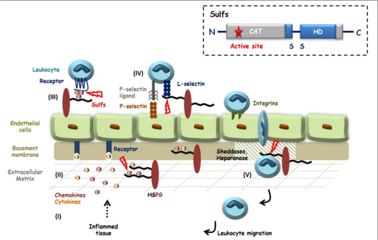
FIGURE 1 | HS in inflammation and potential roles of the Sulfs. Inflammatory stimuli induce the secretion of cytokines and chemokines (I) that activate endothelial cells and blood circulating leukocytes. HS controls the diffusion of these pro-inflammatory proteins, their oligomerization and the establishment of chemotactic gradients (II and III). Activated Leukocytes then adhere and roll over endothelial cells, through interactions of E- and P-selectins with their counter ligands. L-selectin tightens cell-contacts by binding to sLeXdecorated glycoproteins and HSPGs (IV). After passage through the endothelial layer, efficient migration of leukocytes toward
inflammatory sites requires the degradation of basement membrane HSPGs by proteases and Heparanase (V). HS is thus largely involved during inflammation and HS 6-O-sulfation is critical for most of these interactions. Although still poorly investigated, Sulfs could therefore play different roles during inflammation. In the figure, steps during which Sulfs could be implicated are highlighted with red bolts. Inset: schematic representation of Sulf structural organization.
DISCUSSION: POTENTIAL ROLES OF THE
SULFS DURING INFLAMMATION
Over the recent years, Sulfs have emerged as critical regulators of
HS functions, with well documented roles during development
and cancer. However and despite growing evidence, Sulfs have
remained largely unstudied in the context of inflammation (see
Figure 1
).
It is first well established that HS 6-O-sulfation is a
major structural determinant for the interaction with many
chemokines, including CXCL12 (
78
–
80
), CXCL8 (
81
,
82
),
CXCL4 (
83
,
84
) and CCL5 (
85
). For the latter, the resolution
of the CCL5 crystal structure in complex with heparin
provided further evidence of the contribution of 6-O-S
group in the interaction (
85
). Furthermore, HS mimetic
glycopolymers featuring [IdoA(2S), GlcNS(6S)] disaccharides
efficiently inhibited CCL5/heparin interaction (
86
). In this
context, it is therefore most likely that Sulfs would significantly
affect HS binding properties toward many of these chemokines,
with consequences on chemokine oligomerization, storage,
bioavailability and diffusion. Interestingly, a recent study
investigating chemokine inhibition using synthetic sulfated
[IdoA(2S), GlcNS(±6S)]
6dodecasaccharides showed that the
presence of a 6-O-sulfate group on the non-reducing end residue
switched saccharide binding properties from CXCL8 toward
CXCL12 (
87
). In light of this, it was also recently reported
that HSulfs catalyzed the 6-O-desulfation of HS following a
processive and orientated mechanism, starting from S-domain
non-reducing end (
88
). Together, these data thus suggest that
Sulfs could tune HS binding selectivity for chemokines. Despite
these findings and most surprisingly, the effect of Sulfs on
HS/chemokine interaction has so far only been investigated in
one study, which showed the inhibition of CXCL12-α binding to
heparin by the Sulfs in an in vitro immunoassay (
79
).
Studies have reported that the 6-O-sulfation of endothelial
HS was critical for leukocyte rolling (
89
), and that
6-O-sulfation of heparin was necessary to block L-selectin mediated
leukocyte adhesion (
80
,
90
). Sulfs could thus be involved in the
control of leukocyte adhesion and migration on the activated
endothelium. Vascular glycocalyx and especially endothelial HS
undergo significant degradation during inflammation (
91
). A
role of the Sulfs in these mechanisms could thus be postulated,
as suggested by a first study on post-septic mice. Sepsis is
associated with a hyper-inflammatory process, followed by a
delayed period of immunosuppression called compensatory
anti-inflammatory response syndrome (CARS), which can lead
to secondary infections. This study showed that HS of lung
endothelial glycocalyx displayed higher 6-O-sulfation content
after septic injury, which was due to a downregulation of
Sulf-1. Interestingly, the post-septic loss of Sulf-1 was necessary for
CARS to occur, as the administration of exogenous recombinant
Sulf-1 intravenously reversed the immunosuppression phenotype
(
92
). In the same context, Sulfs could also participate in
the degradation of the basement membrane, along with
Heparanase and proteases to facilitate leukocyte migration
toward inflammatory sites. Although there is no evidence of such
an involvement yet, a study preceding the discovery of the Sulfs
showed that activation of endothelial cells with pro-inflammatory
cytokines led to the detection of a sulfatase activity, which was
required for the degradation of the basement membrane (
52
).
Changes in the expression of the Sulfs upon inflammatory
conditions have been reported. An in vivo study on renal
allograft biopsies showed that Sulf-1 expression was repressed
in inflammatory conditions (
93
). In contrast, HSulf-1 was
up-regulated in human fibroblasts upon TNF-α treatment (
94
).
Likewise, TGF-β1 induced the expression of Sulf-2 in renal
epithelial cells (
95
), and of both Sulf-1 and Sulf-2 in lung
fibroblasts (
96
). In line with this, Sulf-2 was overexpressed in
idiopathic pulmonary fibrosis (
97
) and would act as a regulator,
in a negative feedback loop, of TGF-β1 signaling in type 2
alveolar epithelial cells (
96
,
97
). Furthermore, it was shown
that Sulf-2 expression in type II alveolar epithelial cells played
a protective role in epithelial lung injury, inflammation and
mortality (
98
). Surprisingly, Sulf-2 overexpression in human
hepatocellular carcinoma cells promoted TGF-β1 signaling (
99
).
Altogether, these data clearly underline the complex interplay
between Sulf activities and TGF-β1 signaling.
One still intriguing and yet poorly understood issue about
the Sulfs is that the two human forms, HSulf-1 and HSulf-2,
feature very similar enzyme activities in vitro, but show clear
functional discrepancies in vivo. As such, a study on Sulf KO
mice indicated that both forms exhibit redundant or overlapping
functions during development (
100
,
101
), while accumulating
data describe opposite activities in cancer (
75
,
76
). Furthermore,
it has been reported that alternative splicing of Sulf1/Sulf2
genes generated functionally active variants. In Quail, a QSulf-1
variant (QSulf-1B) encoding for a shorter protein form exhibited
opposite activities to full length QSulf-1, QSulf-1B inhibiting
Wnt signaling and promoting angiogenesis (
102
). Mammalian
variants of Sulf1 and Sulf2 were later identified in tumors (
103
,
104
). The occurrence and biological significance of Sulf variants
are still poorly understood. However, some of these variants have
been shown to regulate growth factor signaling pathways and
to display anti-oncogenic and anti-angiogenic properties (
105
).
Noteworthy, the expression of Sulf-1 and Sulf-2 variants was
also reported in the context of inflammation (
105
). Studying the
spatial and temporal expression and activity of the two Sulf forms
as well as their respective splice variants may therefore be critical
to fully understand the role of these enzymes in a given biological
process such as inflammation.
Finally, another interesting observation arose from our recent
article reporting the expression and purification of recombinant
HSulf-2 (
73
). In this work, we showed that the protein
HD domain was very sensitive to proteolytic digestion. This
observation is in agreement with secondary structure predictions
based on amino-acid sequence, which suggests the presence
of large unstructured regions within the HD domain. Most
surprisingly, we found that these degraded forms of
HSulf-2 displayed endosulfatase activity. This is in agreement with
a previous study, which demonstrated that deletion of the
inner region of HSulf-1 HD domain did not abrogate enzyme
activity (
71
). However, partial degradation of the HD domain
may significantly affect HS substrate selectivity and/or the
HD-dependent immobilization of Sulfs on cell-surface HS (
71
). It
could thus be hypothesized that, under inflammatory conditions,
protease-driven processing of the HD could affect both Sulf
biological activity and diffusion throughout tissues.
In conclusion, the structural and functional regulation of HS
by the Sulfs is undeniably a significant topic of interest, which
merits further investigation in the context of inflammation.
Evidence of Sulf involvement in inflammatory processes and in
the modulation of pro-inflammatory effectors are slowly arising,
but progress in the field has been hampered by the complexity
of these enzymes, which markedly distinguishes them from
other eukaryotic sulfatases. Nevertheless, recent access to purified
recombinant protein solved a critical technical bottleneck, which
should boost progress in their study. In this context, we reported
for the first time the intravenous injection of recombinant
enzyme in vivo to analyze an inflammatory process (
92
), and
we anticipate that this study will pave the way to further
investigations in this field.
AUTHOR CONTRIBUTIONS
All authors listed have made a substantial, direct and intellectual
contribution to the work, and approved it for publication.
FUNDING
The authors would like to acknowledge the support of Grenoble
Instruct centre (ISBG; UMS 3518 CNRS-CEA-UJF-EMBL)
with support from FRISBI (ANR-10-INSB-05-02) and GRAL
(ANR-10-LABX-49-01) within the Grenoble Partnership for
Structural Biology (PSB), of the GDR GAG (GDR 3739) and the
Investissements d’avenir program Glyco@Alps
(ANR-15-IDEX-02), and of grants from the Agence Nationale de la Recherche
(ANR-12-BSV8-0023 and ANR-17-CE11-0040) and Université
Grenoble-Alpes (UGA AGIR program).
ACKNOWLEDGMENTS
The authors would like to thank Professor John Gallagher for his
diligent proofreading of the manuscript.
El Masri et al. Potential Roles of Sulfs in Inflammation
REFERENCES
1. Sarrazin S, Lamanna WC, Esko JD. Heparan sulfate proteoglycans. Cold Spring Harb Perspect Biol. (2011) 3:4952. doi: 10.1101/cshperspect.a004952 2. Iozzo RV, Schaefer L. Proteoglycan form and function: a comprehensive
nomenclature of proteoglycans. Matrix Biol. (2015) 42:11–55. doi: 10.1016/j.matbio.2015.02.003
3. Kjellén L, Lindahl U. Specificity of glycosaminoglycan-protein interactions. Curr Opin Struct Biol. (2018) 50:101–8. doi: 10.1016/j.sbi.2017.12.011 4. Li J-P, Kusche-Gullberg M. Heparan sulfate: biosynthesis,
structure, and function. Int Rev Cell Mol Biol. (2016) 325:215–73. doi: 10.1016/bs.ircmb.2016.02.009
5. Gallagher J. Fell-Muir lecture: heparan sulphate and the art of cell regulation: a polymer chain conducts the protein orchestra. Int J Exp Pathol. (2015) 96:203–31. doi: 10.1111/iep.12135
6. Lindahl U, Li JP. Interactions between heparan sulfate and proteins-design and functional implications. Int Rev Cell Mol Biol. (2009) 276:105–59. doi: 10.1016/S1937-6448(09)76003-4
7. Parish CR. The role of heparan sulphate in inflammation. Nat Rev Immunol. (2006) 6:633–43. doi: 10.1038/nri1918
8. Collins LE, Troeberg L. Heparan sulfate as a regulator of inflammation and immunity. J Leukoc Biol. (2019) 105:81–92. doi: 10.1002/JLB.3RU0618-246R 9. Zlotnik A, Yoshie O. The chemokine superfamily revisited. Immunity. (2012)
36:705–16. doi: 10.1016/j.immuni.2012.05.008
10. Lortat-Jacob H, Grosdidier A, Imberty A. Structural diversity of heparan sulfate binding domains in chemokines. Proc Natl Acad Sci USA. (2002) 99:1229–34. doi: 10.1073/pnas.032497699
11. Salanga CL, Handel TM. Chemokine oligomerization and interactions with receptors and glycosaminoglycans: the role of structural dynamics in function. Exp Cell Res. (2011) 317:590–601. doi: 10.1016/j.yexcr.2011.01.004 12. Monneau Y, Arenzana-Seisdedos F, Lortat-Jacob H. The sweet spot: how GAGs help chemokines guide migrating cells. J Leukoc Biol. (2016) 99:935– 53. doi: 10.1189/jlb.3MR0915-440R
13. Proudfoot AE, Handel TM, Johnson Z, Lau EK, LiWang P, Clark-Lewis I, et al. Glycosaminoglycan binding and oligomerization are essential for the in vivo activity of certain chemokines. Proc Natl Acad Sci U A. (2003) 100:1885–90. doi: 10.1073/pnas.0334864100
14. Proudfoot AE. The biological relevance of chemokine-proteoglycan interactions. Biochem Soc Trans. (2006) 34:422–6. doi: 10.1042/BST0340422 15. Miller JD, Clabaugh SE, Smith DR, Stevens RB, Wrenshall LE. Interleukin-2
is present in human blood vessels and released in biologically active form by heparanase. Immunol Cell Biol. (2012) 90:159–67. doi: 10.1038/icb.2011.45 16. Sweeney EA, Lortat-Jacob H, Priestley GV, Nakamoto B, Papayannopoulou
T. Sulfated polysaccharides increase plasma levels of SDF-1 in monkeys and mice: involvement in mobilization of stem/progenitor cells. Blood. (2002) 99:44–51. doi: 10.1182/blood.V99.1.44
17. Middleton J, Neil S, Wintle J, Clark-Lewis I, Moore H, Lam C, et al. Transcytosis and surface presentation of IL-8 by venular endothelial cells. Cell. (1997) 91:385–95. doi: 10.1016/S0092-8674(00)80422-5
18. Middleton J, Patterson AM, Gardner L, Schmutz C, Ashton BA. Leukocyte extravasation: chemokine transport and presentation by the endothelium. Blood. (2002) 100:3853–60. doi: 10.1182/blood.V100.12.3853
19. Sadir R, Imberty A, Baleux F, Lortat-Jacob H. Heparan sulfate/heparin oligosccharides protect stromal cell-derived factor-1 (SDF-1)/CXCL12 against proteolysis induced by CD26/dipeptidyl peptidase IV. J Biol Chem. (2004) 279:43854–60. doi: 10.1074/jbc.M405392200
20. Lortat-Jacob H, Baltzer F, Grimaud JA. Heparin decreases the blood clearance of interferon-gamma and increases its activity by limiting the processing of its carboxyl-terminal sequence. J Biol Chem. (1996) 271:16139– 43. doi: 10.1074/jbc.271.27.16139
21. Jayanthi S, Koppolu BP, Nguyen KG, Smith SG, Felber BK, Kumar TKS, et al. Modulation of interleukin-12 activity in the presence of heparin. Sci Rep. (2017) 7:5360. doi: 10.1038/s41598-017-05382-1
22. Vives RR, Sadir R, Imberty A, Rencurosi A, Lortat-Jacob H. A kinetics and modeling study of RANTES(9-68) binding to heparin reveals a mechanism of cooperative oligomerization. Biochemistry. (2002) 41:14779– 89. doi: 10.1021/bi026459i
23. Rosen SD. Ligands for l-selectin: homing, inflammation, and beyond. Annu Rev Immunol. (2004) 22:129–56. doi: 10.1146/annurev.immunol.21.090501.080131
24. Ivetic A, Hoskins Green HL, Hart SJ. L-selectin: a Major regulator of leukocyte adhesion, migration and signaling. Front Immunol. (2019) 10:1068. doi: 10.3389/fimmu.2019.01068
25. Gotte M. Syndecans in inflammation. Faseb J. (2003) 17:575–91. doi: 10.1096/fj.02-0739rev
26. Nelson RM, Cecconi O, Roberts WG, Aruffo A, Linhardt RJ, Bevilacqua MP. Heparin oligosaccharides bind l- and p-selectin and inhibit acute inflammation. Blood. (1993) 82:3253–58. doi: 10.1182/blood.V82.11.3253.3253
27. Koenig A, Norgard-Sumnicht K, Linhardt R, Varki A. Differential interactions of heparin and heparan sulfate glycosaminoglycans with the selectins. Implications for the use of unfractionated and low molecular weight heparins as therapeutic agents. J Clin Invest. (1998) 101:877–89. doi: 10.1172/JCI1509
28. Luo J, Kato M, Wang H, Bernfield M, Bischoff J. Heparan sulfate and chondroitin sulfate proteoglycans inhibit e-selectin binding to endothelial cells. J Cell Biochem. (2001) 80:522–31. doi: 10.1002/1097-4644(20010315)80:4<522::AID-JCB1006>3.0.CO;2-H
29. Giuffrè L, Cordey AS, Monai N, Tardy Y, Schapira M, Spertini O. Monocyte adhesion to activated aortic endothelium: role of l-selectin and heparan sulfate proteoglycans. J Cell Biol. (1997) 136:945–56. doi: 10.1083/jcb.136.4.945
30. Rops AL, Jacobs CW, Linssen PC, Boezeman JB, Lensen JF, Wijnhoven TJ, et al. Heparan sulfate on activated glomerular endothelial cells and exogenous heparinoids influence the rolling and adhesion of leucocytes. Nephrol Dial Transplant Off Publ Eur Dial Transpl Assoc - Eur Ren Assoc. (2007) 22:1070–7. doi: 10.1093/ndt/gfl801
31. Wang L, Fuster M, Sriramarao P, Esko JD. Endothelial heparan sulfate deficiency impairs l-selectin- and chemokine-mediated neutrophil trafficking during inflammatory responses. Nat Immunol. (2005) 6:902–10. doi: 10.1038/ni1233
32. Bao X, Moseman EA, Saito H, Petryniak B, Petryanik B, Thiriot A, et al. Endothelial heparan sulfate controls chemokine presentation in recruitment of lymphocytes and dendritic cells to lymph nodes. Immunity. (2010) 33:817–29. doi: 10.1016/j.immuni.2010.10.018
33. Fukasawa M, Sekine F, Miura M, Nishijima M, Hanada K. Involvement of heparan sulfate proteoglycans in the binding step for phagocytosis of latex beads by chinese hamster ovary cells. Exp Cell Res. (1997) 230:154–62. doi: 10.1006/excr.1996.3403
34. Dehio C, Freissler E, Lanz C, Gómez-Duarte OG, David G, Meyer TF. Ligation of cell surface heparan sulfate proteoglycans by antibody-coated beads stimulates phagocytic uptake into epithelial cells: a model for cellular invasion by neisseria gonorrhoeae. Exp Cell Res. (1998) 242:528–39. doi: 10.1006/excr.1998.4116
35. Gebska MA, Titley I, Paterson HF, Morilla RM, Davies DC, Gruszka-Westwood AM, et al. High-affinity binding sites for heparin generated on leukocytes during apoptosis arise from nuclear structures segregated during cell death. Blood. (2002) 99:2221–7. doi: 10.1182/blood.V99.6.2221 36. Clasper S, Vekemans S, Fiore M, Plebanski M, Wordsworth P, David G,
et al. Inducible expression of the cell surface heparan sulfate proteoglycan syndecan-2 (fibroglycan) on human activated macrophages can regulate fibroblast growth factor action. J Biol Chem. (1999) 274:24113–23. doi: 10.1074/jbc.274.34.24113
37. Jones KS, Petrow-Sadowski C, Bertolette DC, Huang Y, Ruscetti FW. Heparan sulfate proteoglycans mediate attachment and entry of human t-cell leukemia virus type 1 virions into CD4+ t cells. J Virol. (2005) 79:12692–702. doi: 10.1128/JVI.79.20.12692-12702.2005
38. Sanderson RD, Børset M. Syndecan-1 in b lymphoid malignancies. Ann Hematol. (2002) 81:125–35. doi: 10.1007/s00277-002-0437-8
39. Reine TM, Kusche-Gullberg M, Feta A, Jenssen T, Kolset SO. Heparan sulfate expression is affected by inflammatory stimuli in primary human endothelial cells. Glycoconj J. (2012) 29:67–76. doi: 10.1007/s10719-011-9365-y 40. Berninsone P, Hirschberg CB. Heparan sulfate/heparin
carboxyl half of the holoenzyme. J Biol Chem. (1998) 273:25556–9. doi: 10.1074/jbc.273.40.25556
41. Krenn EC, Wille I, Gesslbauer B, Poteser M, van Kuppevelt TH, Kungl AJ. Glycanogenomics: a CPCR-approach to investigate biological glycan function. Biochem Biophys Res Commun. (2008) 375:297–302. doi: 10.1016/j.bbrc.2008.07.144
42. Carter NM, Ali S, Kirby JA. Endothelial inflammation: the role of differential expression of n-deacetylase/N-sulphotransferase enzymes in alteration of the immunological properties of heparan sulphate. J Cell Sci. (2003) 116:3591– 600. doi: 10.1242/jcs.00662
43. Adhikari N, Rusch M, Mariash A, Li Q, Selleck SB, Hall JL. Alterations in heparan sulfate in the vessel in response to vascular injury in the mouse. J Cardiovasc Transl Res. (2008) 1:236–40. doi: 10.1007/s12265-008-9047-8 44. Rops ALWMM, Loeven MA, van Gemst JJ, Eversen I, Van Wijk XM,
Dijkman HB, et al. Modulation of heparan sulfate in the glomerular endothelial glycocalyx decreases leukocyte influx during experimental glomerulonephritis. Kidney Int. (2014) 86:932–42. doi: 10.1038/ki.2014.115 45. Martinez P, Denys A, Delos M, Sikora AS, Carpentier M, Julien S,
et al. Macrophage polarization alters the expression and sulfation pattern of glycosaminoglycans. Glycobiology. (2015) 25:502–13. doi: 10.1093/glycob/cwu137
46. Ferreras L, Moles A, Situmorang GR, El Masri R, Wilson IL, Cooke K, et al. Heparan sulfate in chronic kidney diseases: exploring the role of 3-O-sulfation. Biochim Biophys Acta Gen Subj. (2019) 1863:839–48. doi: 10.1016/j.bbagen.2019.02.009
47. Sikora A-S, Delos M, Martinez P, Carpentier M, Allain F, Denys A. Regulation of the expression of heparan sulfate 3-O-Sulfotransferase 3B (HS3ST3B) by inflammatory stimuli in human monocytes. J Cell Biochem. (2016) 117:1529– 42. doi: 10.1002/jcb.25444
48. Axelsson J, Xu D, Kang BN, Nussbacher JK, Handel TM, Ley K, et al. Inactivation of heparan sulfate 2-O-sulfotransferase accentuates neutrophil infiltration during acute inflammation in mice. Blood. (2012) 120:1742–51. doi: 10.1182/blood-2012-03-417139
49. Parish CR, Freeman C, Hulett MD. Heparanase: a key enzyme involved in cell invasion. Biochim Biophys Acta. (2001) 1471:M99–108. doi: 10.1016/S0304-419X(01)00017-8
50. Vlodavsky I, Gross-Cohen M, Weissmann M, Ilan N, Sanderson RD. Opposing functions of heparanase-1 and heparanase-2 in cancer progression. Trends Biochem Sci. (2018) 43:18–31. doi: 10.1016/j.tibs.2017.10.007
51. Schmidt EP, Yang Y, Janssen WJ, Gandjeva A, Perez MJ, Barthel L, et al. The pulmonary endothelial glycocalyx regulates neutrophil adhesion and lung injury during experimental sepsis. Nat Med. (2012) 18:1217–23. doi: 10.1038/nm.2843
52. Bartlett MR, Underwood PA, Parish CR. Comparative analysis of the ability of leucocytes, endothelial cells and platelets to degrade the subendothelial basement membrane: evidence for cytokine dependence and detection of a novel sulfatase. Immunol Cell Biol. (1995) 73:113–24. doi: 10.1038/icb.1995.19
53. Digre A, Singh K, Åbrink M, Reijmers RM, Sandler S, Vlodavsky I, Li J-P. Overexpression of heparanase enhances t lymphocyte activities and intensifies the inflammatory response in a model of murine rheumatoid arthritis. Sci Rep. (2017) 7:46229. doi: 10.1038/srep46229
54. Parish CR, Freeman C, Ziolkowski AF, He YQ, Sutcliffe EL, Zafar A, et al. Unexpected new roles for heparanase in type 1 diabetes and immune gene regulation. Matrix Biol J Int Soc Matrix Biol. (2013) 32:228–33. doi: 10.1016/j.matbio.2013.02.007
55. O’Callaghan P, Zhang X, Li J-P. Heparan sulfate proteoglycans as relays of neuroinflammation. J Histochem Cytochem Off J Histochem Soc. (2018) 66:305–19. doi: 10.1369/0022155417742147
56. Buczek-Thomas JA, Nugent MA. Elastase-mediated release of heparan sulfate proteoglycans from pulmonary fibroblast cultures. A mechanism for basic fibroblast growth factor (bFGF) release and attenuation of bfgf binding following elastase-induced injury. J Biol Chem. (1999) 274:25167–72. doi: 10.1074/jbc.274.35.25167
57. Marshall LJ, Ramdin LSP, Brooks T, DPhil PC, Shute JK. Plasminogen activator inhibitor-1 supports IL-8-mediated neutrophil transendothelial migration by inhibition of the constitutive shedding of endothelial
IL-8/heparan sulfate/syndecan-1 complexes. J Immunol Baltim. (2003) 171:2057–65. doi: 10.4049/jimmunol.171.4.2057
58. Nissinen L, Kähäri V-M. Matrix metalloproteinases in inflammation. Biochim Biophys Acta. (2014) 1840:2571–80. doi: 10.1016/j.bbagen.2014.03.007
59. Ihrcke NS, Platt JL. Shedding of heparan sulfate proteoglycan by stimulated endothelial cells: evidence for proteolysis of cell-surface molecules. J Cell Physiol. (1996) 168:625–37.
60. Hayashida K, Parks WC, Park PW. Syndecan-1 shedding facilitates the resolution of neutrophilic inflammation by removing sequestered CXC chemokines. Blood. (2009) 114:3033–43. doi: 10.1182/blood-2009-02-2 04966
61. Wrenshall LE, Cerra FB, Singh RK, Platt JL. Heparan sulfate initiates signals in murine macrophages leading to divergent biologic outcomes. J Immunol Baltim. (1995) 154:871–80.
62. Wrenshall LE, Platt JL. Regulation of t cell homeostasis by heparan sulfate-bound IL-2. J Immunol Baltim. (1999) 163:3793–800.
63. Goodall KJ, Poon IKH, Phipps S, Hulett MD. Soluble heparan sulfate fragments generated by heparanase trigger the release of pro-inflammatory cytokines through TLR-4. PLoS ONE. (2014) 9:e109596. doi: 10.1371/journal.pone.0109596
64. Platt JL, Wrenshall LE, Johnson GB, Cascalho M. Heparan sulfate proteoglycan metabolism and the fate of grafted tissues. Adv Exp Med Biol. (2015) 865:123–40. doi: 10.1007/978-3-319-18603-0_8
65. Akbarshahi H, Axelsson JBF, Said K, Malmström A, Fischer H, Andersson R. TLR4 dependent heparan sulphate-induced pancreatic inflammatory response is IRF3-mediated. J Transl Med. (2011) 9:219. doi: 10.1186/1479-5876-9-219
66. Kodaira Y, Nair SK, Wrenshall LE, Gilboa E, Platt JL. Phenotypic and functional maturation of dendritic cells mediated by heparan sulfate. J Immunol Baltim. (2000) 165:1599–604. doi: 10.4049/jimmunol.165.3.1599 67. Johnson GB, Brunn GJ, Platt JL. Cutting edge: an endogenous pathway
to systemic inflammatory response syndrome (SIRS)-like reactions through toll-like receptor 4. J Immunol Baltim. (2004) 172:20–4. doi: 10.4049/jimmunol.172.1.20
68. Dhoot GK, Gustafsson MK, Ai X, Sun W, Standiford DM, Emerson CP Jr. Regulation of Wnt signaling and embryo patterning by an extracellular sulfatase. Science. (2001) 293:1663–6. doi: 10.1126/science.293.553 5.1663
69. Morimoto-Tomita M, Uchimura K, Werb Z, Hemmerich S, Rosen SD. Cloning and characterization of two extracellular heparin-degrading endosulfatases in mice and humans. J Biol Chem. (2002) 277:49175–85. doi: 10.1074/jbc.M205131200
70. Ai X, Do AT, Kusche-Gullberg M, Lindahl U, Lu K, Emerson CP Jr. Substrate specificity and domain functions of extracellular heparan sulfate 6-O-endosulfatases, QSulf1 and QSulf2. J Biol Chem. (2006) 281:4969–76. doi: 10.1074/jbc.M511902200
71. Frese MA, Milz F, Dick M, Lamanna WC, Dierks T. Characterization of the human sulfatase sulf1 and its high affinity heparin/heparan sulfate interaction domain. J Biol Chem. (2009) 284:28033–44. doi: 10.1074/jbc.M109.035808
72. Tang R, Rosen SD. Functional consequences of the subdomain organization of the sulfs. J Biol Chem. (2009) 284:21505–14. doi: 10.1074/jbc.M109.0 28472
73. Seffouh A, El Masri R, Makshakova O, Gout E, Hassoun ZEO, Andrieu J-P, et al. Expression and purification of recombinant extracellular sulfatase HSulf-2 allows deciphering of enzyme sub-domain coordinated role for the binding and 6-O-desulfation of heparan sulfate. Cell Mol Life Sci CMLS. (2019) 76:1807–19. doi: 10.1007/s00018-019-03027-2
74. El Masri R, Seffouh A, Lortat-Jacob H, Vivès RR. The “in and out” of glucosamine 6-O-sulfation: the 6th sense of heparan sulfate. Glycoconj J. (2017) 34:285–98. doi: 10.1007/s10719-016-9736-5
75. Rosen SD, Lemjabbar-Alaoui H. Sulf-2: an extracellular modulator of cell signaling and a cancer target candidate. Expert Opin Ther Targets. (2010) 14:935–49. doi: 10.1517/14728222.2010.504718
76. Vives RR, Seffouh A, Lortat-Jacob H. Post-Synthetic regulation of HS structure: the yin and yang of the sulfs in cancer. Front Oncol. (2014) 3:331. doi: 10.3389/fonc.2013.00331
El Masri et al. Potential Roles of Sulfs in Inflammation
77. Nishitsuji K. Heparan sulfate s-domains and extracellular sulfatases (Sulfs): their possible roles in protein aggregation diseases. Glycoconj J. (2018) 35:387–96. doi: 10.1007/s10719-018-9833-8
78. Sadir R, Baleux F, Grosdidier A, Imberty A, Lortat-Jacob H. Characterization of the stromal cell-derived factor-1alpha-heparin complex. J Biol Chem. (2001) 276:8288–96. doi: 10.1074/jbc.M008110200
79. Uchimura K, Morimoto-Tomita M, Bistrup A, Li J, Lyon M, Gallagher J, et al. HSulf-2, an extracellular endoglucosamine-6-sulfatase, selectively mobilizes heparin-bound growth factors and chemokines: effects on VEGF, FGF-1, and SDF-1. BMC Biochem. (2006) 7:2. doi: 10.1186/1471-2091-7-2
80. Zhang S, Condac E, Qiu H, Jiang J, Gutierrez-Sanchez G, Bergmann C, et al. Heparin-induced leukocytosis requires 6-O-sulfation and is caused by blockade of selectin- and CXCL12 protein-mediated leukocyte trafficking in mice. J Biol Chem. (2012) 287:5542–53. doi: 10.1074/jbc.M111.314716 81. Spillmann D, Witt D, Lindahl U. Defining the interleukin-8-binding
domain of heparan sulfate. J Biol Chem. (1998) 273:15487–93. doi: 10.1074/jbc.273.25.15487
82. Pichert A, Schlorke D, Franz S, Arnhold J. Functional aspects of the interaction between interleukin-8 and sulfated glycosaminoglycans. Biomatter. (2012) 2:142–8. doi: 10.4161/biom.21316
83. Stringer SE, Gallagher JT. Specific binding of the chemokine platelet factor 4 to heparan sulfate. J Biol Chem. (1997) 272:20508–14. doi: 10.1074/jbc.272.33.20508
84. Pempe EH, Burch TC, Law CJ, Liu J. Substrate specificity of 6-O-endosulfatase (Sulf-2) and its implications in synthesizing anticoagulant heparan sulfate. Glycobiology. (2012) 22:1353–62. doi: 10.1093/glycob/cws092
85. Shaw JP, Johnson Z, Borlat F, Zwahlen C, Kungl A, Roulin K, et al. The X-Ray structure of RANTES; heparin-derived disaccharides allows the rational design of chemokine inhibitors. Struct Camb. (2004) 12:2081–93. doi: 10.1016/j.str.2004.08.014
86. Sheng GJ, Oh YI, Chang S-K, Hsieh-Wilson LC. Tunable heparan sulfate mimetics for modulating chemokine activity. J Am Chem Soc. (2013) 135:10898–901. doi: 10.1021/ja4027727
87. Jayson GC, Hansen SU, Miller GJ, Cole CL, Rushton G, Avizienyte E, et al. Synthetic heparan sulfate dodecasaccharides reveal single sulfation site interconverts CXCL8 and CXCL12 chemokine biology. Chem Commun Camb Engl. (2015) 51:13846–9. doi: 10.1039/C5CC05222J
88. Seffouh A, Milz F, Przybylski C, Laguri C, Oosterhof A, Bourcier S, et al. HSulf sulfatases catalyze processive and oriented 6-O-desulfation of heparan sulfate that differentially regulates fibroblast growth factor activity. Faseb J. (2013) 27:2431–9. doi: 10.1096/fj.12-226373
89. Rops AL, van den Hoven MJ, Baselmans MM, Lensen JF, Wijnhoven TJ, van den Heuvel LP, et al. Heparan sulfate domains on cultured activated glomerular endothelial cells mediate leukocyte trafficking. Kidney Int. (2008) 73:52–62. doi: 10.1038/sj.ki.5002573
90. Wang L, Brown JR, Varki A, Esko JD. Heparin’s anti-inflammatory effects require glucosamine 6-O-sulfation and are mediated by blockade of l- and p-selectins. J Clin Invest. (2002) 110:127–36. doi: 10.1172/JCI0214996 91. Uchimido R, Schmidt EP, Shapiro NI. The glycocalyx: a novel
diagnostic and therapeutic target in sepsis. Crit Care. (2019) 23:6. doi: 10.1186/s13054-018-2292-6
92. Oshima K, Han X, Ouyang Y, El Masri R, Yang Y, Haeger SM, et al. Loss of endothelial sulfatase-1 after experimental sepsis attenuates subsequent pulmonary inflammatory responses. Am J Physiol Lung Cell Mol Physiol. (2019) 317:L667–L77. doi: 10.1152/ajplung.00175.2019
93. Celie JW, Rutjes NW, Keuning ED, Soininen R, Heljasvaara R, Pihlajaniemi T, et al. Subendothelial heparan sulfate proteoglycans
become major l-selectin and monocyte chemoattractant protein-1 ligands upon renal ischemia/reperfusion. Am J Pathol. (2007) 170:1865–78. doi: 10.2353/ajpath.2007.070061
94. Sikora A-S, Hellec C, Carpentier M, Martinez P, Delos M, Denys A, et al. Tumour-necrosis factor-α induces heparan sulfate 6-O-endosulfatase 1 (Sulf-1) expression in fibroblasts. Int J Biochem Cell Biol. (2016) 80:57–65. doi: 10.1016/j.biocel.2016.09.021
95. Alhasan AA, Spielhofer J, Kusche-Gullberg M, Kirby JA, Ali S. Role of 6-O-sulfated heparan sulfate in chronic renal fibrosis. J Biol Chem. (2014) 289:20295–306. doi: 10.1074/jbc.M114.554691
96. Yue X, Li X, Nguyen HT, Chin DR, Sullivan DE, Lasky JA. Transforming growth factor-beta1 induces heparan sulfate 6-O-endosulfatase 1 expression in vitro and in vivo. J Biol Chem. (2008) 283:20397–407. doi: 10.1074/jbc.M802850200
97. Yue X, Lu J, Auduong L, Sides MD, Lasky JA. Overexpression of sulf2 in idiopathic pulmonary fibrosis. Glycobiology. (2013) 23:709–19. doi: 10.1093/glycob/cwt010
98. Yue X. Epithelial deletion of sulf2 exacerbates bleomycin-Induced lung injury, inflammation, and mortality. Am J Respir Cell Mol Biol. (2017) 57:560–9. doi: 10.1165/rcmb.2016-0367OC
99. Chen G, Nakamura I, Dhanasekaran R, Iguchi E, Tolosa EJ, Romecin PA, et al. Transcriptional induction of periostin by a sulfatase 2-TGFβ1-SMAD signaling axis mediates tumor angiogenesis in hepatocellular carcinoma. Cancer Res. (2017) 77:632–45. doi: 10.1158/0008-5472.CAN-15-2556 100. Holst CR, Bou-Reslan H, Gore BB, Wong K, Grant D, Chalasani
S, et al. Secreted sulfatases sulf1 and sulf2 have overlapping yet essential roles in mouse neonatal survival. PLoS ONE. (2007) 2:e575. doi: 10.1371/journal.pone.0000575
101. Lum DH, Tan J, Rosen SD, Werb Z. Gene trap disruption of the mouse heparan sulfate 6-O-endosulfatase gene, sulf2. Mol Cell Biol. (2007) 27:678– 88. doi: 10.1128/MCB.01279-06
102. Sahota AP, Dhoot GK. A novel SULF1 splice variant inhibits wnt signalling but enhances angiogenesis by opposing SULF1 activity. Exp Cell Res. (2009) 315:2752–64. doi: 10.1016/j.yexcr.2009.06.029
103. Gill RB, Day A, Barstow A, Liu H, Zaman G, Dhoot GK. Sulf2 gene is alternatively spliced in mammalian developing and tumor tissues with functional implications. Biochem Biophys Res Commun. (2011) 414:468–73. doi: 10.1016/j.bbrc.2011.09.088
104. Gill RBS, Day A, Barstow A, Zaman G, Chenu C, Dhoot GK. Mammalian sulf1 RNA alternative splicing and its significance to tumor growth regulation. Tumour Biol J Int Soc Oncodev Biol Med. (2012) 33:1669–1680. doi: 10.1007/s13277-012-0423-2
105. Gill RM, Michael A, Westley L, Kocher HM, Murphy JI, Dhoot GK. SULF1/SULF2 splice variants differentially regulate pancreatic tumor growth progression. Exp Cell Res. (2014) 324:157–71. doi: 10.1016/j.yexcr.2014.04.001
Conflict of Interest:The authors declare that the research was conducted in the absence of any commercial or financial relationships that could be construed as a potential conflict of interest.
Copyright © 2020 El Masri, Crétinon, Gout and Vivès. This is an open-access article distributed under the terms of the Creative Commons Attribution License (CC BY). The use, distribution or reproduction in other forums is permitted, provided the original author(s) and the copyright owner(s) are credited and that the original publication in this journal is cited, in accordance with accepted academic practice. No use, distribution or reproduction is permitted which does not comply with these terms.