HAL Id: hal-00593641
https://hal.archives-ouvertes.fr/hal-00593641
Submitted on 16 May 2011
HAL is a multi-disciplinary open access archive for the deposit and dissemination of sci-entific research documents, whether they are pub-lished or not. The documents may come from teaching and research institutions in France or abroad, or from public or private research centers.
L’archive ouverte pluridisciplinaire HAL, est destinée au dépôt et à la diffusion de documents scientifiques de niveau recherche, publiés ou non, émanant des établissements d’enseignement et de recherche français ou étrangers, des laboratoires publics ou privés.
Identification of particulate heavy metal pollution
sources in urban river sediment using scanning electron
microscopy coupled with energy dispersive spectrometer
(SEM-EDS)
Cindy Priadi, Sophie Ayrault, Eric Robin, Philippe Bonte
To cite this version:
Cindy Priadi, Sophie Ayrault, Eric Robin, Philippe Bonte. Identification of particulate heavy metal pollution sources in urban river sediment using scanning electron microscopy coupled with energy dispersive spectrometer (SEM-EDS). 8th World Wide Workshop for Young Environmental Scientists WWW-YES-2008: Urban waters: resource or risks?, May 2008, Créteil, France. �hal-00593641�
8th World Wide Workshop for Young Environmental Scientists WWW-YES-2008: Urban waters: resource
sources in urban river sediment using scanning
electron microscopy coupled with energy dispersive
spectrometer (SEM-EDS)
Cindy R. Priadi, Sophie Ayrault, E. Robin and Philippe Bonté *
* LSCE/IPSL, Laboratoire des Sciences du Climat et de l'Environnement, CEA-CNRS-UVSQ, Domaine du CNRS, Bât. 12, F-91198 Gif-sur-Yvette, France
(E-mail: cindy.priadi@lsce.cnrs-gif.frsophie.ayrault@lsce.cnrs-gif.fr
philippe.bonte@lsce.cnrs-gif.fr )
Abstract
Scanning electron microscopy (SEM) coupled with energy dispersive spectroscopy (EDS) was used to develop a protocol for the identification of trace element carriers in the Seine River. Various Pb, Ni, Zn, V, Cr, and Cu bearing phases were identified on the sediment of the Seine River. Lead and nickel were found to be preferentially bound with iron sulfide particles. This phase is known to be a significant trace metal carrier in a reducing environment. Association of vanadium and calcium was identified which would be a product of road surface runoff. Zinc was also found associated with barite, possibly indicating an urban-related contamination source. Microscopic particle analysis shows to be a powerful tool in the characterization of their different sources and their eventual mobility in the environment.
Keywords
Heavy metal carrier; binding phase; Seine; sediment; Scanning electron microscopy; Energy Dispersive spectrometer; source tracing
INTRODUCTION
The European Water Directive obliges all the water bodies in their territory to fulfill certain water quality criteria. This includes heavy metal concentration, which remains a main concern in anthropogenic watersheds. The ever-growing uses of metal in urban, agricultural and industrial activities have resulted in multiple contaminations in the environment and eventually to living beings. It is a great concern since metals are known to bio-accumulate and can cause long-term health effects. Thus, the understanding of metal cycle in the environment is an important aspect in water basin management.
Metal toxicity is generally defined as bio-available metal, including dissolved and labile metal fraction. The particulate phase plays an indirect, but nevertheless important role because it serves as a binding phase for dissolved metal, thus lowering metal bioavailability. Moreover, this phase may also be a source, acting as temporary storage where metal may be released to the water. It occurs during evolution in physical and/or chemical conditions, such as water discharge, pH and dissolved oxygen. Consequently, the study of particulate phase is important in the understanding of the global metal cycle.
The common sediment phases that exist in the rivers are the following:
• Iron and manganese oxide; this phase is well documented as a metal binding phase in the environment (Horowitz, 1991). These oxides may also exist as thin layers on minerals.
8th World Wide Workshop for Young Environmental Scientists WWW-YES-2008: Urban waters: resource
or risks? 13-16 May 2008
2 / 8
• Carbonate; in a calcareous basin such as the Seine, the carbonate may constitute an important binding phase were metal may be adsorbed on its surface and then incorporated in its crystalline structure on the long term (Elzinga et al., 2006).
• Clay; due to the large specific area, its surface is an important host to precipitation • Quartz; this phase is not necessarily a binding phase but it serves as sites for precipitation
and flocculation of organic matter and secondary mineral.
• Biologic and organic matter; organic matter is known to concentrate up to 10 percent of its volume in metal (Swanson et al., 1966 in Horowitz, 1991).
• Sulfide; in reducing condition, sulfide plays an important role as a binding phase
Aside from association on sediment phases, dissolved metals may also precipitate with anions forming carbonate, hydroxide, sulfate or sulfide particles.
Associations between metal and its binding fraction may take place in various points throughout the particle’s pathway. It may be bound from the source, during the transport (dry or wet), in the water treatment plant, or in the river itself. Trace metal carriers in deposited sediments and suspended particulate matter carry morphologic and geochemical signature that can be linked to specific natural, industrial or urban sources. Studies on these particles, its form and association would allow us to understand its history, including sources and formation. Further observations may help to define metal mobility thus gaining a bigger understanding on their toxicity.
Sequential extraction procedure (Tessier et al., 1979) is widely applied in studies of particulate heavy metal where samples are usually digested with several reagents. Five classes are normally identified, including exchangeable, bound to carbonates, bound to Fe- and Mn-oxides, bound to organic matter and residual fraction. Unfortunately, it is widely known that metals may migrate to different binding fractions during extraction, thus resulting in questionable results and low reproducibility (Garnaud et al., 1999, Webb et al., 2000).
The development of analytical microscopic technology has increased their usage in geosciences. Recent application on urban particles (Bibby and Webster-Brown, 2005, Clozel et al., 2006, El Samrani et al., 2004) show promising results in understanding the binding phenomenon. Although this type of technology demands a longer analysis and interpretation period, the results carry more information and understanding on the particles’ past and composition.
The Seine River Basin is a sedimentary basin which serves as a perfect example of an anthropogenic river which includes agricultural, industrial, and urban sources. Among the large range of potential anthropogenic activities, only mining activities are not presented in the Seine River basin. This study aims to develop a method of direct particle analysis targeted especially for the Seine watershed in order to characterize different natural anthropogenic sources and possible mechanism of formation.
MATERIAL & METHODS
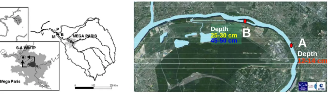
Among a few hundred samples available in the laboratory, sediments taken from 2 sites on the Seine River situated a few km downstream from Paris were chosen (Figure 1). The first one, labeled A, is located right on the outlet of a waste water treatment plant (WWTP) with a depth of 12-14 cm. The second site is approximately 3 km downstream on a dead river branch, labeled B. We analyzed two different sediments in this second site, one from a depth of 25-30 cm, the second was from a depth of 45-50 cm. These depths were chosen due to their high level of Cesium-137 indicating recent deposit (i.e. younger then 1986). The metal concentrations on the
samples were assured as to fulfill the criteria of the limit of detection of the following microscopic analysis by applying total digestion to the samples and analyses on ICP-MS.
A
B
Depth 12-14 cm Depth 25-30 cm 45-50 cmFigure 1 Sampling site on the Seine River for this study
Previously freeze dried samples were grounded on agate mortar. Bulk samples, samples <50 micrometer and magnetic samples (samples separated magnetically) were dispersed in alcohol and filtered on a 0.5 µm nucleopore carbonate filter and posed on a carbon sample holder with a diameter of about 1 cm. The amount of the sample dispersed is approximately 100-200 µg. Prepared filter was checked under microscope to confirm that the particles were evenly dispersed to facilitate the microscopic analysis that follows. Finally, a thin layer of carbon was coated on the filter to allow high electron conductivity during analysis.
Samples were analyzed using a Scanning Electron Microscope (SEM) JEOL JSM 840 coupled with an X-Ray microanalysis system from Princeton Gamma Tech (PGT). A ray of generated electrons of the size of a few angstroms is scanned over a randomly located field. A numerical image is produced from the backscattered electron beam with particles ranging from 0.2 to 20 µm. X-ray spectrum generated from these particles are then acquired (Figure 2) using a high purity Germanium (HPGe) detector and digital pulse processing from PGT. Hundreds of spectrums of particles per sample were analyzed automatically to assure that results are qualitatively representative.
The spectrum is analyzed by the energy dispersive spectrometer (EDS) and then classed according to the pre-defined sediment, as well as metal phases. Predefined sediment phases included various iron and manganese oxides, calcium, SiO2, pyrite and barite. These sediment
phases were chosen according to the capability of the SEM-EDS to identify particles and analyze spectra. Organic matter and amorphous iron oxide are not defined since these phases can not be detected by the SEM. On the other hand, metal classes consisted of zinc, lead, chrome, copper, and vanadium. These metals were chosen due to their high quantity which allows them to be detected on the spectrometer.
8th World Wide Workshop for Young Environmental Scientists WWW-YES-2008: Urban waters: resource
or risks? 13-16 May 2008
4 / 8
Figure 2 An EDS Spectrum of stainless steel
A manual check was done by randomly observing spectra and particles to confirm that they were correctly classed. Interesting particles was further analyzed by producing morphological images and chemical maps of the particles.
RESULTS AND DISCUSSION
To assure the statistical quality of the results, only samples where more than 100 particles analyzed are shown. Due to the statistical limitations that may be caused by microscopic analysis, the results herein will be discussed qualitatively and semi-quantitatively. Graphs are shown only to facilitate results presentations. The horizontal axis indicates the different predefined binding phase. The vertical axe gives the percent value of a certain metal associated with a certain phase. Each color indicates a certain sample (marked on the legend of each figure) and the number in parenthesis next to each sample label indicates the number of particles analyzed containing a given metal. We take for example the Ni association on iron sulfide ( Figure 3, left). Twenty-eight Ni particles were located during the automatic analysis of sediment sample A 12-14 cm. Out of the 28 particles, 26 particles, or 93 %, were found to be associated with iron sulfide.
Pb 0 10 20 30 40 50 60 70
Fe3O4 BaSO4 FeS2 SiO2 Ca other
fractions non-identified p o ur c e nt a g e )A 12-14(32 )B 25-30 (22 )B 45-50 (12
Figure 3 Distribution of nickel binding phase (left) and lead binding phase (right) analyzed by SEM-EDS
Ni (non spinel) 0 10 20 30 40 50 60 70 80 90 100 FeS2 Ca non-identified pou rc e nt a g e )A 12-14 (28 )B 25-30 (27 )B 25-30 50FM (15 )B 45-50 (17 )B 45-50 <50 µm (17
Iron sulfide seems to be a preferable binding site for Ni and Pb ( Figure 3). Given the fact that these sediments were once anoxic, this is a very possible condition as iron sulfide is known to be an important binding fraction in reducing condition. Although the samples were not kept in anoxic conditions, the pyrite may partly oxidize in surface, while the rest remains reduced. This would explain the extraction of the pyrite during the magnetic separation. The association between Ni and iron sulfide was also noted on coastal fresh water lake sediment (Canavan et al., 2007) where Ni and Co is notably associated with pyrite. Sulfide was also observed as a main heavy metal carrier in combined sewer overflow samples (El Samrani et al., 2004)
Analysis on fine fraction (< 50 µm and magnetic fraction of <50 µm) shows a whole different trend where Ni are mostly associated with non-identified phases. Further analysis on the fine fraction (<50 µm) B 45-50 cm shows a 100% association with non-identified phases. This may indicate that Ni is associated with the organic fraction or fine amorphous iron oxide particles a phenomenon also observed by Canavan et al.(2007). This fact may be supported by the analyses of the magnetic fraction of the same sediment. More than two-thirds of the Ni-containing particles in this fraction are also found associated with the non-identified phase, possibly indicating bond with iron oxide, a fairly magnetic particles.
The Paris water treatment plant has been studied by various authors ((Buzier et al., 2006), (Thévenot et al., 2007a) . They cited that due to the moderate removal efficiency for Ni and the addition of Ni-containing chemical during the treatment process, the labile and dissolved Ni concentration from the plant outlet may be comparable to Ni concentrations coming from the outlets upstream. The water on the outlet may create a reducing condition due to the high organic matter content which may promote respiration thus diminishing oxygen level. This reducing environment may promote the formation of sulfides. The high dissolved and labile Ni ions coming out from the WWTP may be found to associate with sulfide particles in the river. Organic matter association is also possible due to this high OM content. In this case, associations would be formed in the WWTP before entering the river. The difference in the formation momentum of these associations is interesting to note when determining choices in source reducing methods.
Figure 3 on the right, shows distribution of Pb on various binding phase. Here, we may note that Pb is also preferentially bonded on pyrite. Pb also show to have a certain affinity for the sulfide fraction and even higher rates as the sediments get deeper. Metal bearing sediment is not stable and the association evolves with time and changing condition. They will have a tendency to migrate from isomorphous replacement into crystalline lattice (Emerson et al., 1983 in (Huerta-Diaz et al., 1998)). The SEM has a weaker capacity in detecting amorphous structure. This may explain that there are higher non-identified phase in the Q 12-14 cm layer, and a higher pyrite associated Pb in the deeper 45-50 cm sediment.
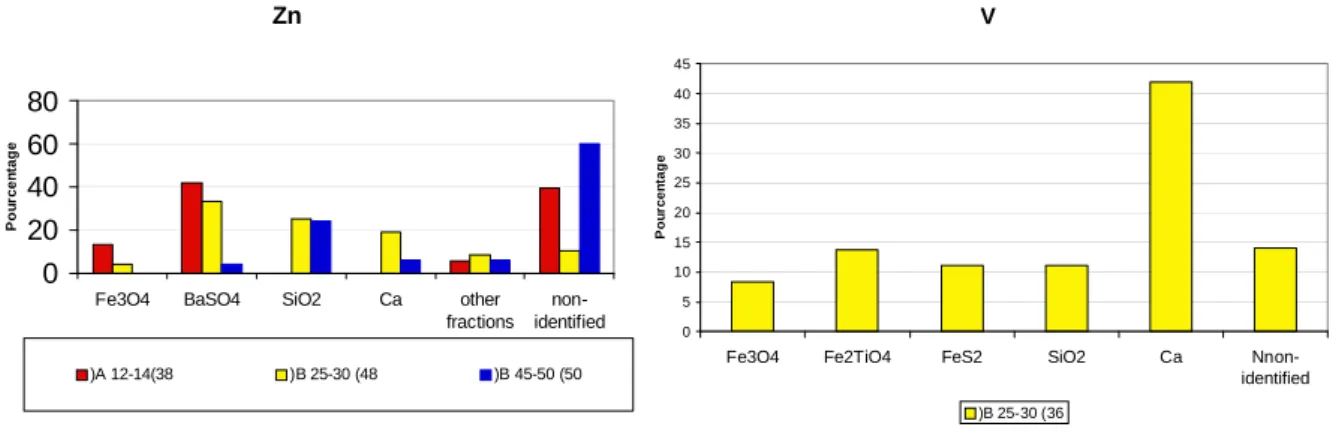
Zn 0 20 40 60 80
Fe3O4 BaSO4 SiO2 Ca other fractions non-identified Po u rc e n ta g e )A 12-14(38 )B 25-30 (48 )B 45-50 (50 V 0 5 10 15 20 25 30 35 40 45
Fe3O4 Fe2TiO4 FeS2 SiO2 Ca
Nnon-identified P o u rcen tag e )B 25-30 (36
Figure 4 zinc (left) and vanadium (right) binding phase analyzed by SEM-EDS
Results of Zn association shows less preferential site (Figure 4, left). We may though observe an important fraction bound with barite, which was less observed for other metals. A recent metal budget was established based on calculations in an urban sewage system from urban and domestic waste in the Seine river watershed by (Thévenot et al., 2007b). It underlined an
8th World Wide Workshop for Young Environmental Scientists WWW-YES-2008: Urban waters: resource
or risks? 13-16 May 2008
6 / 8
important portion of Zn deriving mostly from roof runoff (55 %) where rooftops and rain gutter in Paris are mostly made from Zn. Detailed studies on the runoff by (Gromaire-Mertz et al., 1999) showed that the percentage or Zn bound to particles ranges from 0-73 %, with a median of 9 %. Further “downstream”, street runoff measured 44-96 % of particle-bound Zn, with a median of 75 %. While barite in urban surfaces may derive from the usage of brake in automobiles (Osterle et al., 2001), we may hypothesize that during a rain event, Zn may be washed off from surfaces in a dissolved form at high concentrations. During the flow, the dissolved Zn would associate with particles washed off from road surfaces carrying barite. The association may also well be formed in the sewage system. Similar association between barite and lead were observed in mine wastes where Pb is mobilized in dissolved form (Courtin-Nomade et al., 2008)
The vanadium may also be an indicator of an anthropogenic contamination. SEM observations allowed us to localize specific association between V and calcium (Figure 4, right). V and calcium is produced as slag in steel production. The slag is frequently reused in road construction (Chaurand et al., 2007). This association may indicate the effect of runoff on urban surface which may carry these particles during rainy periods. Another possibility is that V is bound to the calcite in the Seine, which is abundant do the calcareous nature of the watershed. Further comparison with particles directly recovered from road surfaces will be necessary to confirm this hypothesis.
Associations between metal and binding phase may seem complicated as there are an endless variation of metal and binding phase, but surprisingly recent studies in soils by extended X-Ray absorption fine structure spectroscopy have shown a certain restricted uptake mechanism thus restricted associations with phyllosilicates, Fe(oxyhydr)oxides, and Mn-oxides (Manceau et al., 2000). Observations by Transmission Electron Microscopy (TEM)-EDS and SEM-EDS by El Samrani et al. (2004) also support this finding in sewer sediment studies by adding sulfides as an important trace metal carrier. These studies supports the observation as we identify that certain metals seem to be preferentially associated with a certain sediment phase.
A few limitations of this analysis to note is certain undetected phases including the organic matter. This will be compensated in the future by analyzing particles with the TEM. Further detailed study on defined metal bearing phase will also be carried out using local analysis such as EXAFS and XANES to detail the chemical association on the molecular scale and understanding the mobility of the particles.
CONCLUSIONS AND PERSPECTIVES
Scanning electron microscopy proves to be a powerful method in understanding different associations between heavy metal and its carrier phase. Particle analyses of The Seine River bottom sediment downstream of Paris are shown to be effective in determining the sources of contamination. Lead and nickel were found to be preferentially bound with iron sulfide particles. Association of vanadium and zinc indicated an urban-related contamination source through runoff during rainy period.
Its application in the future will be used in understanding monthly, weekly, and even daily dissolved metal variability in the Seine. As the variability seems to be linked to biological activities and changes in redox condition, interesting binding fraction to be analyzed would be the iron and manganese oxide. Calcite bound metal would make an interesting observation as the Seine river system is known to be controlled by its calcium carbonate balance.
REFERENCES
Bibby, R.L. & Webster-Brown, J.G. (2005) Characterisation of urban catchment suspended particulate matter (Auckland region, New Zealand); a comparison with non-urban SPM. Science
of The Total Environment, 343, 177-197.
Buzier, R., Tusseau-Vuillemin, M.-H., dit Meriadec, C.M., Rousselot, O. & Mouchel, J.-M. (2006) Trace metal speciation and fluxes within a major French wastewater treatment plant: Impact of the successive treatments stages. Chemosphere, 65, 2419-2426.
Canavan, R.W., Van Cappellen, P., Zwolsman, J.J.G., van den Berg, G.A. & Slomp, C.P. (2007) Geochemistry of trace metals in a fresh water sediment: Field results and diagenetic modeling.
Science of The Total Environment, 381, 263-279.
Chaurand, P., Rose, J., Briois, V., Olivi, L., Hazemann, L., Proux, O., Domas, J. & Bottero, J.-Y. (2007) Environmental impacts of steel slag reused in road construction: A crystallographic and molecular (XANES) approach. Journal of Hazardous Materials
First International Conference on Engineering for Waste Treatment: Beneficial Use of Waste and By-Products (WasteEng2005), 139, 537-542.
Clozel, B., Ruban, V., Durand, C. & Conil, P. (2006) Origin and mobility of heavy metals in contaminated sediments from retention and infiltration ponds. Applied Geochemistry, 21, 1781-1798.
Courtin-Nomade, A., Soubrand-Colin, M., Marcus, M.A. & Fakra, S.C. (2008) Evidence for the Incorporation of Lead into Barite from Waste Rock Pile Materials. Environ. Sci. Technol. El Samrani, A.G., Lartiges, B.S., Ghanbaja, J., Yvon, J. & Kohler, A. (2004) Trace element carriers in combined sewer during dry and wet weather: an electron microscope investigation.
Water Research, 38, 2063-2076.
Elzinga, E.J., Rouff, A.A. & Reeder, R.J. (2006) The long-term fate of Cu2+, Zn2+, and Pb2+ adsorption complexes at the calcite surface: An X-ray absorption spectroscopy study.
Geochimica et Cosmochimica Acta, 70, 2715-2725.
Emerson, S., Jacobs, L. and Tebo, B. (1983) The behavior of trace metals in marine anoxic waters: Solubilities at the oxygen-hydrogen sulfide interface. In Trace Metals in Sea Water, eds C. S. Wong, E. Boyle, K. W. Bruland, J. D. Burton and E. D. Goldberg, pp. 579--608. Plenum Press,New York.
Garnaud, S., Mouchel, J.-M., Chebbo, G. & Thevenot, D.R. (1999) Heavy metal concentrations in dry and wet atmospheric deposits in Paris district: comparison with urban runoff. The Science
of The Total Environment, 235, 235-245.
Gromaire-Mertz, M.C., Garnaud, S., Gonzalez, A. & Chebbo, G. (1999) Characterisation of urban runoff pollution in Paris. Water Science and Technology, 39, 1-8.
Horowitz, A.J. (1991) A primer on sediment-trace element chemistry, Lewis Publishers, Chelsea. Huerta-Diaz, M.A., Tessier, A. & Carignan, R. (1998) Geochemistry of trace metals associated with reduced sulfur in freshwater sediments. Applied Geochemistry, 13, 213-233.
Manceau, A., Lanson, B., Schlegel, M.L., Harge, J.C., Musso, M., Eybert-Berard, L., Hazemann, J.-L., Chateigner, D. & Lamble, G.M. (2000) Quantitative Zn speciation in smelter-contaminated soils by EXAFS spectroscopy
Osterle, W., Griepentrog, M., Gross, T. & Urban, I. (2001) Chemical and microstructural changes induced by friction and wear of brakes. Wear, 251, 1469-1476.
8th World Wide Workshop for Young Environmental Scientists WWW-YES-2008: Urban waters: resource
or risks? 13-16 May 2008
8 / 8
Swanson, V., Frist, L., Radar Jr. R., & Huffman Jr., C.(1966) Metal sorption by northwest Florida humate, US Geol. Surv. Prof. Paper (1966), pp. 174–177.
Tessier,A., Campbell, P.G.C., & Bisson,M., (1979) Sequential extraction procedure for the speciation of particulate trace metals, Anal. Chem. 51 (1979), pp. 844–851
Thévenot, D.R., Lestel, L., Tusseau-Vuillemin, M.-H., Gonzales, J.-L. & Meybeck, M. (2007a) Plaquette Piren Seine : Les métaux dans le bassin de la Seine, pp. 63. PIREN Seine, Créteil. Thévenot, D.R., Moilleron, R., Lestel, L., Gromaire, M.-C., Rocher, V., Cambier, P., Bonte, P., Colin, J.-L., de Ponteves, C. & Meybeck, M. (2007b) Critical budget of metal sources and pathways in the Seine River basin (1994-2003) for Cd, Cr, Cu, Hg, Ni, Pb and Zn. Science of
The Total Environment Human activity and material fluxes in a regional river basin: the Seine River watershed - Seine Special Issue, 375, 180-203.
Webb, S.M., Leppard, G.G. & Gaillard, J.-F. (2000) Zinc Speciation in a Contaminated Aquatic Environment: Characterization of Environmental Particles by Analytical Electron Microscopy.
Environ. Sci. Technol., 34, 1926-1933.