HAL Id: cea-02370063
https://hal-cea.archives-ouvertes.fr/cea-02370063
Submitted on 26 Nov 2020HAL is a multi-disciplinary open access archive for the deposit and dissemination of sci-entific research documents, whether they are pub-lished or not. The documents may come from teaching and research institutions in France or abroad, or from public or private research centers.
L’archive ouverte pluridisciplinaire HAL, est destinée au dépôt et à la diffusion de documents scientifiques de niveau recherche, publiés ou non, émanant des établissements d’enseignement et de recherche français ou étrangers, des laboratoires publics ou privés.
Rationalizing the diversity of amide–amide H-bonding in
peptides using the natural bond orbital method
Valérie Brenner, Eric Gloaguen, Michel Mons
To cite this version:
Valérie Brenner, Eric Gloaguen, Michel Mons. Rationalizing the diversity of amide–amide H-bonding in peptides using the natural bond orbital method. Physical Chemistry Chemical Physics, Royal Society of Chemistry, 2019, 21 (44), pp.24601-24619. �10.1039/c9cp03825f�. �cea-02370063�
1
Rationalizing the diversity of amide-amide H-bonding in
peptides using the Natural Bond Orbital method
Valérie Brenner*, Eric Gloaguen and Michel Mons*
LIDYL, CEA,CNRS, Université Paris-Saclay, bât 522, CEA Paris –Saclay, 9119 Gif-sur-Yvette, France
Abstract
Natural Bond Orbital (NBO) analysis of electron delocalization in a series of capped isolated peptides is used to diagnose amide-amide H-bonding and backbone-induced hyperconjugative interactions, and to rationalize their spectral effects. The sum of the stabilization energies corresponding to the interactions between NBOs that are involved in the H-bonding is demonstrated as an insightful indicator for the H-bond strength. It is then used to decouple the effects of the H-bond distance to that, intrinsic, of the donor/acceptor relative orientation, i.e., the geometrical approach. The diversity of the approaches brought by the series of peptides studied enables us to illustrate the crucial importance of the approach when the acceptor is a carbonyl group, and emphasizes that efficient approaches can be achieved despite not matching the usual picture of a proton donor directly facing a lone pair of the proton acceptor, i.e., that encountered in intermolecular H-bonds. The study also illustrates the role of backbone flexibility, partly controlled by backbone-amide hyperconjugative interactions, in influencing the equilibrium structures, in particular by frustrating or enhancing the HB for a given geometrical approach. Finally, the presently used NBO-based HB strength indicator enables a fair prediction of the frequency of the proton donor amide NH stretching mode, but this simple picture is blurred by ubiquitous hyperconjugative effects between the backbone and amide groups, whose magnitude can be comparable to that the weakest H-bonds.
2
1. Introduction
The amide-amide H-bonding is a key interaction of proteins and bioinspired polymers, like foldamers, often considered at the heart of the stability of their secondary structures.1-4 During the folding process, proteins NH donor sites seek to satisfy their propensity to form H-bonds (HBs) by finding an acceptor partner CO site leading to intramolecular H-bonding: this feature is even invoked as the main driving force to protein folding.5, 6 Thus early H-bonds biochemists have recognized that the most iconic secondary structures of proteins, such as the -3, 4 or 310 helices4, -turns 7 or 27 ribbons4, 8
are stabilized by H-bonds, namely the so-called C13, C10 or C7 H-bonds (labelled according to the length of the ring formed). Even extended peptide conformations like -strands can be considered as stabilized by such a C5 interaction, as recently surmised from a protein survey.9 Knowledge of these H-bonds has greatly benefited from the characterization of isolated model systems, mostly capped aminoacids or peptide building blocks, composed of complete peptide links, which interact with each other, without perturbations other than those induced by the backbone residues of the sequence.10,
11
In particular such an approach enables to focus on the intrinsic H-bonding, without involvement of neighbouring H-bonds (absence of cooperative effects) or of the environment (no solvent effect). Thus in recent gas phase experiments12 spectroscopists have recorded conformation-selective spectra in the IR and the UV range, making possible the determination of the most stable conformations in flexible molecules, like peptides or foldamer building blocks, and the characterization of their H-bonding content. The wealth of data collected using laser spectroscopy during the past decade10, 11, 13-19 constitutes an extended set of peptide conformations, with both natural14, 20-29 and synthetic -17, 18, 30 or -31, 32peptides, in which two amide groups interact with each other, according to a panel of geometrical approaches modulated by covalent constrains imposed by the peptide backbone. This conformational set can be considered as a sampling of possible amide-amide geometrical approaches, mimicking those encountered in the desolvated inner core of proteins.33 These experimental studies stem from a synergetic action with theoretical approaches, which have greatly improved since the early days of peptide quantum modelling34, 35 and can nowadays efficiently handle dispersive interactions, which are ubiquitous in these systems.36-38 The IR spectra are assigned by comparison with quantum chemistry calculations of vibrational modes and conversely the good agreement usually achieved enables an assessment of the quantum chemistry methods chosen.39 The observable of choice in these gas phase optical experiments is the spectral transition of the NH stretch vibration, since the usual HBs are characterized by unique features,40 namely, i) a red-shifted frequency of the NH stretch reflecting the weakening of the NH covalent bond, ii) an enhancement of the band intensity of this mode due to the polarization of the NH proton donor moiety and iii) a partial electron delocalization (charge transfer) from the HB acceptor to the NH moiety.
A striking observation within the library of H-bonded amides evoked above is that the spectral shifts recorded are sometimes much larger than that measured on a natural reference for two interacting amide groups, namely the dimer of the methylacetamide (MAA) molecule, whose spectroscopy has been recently revisited.41, 42 In this latter system, indeed, the limited contact between the two amides groups allow them to interact in the most efficient way to form a strong, natural HB, which results
3
from an electrostatic-polarization component, also potentially influenced by a dispersive component37, 39 and some partial charge transfer (CT) as well.43, 44 Conversely, when the amides
belong to the same molecule, the set of geometrical approaches available is limited, leading to a significant frustration of the H-bonding. In this respect, the observation of larger red shifts in some intramolecular cases, compared to the intermolecular one, may appear paradoxical since they would correspond to an enhancement of the H-bond with respect to the reference dimer. Moreover, such large red shifts can also correspond to the vibrational signature of free NH bonds, undergoing hyperconjugation (HC) from vicinal or neighbor orbitals45, 46 (Bohlmann effect47) as it has been recently illustrated on isolated model hydrazides.48
The aim of the present benchmark study is to rationalize the diversity of H-bonding strengths observed within a set of peptide secondary structures, to understand why several peptides exhibit unusually red shifted H-bonds and eventually assess the role of the peptide environment (comprising both backbone and main chain) in determining the HB strengths. Beyond the use of vibrational red shifts by spectroscopists, a rigorous analysis of the local electron densities can provide a convenient and effective framework for both diagnosing the presence of H-bond and gauging their strength. The methodology chosen, however, has to meet some essential requirements. First, it must enable a clear diagnostics of the H-bond while being readily applicable to different approaches with a broad spectrum of nearby environments. Second, it must enable the electronic wavefunction to be expressed in a localized language, without making any assumptions about its mathematical form, and should transcript accurate calculations into chemical insights about H-bonding. Finally, the method must distinguish the intrinsic HB features from hyperconjugative effects not directly linked to the HB itself. In this respect, one will notice that, despite HB itself is sometimes viewed or interpreted as an hyperconjugative effect,45, 46 in the present paper the term HC will exclude HB contributions. These criteria lead to choose the natural bond orbital (NBO) method,49-51 in which the electronic wave
function is analyzed in the framework of localized Lewis-like chemical bonds. Based on local block eigenvectors of the one-particle density matrix, and developed to study hybridization and covalency in polyatomic wave functions, this method and its principles have been extended to treat the so-called “van der Waals bonding”, in particular inter- and intra-molecular HBs (see for example: Refs.
52-57). The strategy implemented in this work stems from the NBO analysis49-51 of the electronic
structure of a set of peptide secondary structures, starting from unconstrained amide-amide HBs, as revealed by MAA dimers (a system also previously investigated by Adhikari & Scheiner37 using a NBO analysis), and then comparing them with those encountered in typical peptide building blocks and secondary structures, namely models of the - turn and -strand structures. It will be shown that a general specific feature of the NBO stabilization energies related to the HB formation, namely its dependence with the HB distance, can be used to rank the strengths of the amide-amide HBs observed in peptides and to assess the role of the peptide environment on the structure. Finally, the issue as to whether the NH stretch frequency is a good marker of the HB strength is also addressed, through its correlation with the HB strength, as obtained from NBO analysis, together with the role of hyperconjugative interactions45, 46 between the NH bond considered and vicinal/neighbour orbitals.45,
46, 48
5
2. Methodology
2.1 Benchmark set composition
The present benchmark study is focused on specific conformations of a series of model diamide molecules completed by a few tri- and tetra-amides, all featuring as much as possible isolated HBs, for which the amide bonds involved in the HB considered are either single proton donor or single proton acceptor moieties. It encompasses data on trans-methylacetamide (MAA) dimers,41, 42 basic amino acids (AA), Ala, Gly, Phe, Aib (amino isobutyric acid),14, 20, 23, 27, 29 some di- and tri-peptides of these AAs,21, 22, 24, 25 as well as synthetic AAs (- and -aminoacids).17, 30, 31 All these peptides are capped to form complete amide units, apart from one hydrazide group in an aza-compound.30 The model caps considered are those present in the gas phase experiments as well as more simple counterparts (acetyl and methylamide caps) in order to assess their influence on the peptide structure.
Figure 1: Set of specific conformations of model molecules considered in the present work, closely related to experimental data on t-MAA dimers, capped -, - and -peptides (see text). The stabilizing HBs are indicated by arrows, together with the terminology used (number of atoms in the ring closed by the HB and short names of the molecules).
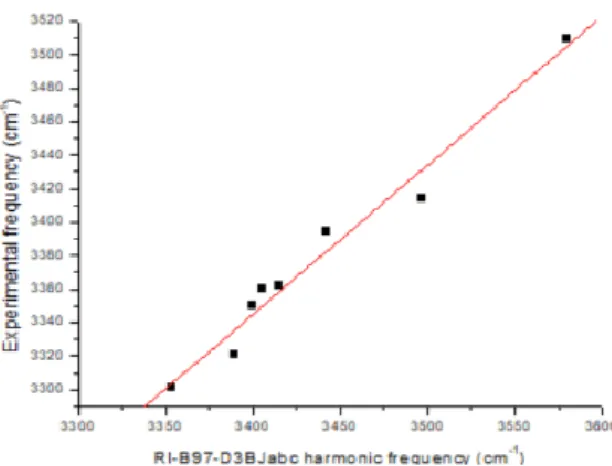
Most of the conformations considered (Fig. 1) were observed in microwave58, 59 or in conformer-selective laser spectroscopic gas phase experiments.14, 17, 20-25, 27, 29-31 The theoretical structures, calculated in the present study, have been determined using a DFT-D method, at the B97D3(BJ)-abc/def2-TZVPPD level of theory,60-62 together with harmonic vibrational frequencies, using the TurboMole package.63 Fig. 2 illustrates the fair correlation obtained this way between experimental NH stretch frequency data and their theoretical counterparts, showing that a good description of the NH stretching mode is achieved at this level of theory.
Figure 2: Correlation between experimental NH stretch frequencies as measured in IR/UV conformer-selective gas phase experiments and harmonic frequencies at the B97D3(BJ)-abc/def2-TZVPPD level of theory.
6 2.2 NBO analyses and diagnostics:
After a brief reminder of the main valency and bonding concepts of the NBO method,45, 46,
49-51
its application to diagnose the presence of a HB will be illustrated on the inverse -turn form of the Ala diamide.59 However, before this, intrinsic amide-backbone hyperconjugative effects, already present in the absence of HB, will be assessed on a relevant conformation of the related model molecule possessing a unique amide group, namely the N-isopropylacetamide.
2.2.1 Nomenclature and computational details:
The NBO-based chemical bonding concepts rest on an expression of the molecular properties in terms of a ‘natural Lewis structure’ depiction of the wavefunction. The wavefunction is optimally transformed into a localized form corresponding to the one-centre (“core” or “lone pair”) and two-center “bond” elements of the chemist’s Lewis structure picture. The various natural localized sets can be considered as resulting from a sequence of transformations of the input orbital basis set. The NBOs are obtained as local block eigenfunctions of the one-electron density matrix and have optimal convergence properties for describing electron density. They are partitioned into high- and low-occupancy orbital types: the small set of most highly-occupied orbitals contains the core and valence (bond-antibond) functions and are distinguished from the weakly occupied “Rydberg” (extra-valence-shell) functions. The symbols “x” and “x*” will be used to refer to filled and unfilled orbitals of the formal Lewis structure, x being either core orbitals (c), lone pairs (n), or bonds (, ), and x* being either or antibonds (*, *) or extra-valence Rydberg (Ry) orbitals. The calculation of the donor-acceptor interactions in the NBO basis is carried out by examining all possible interactions between “filled” (donor) Lewis-type NBOs and “unfilled” (acceptor) non-Lewis NBOs and estimating their energetic importance by 2nd order perturbation theory. The filled NBOs of the “natural Lewis structure” are well adapted to describe covalency effects whereas the unfilled in the formal Lewis and in particular, the antibond NBOs, are well adapted to describe non covalency effects. For each donor NBO (i) and acceptor NBO (j), the stabilization energy E(2) associated with delocalization is calculated as:
where is the donor orbital occupancy, , are diagonal elements (orbital energies) and is the off-diagonal NBO Fock matrix element.
The NBO analysis50 has been carried out on the B97D3(BJ)-abc/def2-TZVPPD equilibrium structures at both the HF/TZVPP and MP2/TZVPP levels using the NBO module64 in the Gaussian 16 package.65 The HF/TZVPP level is only used to generate the first step of the calculation of the stabilization energies; the natural population analysis (NPA) charges and the NBOs occupancies considered in the following being that obtained at the MP2/TZVPP level. It has been demonstrated on a set of small systems that this level of theory is sufficient and gives results similar to those obtained at more sophisticated levels such as MPSDQ or QCISD.48 For the E(2) stabilization energies, the thresholds for considering the interactions as significant were 0.05 kcal/mol for the intermolecular interactions and 0.5 kcal/mol
7
for the intramolecular ones, except in some specific cases (weak interactions), where it was necessary to reduce it down to 0.1 kcal/mol.
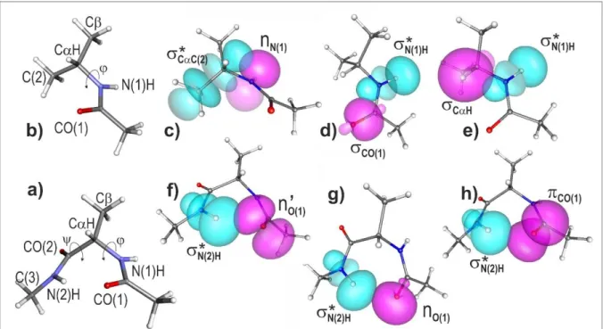
2.2.2 H-bond and hyperconjugation diagnostics: the inverse -turn form of the Ala diamide and the related N-isopropylacetamide conformation.
Before performing the NBO analysis on the inverse -turn form of the Ala diamide, the N-isopropylacetamide (N-propan-2-ylacetamide), a model molecule in which the C-terminal side of the Ala diamide is substituted by a methyl group, was first investigated in order to reveal the contribution of hyperconjugation effects, already present in absence of any H-bonding, and thus to allow in the following a clear discrimination between these HC effects and HB formation (Figure 3). The equilibrium structure of the N-isopropylacetamide conformation, that is directly comparable to the Ala diamide -turn conformation (Figure 3 a and b), exhibits a N-terminal geometry with a C
H ---O(1)C distance of 247 pm, typical of a close contact and comparable to the average contact distance found in crystals : 248 pm (according to the survey by Rowland and Taylor66), together with a Ramachandran dihedral of -86°. NBO analysis (Table S1, in supplementary information) provides evidence for significative hyperconjugative interactions enabling us to rationalize these structural features. The strongest HC interaction (E(2) = 9.0 kcal/mol) involving amide groups and backbone covalent bonds corresponds to an electron delocalization resulting from the overlap of the NBO lone pair (donor) localized on the nitrogen atom, identified as nN(1), with the NBO antibond (acceptor)
localized on the vicinal CC(2) bond, identified as CC(2), referred to as a nN(1) CC(2) interaction
(see Figure 3c). This interaction enables the amide group to strongly affect the orientations of the covalent bonds involving the C atom: an optimum overlap of these two NBOs favors a = -90° geometry and locks the structure of the backbone N-terminal side, in line with the Ramachandran dihedral of = -86° calculated. Two smaller interactions involving the same nitrogen and the C atoms are also detected (HC0 set in Table S1): a nN(1) CH interaction of 2.4 kcal/mol and a nN(1)
CC interaction of 2.2 kcal/mol, due to a lesser NBO overlap.
As far as delocalisation to the N(1)H NBO is concerned, two significant hyperconjugative interactions
are detected, a CO(1) N(1)H interaction of 1.8 kcal/mol and a CH N(1)H interaction of 4.4
kcal/mol (HC1 set in Table S1). The first one is internal to the peptide bond and contributes to the planar structure of the motif, whereas the latter, more importantly describes the tendency of the vicinal NH and CH bonds to adopt an antiparallel orientation (Figure 3d). These interactions result in a modest but significant NBO population of the N(1)H NBO, namely ~34 10-3 e (10-3 e will be
referred to as me, milli-e, in the following). One can also notice that no interaction (with E(2) > 0.1 kcal/mol) corresponding to an eventual CH---O(1)C HB such as a nO(1) CH interaction could be
detected.
The NBO analysis of the inverse -turn form of the Ala diamide (Figure 3 b) demonstrates that delocalization within the N(2)H---O(1)C amide-amide HB results from an overlap between three donor NBOs, located on the CO(1) carbonyl, and the acceptor antibond N(2)H NBO, localized on the
8
N(2)H group. (Table S1, bottom) (one will notice that donor NBOs are located on the HB proton accepting moiety whereas the acceptor orbital pertains to the HB donor NH). Two of the donor NBOs correspond to lone pairs of the O(1) oxygen atom (nO and n’O), which exhibit different mixed sp
characters: the nO NBO is of mixed character, 62% of s character for 38% of p character (Fig. 3g),
whereas n’O is of pure p character (99%; Fig. 3f). The third donor orbital involved in the HB is the
CO(1) NBO, localized on the
Figure 3: Left part: Quantum chemistry structure (at the B97D3(BJ)-abc /def2-TZVPPD level of
theory) of a) the inverse -turn conformation of the Ala residue, with acetyl and methyl amide caps, and of b) the corresponding conformation in the N-isopropylacetamide molecule, in which the C-terminal side of the Ala diamide is substituted by a methyl group. Relevant atomic and dihedral notations are also displayed. Right part : Natural Bond Orbitals, generated from NBO analysis of the two molecules, illustrating the overlaps between electron density donor (magenta) and acceptor (cyan) orbitals corresponding to a series of interactions : in N-isopropylacetamide (top): c) nN(1) CC(2) interaction responsible for the ~ 90° Ramachandran dihedral, d) CO(1)
N(1)H and e) CH N(1)H interactions (magenta), these two latter contributing to a basic
population of the N(1)H NBO. In the Ala inverse -turn model, f-h) the nO(1) N(2)H, n’O(1)
N(2)H and CO(1) N(2)H interactions, which correspond respectively to the three CO(1)-based
donor (magenta) NBOs, donating to the * NBO (cyan) located on the N(2)H covalent bond of the HB donor amide.
CO(1) carbonyl group (Fig. 3g). The overlap of each of these donor NBOs with the acceptor N(2)H
gives rise to significant E(2) stabilization energies of 1.3, 2.5 and 1.0 kcal/mol for the nO(1), n’O(1) and
CO(1) NBOs, respectively. In order to account for all these components, the sum (EHB) of the
9
in the N(2)H---O(1)C HB and obtained from the NBO analysis (4.8 kcal/mol), has been taken as a HB strength indicator.
Additionally, the same analysis also enables us to characterize the strongest hyperconjugative interactions that take place between backbone and/or side chain (SC) NBOs. In the diamide system, we indeed observe hyperconjugative interactions of the same nature than those found in the N-isopropylacetamide model molecule (see Table S1):
(i) three interactions between each of the nN(1) and nN(2) NBO donors and three specific NBO
acceptors. The nN(1) NBO interacts with C atom-based NBOs, the CC(2), CH and CC NBOs,
resulting in a sum of individual E(2) energies, E(2), of 14.4 kcal/mol (the first one being still prominent at 10.1 kcal/mol; Table S1, HC0 set). Similarly the nN(2) NBO donor interacts with NBO’s
involving the C-terminal cap carbon atom (C(3)), the three C(3)H NBOs, resulting in a similar E(2) of
15.4 kcal/mol. These interactions contribute to control of the orientations of the covalent bonds established by the C atoms directly after each amide group along the peptide chain, namely the C atom and the C(3) atom of the C-terminal cap., and
(ii) two interactions involving each of the N(1)H and N(2)H NBO acceptors, namely the CO(1)
N(1)H and CH N(1)H interactions of 1.7 and 3.5 kcal/mol resp. (Table S1, HC1 set) and the CO(2)
N(2)H and CH N(2)H interactions of 1.7 and 3.7 kcal/mol resp. . The CO NH interactions
are internal to the peptide bonds, whereas the CH NH incline the vicinal NH and CH bonds to
adopt an antiparallel orientation.
The HB interactions and the latter hyperconjugative interactions lead together to a N(1)H NBO
population of ~33 me, similar to that obtained for the N-isopropylacetamide molecule, but to an increased population of the N(2)H NBO, ~43 me, providing clear evidence for the N(2)H---O(1)C HB
formation.
Such an analysis and discussion has first been tested on the simplest intermolecular HB system, i.e., the methylacetamide dimer, and then extended to intramolecular HB systems, within a series of model diamide or triamide molecules; the purpose being to discriminate the different HBs from hyperconjugation effects. In addition, the sum EHB as well as the population of the NH involving in
the HB have been used for documenting the behavior of the NH stretch frequency.
3. Intermolecular amide-amide interactions as modelled by MAA dimers
The NBO analysis presented above has been applied to the most natural system in which two non-covalently bound amide groups interact through a HB, namely the trans-methylacetamide dimer. Starting first from the equilibrium structures obtained by quantum chemistry, the analysis procedure has then been extended to a series of elongated or compressed HBs, sharing the same geometrical approach, i.e., the relative orientation of the donor and the acceptor amides.
3.1. MAA dimers
The conformational landscape of the H-bonded dimer of the trans-methylacetamide model molecule (MAA), recently investigated at a high level of theory (MP2/aug-cc-pVDZ ; geometry optimization
10
with counterpoise corrections) by Adhikari and Scheiner37, exhibits several minima, namely two types of H-bonded conformations and one stacked form of lesser stability. The present theoretical investigation (B97D with D3(BJ)-abc corrections), focused on the H-bonded dimers, is in qualitative agreement with these findings. The HB distances, for instance, are found to be closer to those obtained by Adhakari and Scheiner with a counterpoise-corrected optimization than without (see Table S1 and Supp. Info of
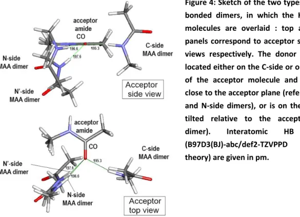
Figure 4: Sketch of the two types of MAA H-bonded dimers, in which the HB acceptor molecules are overlaid : top and bottom panels correspond to acceptor side and top views respectively. The donor molecule is located either on the C-side or on the N-side of the acceptor molecule and has its NH close to the acceptor plane (referred to as C- and N-side dimers), or is on the N-side but tilted relative to the acceptor (N’-side dimer). Interatomic HB distances (B97D3(BJ)-abc/def2-TZVPPD level of theory) are given in pm.
Ref. 37), in agreement with the negligible BSSE expected with the DFT framework associated to this level of basis set (def2-TZVPPD) and justifying geometry optimizations carried out without BSSE-correction in the present work. Two types of H-bonded dimers (Fig. 4) are found: they both exhibit a linear H-bond, with the NH bond of the donor molecule pointing towards the acceptor carbonyl O atom, but they differ by the side of the H-bonding approach. One type has its MAA-donor molecule in close contact with the acetyl moiety of the MAA-acceptor and is referred to as the C-side dimer (right hand-side in Fig. 4.). The donor NH exhibits a side position (CO-H angle CO-H = 118° in the
present data, 120° in ref 37, See Fig. S1 for the definition of the angles and dihedrals), very close to the plane of the proton acceptor amide (H-OCN ~180°, ’ ~0°). In contrast, the other type of dimer has
its donor in contact with the NHMe moiety (left hand-side figure 4). A first N-side conformer exhibits a linear HB, with its donor NH located close to the acceptor amide plane (φH·OCN= -6°, and -11° in Ref
37 and in the present work respectively) but less tilted from the carbonyl axis compared to the C-dimer, (CO-H angle CO-H = 138° and 142° in the present work and in 37 respectively). The method
presently used, however, also leads to a second N-side stable conformer, referred to as N’-side, and characterized by a linear HB (Fig. 4). Its NH donor is significantly shifted above the amide plane ( H-OCN = +44°), but the angle of approach to the acceptor (CO-H = 126°) is comparable to that of the
11
significantly tilted (CN-OC = -40°) compared to the two latter forms, which have their amide planes
nearly perpendicular (CN-OC = -75° and -71°, see Supp. Info). As far as relative stability is concerned,
the two N- and N’-side dimer conformations are found to be nearly isoenergetic (within 0.2 kcal/mol, see Table S2), illustrating the relative flatness of the potential energy surface in the N-side region. One should notice that for the present investigation, focused on the dependence of the H-bonding upon the relative orientation of the donor and acceptor moieties (see Section 3), the issue of the real existence of a minimum at the N’-side geometry, which was not reported by Adhikari and Scheiner, is not critical, since the NBO analysis can be carried out whatever the geometrical conformation, be a minimum or not. Additionally, this N’-side dimer is also interesting for the presently studied issue, since it suggests to consider an alternative orientation of the two amides, differing from that of the C- and N-side dimers.
Concerning the comparison with protein data, the approaches of the donor to the acceptor amide, observed in the dimers, characterized by ‘ angles ranging between 120° (C and N’) and 140°(N) and ‘ between ~0° (C and N) and 35° (N’), are comparable to those obtained from a protein survey33
whose (symmetrized) distributions peak at ’ ~125° and ’ = 0° respectively (See Supp. Info for ’ and ’ angle definition).
From a structural point-of-view, the present results suggest that there exists three possible quasi-isoenergetic HB approaches of an amide to another amide, one with the donor on the C-side of the acceptor, and two on the N-side, characterized by similar HB equilibrium distances (Table S2). The NBO analysis of the three dimers has been carried out in order to characterize the delocalization associated to HB formation. C- and N-side dimers exhibit NBO overlaps between NBO lone pairs of the oxygen atom (nO and n’O) of the CO group of the HB acceptor amide, and the antibond NBO NH
localized on the NH group of the HB donor amide. Each of these interactions gives rise to significant E(2) stabilization energies in the C- (Table S3) and N-dimers: 3.3 and 4.3 kcal/mol resp. for the nO
NH interaction, and 6.6 and 3.8 kcal/mol resp. for the n’O NH interaction. In contrast the
N’-side dimer, with its NH donor located off the amide plane, benefits from three overlaps, those corresponding to the nO/n’O NH, with E(2) = 3.6 and 2.7 kcal/mol resp., and a third one, of CO
NH type, with E(2) = 1.1 kcal/mol (Table S4). The sum of the stabilization energies associated to HB
formation EHB in the three C, N and N’ dimers give rise to decreasing values of: 9.9, 8.4 and 7.4
kcal/mol resp., suggesting decreasing HB strengths. These values are in qualitative agreement with those derived by Adhikari and Scheiner for the MP2 structures of C and N dimers (See Table S2) and are stronger than the value obtained for the inverse -turn of Ala (4.8 kcal/mol ; Section 2.2.2). The EHB values obtained also agree well with the populations of the *NH NBOs, which amount to 48, 45
and 45 me, for the C-, N- and N’-dimers respectively. As expected they are much larger than the reference value for the free amide, i.e. ~34 me in the Ala -turn (see Section 2.2.2) and 31-32 me for the HB acceptor trans-methylacetamide molecule in the C and N’ dimers (Tables S3 and S4). They are even larger than that of the inverse -turn of Ala (43 me). Eventually, the NBO analysis also provides the net charges of both donor and acceptor moieties, which reveal an effective electron transfer of 23 and 19 me at the equilibrium geometry of the C and N’ dimers (Tables S3 and S4), in qualitative agreement with the values of the two indicators, the sumEHB and the *NH NBO population.
12
Coming back to the energetics, one can notice that despite a significant difference in H-bonding strengths, the three dimers exhibit very similar H-bonding distances as well as similar energetics (within less than 0.2 kcal/mol, Table S2). They correspond to different approaches of a donor amide to an acceptor, on either side of the carbonyl acceptor, in or out of the amide plane. Obviously, the several interactions, other than the H-bonds, present in these three species differ substantially. In particular, whereas the C-side dimer donor does not experience any close contact with the acceptor C-side, the situation is different for the N-and N’-side dimers. In the N-side species, an NH in-plane approach of the acceptor with a CO---H angle comparable to that of the C-dimer (120°) would be
hampered by a steric clash between the donor N-atom and the N-terminal methyl of the acceptor, eventually resulting in a larger CO-H (140°). This forbids an approach that would mirror, on the N-side,
that of the C-side dimer, and as a result, the N-side dimer features a lesser delocalization and then a weaker H-bonding. Interestingly, the N’-side dimer can be seen as a trick of the system to overcome this difficulty: tilting the donor NH out of the amide plane in N’ enables the recovery of a small CO-H
angle, while keeping the NBO overlap significant. The similar energetics of the three forms, however, suggests that other attractive interactions in N and N’ compensate for the decrease in HB strength, namely electrostatics, induction and dispersion; the latter being favored by close contacts.
Finally, NH stretch frequency calculations indicate that the C-side dimer exhibit the larger red-shift (by ~ 31 and 36 cm-1 compared to N’ and N-side dimers respectively, see Table S2), suggesting a slightly stronger H-bond than its N- and N’-side counterparts. Likewise, the C-side dimer tends to exhibit a shorter H-bond distance than its N- and N’-side counterparts, by 0.6 or 1.3 pm, depending on the method considered (MP2 in Ref 37 or DFT-D in the present results; Table S2). These theoretical data agree well with the NH-stretch spectroscopic data of MAA in a supersonic expansion, which have been reinterpreted in terms of simultaneous presence of two H-bonded dimers of trans-MAA molecules, whose mid-IR spectral signatures (red-shifted NH bands) differ by 31 cm-1.42
3.2. Ranking the efficiencies of amide-amide approaches: decoupling the H-bonding
strength from the intermolecular distance
The observation of MAA dimers with different geometrical approaches raises the question of the intrinsic strength of these H-bonding approaches and of their relative ranking. Broadly speaking, one can anticipate that the strength of any H-bond depends on both i) the relative orientation of the two amide groups, i.e. the geometrical approach made possible/allowed by the backbone constrains, and ii) the H-bonding distance. Thus ranking the intrinsic efficiencies of the HB approaches requires the decoupling of the HB strength from the distance parameter. For this purpose the dependence of the HB strength upon the intermolecular distance has been obtained for the C- and N’-side MAA dimers, whose geometrical approaches differ radically. A NBO analysis has been carried out to determine the EHB indicator on a series of geometrical structures belonging to the same approach, i.e. for a series
of fixed HB distances around the equilibrium structures, with a frozen relative orientation of the amide groups (acceptor CO-donor-NCamide dihedral), the other degrees of freedom being optimized.
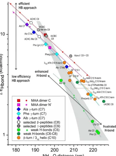
Figure 5 shows that for a given geometry approach, that of the C- or N’-side dimer, the sum of the stabilization energies corresponding to H-bonding (EHB) are nearly aligned along a straight line in a
13
semi-log plot (red and magenta dots) when the HB distance is varied, suggesting an exponential-type behavior in the range of distances considered. Although remarkable, this result is not so easy to rationalize. Each of the two-orbital E(2) interactions obtained from the NBO analysis depends on the overlap between the NBOs involved in the interaction together with the NBO energy difference (expression of E(2) in Section 2.2.1), both depending on the distance in a specific way. The apparent
Figure 5: Semi-log plot of the sum of the HB stabilization energies (EHB), as obtained
from the NBO analysis of the amide-amide HB, in the C- and N’-side dimers of trans-methylacetamide and in a series of conformations of di, tri- or tetra-amide molecules, as a function of the H-O distance (see also Tables S3, S4 and S5). Large symbols stand for equilibrium structures whereas small symbols correspond to squeezed or elongated dimers or C5 structures (see Section 4.2). A full identification of the NH-OC interactions is provided in Table S5 of the Supp. Info., from the abbreviated identifiers. The dotted lines feature a given H-bonding approach, along which structures sharing the same approach are expected to be aligned (see Sections 3.2, 4.1, 4.2 and 5.1): the lower the line, the less efficient the geometrical approach. Within a given approach (dotted lines), equilibrium structures found on the right-hand side correspond to frustrated HBs, and those on the left-hand side to enhanced HBs. The grey area is associated to a neutral region, where the peptide environment is not expected to significantly affect the HB strength (See Discussion in Section 5.2).
quasi-exponential dependence of the sum of those stabilization energies involved in the H-bonding (EHB) presumably results from a subtle sum of specific behaviors involving different pairwise
two-orbital interactions in the particular range of distances considered.
Fig. 5 also shows that both dimers obey parallel dependences in the semi-log plot: the N’-side MAA dimer (magenta dots) lying below that of the C-side dimer (red dots), showing that the former approach is intrinsically less efficient than the latter to establish a HB. As already mentioned above, this apparent penalty can be assigned to the steric hindrance between both amides (in particular the methyl amide side of the HB acceptor and the cloud of the HB donor), which restricts the set of accessible conformations and can forbid geometries most favorable to HB formation. One can notice that, despite a significant difference in HB strength, the dimers remain isoenergetic (Table S2), which justifies focusing our analysis on a local approach, such as the EHB indicator deduced from the NBO
14
The present semi-log plot EHB vs. H-bonding distance of Fig. 5 appears then as an efficient and fast
tool to rank the efficiency of any HB approach relative to the dimers. This can be done by considering the vertical distance of its point to the dimer considered, in the diagram of Fig. 5, whatever the structure chosen to evaluate the H-bond strength of this approach, being an equilibrium geometry or not. In other words, this plot decouples the intrinsic strength of the approach (i.e., its H-bonding efficiency, which is linked to the capability to give rise to electron delocalization and therefore depends upon the approach geometry) from its dependence with the H-bonding distance.
The aim of the present work is to extend the methodological procedure, introduced on the non-covalent dimers, to non-covalently linked amides, e.g. peptides, expecting that consistent series of conformations having similar geometries (case of the Ala, Phe and Aib -turns,36 see Figure 1 and next Section ) are representatives of the same approach and thus exhibit the same HB efficiency. The goal of the following sections will be to collect the HB strengths along such consistent series and to compare them to the dimer behavior in the semi-log plot of Fig. 5. The position of this line relative to those of other approaches should enable a relative ranking. The topology of the covalent link will provide a geometrical approach and thus determine the HB strength efficiency.
4. Intramolecular amide-amide interactions in peptides
4.1.
-turn models of
-peptides
The -turn is the smallest locally folded structure in a diamide model peptide. It has been early recognized as being stabilized by a C7 H-bond that bridges the two ends of the diamide and comes in two variants of opposite folding chirality in -peptides,36 the inverse -turn, and the direct -turn. In
these structures, the environments of the C7 H-bond are different, with a side chain in an equatorial or axial position respectively; the HBs are labelled accordingly (C7 eq and C7ax). The inverse -turn is usually the most stable, by ca. 2 kcal/mol in the case of the alanine diamide, and is the form usually observed in the gas phase.
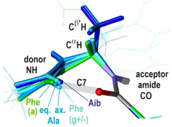
Fig. 6 : Comparative description of the HB approach in -turns of the Ala, Phe (gauche+/-, anti) and Aib series, obtained by superimposing the HB acceptor amides. For the sake of comparison, the Ala C7ax is displayed after a mirror symmetry. Only relevant chemical groups have been highlighted.
a) Assessing the -turn approach
The HB strength has been obtained for a whole series of -turns in their equilibrium geometries, including Ala (C7eq and C7ax forms), Phe (C7eq with the three possible Phe side chain orientations, i.e., anti, gauche+ and gauche-) and the non-proteinogenic amino-isobutyric (Aib) residue (C7, identical to its mirror image). The NBO analysis (see Section 2) carried out on the inverse Ala -turn
15
(C7eq) shows that, despite the delocalization towards the *NH NBO originates from the nO(2) and
n’O(2) lone pair NBOs and the CO(2) NBO (see Fig. 3f-h), this turn nevertheless provides a less efficient
approach than the N’-side dimer as testified by its position (the lower dark blue diamond in Fig. 5) below that of the magenta N’-dimer line in the ranking scale of Fig. 5. The less favorable approach of the inverse -turn compared to this dimer is ascribed to the geometrical constraints imposed by the network of covalent bonds that bridges the amide groups. Figure 5 shows that, along the series of -turns assessed, the efficiency varies only slowly, as testified by the slightly curved line, which joins the several -turn points. It is nearly parallel to those of the dimers at short HB distances (below 200 pm), with an efficiency comparable to that of the N’-side dimer, but takes a slight negative curvature in the long HB region. This latter feature has to be compared with the changes in the approach geometry along the series, as illustrated by Figure 6 , where the Ala and Phe(anti) inverse -turns display large geometrical variations (in particular a lesser HB linearity with smaller NH-O angles). In a context where, by comparison with dimers, a certain approach is expected to give rise to a linear dependence in fig. 5, the increasingly negative curvature of the -turn line in Fig. 5 can be rationalized as a significant change in the geometric approach of the -turn series, at large distances.
b) On the diversity of the equilibrium distances along the -turn family
One of the striking points within the -turn family is the diversity of the equilibrium distances (and hence HB strengths) encountered along the series, which requires a detailed discussion of the interaction at play in these systems.
The inverse -turn: The equilibrium H-bond distance is significantly elongated compared to that of the N-side dimer, in connection with the existence of new constraints, compared to the dimer case, i.e., the interactions responsible for the backbone flexibility.
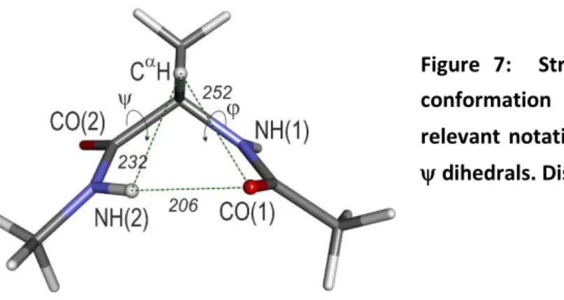
Figure 7: Structure of the inverse -turn C7eq conformation of the Ala diamide model, with relevant notations, including the Ramachandran , dihedrals. Distances are given in pm.
Beyond the H-bond, the Ala C7eq structure (Figure 7) is also characterized by a C(1)-N(1)-C-C(2) Ramachandran dihedralof -82° on the N-terminal side, as well as a short O(1)-H distance (252 pm) in the CO(1)---CH close contact. As described in detail in Section 2, these features result from i) a strong nN(1) CC(2) hyperconjugative interaction (Fig. 3c), which favours ~ 90° Ramachandran
dihedrals, and ii) a significant CH NH HC (Fig. 3d) favoring an antiparallel orientation of the
vicinal NH and CH bonds (see E(2) values in Table S1). These interactions lead in fine to short penalizing O(1)-HC distances and opposes the HB formation. The molecule also experiences an additional CH---N(2)H contact, with an H-H distance (232 pm), significantly shorter than the distance
16
reported in crystal survey (238 pm66), suggesting a repulsive character of this close contact. In conclusion, the C7eq -turn conformation arises from a competition between the C7eq H-bond, on a one hand, and the several interactions which control the backbone flexibility on the other hand, in particular hyperconjugative interactions within the backbone, and to a lesser extent dispersive/close contact interactions involving the H atom of CH and its closest neighbors (Fig. 7), namely the H atom of the N(2)H bond and the O atom of the carbonyl CO(1).
The inverse -turn is also encountered with the phenylalanine (Phe) diamide, with some variability depending upon the orientation of the Phe side chain (Fig. 6 and S2). The two gauche orientations exhibit stronger H-bond strengths, with an HB efficiency close to that of the N’-dimer and H-bond distances, significantly shorter than in the Ala -turn (Fig. 5). In contrast, the anti rotamer of Phe, exhibits a weaker efficiency, together with an increased HB distance. The structures of these species (Figure S2) share the same Ramachandran dihedral, which can be rationalized by the existence of the same hyperconjugation interactions and CH---CO(1) close contacts than with Ala. They differ primarily by the Ramachandran dihedral, which is influenced by interactions between the acceptor CO(2) with the Phe side chain, in particular for the gauche+ and anti rotamers, in which the HB is respectively shortened or elongated.
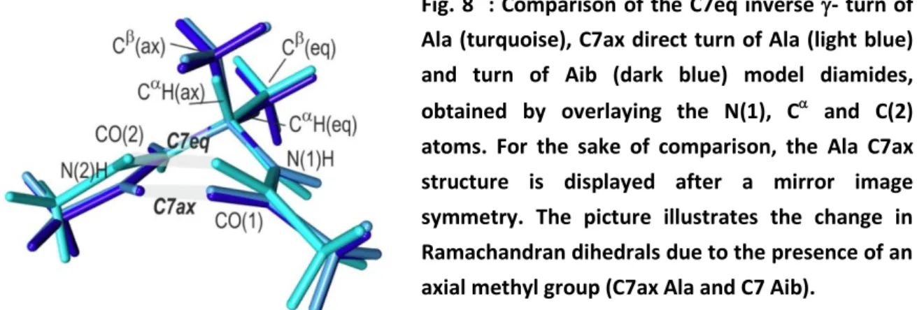
Fig. 8 : Comparison of the C7eq inverse - turn of Ala (turquoise), C7ax direct turn of Ala (light blue) and turn of Aib (dark blue) model diamides, obtained by overlaying the N(1), C and C(2) atoms. For the sake of comparison, the Ala C7ax structure is displayed after a mirror image symmetry. The picture illustrates the change in Ramachandran dihedrals due to the presence of an axial methyl group (C7ax Ala and C7 Aib).
The direct -turn : The direct -turn is a backbone mirror image of the inverse -turn, meaning that the CH covalent bond and the side chain are now in an equatorial and axial position respectively (Figure 8). However, the corresponding C7ax HB is found to be much stronger (the higher dark blue diamond in Fig. 5), with the Ramachandran dihedral adopting a smaller value (72°) compared to the 82° value of the Ala -turn mirror image (Fig. 7), which leads to a shorter HB distance (189 pm) together with a larger value (-54°) (Cf. Ala HB distance = 206 pm, and = -75°). This can be rationalized by considering the effect of the side chain methyl group in the axial position upon the HC effects that describe the backbone flexibility (see the HC sets in Table S1). The methyl group does not affect the strong nN(1) *C(2)CHC term (9.6 kcal/mol; HC0 set in Table S1), which tends to favor
+90° values, but it disrupts the balance between the two other terms of the set in favor of a stronger nN(1) *CC term (5.8 kcal/mol). This latter interaction counterbalances the first term,
leading in fine to a much smaller value (72°) than previously. Interestingly, the presence of the methyl group in axial position also significantly affects HC interactions related to the CH and CMe covalent bonds. When comparing the NBO data of C7eq and C7ax Ala systems (Table S1), one
17
observes: i) a decrease of the HC between the NBO of the CH/CC bond in axial position and the *NH(1) acceptor (red data in HC1 set of Table S1) and concomitantly, ii) an increase of the HC stabilization due to the delocalization from the CCCH bond in equatorial position to the vicinal CN bonds (green data in the HC2 set of Table S1). These changes have two respective consequences: a weakening of the HC, which tended to align the N(1)H bond antiparallel to the CH/CC(ax) bond, and a strengthening of the interaction, which tends to align the C(2)N(2) and C(1)N(1) bonds, antiparallel to the CC/CH(eq) bond, whose consequences in terms of backbone structure (in particular Ramachandran dihedrals) are illustrated in Figure 8. Consequently, in contrast to the C7eq case, the C7ax system no longer exhibits amide-SC close contacts : the CH(ax) H atom interacts only weakly with the CO(1) and N(2)H centers, through elongated interaction distances of 257 and 258 pm respectively.
Similar structural effects are encountered in the two (mirror images) -turns of the achiral amino-isobutyric acid (Aib), whose C atom bears two methyl groups, one equatorial and the other axial with HC features similar to that of the C7ax system (Fig. 6 and 8). Indeed the Aib turn HB is also short (186 pm) and exhibits a larger H-bonding efficiency than the Ala -turn, comparable to that of the N’-side dimer (violet diamond in Fig. 5). The NBO analysis shows that the same HC effects at play in the Ala C7ax are also present in Aib (Table S1). The Aib turn HB, however, is still shorter than the Ala C7ax bond, suggesting that an additional factor is also at play. The structural analysis (see Fig. 8) shows that the simultaneous presence of the two side chain methyl groups distorts the C sp3
hybridization and again favors the presence of close contacts, with shortened H-O and H-H distances compared to C7ax: 247 pm in CH(ax)---CO(1) and 241 pm in CH(ax)---N(2)H respectively.
Finally, the diversity of the equilibrium distances (and hence HB strengths) along the series can be viewed as the manifestation of the effect of the peptide chain environment on the turn structure. The relatively elongated C7eq HB of the Ala inverse -turn, which constitutes a natural HB reference for a -turn in peptides and proteins, results from HCs which tend to stiffen the backbone structure, by imposing the Ramachandran dihedral and by favouring antiparallel orientations of the vicinal N(1)H and CaH bonds. These features are also present in the Phe turns, but the side chain plays here a significant role. The Phe(anti) turn, which exhibits the most elongated HBs (Fig. 6), epitomizes an additional frustration due to unfavorable steric effects, namely a Phe SC-backbone close contact. In contrast, in the Phe(gauche+/-) turns ancillary SC-backbone interactions allow to compensate for this basic hindrance and eventually enhance the HB. At the top of the series, the C7ax bonds of the Ala direct and Aib turns are much stronger. This is ascribed to the side chain methyl group in axial position, which imposes alternative HC interactions that favour parallel CN amide bonds and in fine allow stronger H-bonds to appear. The Aib case is still more constrained due to the simultaneous presence of two bulky side chain methyl groups, which, besides HC effects, also induces additional close contacts.
18
4.2. Extended
-strand-like forms of
-peptides
Local intraresidue interactions, referred to as C5 interactions, are found in extended -strand-like conformations of -peptides (Figs. 1 and 9) and constitute an other iconic approach geometry for two amide groups. Despite the donor amide lies on the C-side of the acceptor, like in the C-side dimer, the fact that donor N(1)H and acceptor CO(2) bonds are facing each other in a quasi-antiparallel disposition because of the strong constrain induced by the small C spacer, the non-linear approach is not favorable to H-bonding. The H-bonding nature of the interaction has even been questioned, despite the recent IUPAC broad redefinition of the HB,67 which qualifies it as a HB. The C5 HBs of Ala and Phe(anti) residues appear quite weak according to the strength scale of Fig. 5 (EHB = 1.1 kcal/mol), i.e., with strengths typically 3 to 4 times weaker than that of a dimer at the
same distance, featuring an intrinsically poor HB efficiency of the C5 approach, obviously strongly impaired by the quasi parallel disposition of the donor and the acceptor. As a direct consequence, the main stabilization arises from the n’O(2) NBO (Cf. Fig. 3.f.), which possesses the right p character to
optimize the overlap with the *NH(1) NBO in such an approach. The *N(1)H population, at ~ 36 me,
exhibits a
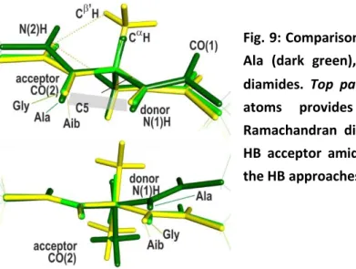
Fig. 9: Comparisons of the C5 extended conformations of Ala (dark green), Gly (green) and Aib (yellow) model diamides. Top panel: overlapping the N(1), C and C(2) atoms provides insights on the differences in Ramachandran dihedrals; bottom panel: overlaying the HB acceptor amides illustrates the differences between the HB approaches.
enhancement compared to the ~31-32 me reference for a free amide NH, taken from the HB acceptor trans-methylacetamide molecule in the C and N’ dimers (see Tables S3 and S4), but remains smaller than that of Ala C7 (~ 43 me). It should be noted that, besides the HB delocalization, the *N(1)H NBO also benefits from significant HC interactions from the vicinal CH and, to a lesser extent,
CCNBOs, with E(2) values of 2.6 and 0.6 kcal/mol respectively (Table S1). In this perspective, the
spectral shift, usually assigned to the C5 HB interaction,16 should also be considered as greatly influenced by a substantial HC relative contribution.
Comparison of the Ala C5 structure to its Gly counterpart is interesting since the achiral property of Gly leads to a backbone conformation of Cs symmetry (Figure 9) in contrast to Ala. Examination of the NBO interactions (Table S1) shows that, as in -turns, the strong HC interactions which control the Ramachandran dihedral angle, i.e., the delocalization from the nN(1) lone pair, is also present in
19
both Ala and Gly species and occurs towards the *CH and *CH’ NBOs for Gly and *CH and
*CC NBOs for Ala. In Gly, the HC interactions are the same (9.0 kcal/mol) and lead to and
Ramachandran dihedrals of 180°; the situation is asymmetric in Ala, where the main delocalization from the nN(1) lone pair goes to the side chain *CC NBO (E(2) value of -9.2 kcal/mol), favoring a
situation where the C(1)N(1)CC dihedral (80°) is closer to 90°, eventually leading to = -158° and = 159° Ramachandran dihedrals. This distortion induces a slight increase of the C5 HB distance compared to Gly, which is directly correlated to the changes in stabilization energy (1.1 vs. 1.4 kcal/mol for Ala and Gly resp.; Figure 5).
Another interesting comparison arises from the C5 conformation of the achiral Aib, whose amide network is also planar (Fig. 9). The striking point, however, is the HB enhancement in Aib (EHB = 3.5
kcal/mol) and the shortening of its equilibrium HB distance (Fig. 5). Additionally the proximity of C5 Aib to the dimer lines in the ranking scale of Figure 5, compared to C5 Ala and Gly, features a higher HB efficiency of the C5 approach in Aib. The trend is confirmed when considering squeezed (out-of-equilibrium) conformations of the Ala and Gly C5 structures obtained through full optimization when constraining the HB distance to a series of fixed values (green points in Figure 5). Along this series, the HB strength follows a curved line (green dots) in Fig. 5, mimicking the long distance behavior already observed in the C7 series. Like for this latter geometry, one can notice a significant change in the approach geometry along the C5 series, as depicted in Figure 9, which can account for the negative curvature of the C5 approach line at large HB distances.
The C5 series also shares another feature with the C7 series, i.e., a significant spread of the equilibrium H-distances observed, especially between Ala and Gly residues compared to Aib. A detailed structural analysis shows that in this latter species a repulsive close contact occurs between the acceptor amide N(2)H and the two side chain C’H H atoms (226 pm, see Fig. 9, left panel, much less than the 238 pm value in crystals66). It pushes the HB acceptor CO(2) towards the N(1)H donor, leading to a strong HB shortening and enhancement compared to the Ala and Gly residues, where the close contact does not occur. Finally, it should be noted that the Aib molecule presents the same features as diethylglycine, the model molecule chosen by Newberry and Raines9 to provide evidence for a C5 H-bond in peptides. The present analysis shows that, because of the occurrence of the above mentioned repulsive close contact, the model chosen by these authors grossly overestimates the strength of the C5 HB compared to peptides and proteins.
Besides C5 bonds, other examples of intraresidue HBs can be found in flexible species, such as the C6 HB in extended forms of the Ac-3hPhe-NHMe -peptide17
(see Fig. 1). The increased flexibility of this backbone ensures a much better efficiency of this C6 approach, compared to C5, closer to that of the weakest C7 -turns (Fig.5, EHB indicator ~3.3 kcal/mol with a *NH population of ~ 40 me).
4.3. Intramolecular amide-amide interactions in models of
-turn and 3
10helices
-turns are essential secondary structures, stabilized by C10 HBs, which bridge two amide groups two residues apart, and contain a central amide group.22 Incipient 310 helices are minimalist helical
models, which feature two C10 HBs, one on the C-side terminal and the other on the N-side.24 They are nearly parallel, similar to those of the -turns although slightly distorted. Examples of such C10
20
HB in the gas phase have been taken from experimentally observed -turns (Ac-Gly-Phe-NHMe, type I and II’),68 the incipient 310 Ac-Ala-Phe-Ala-NH2 helix observed experimentally24 as well as the
Ac-(Ala)3-NHMe and Ac-(Aib)3-NHMe model 310 helices. Each helix provides two examples of C10 HB,
labelled N-terminal or C-terminal accordingly to their location along the sequence.24 They slightly differ in geometrical approach and distance because of distortions of the helical structure, either due to the small size of the helix or to the presence of the Phe residue in the sequence.
Figure 10: Comparisons of the C10 H-bonds of the type I Ac-Gly-Phe-NHMe -turn (orange), of the Ac-Ala-Phe-AlaNH2 and of the Ac-(Ala)3-NHMe and
Ac-(Aib)3-NHMe helices (bright and dark brown for
the N- and C-terminal sides respectively). For the sake of clarity the side chains are displayed on a line style. The overlay of the HB acceptor amides emphasizes the diversity of HB approaches.
The -turn HBs exhibit EHB values of the order of ~4 kcal/mol with *NH populations around 42 me.
Together with their relatively large HB distance (208 pm), this characterizes them as medium range HBs (Fig. 5), with a modest HB efficiency, as illustrated by the orange eye guide of Fig. 5, passing through the -turn points, parallel to but lying well below the dimer lines (red and magenta dots). Concerning the helix C10s, a large diversity of HB strengths is observed, ranging from 2 to 5 kcal/mol, with *NH populations between 39 and 47 me., to be correlated with the diversity in terms of
geometrical approach within the series (Fig. 10). The N-terminal helix C10s, all lying above the orange line, present a higher efficiency than the -turn approach, in contrast to the C-terminal C10s, which exhibit a reverse trend. The large diversity in the HB distances within the helix C10 set is ascribed to specific backbone distortions in the helices as noticed earlier and epitomizes the effect of the environment upon the H-bonding through both the control of the HB approach efficiency and the possible HB frustration within the constrained approach.
4.4. Intramolecular amide-amide interactions in di-amide models of
- and
-peptides
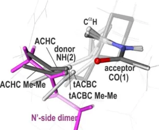
Figure 11: Comparisons of the H-bonds of the tACBC (dark grey; C8 H-bond) and ACHC (grey; C9 H-bond) diamide models with the N’-side dimer of MAA (magenta), which presents a HB approach on the same side of the acceptor. The overlay of the HB acceptor amides emphasizes the differences between the several HB approaches. The tACBC/ACHC acronyms stand for the systems experimentally studied and described in Fig. 1; names followed by Me-Me
21
stand for model systems with minimalist caps (with acetyl and methylamide groups). For the sake of clarity side chains irrelevant to the discussion are displayed on a line style.
The most stable conformations observed for the ACHC31 and tACBC30 backbones are stabilized by C8 and C9 bonds respectively (grey and white disks in Fig. 5), whose efficiencies are found to be as large as that of the C-type dimer. They show EHB and *N(1)H population, in the 13-10 kcal/mol and 50-56
me range respectively, comparable to those of C-side dimers at the same HB distance (Fig. 5). These conformations both exhibit the same CH--- CO(1) close contact than that encountered in the -turns of -peptides, which is caused by the same strong nN(1) *CC and CH *N(1)H hyperconjugation
interactions as in C7eq -turns (respective E(2) of 8.2 and 4.6 kcal/mol in ACHC, and 13.5 and 4.3 kcal/mol in tACBC, to be compared with 10.1 and 3.7 kcal/mol in Ala C7eq). However, in contrast to the -turns, the additional flexibility, provided by the longer covalent spacers between the amides, together with a favorable range of amide-amide distances, enforced by the cyclic constrain, endows these chains with both an efficient approach and smaller H-bonding distances.
At this stage, one can notice that these species also enable us to document the capacity of apparently ancillary interactions, present in the molecule, in amending the approach and eventually the structure.
In the -peptide tACBC case, the efficiency is as high as that of the C-side dimer, and the HB distances of these species are very similar, suggesting a priori minimal frustrating/enhancing constrains due to the tACBC backbone (grey disk in Fig. 5 at EHB = 9.6 kcal/mol). One should however notice that the
bulky caps of this model interact with each other and contribute to the structure. Indeed, a substitution of these caps by smaller methyl groups (tACBC Me Me species) does not affect the HB efficiency (same approach) but decreases the HB distance (grey disk at EHB = 11.1 kcal/mol; tACBC
Me Me label in Fig. 5). Being smaller than the bulky caps of t-ABCB, the methyl groups do no longer interact with each other, which increases the contribution of backbone intrinsic folding to the H-bonding: the shorter HB distance of the tACBC Me-Me model compared to the dimer illustrates the enhancing role of the -peptide backbone, in particular due to the trans-disposition of the amides on the cyclobutyl ring of this compound.
In contrast, in the experimentally studied ACHC -peptide, strong dispersion interactions between the benzyl end and the cyclic side chain tend to make the structure compact with a small HB distance (white disk at EHB = 13.1 kcal/mol in Fig. 5). When substituting the benzyl end by a methyl group in
the present calculations, the system adopts a looser structure, with a longer H-bond, a smaller EHB
(white disk at EHB = 9.5 kcal/mol; ACHC MeMe label in Fig. 5) as well as a weaker efficiency due to a
lesser linearity of the approach (in particular a smaller NH-O angle; see Fig. 11). The efficiency then drops to that of the N’-side dimer, in agreement with the comparable approach in these species as illustrated by Fig. 11. The significantly shorter equilibrium distance compared to the N’-side dimer, however, demonstrates an HB enhancement induced by the constrained -peptide backbone.
22
Interestingly, among all the species studied, only one exhibits a stronger efficiency than the C-side dimer : this is the so-called hydrazino-turn conformation of an Aza -peptide,30
whose HB presents a C8+C5 bifurcated nature, due to the presence of a N-heteroatom within the main chain, explaining its apparent extra efficiency (dark grey point above the C-dimer line in Fig. 5). However, in contrast to
tACBC, despite this large efficiency, the HB remains relatively modest due to the large H-distance
imposed by the rigidity of the hydrazino-turn structure.
Besides these highly efficient HB approaches, the cis-ACBC -peptide30 constitutes a dramatic counterexample. The cis disposition of the amide about the cyclobutyl ring forces the amides to be relatively close and to adopt nearly stacked conformations, which hampers access to efficient HB approaches. Consequently the most stable conformation is stabilized by a C8 non-linear HB, with a much more modest HB efficiency (similar to those of C10 HBs) and a very elongated HB distance, epitomizing the dramatic effect of the steric constraints in this cyclic backbone.
5. Synthesis: ranking the H-bond approaches and assessing frustrating/enhancing effects on
the equilibrium structures
5.1. The approaches
The extended set of conformations considered in the study, with or without covalent linkages between the amide groups, offers various geometrical approaches illustrative of the diversity met in proteins. The EHB - distance semi-log plot derived from NBO analyses (Fig. 5) enables us to rank the
corresponding approaches according to their efficiency. The HB strength is found to follow a roughly linear behavior in this plot, as the HB distance varies along a fixed approach. Demonstrated for the dimers, this behavior seems to hold, for each type of secondary structure studied, as long as the geometrical approach does not change significantly along the series considered, as illustrated by -turns, -turn/3-10 helices approaches. The order of the corresponding parallel lines then provides a relative ranking of the approaches, independently from the species that serve to rank them (Cf. vertical arrows in Fig. 5).
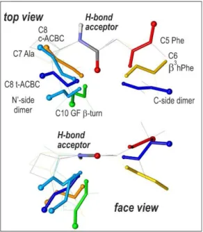
Fig. 12: Various geometrical HB approaches illustrated by selected representative structures studied in the present survey. The HB acceptor amides are overlaid (top: top view for the acceptor amide; bottom: side view), the donor amides are displayed according to a rainbow color code corresponding to increasing HB efficiencies (and not necessary HB strength) when going from red to blue; The amide carbonyl O atom and NH H atom are indicated by large and small balls respectively); for the sake of clarity, the rest of the backbone and side chain have been hidden.