HAL Id: inserm-00380055
https://www.hal.inserm.fr/inserm-00380055
Submitted on 5 May 2009HAL is a multi-disciplinary open access archive for the deposit and dissemination of sci-entific research documents, whether they are pub-lished or not. The documents may come from teaching and research institutions in France or
L’archive ouverte pluridisciplinaire HAL, est destinée au dépôt et à la diffusion de documents scientifiques de niveau recherche, publiés ou non, émanant des établissements d’enseignement et de recherche français ou étrangers, des laboratoires
The CD38-independent ADP-ribosyl cyclase from mouse
brain synaptosomes: a comparative study of neonate
and adult brain.
Claire Ceni, Nathalie Pochon, Michel Villaz, Hélène Muller-Steffner, Francis
Schuber, Julie Baratier, Michel de Waard, Michel Ronjat, Marie-Jo Moutin
To cite this version:
Claire Ceni, Nathalie Pochon, Michel Villaz, Hélène Muller-Steffner, Francis Schuber, et al.. The CD38-independent ADP-ribosyl cyclase from mouse brain synaptosomes: a comparative study of neonate and adult brain.: CD38-independent ADP-ribosyl cyclase from brain synaptosomes. Bio-chemical Journal, Portland Press, 2006, 395 (2), pp.417-26. �10.1042/BJ20051321�. �inserm-00380055�
The CD38-independent ADP-ribosyl cyclase from mouse brain synaptosomes : a comparative study of neonate and adult brain.
Claire CENI*, Nathalie POCHON†, Michel VILLAZ*, Hélène MULLER-STEFFNER‡, Francis SCHUBER‡, Julie BARATIER§, Michel DE WAARD†, Michel RONJAT†, and Marie-Jo MOUTIN†.
*Laboratoire Canaux Ioniques et Signalisation, INSERM E9931, DRDC-CEA (UJF Grenoble), 17 avenue des martyrs, 38051 Grenoble Cedex 9 , France.
†Laboratoire Canaux Calciques, Fonctions et Pathologies, INSERM U607, DRDC-CEA (UJF Grenoble), 17 avenue des martyrs, 38051 Grenoble Cedex 9 , France.
‡ Laboratoire de Chimie Bioorganique, UMR7514 CNRS-ULP, Faculté de Pharmacie, 74 route du Rhin, 67400 Strasbourg-Illkirch , France
§ Laboratoire du Cytosquelette, INSERM U366, DRDC-CEA (UJF Grenoble), 17 avenue des martyrs, 38051 Grenoble Cedex 9 , France
Corresponding author :
Marie-Jo Moutin
Laboratoire Canaux Calciques, Fonctions et Pathologies, INSERM-U607, DRDC-CEA, 17 avenue des martyrs, 38051 Grenoble Cedex 9 , France.
Tel: 33 4 38 78 55 93 ; fax : 33 4 38 78 50 41; e.mail: marie-jo.moutin@cea.fr
Short title :
CD38-independent ADP-ribosyl cyclase from brain synaptosomes
Abbreviations footnote :
SYNOPSIS
Cyclic ADP-ribose (cADPR), a metabolite of NAD+, is known to modulate intracellular calcium levels and to be involved in calcium-dependent processes including synaptic transmission, plasticity and neuronal excitability. However, the enzyme responsible for producing cADPR in the cytoplasm of neural cells - and particularly at the synaptic terminals of neurons – remains unknown. In the present work, we show that endogenous concentrations of cADPR are much higher in embryonic and neonate mouse brain compared to the adult tissue. We also demonstrate, by comparing wild-type and Cd38-/- tissues, that brain cADPR content is independent from the presence of CD38 (the best
characterized mammalian ADP-ribosyl cyclase) not only in adult but also in developing tissues. We show that Cd38-/- synaptosomes preparations contain high ADP-ribosyl cyclase activities, which are more important in neonate than in adult, in line with the levels of endogenous cyclic nucleotide. By using an HPLC method and adapting the cycling assay initially developed to study endogenous cADPR, we accurately examined the properties of the synaptosomal ADP-ribosyl cyclase. This intracellular enzyme has estimated Km for NAD+ of 21 µM, a broad optimal pH at 6.0-7.0, and concentration of free calcium has no major effect on its cADPR production. It binds NGD+, which inhibits its NAD+ metabolizing activities (Ki = 24 µM), despite its incapacity to cyclize this analogue. Interestingly, it is fully inhibited by low (micromolar) concentrations of zinc. We propose that this novel mammalian ADP-ribosyl cyclase regulates the production of cADPR and therefore calcium levels within brain synaptic terminals. In addition, this enzyme might be a potential target of neurotoxic Zn2+.
Key words
INTRODUCTION
Calcium is an intracellular messenger relaying information within cells to regulate their activity. Increases in cytosolic calcium concentration involve several calcium channels from both the cell surface and the intracellular membranes giving access to the two available sources of Ca2+, external medium and intracellular Ca2+ stores. Several metabolites of NAD+ have been identified as second messengers contributing to the complex and versatile cell calcium signaling. Cyclic ADP-ribose (cADPR) was shown to modulate calcium movements from ryanodine-sensitive endoplasmic reticulum stores (for review see [1, 2]). Nicotinic acid adenine dinucleotide phosphate (NAADP+) was shown to act on as yet to be identified intracellular stores of calcium (for review see [3-5]). ADP-ribose (ADPR) was shown to modulate the activity of a plasma membrane Ca2+ channel, TRPM2, a member of the large Transient Receptor Potential (TRP) channels family [6]. Very recently, cADPR was also shown to synergize with ADPR in the activation of TRPM2 channels [7]. Therefore, enzymes capable of producing these NAD+ metabolites within cells undoubtedly have key roles in the control of fundamental calcium-dependent cellular processes.
In neural cells, like in other cells, calcium controls a large number of functions. Cyclic ADP-ribose, which is abundant in the brain [8, 9], was shown to modulate calcium movements from ryanodine-sensitive stores both in neurons [10-13] and astrocytes [14]. It is involved in calcium-dependent neural processes including synaptic transmission, plasticity and neuronal excitability [13-16]. The cellular events that allow cADPR to control ryanodine-sensitive stores or TRPM2-channels remain to be clarified. But, there is growing evidence that cADPR could be produced in response to the activation of some metabotropic receptors [17-23] and play a critical role at synaptic terminals. However, the identity of the cADPR synthesizing enzyme involved in such signalling pathways remains unknown.
Cyclic ADP-ribose synthesizing enzymes known to date form a growing family of proteins that are highly conserved at both genetic and structural levels. All these enzymes produce both cADPR (ADP-ribosyl cyclase activity) and ADPR (NAD+ glycohydrolase, or NADase activity) from NAD+, but in considerably varying proportions [24]. The first identified enzyme of this family, mainly endowed with ADP-ribosyl cyclase activity, was isolated from the mollusk Aplysia californica [25], where it is present in the cytosolic compartment of synaptic terminals [15]. In mammals, two proteins sharing about 30% sequence identity with the Aplysia cyclase have then been identified, CD38 [26] and CD157 (or BST-1) [27]. Only CD38, which is a membrane-associated protein and
which, unlike the Aplysia enzyme, mainly exhibits an NADase activity, is expressed in the nervous system [28, 29]. Recently, we have shown that CD38 is mostly ectocellular in the brain tissue but also has intracellular location. However, as it is principally located in the soma of neural cells [30], CD38 activity is unlikely to play a significant role in synaptic transmission in relation with ryanodine sensitive internal stores. The discovery that the endogenous level of cADPR of adult brain tissue is not changed in CD38 knock-out mice [9] is consistent with this conclusion. Moreover, we have recently revealed the existence of an ADP-ribosyl cyclase in neural cells from Cd38-/- mice [31]. This novel enzyme which produces intracellular cADPR and ADPR, is present in brain during development and in the adult, and exhibits low basal activity which can be enhanced by GTP-γ-S application. This novel ADP-ribosyl cyclase could be a relay between extracellular signals and intracellular calcium-dependent events occurring at synaptic terminals.
In the present study, we show that synaptosomes from Cd38-/- mouse brains have high intracellular ADP-ribosyl cyclase activity, and that the highest activities are detected in synaptosomes purified from neonate compared to synaptosomes purified from adult animals. This result is consistent with the observation that endogenous brain cADPR content, which is definitely not related to the presence of the CD38 protein, is higher in the developing brain and declines in the adult tissue. We then characterized the ADP-ribosyl cyclase from brain synaptosomes. Interestingly, we found that this ADP-ribosyl cyclase is sensitive to micromolar concentrations of zinc ions, which were described as modulators of synaptic activities [32] and whose homeostasis imbalance was shown to generate brain injury and defects in cognitive development [33, 34].
We propose that the novel mammalian ADP-ribosyl cyclase participates to the production of the cADPR messenger, and also of ADPR, within nerve terminals and might therefore be involved in crucial calcium-dependent pathways in brain development and functioning.
EXPERIMENTAL Materials
ADP-ribosyl cyclase from Aplysia californica, nucleotide pyrophosphatase from Crotalus atrox venom, NAD+ glycohydrolase from Neurospora crassa, alcohol dehydrogenase from Bakers yeast, diaphorase from Clostridium kluyveri and resazurin were purchased from Sigma. Alkaline phosphatase from calf intestine was purchased from Roche Diagnostics Corporation. 14C-NAD+ (nicotinamide [U-14C]adenine dinucleotide, 300 mCi/mmol) was from Amersham Pharmacia Biotech Europe. Monoclonal antibodies, anti-synapsin IIa and anti-PSD95, were purchased from BD Transduction Laboratories.
Preparation and Western-blot analysis of brain synaptosomes
Synaptosomes were prepared from total brain isolated from adult and neonate CD38 deficient mice using a one-step preparation based on the known isopycnic densities of cellular components, as described in [35]. They were subsequently either directly frozen in liquid nitrogen, or dialysed against solution D (containing 10 mM Tris,HCl pH 7.5, 2.2 mM CaCl2, 0.5 mM Na2HP04, 0.4 mM KH2PO4, 4 mM NaHCO3 and 80 mM NaCl) for 2 hours at 4°C and then frozen in liquid nitrogen. Using this protocol, we were able to purify from neonate brains (postnatal days 1 or 2) approximately one third of the quantity of synaptosomes purified with the same weight of adult brains. Therefore, about 1% (w/w) of the total protein content of adult tissue corresponds to the purified synaptosomes proteins, whereas only about 0.3-0.4% (w/w) of the total protein content of neonate tissue represent the synaptosomes proteins.
Synaptosomes enrichment was tested by Western-blot analysis of the associated proteins using monoclonal antibodies against two synaptic proteins, i.e. synapsin IIa and PSD95 (at a dilution of 1/2000 and 1/250 respectively). Proteins (6 µg per well) were separated on 12 % (wt/wt) polyacrylamide denaturing gels, followed by an electrotransfer for 2h at 500 mA to Immobilon P sheets (Millipore). After 1 h incubation with 4% non-fat dry milk (Bio-Rad), the primary antibody was added and incubated overnight at 4°C. The blots were then stained for 3 h at room temperature with horseradish peroxidase-conjugated anti-mouse IgG (Cappel). After washing the blots, the reactive proteins were detected using the Renaissance chemiluminescent reaction (NEN Life Science Products) followed by exposure to Biomax films (Kodak).
Measurement of endogenous cyclic ADP-ribose
The measurement of cADPR from brain tissues was performed essentially as already described [36], except that fresh tissues were used instead of frozen tissues. We found that the use of fresh tissue did not modify the results obtained by comparing the cADPR content of half-brain homogenates, one half-brain being frozen and pulverized to powder prior to the addition of perchloric acid and the other half being directly mixed with the acid : same results were obtained with both types of homogenates (not shown). For each purification performed, half-brains were used in the case of adult mice, whereas entire brains were used for embryos and neonates.
The fresh brain tissues from wild-type mice (of known weight) were directly homogenized on ice with a Potter in 0.6 M perchloric acid. The fresh brain tissues from CD38 deficient mice were first homogenized on ice with 100 mM NaH2PO4 buffer at pH 8.0, aliquots were removed to determine the protein content according to Bradford (using bovine serum albumin as standard), and then (4 min after adding the buffer) the PCA was added to a final concentration of 0.6 M. This slightly different protocol, allowing the measurement of protein content in homogenate, was used with the brain tissue from CD38 deficient mice since we have observed that the cADPR level was stable in this tissue during the first 4 minutes after buffer addition. This was not the case with wild-type tissues, in which the cADPR level decreases quickly (not shown). The acid was eliminated by extraction with 1,1,2-trichlorotrifluoroethane (TFE) and tri-n-octylamine (TNO), and finally 20 mM NaH2PO4 at pH 8.0 was added to the samples. Samples were then reacted one night at 37°C with a mix of nucleotide pyrophosphatase, alkaline phosphatase and NAD+-glycohydrolase in the presence of magnesium as in [36] in order to remove all contaminating nucleotides. Enzymes were eliminated on ultrafiltration units (nanosep 10 K from Pall Corporation).
Cyclic ADP-ribose present in samples was then quantified. The conversion of cADPR into NAD+ was performed by incubating the sample 1 hour at room temperature with 0.3 µg/ml Aplysia ADP-ribosyl cyclase and 10 mM nicotinamide in 35 mM NaH2PO4 at pH 8.0. The cycling reaction was then allowed to proceed in 60 mM NaH2PO4 at pH 8.0 in the presence of 0.8% ethanol, 10 mM nicotinamide, 40 µg/ml bovine serum albumin, 20 µg/ml diaphorase (extemporaneously treated with charcoal), 20 µg/ml alcohol dehydrogenase, 10 µM FMN and 20 µM resazurin. The increase in resorufin fluorescence (excitation at 544 nm and emission at 590 nm) was measured every minute during 2 or 3 hours using a fluorescence plate reader (FluoStar from BMG Labtechnologies Inc.) and compared to standard solutions of cADPR and NAD+.
In order to calculate the cADPR content of the samples isolated from wild-type mice, in pmoles range per mg of protein, we determined the ratio [quantity of protein / quantity of tissue] in both neonate and adult brain by weighing brains and analyzing their protein content using the Bradford assay. The ratios obtained were of 36 µg of protein/mg of tissue in neonate (n=4) and 52 µg of protein per mg of tissue in adult (n=3). In the case of CD38 deficient mice brains, cADPR content calculation was made using the brain homogenate protein content also measured with the Bradford assay.
Enzyme assays with 14C-NAD+ and analysis by HPLC
Synaptosomes (1 mg of protein/ml) were incubated at 32 or 37°C in 20 mM buffer (citrate-HCl at pH 4.0, MES-NaOH at pH 5.0 and 6.0, and Tris,HCl at pH 7.0 and 8.0) with 60 µM 14C-NAD+ (8 mCi/mmole), 4 mM EDTA (to inhibit the nucleotide pyrophosphatases, see [31]), and with or without 1% (wt/vol) Triton X-100. Reactions were stopped at given times (see legends to figures) by adding 10% (vol/vol) perchloric acid, followed by neutralization with K2CO3.
HPLC analysis and standards preparation (14C-cADPR, 14C-ADPR and 14C-AMP) were performed essentially as previously described [31]. Briefly, HPLC separations were performed isocratically on a C18 column using 10 mM ammonium phosphate buffer at pH 5.5 and 1% (vol/vol) acetonitrile. Radiodetection of the products was performed with an on-line radioactivity monitor after mixing eluting products with a scintillation liquid. Reaction products of NAD+ (ADPR, cADPR, AMP) were identified on the chromatograms by separately injecting standards.
Enzymatic tests using the cycling assay
Synaptosomes (0.2-0.4 mg of protein/ml) were generally incubated in solution D with 200 µM NAD+ in the absence or presence of 1% (wt/vol) of the detergent polyoxyethylene-10-tridecyl ether (Emulphogen). However, in the experiments with varying calcium concentrations, synaptosomes were first centrifuged (30 min at 540,00xg), rinsed and resuspended in solutions containing 50 mM Tris,HCl at pH 7.5 and free calcium at 1 nM (2 mM EGTA and 52 µM CaCl2), or 1 µM (1 mM EGTA and 1.93 mM CaCl2), or 1 mM (2 mM EGTA and 3 mM CaCl2). They were subsequently incubated (at 0.4 mg of protein/ml) with NAD+ and Emulphogen.
Reactions were stopped at given times (see legends to figures) by adding 0.6 M perchloric acid, followed by neutralization with K2CO3. The contaminating nucleotides were eliminated by the use of a mix of the three nucleotide hydrolytic enzymes as detailed above except that the overnight
reaction was performed in solution D. We have controlled that the substrate used in our experiments (200 µM NAD+) was completely degraded during this step (not shown).
Then, the quantity of cADPR produced in the samples was determined : the conversion of cADPR into NAD+ and the cycling reaction were allowed to proceed as detailed above except that the NaH2PO4 buffer was replaced by solution D. Results were compared to standard solutions of cADPR and NAD+ analysed in the same experimental conditions, i.e. metabolites were diluted in solution D. The recovery of cADPR content after extraction with perchloric acid and neutralization with K2CO3 was estimated to 68% of control by tests performed with standard solutions of cADPR.
Assays of GDP-ribosyl cyclase activity
The conversion of NGD+ to cyclic GDP-ribose (cGDPR) was followed fluorometrically as already described [30]. The enzyme reaction was started by adding 100 µM NGD+ to synaptosomes (500 µg/ml) purified from adult or neonate Cd38-/- mouse brains that were incubated at 37°C in solution D in the presence of Emulphogen (0.25% wt/vol). After 2 hours of incubation at 37°C, Aplysia cyclase (3 µg/ml) was added as a control in order to transform all remainder NGD+ into cGDPR. Before each measurement, all materials including quartz cuvette, injection syringes and mixer were meticulously washed and incubated 15 minutes with boiling water in order to totally eliminate contaminating
Aplysia cyclase.
Kinetic analysis
The initial rates of substrate conversion were calculated from a polynomial regression curve. Kinetic parameters were determined from the plot of the initial rates as a function of substrate concentrations using a non-linear regression program (Graph Pad Prism).
RESULTS
Brain tissues contain higher cADPR levels during their development
Previous studies have estimated the cADPR content of brain homogenates isolated from adult mice [9, 37], however, the cADPR level of the developing tissue had never been studied yet. In the present work, we have examined the cADPR content of brain tissue from embryonic (days 15 or 16), neonate (postnatal day 1 or 2), and adult wild-type and CD38 deficient mice (Fig. 1). Using the highly sensitive cycling assay recently developed by Graeff and Lee [36], we have first confirmed the results of literature, showing that the cADPR content of adult brain homogenates prepared from
Cd38-/- deficient mice is not significantly different from the one of wild-type adult brain homogenates [9]. The cADPR content of adult mouse brain we have measured (3.0 ± 0.8 pmol/mg) was similar to the adult mouse brain content found with a radioimmunoassay (3.86 ± 0.87 pmol/mg, [9]) and to the adult rat brain content measured by a Ca2+-release bioassay (2.75 ± 0.35 pmol/mg, [8]). Using the cycling assay, Graeff and Lee [36] estimated the cerebrum and cerebellum cADPR contents of respectively 2.2 ± 0.3 and 2.1 ± 0.3 pmol/mg, values which are close to those we have obtained with the entire brain.
Very interestingly, the cADPR contents measured during the development of the brain, in embryos and in neonates, were found to be notably higher that the contents of the adult tissue. Indeed, the embryonic cADPR content was estimated to 7.3 ± 1.1 pmol/mg, and the neonate content was 5.9 ± 0.4 pmol/mg. Moreover, neonate CD38 deficient brain tissue showed cADPR content equivalent to neonate wild-type tissue, and higher than the content of adult Cd38-/- brain tissue.
In conclusion, our results indicate the presence of higher cADPR contents in the developing brain than in the adult tissue : the level being 2.5- and 2-fold higher respectively in embryos and in neonates. Besides, as in the adult tissues [9], the cADPR contents of developing brain are independent of the presence of the CD38 protein.
An intracellular ADP-ribosyl cyclase is present in brain synaptosomes isolated from CD38 deficient mice.
Cyclic ADPR production was shown to occur in response to the activation of metabotropic receptors in several neural cell types [17-23], and was involved in different neural processes occurring at synaptic connections [14-16]. However, the enzyme responsible for producing cADPR in the cytoplasm of neural cells - and particularly at the synaptic terminals of neurons – remains unknown.
Interestingly, we have recently demonstrated the presence in brain of a new ADP-ribosyl cyclase, different from the known ADP-ribosyl cyclases CD38 and CD157 (see Introduction).
For these reasons, we wished to determine whether the novel ADP-ribosyl cyclase was present in brain synaptic terminals. We have thus isolated synaptosomes from adult and neonate Cd38-/- mice
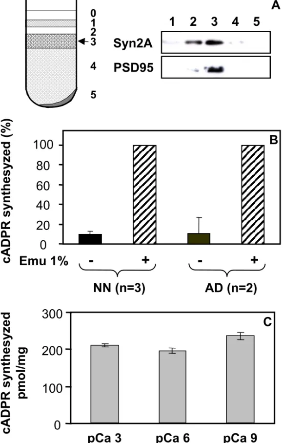
brains according to Phillips et al. [35], and have examined the occurrence of an ADP-ribosyl cyclase activity within these subcellular structures. Synaptosomes enrichment in the preparation was controlled by Western-blot using antibodies against two typical synaptic proteins: synapsin IIa which is a synaptic vesicles-associated protein, and PSD95 which is a protein from the post-synaptic density. As shown in Fig 2A, the fraction 3 which is found at the 1.25 M/1.0 M sucrose interface, is highly enriched in these markers. The ADP-ribosyl cyclase activity was tested in this synaptosomes enriched fraction using two sensitive assays, i.e. a radio-HPLC method to follow the transformation of 14C-NAD and identify its reaction products [31, 38] and a fluorimetric cycling assay which quantifies the production of cADPR from NAD+ [36]. It should be noted here that we have adapted this latter assay, initially developed to study the cADPR contents in tissues [36], to accurately determine an ADP-ribosyl cyclase activity.
Brain synaptosomes (fraction 3; 0.2-0.4 mg/ml) were tested for their capacity to transform NAD+ (200 µM) specifically into cADPR after incubation at 37°C in their conservation medium, i.e. at pH 7.5 and in the presence of millimolar concentrations of CaCl2. Fig. 2B shows the results obtained with synaptosomes isolated from adult and neonate CD38 deficient brain tissues which were either intact or treated with the detergent Emulphogen (an optically neutral detergent, equivalent to Triton X-100). The cADPR production was analyzed using the fluorimetric cycling assay. Under the experimental conditions used (1 hour incubation for neonate, 2 hours incubation for adult), with both adult and neonate brain tissues, the cADPR produced by intact synaptosomes was only approximately 10% of the cADPR synthezised by detergent-treated synaptosomes.
Brain synaptosomes (1 mg/ml) were also tested for their ability to transform 14C-NAD (60 µM) into cADPR and ADPR after incubation at 32°C and pH 6.0 in the presence of 4 mM EDTA (experimental conditions similar to those already used with brain cells and membrane extracts, in which EDTA was added to inhibit the pyrophosphatases cleaving NAD+ metabolites such as ADPR, see [31]). Fig. 3 shows representative HPLC radiochromatograms obtained with intact (control) and Triton X-100-treated synaptosomes isolated from adult (A,B) and neonate (C,D) Cd38-/- brain tissues
after an incubation of 8 hours. In the reactions with intact synaptosomes, peaks corresponding to ADPR were observed (4% of NAD+ in the adult, and 6.7% of NAD+ in the neonate), whereas no
cADPR peaks could be detected. In sharp contrast, when the same reactions were performed in the presence of detergent, both ADPR and cADPR peaks were observed: 29% of NAD+ was converted to ADPR and 2% to the cyclic compound in adult synaptosomes; 52% of NAD+ was converted to ADPR and 4% to cADPR in neonate synaptosomes.
We next examined, using the cycling assay, the effect of calcium concentration on the synaptosomes cADPR synthesis activity. To that end, adult synaptosomes (0.4 mg/ml) were incubated 2 hours with NAD+ and Emulphogen in solutions containing varying free calcium concentrations at pH 7.5 (Fig. 2C). We have observed less than 12% difference between the cADPR production measured at 1 nM, 1 µM and 1 mM free calcium.
Thus, by the use of two different methods, we have demonstrated the presence of an ADP-ribosyl cyclase in synaptosomes isolated both from neonate and adult CD38 deficient brain tissues. We detected much larger ADP-ribosyl cyclase and NADase activities in detergent-treated synaptosomes compared to intact structures, a result indicative of an intracellular location of the enzyme catalytic site. The low activities observed with intact synaptosomes are indeed very likely due to the presence of leaky synaptosomes in our preparations. Moreover, free calcium appears to have no major effect on the cADPR production by the synaptosomal enzyme. The enzyme detected in synaptosomes certainly corresponds to the novel enzyme which we have recently detected within brain cells and membrane extracts (extracts P540 obtained at high speed centrifugation andenriched in endoplasmic reticulum and plasma membranes) isolated from Cd38-/- mouse [31].
In order to have access to the full synaptosomal activity, the subsequent experiments were all performed with detergent-treated synaptosomes, i.e. in the presence of 1% (wt/vol) Triton X-100 or Emulphogen.
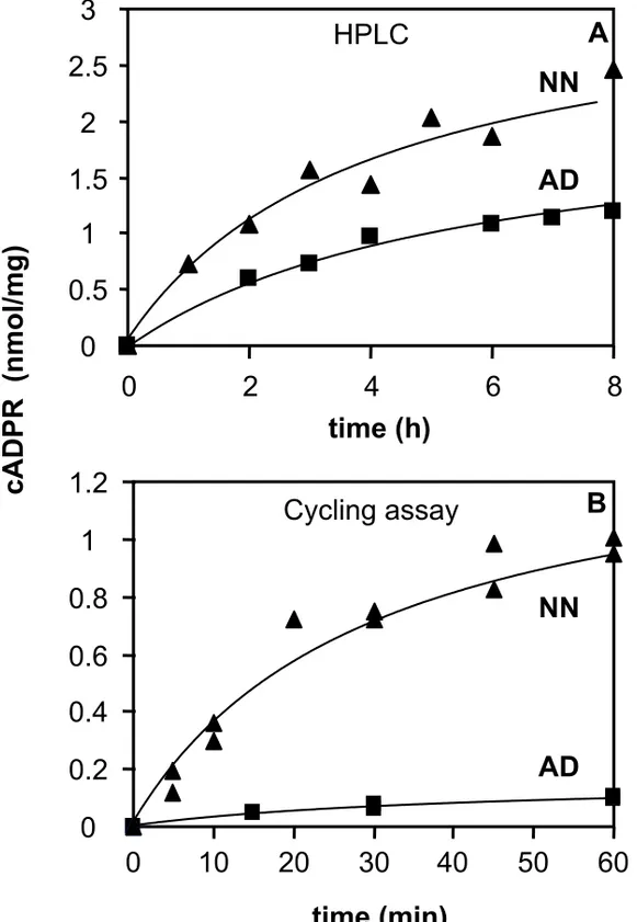
Reaction progress curves of the ADP-ribosyl cyclase activity of synaptosomes isolated from neonate and adult Cd38-/- mouse brain.
We have analyzed the kinetics of cADPR synthesis by brain synaptosomes from CD38 deficient neonate and adult mice by the HPLC method (Fig. 4A) and the cycling assay (Fig. 4B). Experimental protocols used here were the same as those described above. Incubation times were comprised between 1 and 8 hours for the HPLC experiments, and between 5 minutes and 1 hour in the cycling assays which are much more sensitive.
The estimated initial rates of the cADPR production obtained in the presence of 60 µM NAD+ at pH 6.0 and 32°C (HPLC) are 4.1 pmol/mg/min for the adult brain synaptosomes and 8.4 pmol/mg/min
for the neonate brain synatosomes. The initial rates of the reactions performed with 200 µM NAD+ at pH 7.5 and 37°C (cycling assay) are 2.3 pmol/mg/min for adult brain synaptosomes and 36 pmol/mg/min for neonate brain synatosomes.
With both methods, we revealed significantly lower activities in the adult synaptosomes compared to neonate synaptosomes, a result which is consistent with the lower cADPR levels observed in adult tissues compared to the developing tissues (Fig. 1). With the cycling assay, which is much more precise than the HPLC method, we found that neonate synaptosomes have about 15-times higher initial rates of cADPR synthesizing activity than the adult synaptosomes.
Moreover, the initial rate of reaction obtained with neonate synaptosomes using the cycling assay (36 pmol/mg/min) is about 10-fold higher than the rate evaluated under similar experimental conditions (200 µM reactive NAD+ at pH 7.5 and 37°C) with cells isolated from neonate cerebral hemispheres (3.2 pmol/mg/min, [31]).
Altogether, these results show a higher cADPR synthezising activity in brain synaptosomes from CD38 deficient mouse compared to entire brain cells, and thus an enrichment of the novel ADP-ribosyl cyclase in terminals of brain neural cells. Moreover, higher activities were found in synaptosomes from neonate compared to synaptosomes from adult.
Properties of the adult brain synaptosomal ADP-ribosyl cyclase : Km for NAD+, competition
between NAD+ and NGD+, pH dependence.
We further studied some properties of the synaptosomal ADP-ribosyl cyclase from CD38 deficient mouse brain and determined the apparent Km for the transformation of NAD+ into cADPR using the cycling assay. To that end, adult synaptosomes (0.4 mg/ml) were incubated 15 or 30 min at pH 7.5 and 37°C in the presence of increasing concentrations of the substrate in order to determine the initial rates of cADPR synthesis for each NAD+ concentration. The experimental data were fitted assuming a hyperbolic NAD+ concentration dependence. From the plot of these initial rates (Fig 5A) a Km of 21 µM was estimated for NAD+, i.e. a value within the range of the Michaelis constants determined for the other known mammalian enzymes of the cyclases/NADase family [39, 40], and which is slightly higher than the Km estimated for the Aplysia cyclase of 4.6 µM [41]. The Vm was around 6 pmoles of cADPR formed/min/mg with adult synaptosomes under these experimental conditions.
In a previous work [31], we have demonstrated that, in contrast to all other known ADP-ribosyl cyclases including mammalian CD38 and CD157, and Aplysia cyclase, the novel cyclase detected in
a membrane extract from CD38 deficient brain was unable to transform NGD+ (a surrogate NAD+ analogue) into cyclic GDP-ribose (a fluorescent analogue of cADPR). In the present study, we have tested the ability of NGD+ to compete with the endogenous substrate NAD+ for binding to the active site of the synaptosomal enzyme. Therefore, the apparent Km of NAD+ was determined under the same experimental conditions as above, but in the presence of NGD+ (at 100 and 300 µM). NGD+ was shown to inhibit the synthesis of cADPR from NAD+, leading to an increase of the apparent Km for NAD+ (Fig. 5A). The apparent Km values for NAD+ were plotted as a function of the NGD+ concentration (Fig. 5B). This secondary plot is reasonably linear, indicating that NGD+ is able to displace NAD+ from the active site in a competitive manner. The deduced K
i for NGD+ was estimated to be of 24 µM, i.e. in the same range as the Km for NAD+.
We confirmed this inhibitory effect of NGD+ by the use of the HPLC method. Thus, NGD+ (1 mM) was shown to inhibit cADPR and ADPR production catalyzed by the adult synaptosomal cyclase when using 60 µM substrate. Both metabolites production was reduced by about 50-70 % under the experimental conditions used, which include long incubation times in order to have sufficient sensitivity to observe the cADPR production by the HPLC method.
We have then tested the possibility that synaptosomes from Cd38-/- brain could utilize NGD+ as a substrate. To test a possible transformation of NGD+ into cGDPR, fluorimetric measurements with both adult (not shown) and neonate (see Fig. 6) Cd38-/- synaptosomes were performed.
Synaptosomes were incubated (at 0.5 mg/ml) for 2 hours at 37°C with Emulphogen in the presence of NGD+ (100 µM). Representative results presented in Fig. 6 show that a GDP-ribosyl cyclase activity was barely measurable. The final addition of Aplysia cyclase, which converts the residual substrate into cGDPR, indeed demonstrated that less than 1% (limit of detection) of NGD+ may have been transformed into cGDPR by the synaptosomal enzyme within the time period studied. Additional experiments were performed using an HPLC method (detailed in [42]), which permits the detection of both products, cGDPR and GDPR. Adult and neonate synaptosomes (0.4 mg/ml) were incubated with NGD+ (100 µM) for 2and 4hhours at 37°C. The HPLC chromatograms show that a very small amount of NGD+ could be transformed into GDPR (about 4-5%), while no cGDPR formation could be detected (not shown). Thus, even if the synaptosomal preparation represents an enriched preparation compared to the membrane fraction previously studied [31], we were unable to observe any cyclisation of NGD+ by the use of two different methods. The synaptosomal enzyme from CD38 deficient mouse brain appears, therefore, to be a very poor GDP-ribosyl cyclase.
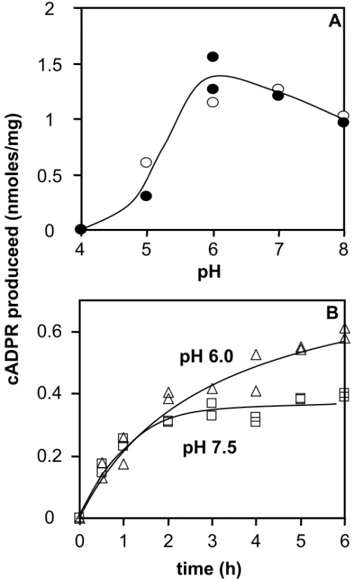
We then examined the pH-dependence of the synaptosomal enzyme activity using the HPLC method. To that end, the cADPR production by adult synaptosomes was studied after incubations of either 6 hours at 37°C or 7 hours at 32°C in the presence of 60 µM NAD+. Fig. 7A shows that similar results were observed using both experimental conditions : the ADP-ribosyl cyclase activity, which is optimal at pH between 6 and 7, is strongly reduced by acidic pHs but is only slightly affected at pH 8.0. Reaction progress curves of adult synaptosomes were obtained using the cycling assay at pH 6.0 and at pH 7.5 in the presence of 60 µM of NAD+ (Fig. 7B). We observed that the initial rates of cADPR production are similar at these two pH values. However, the shape of the kinetics curves were different : at pH 7.5 the cADPR production is clearly slowed down after 2 hours of incubation, whereas production is still progressing at pH 6.0 after this time. Such an effect is reminiscent of the paracatalytic inactivation, observed at pH > 7.0, for the NAD+ glycohydrolase activity of other members of that enzyme family [39].
Altogether, these results show that the brain synaptosomal ADP-ribosyl cyclase has a broad optimal pH at 6.0-7.0. They provide a Km of 21 µM for NAD+. Moreover, they demonstrate that NGD+, despite its inability to be a substrate for the formation of cGDPR, can bind with a good affinity the active site of the novel enzyme and inhibit the transformation of NAD+ (Ki = 24 µM).
Micromolar concentrations of zinc ions inhibit the ADP-ribosyl cyclase detected in Cd38-/- brain
synaptosomes.
We then studied the effect of zinc ions, which play major roles on the synaptic activity and during brain development (for review see [32, 34, 43, 44]), on the Cd38-/- synaptosomal ADP-ribosyl cyclase activity.
We have therefore examined the effect of increasing concentrations of Zn-acetate on the cADPR production by neonate and adult brain synaptosomes after incubation with 200 µM NAD+. The results presented in Fig. 8 show a decrease of the cADPR production with an IC50 of 1.8 µM with neonate synaptosomes and 0.9 µM with adult synaptosomes under these experimental conditions. To confirm these results, first we have verified that zinc does not affect the cADPR determination by the cycling assay. To that end, we have analysed by the cycling assay standard solutions of cADPR (10 nM) containing varying concentrations of zinc (0, 0.2, 2, 20 and 200 µM). We found that the presence of zinc at these concentrations did not modify the signal (slope of the increase in fluorescence at 590 nm) obtained (not shown). Next, we have also controlled that no endogenous zinc from brain tissue remained in our synaptosomes preparations. Thus, a zinc chelating agent
(N,N,N’,N’-tetrakis (2-pyridylmethyl)-ethylenediamine or TPEN) was added to brain synaptosomes which were tested for their ability to synthesize cADPR : the same cADPR production was observed when incubating the adult synaptosomes with 60 µM NAD+ in the presence of 0, 10 or 25 µM TPEN (not shown). Finally, we also have checked that the decrease in the cADPR level was not due to an increase of the cADPR hydrolysis. To that end, cADPR (100 nM or 1 µM) was incubated (at 37°C for 2 hours) in the presence of adult synaptosomes (0.4 mg/ml) without or with 10 µM Zn2+, and then worked-up as in the preceding experiments (precipitation with perchloric acid and neutralization, treatment with the three hydrolases overnight to eliminate the contaminating nucleotides). The resulting cADPR was then estimated and compared to untreated cADPR. We found that neither the presence of the synaptosomes, nor the treatment overnight with the three hydrolases did change the cADPR level (not shown). Such control experiments demonstrate that the effect of zinc ions is definitely due to an inhibition of the cADPR synthesis catalysed by the non-CD38 ADP-ribosyl cyclase.
We also have tested the effect of two other divalent ions, Cu2+ and Ni2+, on the synaptosomes cyclase activity. In experimental conditions similar to those used to study the effect of zinc ions (see legend to Fig. 8), we observed that 10 µM of copper ions completely inhibited the formation of cADPR by adult brain synaptosomes, whereas 10 µM of Ni2+ did not affect this activity (not shown). These divalent ions where shown to modify the ADP-ribosyl cyclase activity of known mammalian enzymes at millimolar concentrations: Zn2+, Cu2+ and Ni2+ activate CD38, whereas Zn2+ activates and Cu2+ inhibits CD157. The effects observed on the synaptosomal cyclase are thus clearly different, except the similar inhibitory effect of copper but which was only observed at higher concentrations on CD157 cyclase activity.
Thus, our results show, for the first time, a micromolar sensitivity to zinc (and copper) of an intracellular ADP-ribosyl cyclase from brain, an enzyme which we found abundant within synaptosomes.
DISCUSSION
In the present work, we show for the first time that endogenous levels of cADPR are largely higher in developing brain compared to the adult tissue. We also demonstrate that these levels are totally independent from the presence of the CD38 protein, not only in the adult as already shown by Partida-Sanchez et al. [9], but also during development. Interestingly, we have previously revealed the existence of a novel ADP-ribosyl cyclase in the CD38-deficient mouse brain whose activity was shown to be largely superior in developing brain than in the adult tissue [31]. The non-CD38 enzyme, expressed at all developmental stages, is thus undeniably in charge of the brain cADPR level, which is down regulated during development. Moreover, the high amounts of cADPR detected in embryonic and neonate brain certainly reflect a key role of this cyclic metabolite during developmental processes of the central nervous system.
In addition, we show that the novel ADP-ribosyl cyclase detected in Cd38-/- brain is abundant within synaptosomes purified from both adult and neonate mouse brains. This result is remarkable since cADPR is believed to be involved in processes occurring at synaptic connections, including neurotransmitter release and long-term synaptic depression [15, 16, 45]. In this context, it must be taken into consideration that, although functional synapses are known to be completely fashioned after birth (around postnatal day 2), the first morphologically identified synapses appear earlier in mouse brain, around E16 in the mouse [46]. The amount of synapses increases then continuously, and they are plentiful in the neonate brain. Since we found that the novel ADP-ribosyl cyclase is enriched in terminals of brain neural cells and that its activity is higher in neonate synaptosomes compared to adult synaptosomes, our results raise the question of the role of cADPR and its synthesizing enzyme in morphologically shaped but non-totally efficient synapses. The ADP-ribosyl cyclase and the metabolites it produces (cADPR, but also ADPR) may, thus, be involved in synaptogenesis, via regulation of calcium signals necessary to the appearance of novel constituents of the synapse or to the correct placement of these constituents in the mature synapse.
We have further characterized ADP-ribosyl cyclase of brain synaptosomes. We showed that it functions best at pH between 6.0 and 7.0 and that the concentration of free calcium has no major effect on its cADPR production. We also showed that it shares some of the properties of other known enzymes of that family (including mammalian CD38 and CD157, and the Aplysia cyclase), such as the ability of synthesizing both cADPR and ADPR, an affinity for NAD+ in the micromolar range (Km = 21 µM) and the ability to bind with a similar affinity the NAD+ analogue, NGD+ (Ki =
24 µM). This later result is intriguing since the novel enzyme happens to be unable to transform NGD+ into the cyclic compound, cGDPR ([31] and present manuscript), in contrast to all other known enzymes of the family. The inability of the enzyme to cyclize the NGD+ molecule may be due to a different organization of its active site. Thus, we describe an ADP-ribosyl cyclase that is competent to transform NAD+ to cADPR, that is in charge of the endogenous cADPR level found in brain, but that is unable to transform NGD+ to cGDPR. Such results may have an essential impact on the analysis of the physiological role of cADPR, and on “non-classical” ADP-ribosyl cyclases which definitely may not only be studied using the NAD+ analogue, NGD+, to establish cyclization reactions.
Given our previous report with brain membrane extracts [Ceni, 2003 #525], we have also tested the effect of GTP-γ-S on the production of NAD+ metabolites by Cd38-/- brain synaptosomes. Results turned out to be more complex to analyze. Thus, in contrast to our previous work, we were unable to detect, under any conditions tested, a significant modification of the cADPR production after addition of GTP-γ-S. On the other hand, ADPR production was clearly increased (3- to 7-fold, depending on the medium and time of incubation) after addition of GTP-γ-S in the absence of EDTA (data not shown), in agreement with what was observed with the less purified brain membrane extracts. These results deserve, however, further investigations since new unidentified NAD+ metabolites were also detected on the chromatograms (data not shown).
Another major finding of the present work is that the ADP-ribosyl cyclase from brain synaptosomes is very sensitive to Zn2+, being totally inhibited by micromolar concentrations of that ion. The ADP-ribosyl cyclase activities of CD38 and CD157 were shown, in contrast, to be activated by high (millimolar) concentrations of zinc [47, 48]. This opposing effect of zinc on the synaptosomal enzyme is another element strongly suggesting essential structural differences between the novel enzyme and the other known mammalian ADP-ribosyl cyclases.
Moreover, the sensitivity of the synaptosomal ADP-ribosyl cyclase to zinc is opening new research avenues because zinc has critical roles in the central nervous system, both on the synaptic activity and during brain development and aging (for review see [32, 34, 43, 44]). The nervous system contains high concentrations of zinc (average total brain zinc : 150 µM [49]), which is distributed into three cellular pools: the largest exists in tight association with intracellular proteins (mostly metallothioneins), a second pool is sequestered in presynaptic vesicles of certain neurons (principally excitatory glutamatergic neurons of the forebrain), the third pool is constituted by the free ions in the cytoplasm. Zinc is known to exert a neuromodulatory action at excitatory synapses of
‘gluzinergic neurons’ when released during synaptic transmission. This ion acts not only as an intercellular signalling messenger directly binding to post-synaptic receptors and ionic channels, but also as an intracellular signal by entering into post-synaptic neurons. Fluctuations of free zinc intracellular concentrations, [Zn]i, in the cytoplasm of neuronal and glial cells appear to represent important signals for these cells. Intracellular Zn2+ is indeed known to act on several cellular pathways, in order to enhance the anti-oxydant response, the activity of poly-ADP-ribose polymerase-1 and of protein kinase C enzymes, … etc. Moreover, although [Zn]i in resting cells is thought to be maintained at very low levels (in the subnanomolar range), it may be elevated (in the micromolar range) during injurious stimuli [33, 50].Evidences have indicated that the entry of zinc into post-synaptic neurons contribute to the toxic excess occurring during seizure and traumatic brain injury. It has been suggested that high intracellular free zinc promotes neuronal death by inhibiting cellular energy production [43].
We propose that the brain synaptosomal ADP-ribosyl cyclase is a new intracellular target of neurotoxic Zn2+. By acting on this enzyme, zinc ions may modify neural cells calcium homeostasis. Altogether, our results strongly suggest a crucial role of the synaptosomal ADP-ribosyl cyclase and of the metabolites it produces, cADPR and ADPR, in Ca2+-dependent processes necessary for the development of the central nervous system and also in the adult brain. Moreover, their action may be linked to Zn2+ cellular signals, particularly during brain injury.
ACKNOWLEDGEMENTS
We thank Dr Frances Lund for precious discussions and comments on the manuscript. This work was supported by recurrent grants from the French INSERM. We thank the zootechnicians of the CEA-SDV-DRDC Department.
REFERENCES
1 Guse, A. H. (1999) Cyclic ADP-ribose: a novel Ca2+-mobilising second messenger. Cell Signal 11, 309-16.
2 Higashida, H., Hashii, M., Yokoyama, S., Hoshi, N., Chen, X. L., Egorova, A., Noda, M. and Zhang, J. S. (2001) Cyclic ADP-ribose as a second messenger revisited from a new aspect of signal transduction from receptors to ADP-ribosyl cyclase. Pharmacol. Ther. 90, 283-96. 3 Rutter, G. A. (2003) Calcium signalling: NAADP comes out of the shadows. Biochem. J.
373, e3-4
4 Kinnear, N. P., Boittin, F. X., Thomas, J. M., Galione, A. and Evans, A. M. (2004) Lysosome-sarcoplasmic reticulum junctions. A trigger zone for calcium signaling by nicotinic acid adenine dinucleotide phosphate and endothelin-1. J. Biol. Chem. 279, 54319-26
5 Dammermann, W. and Guse, A. H. (2005) Functional ryanodine receptor expression is required for NAADP-mediated local Ca2+ signaling in T-lymphocytes. J. Biol. Chem. 280, 21394-9
6 Perraud, A. L., Fleig, A., Dunn, C. A., Bagley, L. A., Launay, P., Schmitz, C., Stokes, A. J., Zhu, Q., Bessman, M. J., Penner, R., Kinet, J. P. and Scharenberg, A. M. (2001) ADP-ribose gating of the calcium-permeable LTRPC2 channel revealed by Nudix motif homology. Nature 411, 595-9.
7 Kolisek, M., Beck, A., Fleig, A. and Penner, R. (2005) Cyclic ADP-ribose and hydrogen peroxide synergize with ADP-ribose in the activation of TRPM2 channels. Mol. Cell. 18, 61-9
8 Walseth, T. F., Aarhus, R., Zeleznikar, R. J., Jr. and Lee, H. C. (1991) Determination of endogenous levels of cyclic ADP-ribose in rat tissues. Biochim. Biophys. Acta. 1094, 113-20 9 Partida-Sanchez, S., Cockayne, D. A., Monard, S., Jacobson, E. L., Oppenheimer, N., Garvy,
B., Kusser, K., Goodrich, S., Howard, M., Harmsen, A., Randall, T. D. and Lund, F. E. (2001) Cyclic ADP-ribose production by CD38 regulates intracellular calcium release, extracellular calcium influx and chemotaxis in neutrophils and is required for bacterial clearance in vivo. Nat Med. 7, 1209-16.
10 Hua, S. Y., Tokimasa, T., Takasawa, S., Furuya, Y., Nohmi, M., Okamoto, H. and Kuba, K. (1994) Cyclic ADP-ribose modulates Ca2+ release channels for activation by physiological Ca2+ entry in bullfrog sympathetic neurons. Neuron 12, 1073-9
11 Empson, R. M. and Galione, A. (1997) Cyclic ADP-ribose enhances coupling between voltage-gated Ca2+ entry and intracellular Ca2+ release. J. Biol. Chem. 272, 20967-70.
12 Hashii, M., Minabe, Y. and Higashida, H. (2000) cADP-ribose potentiates cytosolic Ca2+ elevation and Ca2+ entry via L-type voltage-activated Ca2+ channels in NG108-15 neuronal cells. Biochem. J. 345 Pt 2, 207-15
13 Budde, T., Sieg, F., Braunewell, K. H., Gundelfinger, E. D. and Pape, H. C. (2000) Ca2+ -induced Ca2+ release supports the relay mode of activity in thalamocortical cells. Neuron 26, 483-92.
14 Verderio, C., Bruzzone, S., Zocchi, E., Fedele, E., Schenk, U., De Flora, A. and Matteoli, M. (2001) Evidence of a role for cyclic ADP-ribose in calcium signalling and neurotransmitter release in cultured astrocytes. J. Neurochem. 78, 646-57.
15 Mothet, J. P., Fossier, P., Meunier, F. M., Stinnakre, J., Tauc, L. and Baux, G. (1998) Cyclic ADP-ribose and calcium-induced calcium release regulate neurotransmitter release at a cholinergic synapse of Aplysia. J. Physiol. 507, 405-14.
16 Reyes-Harde, M., Empson, R., Potter, B. V., Galione, A. and Stanton, P. K. (1999) Evidence of a role for cyclic ADP-ribose in long-term synaptic depression in hippocampus. Proc. Natl. Acad. Sci. U S A. 96, 4061-6.
17 Higashida, H., Yokoyama, S., Hashii, M., Taketo, M., Higashida, M., Takayasu, T., Ohshima, T., Takasawa, S., Okamoto, H. and Noda, M. (1997) Muscarinic receptor-mediated dual regulation of ADP-ribosyl cyclase in NG108-15 neuronal cell membranes. J. Biol. Chem. 272, 31272-7.
18 Morita, K., Kitayama, S. and Dohi, T. (1997) Stimulation of cyclic ADP-ribose synthesis by acetylcholine and its role in catecholamine release in bovine adrenal chromaffin cells. J. Biol. Chem. 272, 21002-9.
19 Pollock, J., Crawford, J. H., Wootton, J. F., Seabrook, G. R. and Scott, R. H. (1999) Metabotropic glutamate receptor activation and intracellular cyclic ADP-ribose release Ca2+ from the same store in cultured DRG neurones. Cell Calcium 26, 139-48
20 Hotta, T., Asai, K., Fujita, K., Kato, T. and Higashida, H. (2000) Membrane-bound form of ADP-ribosyl cyclase in rat cortical astrocytes in culture. J. Neurochem. 74, 669-75.
21 Noda, M., Yasuda, S., Okada, M., Higashida, H., Shimada, A., Iwata, N., Ozaki, N., Nishikawa, K., Shirasawa, S., Uchida, M., Aoki, S. and Wada, K. (2003) Recombinant
human serotonin 5A receptors stably expressed in C6 glioma cells couple to multiple signal transduction pathways. J. Neurochem. 84, 222-32
22 Morikawa, H., Khodakhah, K. and Williams, J. T. (2003) Two intracellular pathways mediate metabotropic glutamate receptor-induced Ca2+ mobilization in dopamine neurons. J. Neurosci. 23, 149-57
23 Higashida, H., Zhang, J. S., Mochida, S., Chen, X. L., Shin, Y., Noda, M., Hossain, K. Z., Hoshi, N., Hashii, M., Shigemoto, R., Nakanishi, S., Fukuda, Y. and Yokoyama, S. (2003) Subtype-specific coupling with ADP-ribosyl cyclase of metabotropic glutamate receptors in retina, cervical superior ganglion and NG108-15 cells. J. Neurochem. 85, 1148-58
24 Schuber, F. and Lund, F. E. (2004) Structure and enzymology of ADP-ribosyl cyclases: conserved enzymes that produce multiple calcium mobilizing metabolites. Curr. Mol. Med. 4, 249-61
25 Glick, D. L., Hellmich, M. R., Beushausen, S., Tempst, P., Bayley, H. and Strumwasser, F. (1991) Primary structure of a molluscan egg-specific NADase, a second-messenger enzyme. Cell Regul. 2, 211-8
26 States, D. J., Walseth, T. F. and Lee, H. C. (1992) Similarities in amino acid sequences of Aplysia ADP-ribosyl cyclase and human lymphocyte antigen CD38. Trends Biochem. Sci.
17, 495.
27 Itoh, M., Ishihara, K., Tomizawa, H., Tanaka, H., Kobune, Y., Ishikawa, J., Kaisho, T. and Hirano, T. (1994) Molecular cloning of murine BST-1 having homology with CD38 and Aplysia ADP-ribosyl cyclase. Biochem. Biophys. Res. Commun. 203, 1309-17.
28 Koguma, T., Takasawa, S., Tohgo, A., Karasawa, T., Furuya, Y., Yonekura, H. and Okamoto, H. (1994) Cloning and characterization of cDNA encoding rat ADP-ribosyl cyclase/cyclic ADP-ribose hydrolase (homologue to human CD38) from islets of Langerhans. Biochim. Biophys. Acta. 1223, 160-2.
29 Yamada, M., Mizuguchi, M., Otsuka, N., Ikeda, K. and Takahashi, H. (1997) Ultrastructural localization of CD38 immunoreactivity in rat brain. Brain Res. 756, 52-60.
30 Ceni, C., Pochon, N., Brun, V., Muller-Steffner, H., Andrieux, A., Grunwald, D., Schuber, F., De Waard, M., Lund, F. E., Villaz, M. and Moutin, M. J. (2003) CD38-dependent ADP-ribosyl cyclase activity in developing and adult mouse brain. Biochem. J. 370, 175-183 31 Ceni, C., Muller-Steffner, H., Lund, F., Pochon, N., Schweitzer, A., De Waard, M., Schuber,
cyclase/NAD+-glycohydrolase in brain from CD38-deficient mice. J. Biol. Chem. 278, 40670-8
32 Smart, T. G., Hosie, A. M. and Miller, P. S. (2004) Zn2+ ions: modulators of excitatory and inhibitory synaptic activity. Neuroscientist 10, 432-42
33 Choi, D. W. and Koh, J. Y. (1998) Zinc and brain injury. Annu. Rev. Neurosci. 21, 347-75 34 Bhatnagar, S. and Taneja, S. (2001) Zinc and cognitive development. Br. J. Nutr. 85 Suppl 2,
S139-45
35 Phillips, G. R., Huang, J. K., Wang, Y., Tanaka, H., Shapiro, L., Zhang, W., Shan, W. S., Arndt, K., Frank, M., Gordon, R. E., Gawinowicz, M. A., Zhao, Y. and Colman, D. R. (2001) The presynaptic particle web: ultrastructure, composition, dissolution, and reconstitution. Neuron 32, 63-77
36 Graeff, R. and Lee, H. C. (2002) A novel cycling assay for cellular cADP-ribose with nanomolar sensitivity. Biochem. J. 361, 379-384.
37 Walseth, T. F., Wong, L., Graeff, R. M. and Lee, H. C. (1997) Bioassay for determining endogenous levels of cyclic ADP-ribose. Methods Enzymol. 280, 287-94.
38 Augustin, A., Muller-Steffner, H. and Schuber, F. (2000) Molecular cloning and functional expression of bovine spleen ecto-NAD+ glycohydrolase: structural identity with human CD38. Biochem. J. 345, 43-52.
39 Price, S. R. a. P., P. H. (1987) in Pyridine nucleotide coenzymes: chemical, biochemical and medical aspects, pp. 513-548
40 Cakir-Kiefer, C., Muller-Steffner, H., Oppenheimer, N. and Schuber, F. (2001) Kinetic competence of the cADP-ribose-CD38 complex as an intermediate in the CD38/NAD+ glycohydrolase-catalysed reactions: implication for CD38 signalling. Biochem. J. 358, 399-406.
41 Cakir-Kiefer, C., Muller-Steffner, H. and Schuber, F. (2000) Unifying mechanism for Aplysia ADP-ribosyl cyclase and CD38/NAD(+) glycohydrolases. Biochem. J. 349, 203-10. 42 Goodrich, S. P., Muller-Steffner, H., Osman, A., Moutin, M. J., Kusser, K., Roberts, A.,
Woodland, D. L., Randall, T. D., Kellenberger, E., LoVerde, P. T., Schuber, F. and Lund, F. E. (2005) Production of calcium-mobilizing metabolites by a novel member of the ADP-ribosyl cyclase family expressed in Schistosoma mansoni. Biochemistry 44, 11082-97
43 Dineley, K. E., Votyakova, T. V. and Reynolds, I. J. (2003) Zinc inhibition of cellular energy production: implications for mitochondria and neurodegeneration. J. Neurochem. 85, 563-70
44 Mocchegiani, E., Bertoni-Freddari, C., Marcellini, F. and Malavolta, M. (2005) Brain, aging and neurodegeneration: role of zinc ion availability. Prog. Neurobiol. 75, 367-90
45 Brailoiu, E. and Miyamoto, M. D. (2000) Inositol trisphosphate and cyclic adenosine diphosphate-ribose increase quantal transmitter release at frog motor nerve terminals: possible involvement of smooth endoplasmic reticulum. Neuroscience 95, 927-31
46 Bouwman, J., Maia, A. S., Camoletto, P. G., Posthuma, G., Roubos, E. W., Oorschot, V. M., Klumperman, J. and Verhage, M. (2004) Quantification of synapse formation and maintenance in vivo in the absence of synaptic release. Neuroscience 126, 115-26
47 Kukimoto, I., Hoshino, S., Kontani, K., Inageda, K., Nishina, H., Takahashi, K. and Katada, T. (1996) Stimulation of ADP-ribosyl cyclase activity of the cell surface antigen CD38 by zinc ions resulting from inhibition of its NAD+ glycohydrolase activity. Eur. J. Biochem.
239, 177-82.
48 Hirata, Y., Kimura, N., Sato, K., Ohsugi, Y., Takasawa, S., Okamoto, H., Ishikawa, J., Kaisho, T., Ishihara, K. and Hirano, T. (1994) ADP ribosyl cyclase activity of a novel bone marrow stromal cell surface molecule, BST-1. FEBS Lett. 356, 244-8.
49 Takeda, A. (2000) Movement of zinc and its functional significance in the brain. Brain Res. Brain Res. Rev. 34, 137-48
50 Outten, C. E. and O'Halloran, T. V. (2001) Femtomolar sensitivity of metalloregulatory proteins controlling zinc homeostasis. Science 292, 2488-92
FIGURES LEGENDS
Figure 1 Endogenous cADPR content in developing and adult brain from wild-type and CD38 deficient mice.
Extracts were prepared from tissues isolated from embryos (EM, days 15 or 16), from neonates (NN, days 1 or 2), or adults (AD), and analyzed for cADPR content using the cycling assay as described under Materials and Methods.
Figure 2 Analysis of the ADP-ribosyl cyclase activity from Cd38-/- mouse brain synaptosomes by the cycling assay - Effect of detergent and free calcium concentration.
A: Synaptosomes preparation by centrifugation of brain homogenate on a discontinuous sucrose gradient and Western-blot analysis of the collected fractions containing proteins (6 µg/well) using anti-synapsin IIa and anti-PSD95. Synaptosomes were collected at the 1.25 M/1.0 M sucrose interface (fraction 3).
B: Synaptosomes (0.4 mg/ml for adult and 0.2 mg/ml for neonate) were incubated (2 hours for adult and 1 hour for neonate) with 200 µM NAD+ in solution D (containing 10 mM Tris,HCl pH 7.5, 2.2 mM CaCl2, 0.5 mM Na2HPO4, 0.4 mM KH2PO4, 4 mM NaHCO3, and 80 mM NaCl) in the absence (-) or presence (+) of the detergent Emulphogen (1%, wt/vol). Reactions were stopped by addition of perchloric acid followed by neutralization. The formation of cADPR was detected using the cycling assay as described under Materials and Methods. The results obtained in the absence of detergent was expressed in percent of the result obtained in the presence of the detergent.
C: Adult synaptosomes (0.4 mg/ml) were incubated 2 hours in 50 mM Tris,HCl at pH 7.5 in the presence of 200 µM NAD+ and 1% (wt/vol) Emulphogen, and with either 1 nM (pCa 9), 1 µM (pCa 6) or 1 mM (pCa 3) free calcium. The experiment was reproduced twice with two different synaptosomal preparations (n = 4). Analysis of the cADPR production was performed using the cycling assay.
Figure 3 Analysis of NAD+ metabolizing activities of synaptosomes from adult and
neonate Cd38 -/- mice brain by HPLC - Effect of detergent.
Synaptosomes (1 mg/ml) prepared from brain tissue of adult and neonate Cd38-/- mice were
incubated for 8 hours in 20 mM MES-NaOH at pH 6.0 and 32°C with 14C-NAD+ (8 mCi/mmol) and NAD+ (final concentration 60 µM) in the presence of 4 mM EDTA, with or without 1% (wt/vol)
Triton X-100 (TX). Reactions were stopped by addition of perchloric acid followed by neutralization. Products analysis was performed by HPLC using an on-line radioactivity detector as described under Materials and Methods.
A : Products obtained with adult synaptosomes.
B : Products obtained with detergent-treated adult synaptosomes. C : Products obtained with neonate synaptosomes.
D : Products obtained with detergent-treated neonate synaptosomes.
Figure 4 Kinetics of the ADP-ribosyl cyclase activity of synaptosomes from adult and neonate Cd38-/- brains, measured using both HPLC (A) and the cycling assay (B).
A : synaptosomes (1 mg protein/ml) were incubated at 32°C for the given times with 14C-NAD+ (8 mCi/mmol) and NAD+ (final concentration 60 µM), 4 mM EDTA, 1% (wt/vol) Triton X-100 in 20 mM MES-NaOH at pH 6.0.
B : synaptosomes (adult 0.4 mg/ml, neonate 0.2 mg/ml) were incubated for the given times with 200 µM NAD+ in solution D (see legend to Fig. 3) in the presence of 1% (wt/vol) Emulphogen.
Figure 5 Properties of the CD38-independent ADP-ribosyl cyclase : determination of the apparent Km of NAD+ and of the inhibition constant of NGD+.
A and B : adult brain synaptosomes (0.4 mg/ml) were incubated 15 or 30 minutes at 37°C in solution D (pH 7.5) with 1% (wt/vol) Emulphogen and varying NAD+ concentrations, in the absence (◊) or in the presence (x) of 100 µM NGD+. Then, the cADPR formation was analysed by the cycling assay and reaction rates were estimated. B : secondary plot of the apparent Km for NAD+ versus NGD+ concentration (with n=3 at 0 NGD+; n=1 at 100 µM NGD+, n=2 at 300 µM NGD+).
C and D: adult brain synaptosomes (1.1 mg of protein /ml) were incubated 2 or 4 hours at 37°C in the presence of 14C-NAD+ (4 mCi/mmol) and NAD+ (60 µM), 4 mM EDTA, 1% (wt/vol) Triton X-100 in 20 mM Tris,HCl at pH 7.0 either in the absence or in the presence of 1 mM NGD+. Product analysis was performed by HPLC.
Figure 6 Absence of cGDPR formation by the ADP-ribosyl cyclase from Cd38-/- brain synaptosomes.
NGD+ (100 µM) was added to 0.5 mg/ml neonate brain synaptosomes from Cd38-/- mouse incubated at 37°C in solution D. Formation of cGDPR was detected by fluorescence at 420 nm upon excitation at 300 nm. After 2 hours of incubation, Aplysia cyclase (0.3 µg/ml) was added to fully transform the residual NGD+ into cGDPR (control).
Figure 7 pH effects on the CD38-independent synaptosomal ADP-ribosyl cyclase activity.
A : adult brain synaptosomes (1 mg of protein /ml) were incubated 6h at 37°C or 7h at 32°C in the presence of 14C-NAD+ (4 mCi/mmol) and NAD+ (60 µM), 4 mM EDTA, 1% (wt/vol) Triton X-100 in 20 or 40 mM citrate-HCl (pH 4.0), MES-NaOH (pH 5.0 and 6.0), or Tris,HCl (pH 7.0 or 8.0). Products analysis was performed by HPLC.
B : synaptosomes (adult 0.4 mg/ml) were incubated for the given times with 60 µM NAD+ in solution D (see legend to Fig. 3) containing either 30 mM Tris,HCl (pH 7.5) or 80 mM MES-NaOH (pH 6.0) in the presence of 1% (wt/vol) Emulphogen. The cADPR formation was analysed by the cycling assay.
Figure 8 Effect of zinc ions on the ADP-ribosyl cyclase activity of synaptosomes from neonate and adult CD38-deficient mice.
Synaptosomes (0.4 mg/ml for adult and 0.2 mg/ml for neonate) were incubated (2 hours for adult and 1 hour for neonate) with 200 µM NAD+ in solution D (see legend to Fig. 2) containing 1% (wt/vol) Emulphogen and varying concentrations of zinc-acetate. The cADPR formation was analysed using the cycling assay.