HAL Id: hal-00704853
https://hal.archives-ouvertes.fr/hal-00704853
Submitted on 6 Jun 2012
HAL is a multi-disciplinary open access
archive for the deposit and dissemination of
sci-entific research documents, whether they are
pub-lished or not. The documents may come from
teaching and research institutions in France or
abroad, or from public or private research centers.
L’archive ouverte pluridisciplinaire HAL, est
destinée au dépôt et à la diffusion de documents
scientifiques de niveau recherche, publiés ou non,
émanant des établissements d’enseignement et de
recherche français ou étrangers, des laboratoires
publics ou privés.
of Cell Nuclei in Actin-Tagged Confocal Microscopy
Images
Leila Meziou, Aymeric Histace, Frédéric Precioso, Bogdan Matuszewski,
Franck Carreiras
To cite this version:
Leila Meziou, Aymeric Histace, Frédéric Precioso, Bogdan Matuszewski, Franck Carreiras. Fractional
Entropy Based Active Contour Segmentation of Cell Nuclei in Actin-Tagged Confocal Microscopy
Im-ages. Conference on Medical Image Understanding and Analysis, Jul 2012, Swansea, United Kingdom.
pp.117-123. �hal-00704853�
PAPER 13: UNSUPERVISED SEGMENTATION OF CONFOCAL MICROSCOPY IMAGES
Fractional Entropy Based Active Contour
Segmentation of Cell Nuclei in Actin-Tagged
Confocal Microscopy Images
Leila Meziou1 [email protected] Aymeric Histace1 [email protected] Frédéric Precioso2 [email protected] Bogdan J. Matuszewski3 [email protected] Franck Carreiras4 [email protected] 1ETIS - UMR 8051 - CNRS University of Cergy-Pontoise/ENSEA, Cergy-Pontoise, France 2I3S - UMR 6070 - CNRS University of Nice/Sophia-Antipolis, Nice, France
3ADSIP Research Centre University of Central Lancashire Preston, UK
4ERRMECe
University of Cergy-Pontoise Cergy, France
Abstract
In the framework of cell structure characterization for predictive oncology, we pro-pose in this paper an unsupervised statistical region based active contour approach in-tegrating an original fractional entropy measure for single channel actin tagged fluo-rescence confocal microscopy image segmentation. Following description of statistical based active contour segmentation and the mathematical definition of the proposed frac-tional entropy descriptor, we demonstrate comparative segmentation results between the proposed approach and standard Shannon’s entropy obtained for nuclei segmentation. We show that the unsupervised proposed statistical based approach integrating the frac-tional entropy measure leads to very satisfactory segmentation of the cell nuclei from which shape characterization can be subsequently used for the therapy progress assess-ment.
1
Introduction
Segmentation of cellular structures is an essential tool in cell microscopic imaging as it enables measurements which can be used to track cell divisions or help to reconstruct cor-responding cell lineage trees providing data for calculation of different parameters like cell proliferation rate for instance. More specifically, the work presented in this paper has been carried out in a context of analyzing changes of cell cytoskeleton properties in a response to ionizing radiation insult. The final goal of this research effort is to better understand cell bio-mechanical responses during cancer radiation therapy. In this context, we propose an unsupervised segmentation approach of fluorescence confocal microscopy images which
c
2012. The copyright of this document resides with its authors. It may be distributed unchanged freely in print or electronic forms.
– in order to effectively extract nuclei as a first step for providing spatial reference frame for analyzing cytoskeleton changes.
To date, only few methods have been proposed to address direct segmentation (without any denoising preprocessing of acquired images) of cell structures in fluorescence confo-cal microscopy images. In former approaches proposed in [8] and [12], authors focused on nuclei segmentations. In [14], authors proposed cell segmentation in 2D-fluorescent im-ages with two channels (actin and nucleus tagging) using a multiphase level-set combining Chan-Vese [2] and geodesic active contour models, together with repulsive force introduced to prevent segmented cells from overlapping. In [7, 15] automated 3D cell segmentation from a 3D confocal acquisition of early Zebrafish embriogenesis is proposed; Two different fluorescent markers (red for nuclei and green for membrane) are used to easily discriminate nuclei from cell membranes. In [15], authors introduced an adapted version of the subjec-tive surface technique [13] for surface reconstruction from missing boundary information whereas [7] use a multiphase level-set based on probability correlation functions.
Within a level set framework as in [7, 14], our method aims at a different objective: segmentation of microscopic 2D images extracted from a full single channel confocal acqui-sition with only one fluorescent marker used for actin tagging. The filament actin (F-actin) is believed to play a vital role in cell structure [3]. As Actin is one of the three most common proteins in human cytoskeleton, analysing its changes and properties could be instrumen-tal in analysis of cell properties. For example this can be associated with cancer evolution. Nevertheless, due to a highly complex actin appearance, a high level of noise and a strong non-homogeneity of intensity and gradient information, the segmentation of cell structures in such imaging data, is a very challenging task. Moreover, a particular attention is given to completely avoid any enhancement preprocessing [10] and to reduce to its minimum, manual interventions during the whole segmentation process.
The data used in this paper were obtained from human prostate cells (PNT2). Actin were labelled with phalloidin-FITC and all imaging was carried out using a Zeiss LSM510 confocal microscope. Fig.1shows different slices from the microconfocal acquisition of the monolayer PNT2 cell culture. The stack volume is defined on the 512 × 512 × 98 grid of pixels each 0.21 µm × 0.21 µm × 0.11 µ in size. Actin is mostly present at the periphery of the cells and within the cytoplasm. We can then notice that high intensities of actin tagged confocal images allows biologists to roughly delineate cell membranes whereas darkest areas are identified as nuclei. Owing to the high level of Poisson noise corrupting these images and the particular texture of actin, classic region based active contour approach, like the Chan and Vese one [2], fails even in segmenting properly the boundaries of nuclei corresponding to each cell [6]: We then propose to tackle this segmentation using statistical based active contour (see [5] for an overview on the work on this area) more adapted to this particular context than classic region based ones.
The remaining of this article is organized as follows: in Section 2, the framework of statistical based active contour using entropy estimation is first presented, subsequently the corresponding Partial Differential Equation (PDE) which steers the evolution of the contour is described and finally attention is focused on the particular proposed fractional entropy descriptor; Section 3 focuses on experiments on microscopic images followed by conclusions and perspectives drawn in Section 4.
PAPER 13: UNSUPERVISED SEGMENTATION OF CONFOCAL MICROSCOPY IMAGES
(a) (b) (c)
Figure 1: Examples of actin tagged fluorescence confocal microscopy images extracted from a 3D microconfocal acquisition of the monolayer PNT2 cell culture.(a)Lower slice (with low z-stack index), (b)Mid-slice with the lowest level of structural noise (a “hole” is high-light in yellow which should not to be confused with a nucleus),(c)Upper slice with non-homogeneity of the fluorescent marker on the left hand side.
2
Active contour segmentation using a fractional entropy
descriptor
Formerly introduced in [1], integral based features active contour methods are derived from traditional region based approaches by utilizing integral statistics as descriptors of the inner (Ωin) and outer (Ωout) regions delimited by the active curve Γ at a given iteration τ of the
segmentation process.
First, let H(Ωi) denotes an integral entropy estimation associated to a particular region
Ωiwithin image such as
H(Ωi) =
Z
Ωi
ϕ ( ˆp(I(x), Ωi)) dx , (1) with ϕ a monotonic increasing function, I(x) the luminance of pixel x = (x, y) and ˆpthe non-parametrically estimated Probability Density Function (PDF) of region Ωi. More precisely,
usually PDF ˆpis estimated using Parzen window technique such as:
ˆ p(I(x), Ωi) = 1 |Ωi| Z Ωi Gσ(I(x) − λ ) dx, (2) where λ ∈ [0...2n− 1], n is the quantization level of image intensity function, and G
σis the
Gaussian kernel of standard deviation σ . In the framework of statistical region based active contour segmentation, corresponding functional to minimize HT is defined as a competition
between inner and outer regions characterized by the considered entropy descriptor H of Eq. (1) such as:
HT= H(Ωin) + H(Ωout) + g
Z
Γ
ds, (3)
where g is a positive real value and s standard arclength of the curve. This functional com-bines measures of the considered entropy descriptor of inner Ωinand outer Ωout regions of
the curve for a given iteration τ of the segmentation with an additional regularization term minimizing the curve length. The Euler derivative of Eq. (3) and usual minimization scheme leads to the Partial Differential Equation (PDE) steering the evolution in the orthogonal di-rection N of the active curve Γ :
∂ Γ
A(s, Ωi) = −
1 |Ωi|
Z
Ωi
ϕ0( ˆp(I(x), Ωi))[ ˆp(I(x), Ωi) − Gσ(I(x) − I(s))] dx . (5)
For illustration, let’s consider the particular case of Shannon’s entropy: ϕ function is given by ϕ(r) = −r.log(r) and then H(Ωi) = −
R
Ωip(I(x), Ωˆ i)log( ˆp(I(x), Ωi))dx. As it will
be shown in the Experiment section, standard Shannon’s entropy have some limitations in terms of segmentation performance: more specifically, this measure makes segmentation of corrupted (Gaussian noise) textured images challenging [4], in the case of high level of struc-tural noise, the segmentation results are not that satisfactory. This can be explained by the fact that Shannon’s entropy tassumes that the corrupting noise (and then the corresponding PDF ˆp) can be parametrically modeled within the exponential family [5] which is not true when considering confocal microscopy data. In this particular context, fractional entropy like the Rényi, given by:
HR(Ωi) = 1 1 − αlog Z Ωi ˆ p(I(x), Ωi)αdx , (6)
is of primary interest. Rényi’s entropy shows some relaxation possibilities regarding the shape of the PDF ˆp which can be used by a judicious setting of α (considered as strictly positive and lower than one in this study [11]). Unfortunately, Rényi’s entropy as expressed in Eq. (6) is part of the non-integral entropy family that can not be easily associated to a region-based criterion in a classic active contour based segmentation. Nevertheless, taking benefits of the properties of Rényi’s entropy, we propose to define a fractional entropy mea-sure adapted to the framework of statistical region-based active contour segmentation. For this, let consider Eq. (1) with ϕ function and its derivative given by:
ϕ (r) = − 1 1 − αlog(r
α) and ϕ0(r) = − α
(1 − α)r. (7)
Considering ϕ function of Eq. (7), we obtain an integral entropy measure integrating a fractional parameter.
3
Experiments and results on nuclei segmentation
In this section, comparative segmentation results obtained are first described for the unsu-pervised nuclei segmentation within the mid-slice of the considered single channel confocal microscopic acquisition (Fig.1(b)).
The PDE from Eq. (4) is implemented in the level-set framework in order to be able to automatically handle topological changes [9]. The initialization of the active contour is a set of small circles uniformly distributed all over image which allows an easy initialization of the algorithm. Classic AOS scheme is used for implementation in order to obtain a reasonably fast convergence segmentation.
Fig.2shows results obtained with the standard Shannon’s entropy criterion and the pro-posed fractional entropy descriptor. Considering experiments based on Shannon’s entropy (Fig.2 (left)), as one can notice, the method does not lead to satisfying results . Figs. 2
(middle and right) shows results of nuclei segmentation on the same slice, but with the pro-posed fractional entropy criterion: the nuclei segmentation is definitely improved. As one can notice, as actin is a complex structure, some artifacts could appear. It is possible to
PAPER 13: UNSUPERVISED SEGMENTATION OF CONFOCAL MICROSCOPY IMAGES
Figure 2: Comparative results of nuclei segmentation. Left: Shannon’s entropy ; Middle and Right: Fractional entropy descriptor with α = 0.5 (middle) and α = 0.7 (right) ; for all experiments g = 10.
overcome this drawback with an adapted choice of α parameter. As one can see in Fig.2, for α = 0.5, smaller number of artifacts related to α value and those results show that this parameter plays an important role in the sensitivity of the criterion to the level of corrupting noise. Moreover, it is important to notice that the proposed fractional entropy measure can also distinguish a hole from a nucleus (which method based on Shannon’s criterion was not able to achieve), whereas the associate PDFs are statistically very similar.
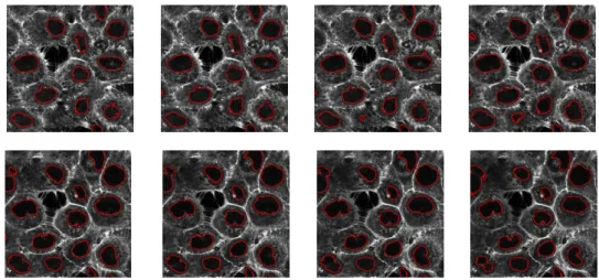
Finally, Fig.3shows some segmentation results obtained on the whole stack of acquired images. Results shown are obtained with α = 0.5, and g = 10. To obtain these results, a propagation initialization strategy, starting on middle slice is used which makes integration of some spatial coherence within the segmentation scheme to avoid propagation of false detection due to complex appearance of actin.
Figure 3: Segmentation of nuclei made on upper (upper row) and lower (bottom row) slices of the all stack, mid-slice of Fig.2being the initialization level). α = 0.5 and g = 10.
These results have been qualitatively considered as very satisfactory from an expert point of view and a very good start for further investigations on that particular data.
The contribution of the segmentation approach presented in this article is twofold: (i) An unsupervised cell nuclei segmentation method is proposed for single channel actin tagged acquisitions without any enhancement or denoising preprocessing of the considered images. (ii) Whereas in the framework of statistical based active contour methods standard Shannon’s entropy is most often considered as the region descriptor, we proposed an original fractional entropy measure inspired from Rényi’s entropy making possible a relaxation of the sensibil-ity of the descriptors to strong variations of the shapes of the non parametrically estimated related PDF. Main motivation was to overcome the limitations of Shannon’s entropy which appeared not adapted to our segmentation problem. We are currently working on locally relating the optimal choice of α parameter with the level of noise and/or the type of texture characterizing the image to segment. From an application point of view, we are finalizing a 3D version of our slice-by-slice segmentation approach to have a direct visualization of the 3D shape of the nuclei. Membrane segmentations will be the next step.
Acknoledgements
This work was supported by the UK Engineering and Physical Sciences Research Council [TeRaFS project, grant number EP/H024913/1].
References
[1] G. Aubert, M. Barlaud, O. Faugeras, and S. Jehan-Besson. Image segmentation using active contours: Calculus of variations or shape gradients? SIAM Journal on Applied Mathematics, 63:2128–2154, June 2003.
[2] T. F. Chan and L. A. Vese. Active contours without edges. IEEE transactions on Image Processing, 10(2):266–277, February 2001.
[3] A. Hall. The cytoskeleton and cancer, volume 28. Springer Netherlands, Philadelphia, PA, USA, 06 2009. ISBN 0167-7659.
[4] A. Herbulot, S. Jehan-Besson, S. Duffner, M. Barlaud, and G. Aubert. Segmentation of vectorial image features using shape gradients and information measures. Journal of Mathematical Imaging and Vision, 25(3):365–386, 2006.
[5] F. Lecellier, M.J. Fadili, S. Jehan-Besson, G. Aubert, M. Revenu, and E. Saloux. Region-based active contours with exponential family observations. Journal of Mathe-matical Imaging and Vision, 36(1):28–45, January 2010.
[6] L. Meziou, A. Histace, F. Precioso, B. Matuszewski, and M. Murphy. Confocal Mi-croscopy Segmentation Using Active Contour Based on Alpha-Divergence. In Pro-ceedings of IEEE ICIP conference, pages 3138–3141, 09 2011.
[7] K. Mosaliganti, A. Gelas, A. Gouaillard, R. Noche, N. Obholzer, and S. Mega-son. Detection of spatially correlated objects in 3d images using appearance mod-els and coupled active contours. In Proceedings of MICCAI, pages 641–648, London, UK, 2009. Springer-Verlag. ISBN 978-3-642-04270-6. doi: http://dx.
PAPER 13: UNSUPERVISED SEGMENTATION OF CONFOCAL MICROSCOPY IMAGES
doi.org/10.1007/978-3-642-04271-3_78. URLhttp://dx.doi.org/10.1007/
978-3-642-04271-3_78.
[8] C. Ortiz De Solorzano, E. Garcia Rodriguez, A. Jones, D. Pinkel, J. W. Gray, D. Sudar, and S. J. Lockett. Segmentation of confocal microscope images of cell nuclei in thick tissue sections. Journal of Microscopy, 193(3):212–226, 1999.
[9] S. Osher and J. A. Sethian. Fronts propagating with curvature dependent speed: Al-gorithms based on hamilton-jacobi formulations. Journal of Comp. Phy., 79:12–49, 1988.
[10] S. Pop, A. Dufour, and J-C. Olivo-Marin. Image filtering using anisotropic structure tensor for cell membrane enhancement in 3d microscopy. In Proceedings of IEEE-ICIP, pages 2085–2088, September 2011.
[11] A. Rényi. On measures of entropy and information. 4th Berkeley Symposium on Math-ematical Statistics and Probability., 1:547–561, 1960.
[12] A. Sarti, C. Ortiz De Solorzano, S. Lockett, and R. Malladi. A geometric model for 3d confocal microscope image analysis. IEEE Transactions on Biomedical Engineering, 47:1600–1609, 2000.
[13] A. Sarti, R. Malladi, and J. A. Sethian. Subjective surfaces: A geometric model for boundary completion. International Journal on Computer Vision, 46(3):201–221, February 2002. ISSN 0920-5691.
[14] P. Yan, X. Zhou, M. Shah, and S. T. C. Wong. Automatic segmentation of high through-put rnai fluorescent cellular images. IEEE Transactions on Information Technology in Biomedicine, 12(1):109–117, January 2008.
[15] C. Zanella, M. Campana, B. Rizzi, C. Melani, G. Sanguinetti, P. Bourgine, K. Mikula, N. Peyriéras, and A. Sarti. Cells segmentation from 3d confocal images of early zebrafish embryogenesis. IEEE transactions on Image Processing, 19(3):770–781, March 2010. ISSN 1057-7149.