HAL Id: hal-03088294
https://hal.archives-ouvertes.fr/hal-03088294
Submitted on 19 Jan 2021
HAL is a multi-disciplinary open access
archive for the deposit and dissemination of
sci-entific research documents, whether they are
pub-lished or not. The documents may come from
teaching and research institutions in France or
abroad, or from public or private research centers.
L’archive ouverte pluridisciplinaire HAL, est
destinée au dépôt et à la diffusion de documents
scientifiques de niveau recherche, publiés ou non,
émanant des établissements d’enseignement et de
recherche français ou étrangers, des laboratoires
publics ou privés.
In vivo time-course biocompatibility assessment of
biomagnetic nanoparticles-based biomaterials for tissue
engineering applications
Fernando Campos, Ana Belén Bonhome-Espinosa, Carmona Ramón, Juan
D.G. Durán, Pavel Kuzhir, Miguel Alaminos, Modesto Lopez-Lopez, Ismael
Rodriguez, Victor Carriel
To cite this version:
Fernando Campos, Ana Belén Bonhome-Espinosa, Carmona Ramón, Juan D.G. Durán, Pavel Kuzhir,
et al.. In vivo time-course biocompatibility assessment of biomagnetic nanoparticles-based
biomate-rials for tissue engineering applications. Matebiomate-rials Science and Engineering: C, Elsevier, 2021, 118,
pp.111476. �10.1016/j.msec.2020.111476�. �hal-03088294�
1
In vivo time-course biocompatibility assessment of biomagnetic nanoparticles-based biomaterials for tissue
1
engineering applications
2
3
Fernando Campos1,5, Ana Belén Bonhome-Espinosa3, Ramón Carmona4, Juan de Dios García López-Durán3,5,
4
Pavel Kuzhir6, Miguel Alaminos1,5, Modesto Torcuato López-López3,5*, Ismael Angel Rodriguez1,2,a*, Víctor
5
Carriel1,5,a
6
7
1-Department of Histology, Tissue Engineering Group, Faculty of Medicine, University of Granada, Granada,
8
Spain.
9
2-Department of Histology, Faculty of Dentistry, Nacional University of Cordoba, Cordoba, Argentina.
10
3-Department of Applied Physics, University of Granada, Avenida de la Fuente Nueva, 18071 Granada, Spain.
11
4-Department of Cell Biology, Faculty of Sciences, University of Granada, Campus Fuentenueva s/n, Granada,
12
Spain.
13
5-Instituto de Investigación Biosanitaria ibs.GRANADA, Granada, Spain.
14
6-University Côte d’Azur, CNRS UMR 7010, Institute of Physics of Nice, Parc Valrose, 06108, Nice, Cedex2,
15
France.
16
a These authors contributed equally: Víctor Carriel, Ismael Angel Rodriguez.
17
18
* Corresponding: Prof. Dr. Modesto Torcuato López-López & Prof. Dr. Ismael Rodriguez, Department of Applied
19
Physics and Department of Histology, Tissue Engineering Group, University of Granada, Granada, Spain. Email:
20
modesto@ugr.es; ismael.rodriguez@unc.edu.ar21
22
23
Abstract:24
Novel artificial tissues with potential usefulness in local-based therapies have been generated by tissue engineering
25
using magnetic-responsive nanoparticles (MNPs). In this study, we performed a comprehensive in vivo
26
characterization of bioengineered magnetic fibrin-agarose tissue-like biomaterials. First, in vitro analyses were
27
performed and the cytocompatibility of MNPs was demonstrated. Then, bioartificial tissues were generated and
28
subcutaneously implanted in Wistar rats and their biodistribution, biocompatibility and functionality were analysed
29
at the morphological, histological, haematological and biochemical levels as compared to injected MNPs.
30
Magnetic Resonance Image (MRI), histology and magnetometry confirmed the presence of MNPs restricted to the
31
grafting area after 12 weeks. Histologically, we found a local initial inflammatory response that decreased with
32
time. Structural, ultrastructural, haematological and biochemical analyses of vital organs showed absence of
33
damage or failure. This study demonstrated that the novel magnetic tissue-like biomaterials with improved
34
biomechanical properties fulfil the biosafety and biocompatibility requirements for future clinical use and support
35
the use of these biomaterials as an alternative delivery route for magnetic nanoparticles.
36
37
Key words: Tissue Engineering, Magnetic nanoparticles, Biomaterials, Bio-distribution, In vivo biocompatibility
38
39
40
41
2
1. Introduction
42
During the last years, magnetic nanoparticles (MNPs) have been evaluated in biomedicine for hyperthermia
43
induction [1], cell labelling and separation [2], DNA separation [3], magnetic resonance imaging [4] and for drug
44
or gene therapies [5, 6]. Iron oxide MNPs are the most commonly used, especially Fe3O4 (magnetite) and ᵧFe2O3
45
(maghemite), because these are stable from a thermal, chemical and colloidal standpoint. In addition, based on the
46
MNPs magnetic properties, it was hypothesized that these particles could be guided to specific in vivo locations
47
using a magnetic field gradient. This could be useful as an alternative method to concentrate growth factors, drugs
48
or cells associated to the particles [7-9], and it has been postulated that MNPs could be useful tools for theranostic
49
[10] and local-based tissue engineering applications [11-15]. MNPs were previously tested for the generation of
50
bioengineered magnetic tissue-like substitutes with improved properties without affecting cell adhesion,
51
proliferation, viability or differentiation in vitro [16, 17], showing a significant improvement of the biomechanical
52
properties of these biomaterials [13, 17].
53
Concerning the in vivo biodistribution of MNPs, it is clear that the administration route is a critical factor
54
determining bioavailability and in vivo functionality of MNPs [10]. To the date, several studies focused on
55
determining the fate of these particles when injected into the bloodstream [10] and results demonstrated that
56
injected MNPs have a short lifespan, tend to accumulate in different organs and may have a certain degree of
57
cytotoxic effects [10, 18]. However, the in vivo biodistribution of particles used within biomaterials needs further
58
characterization [14], and in vivo studies evaluating the cellular and molecular processes related to biocompatibility,
59
biodegradability and biodistribution of implanted magnetic hydrogels are in need. Our group previously developed
60
a fibrin-agarose hydrogel (FAH), which was successfully used in numerous tissue engineering applications
[19-61
26] and is currently used in clinical trials with the approval of the Spanish Agency of Medicines and Medical
62
Devices (AEMPS) according to the EU guidelines for clinical use [27]. Therefore, FAH can be a useful carrier
63
candidate to be combined with MNPs in order to generate novel biocompatible magnetic tissue-like biomaterials
64
[12, 13, 17].
65
The aim of this study is to determine the biocompatibility of FAH-based magnetic tissue-like biomaterials
66
containing MagNP-OH magnetic nanoparticles and to study their in vivo biodistribution in a rat model. First, the
67
structure and biocompatibility of the magnetic hydrogels were determined in vitro. Then, magnetic scaffolds and
68
scaffold-free MNPs were subcutaneously grafted in animals and the host response was evaluated by magnetic
69
resonance imaging, laboratory testing, histology and magnetometry after 12 weeks in vivo.
70
71
2. Materials and methods
72
2.1 In vitro analyses
73
2.1.1 Magnetic nanoparticles (MNPs) characterization
74
In this study, we used commercially available MNPs (Nanomyp, Granada, Spain) referred to as MagNP-OH. These
75
MNPs are composed by a polycrystalline magnetite core coated with methyl methacrylate-co-hydroxyl ethyl
76
methacrylate-co-ethylene glycol dimethacrylate (MMA-co-HEMA-co-EGDMA). The MagNP-OH particles were
77
prepared for analyses following previously described procedures [13, 17].
78
The ultrastructure and dimensions of the MagNP-OH were determined by using a LIBRA 120 PLUS Carl Zeiss
79
(Carl Zeiss, Oberkochen, Germany) transmission electron microscope (TEM). The magnetic properties of the
80
MagNP-OH were characterized by a vibrating sample magnetometer VSM 4500 (EG&G Princeton Applied
81
Research, NJ).
3
2.1.2 Analysis of biocompatibility of the MagNP-OH on 2D cell cultures
83
2.1.2.1 Cell culture and cell-MagNP-OH interaction model
84
Human fibroblast, primary cultures obtained from human oral mucosa biopsies were cultured for 24 h in 24-well
85
plates (2x10⁴ cells/well) with Dulbecco’s Modified Eagle Medium (DMEM) with 10 % Fetal Bovine Serum (FBS)
86
and antibiotics/antimycotics commercial cocktail solution (all cell culture reagents from Sigma-Aldrich, Steinheim,
87
Germany) at 37°C with 5% of CO2. MagNP-OH particles were added to cultured cells at a concentration of 0.5%
88
and 1% (w/v) in DMEM (without FBS and antibiotics) and were kept in culture for 24 h, and biocompatibility was
89
determined after this time. As positive controls of live cells (100% cell viability), the same cells were cultured
90
without MagNP-OH particles. As negative controls (100% cytotoxicity), cells were incubated in the same medium
91
with 1% of triton X-100 (PanReac AppliChem, Barcelona, Spain). Biocompatibility was analysed in six
92
independent samples and viability was evaluated 4 times in each sample (24 measures per test and condition).
93
94
2.1.2.2 In vitro assessment of MagNP-OH cytocompatibility
95
Cytocompatibility was evaluated using a combination of morphological analyses, functional WST-1 cell
96
viability/proliferation assay and quantification of free DNA released from dead cells (able to detect cell membrane
97
structural integrity), as previously described [20, 28, 29]. First, the morphological changes associated with the
98
presence of MagNP-OH were determined by phase contrast microscopy. Then, we analysed the metabolic activity
99
of the human cells using commercially available WST-1 assays (Roche Diagnostic, Mannheim, Germany) using a
100
Microplate Reader (Biochrom® Asys UVM340, Cambridge, UK) at a wavelength of 450–690 nm [20, 28]. Finally,
101
the DNA-released as a consequence of irreversible cell membrane damage was quantified by using a NanoDrop
102
2000 UV-vis spectrophotometer (Thermo Fisher Scientific, Waltham, MA, USA) [14, 20].
103
104
2.1.2.3 Preparation of the magnetic tissue-like biomaterials
105
In this study, we prepared three types of scaffolds: non-magnetic FAH, and two types of magnetic FAH: FAH
106
containing MagNP-OH (FAH-MNPs), and FAH containing MagNP-OH with the application of a definite magnetic
107
field during gelation (FAH-MNPs-F). For the preparation of FAH and the magnetic scaffolds (FAH-MNPs and
108
FAH-MNPs-F), we used a variation of a previously described method for non-magnetic FAH [19, 20, 22, 29, 30].
109
Briefly, hydrogels were generated by mixing 70% of human plasma, 13.5% of PBS (0.1M, pH 7.2-7.4) containing
110
or not MagNP-OH (0.5% v/v of final hydrogel volume) and 1.5% of tranexamic acid (Amchafibrin,
Fides-111
Ecofarma, Valencia, Spain). This solution was carefully mixed and then, a 2% solution of CaCl2 was added (10%
112
of the final volume) to promote fibrin gelation, followed by 5% volume of melted 2% type VII agarose (both by
113
Sigma-Aldrich, Steinheim, Germany) in PBS. This mixture was aliquoted and kept in a cell incubator using
114
standard culture conditions until complete gelation [13]. In the case of FAH-MNPs-F, the mixture was subjected
115
to a vertical magnetic field (48 kA/m) during the first 5 minutes of the process of jellification to obtain an
116
anisotropic biomaterial composed by aligned fibres as previously reported [13]. The process, by which the
117
hydrogel is formed, has been previously described [20, 22, 28]. Concisely, an addition of CaCl2 can activate the
118
blood coagulation factors of the human plasma, resulting in the cleavage of fibrinogen by thrombin, with the
119
subsequent polymerization of fibrin monomers into an insoluble fibrin gel [31]. At the same time, the agarose
120
polysaccharides jellify by forming hydrogen bonds on the fibrin fibres as temperature decreases [32, 33].
121
122
123
4
2.1.2.4 Analysis of biomechanical properties of the magnetic tissue-like biomaterials
124
Magnetic and non-magnetic tissue-like biomaterials were subjected to oscillatory shear strains of increasing
125
amplitude and fixed frequency (1 Hz), and the corresponding oscillatory shear stress was assessed using a Haake
126
MARS III (Thermo Fisher Scientific, Waltham, MA, USA) rheometer at 37°C. The measuring system geometry
127
was a 3.5 cm diameter parallel plate set with rough surfaces to avoid wall slip, and the rotating plate was adjusted
128
to a normal force of 5 N. Measurements were conducted under oscillatory shear strains and the biomechanical
129
properties of the different tissue-like biomaterials were studied by determining the complex viscoelastic modulus
130
of each sample.131
132
2.2 In vivo analyses133
2.2.1 Laboratory animals134
In this study, a total of eighty-five 12-week-old adult male Wistar rats weighing 250–300 g were used. Animals
135
were maintained in the Experimental Unit of the University Hospital Virgen de las Nieves in Granada (Spain).
136
Animals were housed in a temperature-controlled room (21 ± 1°C) on a 12 h light/dark cycle with ad libitum access
137
to tap water and standard rat chow. These studies were performed according to the European Union and Spanish
138
Government guidelines for the ethical care of animals (EU Directive No. 63/2010, RD 53/2013) and this project
139
was approved by the CEEA ethical committee for animal experimentation (approval number: 03-7-15-311).
140
141
2.2.2 Surgical procedure and experimental groups
142
For the in vivo biocompatibility evaluation of magnetic tissue-like biomaterials, and to study the biodistribution of
143
the MagNP-OH, animals were deeply anaesthetized by intraperitoneal injection of a mixture of acepromazine
144
(Calmo-Neosan®, 0.001 mg/g of the weight of the animal, Boehringer Ingelheim, Ingelheim am Rhein, Germany)
145
and ketamine (Imalgene 1000®, 0.15 mg/g of the weight of the animal,Boehringer Ingelheim). Each animal was
146
randomly assigned to one of the following experimental groups (n = 20 in each except for the control group):
147
- (i) FAH group: once anaesthetized, a 1 cm-long incision was made in the forearm skin of each animal, a
148
FAH tissue-like substitute was subcutaneously grafted, and the injury was repaired using absorbable sutures. These
149
animals were used as a control group.
150
- (ii) FAH-MNPs group: in these animals, FAH containing 0.5% (v/v) MagNP-OH were implanted
151
following the same procedure described for the FAH group.
152
- (iii) FAH-MNPs-F group: FAH containing 0.5% (v/v) MagNP-OH subjected to a magnetic field during
153
gelation were implanted.
154
- (iv) MNPs-INJ group: in this case, MNPs were injected in the same area of groups i, ii and iii (forearm
155
subcutaneous tissue). In this sense, subcutaneous injection of a solution containing the MNPs was given in both
156
forearms of each rat (250 µl of a sterile physiological solution containing 12.5 mg of MagNP-OH).
157
- (v) CTR group: five healthy animals were used as controls.
158
Animals were euthanized after 1, 3, 5 or 12 weeks (n = 5 in each period) by using an overdose of anaesthetics
159
followed by intracardiac perfusion of fixative.
160
161
2.2.3 Magnetic resonance imaging (MRI)
162
Magnetic Resonance Image (MRI) analysis was used to identify the grafted materials in each animal and to assess
163
the effects of these materials on the morphology of some major body organs. For this purpose, 3 animals
5
corresponding to each experimental group (CTR, FAH, FAH-MNPs, FAH-MNPs-F and MNPs-INJ) were
165
analysed after 1, 3, 5, and 12 weeks of the surgical procedure using a Biospec TM 70/20 USR device equipped
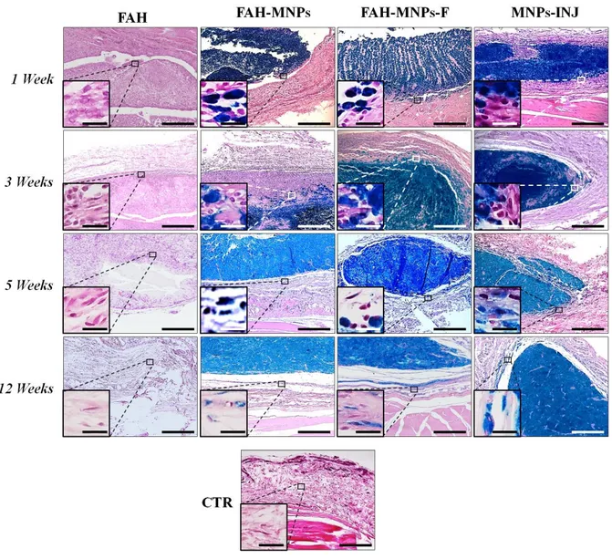
166
with 7 Tesla Ultrashield Refrigerated magnets (Bruker, Billerica, MA). This device was designed and optimized
167
for the analysis of small experimentation animals and is available at the Scientific Research Facility of the
168
University of Granada, Spain. First, animals were anesthetized with isoflurane using a Ohmeda veterinary
169
anaesthesia unit and immobilized in a MRI-compatible cradle. Temperature was kept at 37°C using a water bath
170
circulation system. Then, a whole-body scan was performed on each animal, and the morphology of liver, kidneys,
171
lymph nodes and spleen was evaluated, and the grafting site was specifically analysed to determine the MRI
172
morphology of the implant site and possible migration of the particles to local and regional tissues. In all cases,
173
high resolution axial T2-weighted images were acquired through a T2-TurboRARE sequence using the following
174
settings and experimental conditions: echo time = 23 ms; rare factor = 8 and slice thickness = 1 mm; repetition
175
time = 1371.177 ms with average = 8.
176
177
2.2.4 Haematological and biochemical studies
178
1.5 mL of blood was collected from 5 animals corresponding to each experimental condition at 1, 3, 5 and 12
179
weeks of in vivo follow-up. Blood was stored in Eppendorf tubes containing 5% heparin. For complete blood
180
count, a Sysmex KX-21N automatic analyser (Roche Laboratories, Basel, Switzerland) was used as previously
181
described [32] to determine the following haematological parameters: concentration of haemoglobin (HGB),
182
erythrocytes count (RCB), haematocrit count (HCT), platelets (PLT), white blood cells (WBC), lymphocytes
183
(LYM), neutrophils (NEUT), monocytes-basophils-eosinophils (MXD) [34, 35].
184
For biochemical tests, blood was centrifuged for 15 min at 3,500 rpm and the supernatant serum was collected for
185
analysis using a clinical chemistry analyser Cobas c311 (Roche Laboratories, Basel, Switzerland). The following
186
biochemical parameters were analysed in each sample: Alanine aminotransferase (ALT), urea (UREA), creatinine
187
(CREJ2), iron (IRON2) (all RTU kits from Roche Laboratories) [34, 35].
188
189
2.2.5 Histological and histochemical studies
190
For the histological analyses, animals were deeply anaesthetized and perfused with 4% neutral buffered
191
paraformaldehyde. For all animals included in the study, the area of the implant (FAH, FAH-MNPs,
FAH-MNPs-192
F and MNPs-INJ) was carefully dissected and post-fixed in 4% neutral buffered formaldehyde for 24 h, dehydrated
193
and embedded in paraffin. For animals corresponding to 1 and 12 weeks of follow-up, four vital organs (liver,
194
kidneys, lymph nodes and spleen) were also extracted, post-fixed and embedded in paraffin. Tissue blocks were
195
sectioned at 5 µm of thickness, rehydrated and stained with haematoxylin-eosin (H&E) for histological evaluation.
196
In addition, different histochemical techniques were used to determine tissue-specific normal parameters. The
197
periodic acid-Schiff histochemical method (PAS) was used to evaluate the glycogen content of the liver, the
198
glomerular basement membrane of the kidney and the basement membranes of each tissue and organ. To identify
199
the presence of ferric iron, all tissues and organs were stained with Perls (Prussian blue reaction) histochemical
200
method contrasted with H&E as described previously [14].
201
The percentage of positive area for Perls-positive histochemical reaction in the spleen was determined with ImageJ
202
software (National Institutes of Health, USA) from each group at 1 and 12 weeks following a previously described
203
methodology [28, 36].
204
205
6
2.2.6 Ultrastructural analyses
206
For the ultrastructural analyses, tissue samples corresponding to the 12 weeks follow-up period were obtained
207
from each animal included in the in vivo study at the moment of the euthanasia. Samples were fixed in 2.5%
208
glutaraldehyde, washed three times in cacodylate buffer and post-fixed in 1% osmium tetroxide. Tissues were then
209
dehydrated and embedded in epoxy resin, and sectioned. Ultrathin sections were stained with aqueous uranyl
210
acetate and lead citrate, mounted on grids and analysed in a Carl Zeiss EM902 transmission electron microscope.
211
The presence of iron atoms in tissues was identified by Electron Energy-Loss Spectroscopy (EELS). Analysis of
212
the heterogeneous pattern of the FAH, FAH-MNPs and FAH-MNPs-F was carried out by scanning electron
213
microscopy (SEM) using a FEI Quanta 200 equipment (Hillsboro, OR) as previously described [20, 22, 28].
214
215
2.2.7 Magnetometry
216
After the euthanasia, we tracked the presence of particles at the site of implantation and vital organs. We checked
217
the magnetic response of the samples with a neodymium magnet in a magnetic field gradient of 10 mT/mm and a
218
maximum field of 470 mT. In addition, we quantified the magnetic response of the samples through magnetometry
219
measurements using a vibrating sample magnetometer VSM 4500 (EG&G Princeton Applied Research, NJ) at
220
room temperature. For this, similar volumes of tissue (equivalent to 50 mg of mass) were used from each condition.
221
The detection limit of the magnetometer was 0.001 emu/g, which corresponds to approximately 30 µg of particles.
222
These analyses were performed in animals corresponding to 12 weeks of in vivo follow-up.
223
224
2.2.8 Statistical analyses
225
In this study, all variables were subjected to the Shapiro-Wilk test of normality and resulted to be non-normally
226
distributed. Therefore, Fisher Exact Test and Mann-Whitney U test were used to determine statistical differences
227
between comparison groups. All variables were analysed by using the software SPSS 16.00 (IBM Company,
228
Armonk, NY) and results were shown as mean ± standard deviation (SD). In this study, p < 0.05 was considered
229
as statistically significant in two-tailed tests.
230
231
3. Results and Discussion
232
3.1 In vitro characterization and biocompatibility
233
First, our ultrastructural analysis of the MagNP-OH used in this study allowed us to confirm that the diameter of
234
the particles was 70 ± 18 nm, which is higher than MNPs frequently used by other authors in tissue engineering
235
applications (30-40 nm) [15, 16, 37-41]. Interestingly, particles tended to form polycrystalline aggregates
236
externally coated by a polymeric matrix surrounding each aggregate (Figure 1A). Magnetic characterization of the
237
MagNP-OH revealed the typical soft ferromagnetic character of these MNPs, with negligible remnant
238
magnetization, and a saturation magnetization of 161 ± 7 kA/m (Figure 1B). It is well known that the size of the
239
MNPs is directly related to the magnetic response of the particles, and multidomain MNPs (> 50 nm) commonly
240
show higher magnetic response as compared to small particles [38]. However, it has been previously demonstrated
241
that biointegration of large MNPs in a hydrogel mesh tend to be incomplete due to gravitational settling and lower
242
total surface area of these MNPs [14]. Although MNPs with a larger diameter will show stronger magnetic
243
response [14], the present work was carried out with smaller nanoparticles in order to favour biointegration in the
244
fibrin-agarose mesh. Further research is in need to determine the optimal size of the MNPs for use in the generation
7
of magnetic hydrogels, and the possibility of using particles of different sizes in the same hydrogel. In fact, the in
246
vivo biological effects of MNPs have been demonstrated to be size-dependent [42].
247
Regarding in vitro cytocompatibility of human cells cultured in the presence of MagNP-OH, we carried out a
248
multi-level approach analysis at three levels: cell morphology, cell function and integrity of the cell membrane.
249
Results show that, in general, MNPs are highly cytocompatible. In the first place, co-culture of these MNPs with
250
human cells was not able to modify the typical elongated spindle-like shape of viable human fibroblasts, suggesting
251
that these cells remained highly viable, and MNPs were mostly found homogeneously distributed in the
252
extracellular space of the cells (Fig. 1C). In fact, the morphology of these cells was comparable to positive control
253
cells cultured without MNPs and very different from the small, rounded-shape appearance of negative control dead
254
cells. To confirm these results at the functional level, we then analysed the functionality of the cellular
255
mitochondrial dehydrogenase by WST-1 assay. Results showed high levels of metabolic activity in cells containing
256
MagNP-OH (at the concentration of 1% and 0.5%) with values comparable (p > 0.05) to the positive control group.
257
Positive controls and both experimental groups containing MagNP-OH showed significantly higher WST-1 values
258
as compared to the negative control group (p < 0.0001) (Figure 1D), suggesting high metabolic activity [20, 28].
259
Finally, viability was analysed at the structural level by quantifying released DNA from cells cultivated in the
260
presence of the MagNP-OH (Figure 1E), which is unfailingly associated to a cell membrane disruption [20, 28].
261
Results showed low cell mortality in positive controls and both experimental groups (1% and 0.5%), with no
262
differences between positive controls and cells cultured with MagNP-OH (p > 0.05). However, mortality was
263
significantly higher in negative controls (p = 0.0020). These results are in agreement with the high
264
cytocompatibility previously observed in hydrogels containing MagNP-OH [17], especially when these particles
265
were coated with a polymer such as polyethylene glycol (PEG) [14]. The polymeric coating of MNPs provides
266
hydrophilic properties that may improve stabilization in colloidal suspension and increase biocompatibility [10,
267
43]. Altogether, these results support the high in vitro biocompatibility of the coated MagNP-OH used in this
268
study.
269
Once the cytocompatibility of MagNP-OH was evaluated in vitro, we generated magnetic tissue-like biomaterials
270
containing these particles. The rheological characterization of FAH-MNPs and FAH-MNPs-F revealed that
271
incorporation of MagNP-OH considerably increased the strength of the tissue-like biomaterials as compared to
272
control nonmagnetic FAH for the complex viscoelastic modulus
( ) ( )
2 2
12''
'
*
G
G
G
=
+
(Figure 1F), for both273
the biomaterials subjected to magnetic fields during the polymerization and materials devoid of these fields. In
274
agreement with our previous studies [20, 28, 36], SEM images confirmed that the use of a magnetic field induced
275
the alignment of the MNPs within the fibrin-agarose network (Figure 2). These findings are in agreement with
276
previous reports showing that MNPs can considerably improve the biomechanical properties of FAH, thus
277
increasing their putative usefulness in tissue engineering and regenerative medicine [13, 14, 17]. Previous studies
278
suggest that the biomechanical improvement observed by the incorporation of MNPs within FAH can be explained
279
by two reasons: on the one hand, the strong electrostatic interactions established between the biomaterial network
280
and the MNPs, and on the other hand, the formation of MNPs clusters within the biomaterials network [17]. The
281
electrostatic interactions increase the overall molecular interaction forces of the resulting biomaterial network,
282
therefore explaining the biomechanical improvement. Furthermore, it was demonstrated that the MNPs clusters
283
act as knots, sites from where the fibrin network is organized in more compact, thicker and aligned fibrin strands
8
which create a biomechanically and structurally more efficient network as compared to the classical nonmagnetic
285
hydrogels [17].286
287
288
289
Figure 1. In vitro characterization of the MagNP-OH particles used in this study. A) Transmission electron
290
microscopy ultrastructural analysis of MagNP-OH particles. Scale bar: 200 nm (left) and 100 nm (right). B)
291
Magnetization curve of MagNP-OH particles. C) Phase contrast microscopy image of human fibroblasts cultured
292
with MagNP-OH particles. Cells are labelled with black arrows and MagNP-OH particles are highlighted with
293
white arrows. D) Results of the cellular metabolic activity as determined by WST-1 assay. E) Analysis of cell
294
membrane integrity as determined by DNA quantification. In D and E, values correspond to averages and standard
9
deviations.* Results are statistically different from all the other study groups. PC: positive controls; NC: negative
296
controls. F) Biomechanical properties are shown as the complex viscoelastic modulus of nonmagnetic FAH,
FAH-297
MNPs and FAH-MNPs-F.298
299
300
301
Figure 2. Representative images corresponding to the SEM analysis of materials used in this study. FAH:
302
fibrin-agarose hydrogels; FAH-MNPs: FAH containing MagNP-OH; FAH-MNPs-F: FAH containing MagNP-OH
303
subjected to a magnetic field during gelation. Scale bar 10 µm
304
305
3.2 In vivo evaluation
306
Accurate monitoring and analysis of the fate of MNPs grafted in vivo in laboratory animals is essential for their
307
future clinical translation and practical application [44]. Ideally, the biocompatibility of grafted MNPs should be
308
assessed by using a combination of invasive and non-invasive methods allowing a precise evaluation of the fate
309
of the MNPs and the potential metabolic pathways and organs involved in MNPs metabolism and biodegradation.
310
Consequently, animals grafted with the different materials were subjected to an array of highly accurate evaluation
311
methods that included MRI, magnetometry, haematological, biochemical and histological analyses to determine
312
biocompatibility and the outcome of the grafted materials. In addition, analyses were performed at the local and
313
the distal level as requested by most National Medicines Agencies for Advanced Therapies Medicinal Products
314
[45]. In this sense, we first analysed the local graft site to determine in situ biosafety, temporal stability and
315
migration to neighbour tissues. Then, we analysed four key distal organs to shed light on the possible distal effects
316
of the grafted biomaterials, including distal organ migration. In general, all these analyses allowed us to
317
demonstrate that the biomaterials used in the present work were safe and complied with the main biosafety
318
requirements for future clinical use.
319
In situ time-course analysis of the implant site using MRI (Fig. 3) showed that non-magnetic FAH remained at the
320
grafting site and was locally metabolized and reabsorbed in 12 weeks. In contrast, magnetic biomaterials
321
containing MagNP-OH, as well as the injected MagNP-OH remained located in the implant site after 12 weeks of
322
in vivo follow-up (Fig. 3). These results coincide with the magnetometric analyses showing that controls and
323
animals grafted with FAH were negative, whereas all animals with grafted MagNP-OH showed a highly positive
324
magnetic response at the grafting site after 12 weeks of follow-up. Furthermore, the MRI analysis of distal organs
325
did not reveal any sign of damage, inflammation or organ failure in any of the experimental groups during the
326
whole follow-up period (Supplementary Fig. S1).
327
328
329
10
330
331
Figure 3. Magnetic Resonance Images (MRI) analysis of animals with the different materials grafted at each
332
study time. Images were taken at the grafting site. White circles correspond to hyperintense areas corresponding
333
to MagNP-OH accumulation at the implantation site. CTR: control animals; FAH: fibrin-agarose hydrogels;
FAH-334
MNPs: FAH containing MagNP-OH; FAH-MNPs-F: FAH containing MagNP-OH subjected to a magnetic field
335
during gelation; MNPs-INJ: MNPs injected subcutaneously.
336
337
Then, the implantation site was analysed histologically. In this regard, most animals showed an initial mild local
338
inflammatory reaction restricted to the tissues surrounding the implanted materials, but no signs of necrosis,
339
infection, rejection or malignant transformation were found in any of the groups (Fig. 4). This reaction was similar
340
in all groups grafted with biomaterials (FAH, FAH-MNPs and FAH-MNPs-F), and tended to decrease over time.
341
In the FAH group, we found that the number of inflammatory cells, especially macrophages, decreased with time
342
and almost disappeared after 12 weeks. An interesting finding of our study was that the three groups in which
343
MNPs were grafted tended to encapsulate the grafts, with the formation of a central Perls-positive nucleus
344
surrounded by a connective tissue capsule. Although we found a single nucleus in the MNPs and
FAH-345
MNPs-F groups, particles tended to form several independent nuclei in the MNPs-INJ group, at least during the
11
first 5 weeks (Fig. 4). These results confirm the absence of an increased inflammatory reaction driven by the MNPs
347
implant and point out the usefulness of the FAH-MNPs model as a straightforward way of providing the host tissue
348
with MNPs that could exert a positive clinical effect [46, 47]. As compared to injection, surgical implantation of
349
a tissue-like magnetic material allowed a more efficient control of the grafting site and, according to our results,
350
favoured the MNPs containment and enhanced encapsulation of the whole grafted mass in a single nucleus, thus
351
preventing the connective tissue infiltration and grafts disaggregation or dispersion found in the MNPs-INJ group.
352
In addition, the MNPs corresponding to the FAH-MNPs-F group showed a clear definite alignment and orientation
353
of the MNPs during the first weeks, even though this orientation was lost after 5 weeks. Interestingly, the loss of
354
this characteristic alignment pattern obtained by the use of a magnetic field coincided with the in vivo
355
biodegradation process of the FAH [34]. In this regard, we recently demonstrated that FAH biomaterials are
356
progressively remodelled and infiltrated by host immune cells, mainly macrophages, being completely degraded
357
after 5 to 9 weeks of in vivo implantation [23, 34]. We may hypothesize that the progressive degradation and
358
remodelling of the FAH network supporting the aligned particles resulted in a loss of structural cohesion of the
359
aligned MNPs clusters with the consequent loss of its aligned structural pattern [23, 34].
360
The three-dimensional orientation of biomaterials is one of the goals of current tissue engineering, since most
361
human tissues are characterized by a nonlinear and anisotropic mechanical behaviour [48] due to the non-random
362
distribution of its components, and this distribution is essential for its proper in vivo function. The use of
12
MNPs-F could contribute to obtaining MNPs-based bioartificial tissues with defined alignment with added value
364
for use in regenerative medicine [49], as shown in Fig. 2.
365
366
367
368
Figure 4. Perls histochemical results of grafted biomaterials and injected MNPs at 1, 3, 5 and 12 weeks in
369
vivo. CTR: control animals; FAH: fibrin-agarose hydrogels; MNPs: FAH containing MagNP-OH;
FAH-370
MNPs-F: FAH containing MagNP-OH subjected to a magnetic field during gelation; MNPs-INJ: MNPs injected
371
subcutaneously. The inserts images correspond to higher magnifications of the same images. Scale bars: 300 µm
372
(large images) and 20 µm (inserts).
373
374
Furthermore, we carried out TEM analyses to determine the biocompatibility of each biomaterial at the
375
ultrastructural level after 12 weeks of the surgical procedure. On the one hand, host tissues corresponding to the
376
FAH group showed some macrophages with intracellular phagosomes containing rests of the biomaterial, along
377
with extracellular matrix mainly consisting of collagen fibres that were comparable to control tissues (Fig. 5). On
378
the other hand, animals grafted with MNPs showed very similar behaviour regardless of the specific group
379
considered (FAH-MNPs, FAH-MNPs-F and MNPs-INJ). In all these groups, we found numerous host cells
380
compatible with macrophages containing abundant intracellular MNPs that tended to keep their original
13
polycrystalline aggregate pattern inside the cells. Most of the particles gathered in cytoplasmic vesicles that could
382
correspond to endosomes or secondary lysosomes, as well as large phagosomes. MagNP-OH were also found in
383
the extracellular space. No signs of necrosis or cell alterations were detected in any of the study groups. These
384
findings confirm the high biocompatibility of the different materials used in this study. In agreement with previous
385
reports, our results suggest that iron oxide nanoparticles are mostly engulfed within the human cells and do not
386
cause any detectable alterations in these cells [10, 50].
387
388
389
Figure 5. Representative images corresponding to the ultrastructural analysis of materials grafted in vivo
390
for 12 weeks. CTR: control animals; FAH: fibrin-agarose hydrogels; FAH-MNPs: FAH containing MagNP-OH;
391
MNPs-INJ: MNPs injected subcutaneously. Black arrow: phagosome containing agarose; white arrow: MNPs. The
392
inserts images correspond to higher magnifications of the same images. Scale bars: 300 µm (large images) and 20
393
µm (inserts).
394
395
396
Once the local implant site was analysed, we evaluated the morphology, structure and function of several major
397
distal organs of each animal to determine the possible distal effects of each biomaterial as part of the global
398
biocompatibility and biosafety assessment required for future clinical use [45]. In this regard, the whole-body MRI
399
scan analysis of each animal and the specific analysis of four key organs playing a role in metabolizing and
400
processing a biomaterial grafted in vivo -liver, kidneys, lymph nodes and spleen- revealed a perfectly normal
401
morphology devoid of detectable alterations (Supplementary Fig. 1). No MNPs were detected by MRI in any of
402
these organs. Similarly, analysis of the different organs using magnetometry, revealed a negative signal in all
403
animals after 12 weeks of in vivo follow-up. These results suggest that MagNP-OH stayed at the grafting site and
404
did not tend to migrate, supporting the stability of this type of MNPs.
405
At the histological level, the structural analysis of distal organs confirmed the absence of alterations during the
406
whole study. Indeed, no signs of inflammation, fibrosis, necrosis or other detectable tissue alterations were
407
observed in histological sections of liver, kidneys, lymph nodes and spleen stained with H&E in any of the groups
408
(Supplementary Fig. S2). Similarly, PAS staining analysis (Supplementary Fig. S3) confirmed that the content of
14
glycogen was normal in hepatocytes of all groups of animals, and the glomerular and non-glomerular basement
410
membranes were also free from detectable alterations. These results are in agreement with previous studies
411
demonstrating that this type of MNPs coated with different polymers can be safely used without significant
412
histological alterations of vital organs [14, 51], whereas other types of particles were associated to histological
413
lesions in liver and kidney [52].
414
To identify any possible particle migration to distal organs, we also analysed these organs using the Perls
415
histochemical technique, which is specific for detection of iron in cells and tissues. Results showed very few or no
416
particles in the liver and kidney at 1 and 12 weeks, but a positive reaction was found in spleen and lymph nodes at
417
both analysed times (Fig. 6). These findings are in agreement with previous reports demonstrating that
418
nanoparticles of different nature tend to be massively captured by cells of the mononuclear phagocyte system [49].
419
Hence, we found that lymph nodes were Perls-negative in CTR and FAH group, whereas animals with grafted
420
MNPs (FAH-MNPs, FAH-MNPs-F and MNPs-INJ) showed small Perls-positive areas after 1 week, and moderate
421
to intense Perls-positive areas after 12 weeks, especially in the case of the injected MNPs. Then, we analysed both
422
components of the rat spleen (the red and the white pulp), and we found that the staining area and intensity were
423
higher in this organ than in the rest of organs. As expected, the red pulp of the spleen was very positive to the Perls
424
method in all groups, including controls, but this reaction became significantly more intense after 12 weeks in
425
FAH-MNPs, FAH-MNPs-F and MNPs-INJ groups (p < 0.05). These findings are consistent with the primary
426
function of the red pulp, which is related to filtering peripheral blood from antigens, foreign bodies and all kinds
427
of substances that may arrive in the blood, blood iron turnover, as well as serving as a huge reservoir of monocytes
428
[18]. The white pulp, however, was mostly negative for this staining technique, although the marginal zone (in
429
which antigen-presenting cells, such as dendritic cells and macrophages exists) was positive at 12 weeks in
FAH-430
MNPs, FAH-MNPs-F and MNPs-INJ groups. Quantification of the Perls-positive areas in the red pulp (Fig. 7)
431
demonstrated no significant differences among samples at 1 week (p > 0.05), but a significant increase was found
432
at 12 weeks in the FAH-MNPs and MNPs-INJ groups (p < 0.05). In fact, the three groups in which MNPs were
433
grafted in animals, showed significant differences vs. control at week 12 (p < 0.05). These results were
434
corroborated at the ultrastructural level by TEM analysis confirming the presence of macrophages containing
435
electrodense iron-rich granular material identified as iron by EELS in all experimental groups (Fig. 7). In contrast
436
with the macrophages observed within grafted biomaterials, in spleen, these cells contained iron-rich granular
437
intracytoplasmic vesicles, but polycrystalline aggregates were not detected.
438
The presence of abundant cells containing iron in the spleen of all animal groups could be explained by the
439
important role that the spleen plays in mechanical filtration of red blood cells and haemoglobin iron recycling and
440
turnover [18]. The increase observed in animals in which MagNP-OH were grafted, strongly suggests that MNPS
441
could progressively reach to the spleen through blood circulation, as other authors demonstrated by intraperitoneal
442
injection of RITC-labeled MNPs [10, 50]. The fact that our magnetometry and MRI analyses were negative could
443
probably be explained by the low concentration of MagNP-OH that reached the spleen, which was probably below
444
than 30 µg, which is the minimum concentration required for detection by magnetometry and MRI. Another
445
possibility is that MagNP-OH were progressively transformed into non-magnetic iron forms by host cells as
446
previously suggested [47]. Interestingly, previous works showed that superparamagnetic iron-oxide NPs show
447
identical distribution pattern when administered in vivo [51].
448
In consequence, our histological results, in line with results published by other authors, highlight the relevance of
449
the administration route and NPs size in the subsequent organic biodistribution [10], and confirm that MagNP-OH
15
tend to remain stable at the implant site, with some particles biodistributed to lymphoid organs, without altering
451
their histological structure and function.
452
453
454
455
Figure 6. Perls histochemical results of distal organs at 1 and 12 weeks. FAH: fibrin-agarose hydrogels;
FAH-456
MNPs: FAH containing MagNP-OH; FAH-MNPs-F: FAH containing MagNP-OH subjected to magnetic field
457
during gelation; MNPs-INJ: MNPs injected subcutaneously; CTR: control animals. Scale bar 100 µm.
16
459
Figure 7. Quantitative results of Perls positive histochemical reaction and EELS in the red pulp of the spleen
460
in each group and control. The table on top shows the results of the quantification of the percentage of the area
461
corresponding to Perls-positive signal. The figures below show illustrative ultrastructural images of macrophages
462
containing iron in intracellular phagosomes corresponding to two groups of animals (FAH and FAH-MNPs), along
463
with an EELS spectrum with a peak corresponding to intracellular iron. CTR: control animals; FAH: fibrin-agarose
464
hydrogels; FAH-MNPs: FAH containing MagNP-OH; FAH-MNPs-F: FAH containing MagNP-OH subjected to
465
a magnetic field during gelation; MNPs-INJ: MNPs injected subcutaneously. Values in the table are shown as
466
mean ± standard deviation for each group of animals and each follow-up time, and the statistical p values
467
corresponding to the comparison of these values with CTR are shown in brackets. In the last row, the statistical p
468
values corresponding to the comparison of values at 1 week vs. values at 12 weeks are shown for each study group.
469
Significant differences (p < 0.05) are highlighted with asterisks (*). Scale bar for FAH = 1 µm and scale bar for
470
FAH-MNPs = 2 µm.471
472
473
HAEMOGRAMGeneral count WBC/White cells
WBC (103) RBC (106) HGB HCT PLT (105) LYM MXD NEUT Weeks µL-1 µL-1 g/dL % µL-1 % % % FAH 3.04 ± 1.51* 7.14 ± 0.22* 13.5 ± 0.52 39.54 ± 1.1 5.16 ± 1.99 45.68 ± 23.56 32.56 ± 22.96 21.76 ± 4.64 1 4.56 ± 3.31 7.61 ± 0.06 14.14 ± 0.51 42.28 ± 0.66 4.60 ± 2.59 64.38 ± 14.8 12.96 ± 0.86 22.66 ± 14.22 3 2.75 ± 0.26* 8.04 ± 0.4 14.15 ± 0.27 43.78 ± 1.3 6.57 ± 0.23 77.63 ± 4.06 9.25 ± 3.02 13.13 ± 5.75 5 9.74 ± 3.89 8.15 ± 0.38 14.32 ± 0.2 44.06 ± 1.93 5.11 ± 3.63 68.5 ± 16.18 9.32 ± 3.65 22.18 ± 13.58 12 FAH-MNPs 2.4 ± 1.27 7.47 ± 0.64 13.22 ± 1.48 40.02 ± 4.35 5.63 ± 2.96 42.08 ± 26.77 33.02 ± 25.75 24.84 ± 12.87 1 5.18 ± 3.11 7.51 ± 0.3 14.16 ± 0.76 42.26 ± 2.17 3.82 ± 2.16 62.02 ± 15.52 13.34 ± 2.57 24.64 ± 16.25 3 2.48 ± 1.09 8.11 ± 1.48* 14.18 ± 9.93 44.27 ± 1.72 6.24 ± 2.57 72.7 ± 16.81 5.92 ± 1.78 21.08 ± 9.9 5 2.94 ± 3.52 7.83 ±0 .68 14.16 ± 0.76 42.48 ± 4.15 8.74 ± 0.84 80.32 ± 3.35 * 15.28 ± 4.59 2.56 ± 5.96 12
17
FAH-MNPs-F 3.63 ± 2.1 7.76 ± 0.38 13.83 ± 0.25 42.43 ± 1.85 5.61 ± 1.21 66.25 ± 8.58 13.83 ± 5.93 19.93 ± 6.41 1 2.46 ± 0.46 7.62 ± 0.44 13.58 ± 0.39 41.18 ± 1.69 6.31 ± 0.72 76.02 ± 3.35 11.04 ± 1.35 12.94 ± 2.11 3 5.36 ± 4.05 7.77 ± 4.09* 13.91 ± 0.6 42.73 ± 1.81 3.62 ± 2.48 68 ± 12.91 16.02 ± 7.07 15.98 ± 6.97 5 5.72 ± 1.73 8.46 ± 0.62* 14.52 ± 0.6 45.66 ± 3.1 3.78 ± 4.1 69 ± 13.41 12.64 ± 2.97 18.36 ± 11.6 12 MNPs-INJ 2.48 ± 0.43 7.82 ± 0.24 13.78 ± 0.42 42.8 ± 1.86 4.88 ± 2.75 72.08 ± 6.03 12.53 ± 1.24 15.4 ± 6.02 1 1.92 ± 0.64 7.54 ± 0.26 13.5 ± 0.24 41.22 ± 1.51 6.26 ± 0.73 73.2 ± 7.71 14.92 ± 5.72 11.88 ± 2.58 3 1.78 ± 0.29 8.03 ± 0.23 * 14.08 ± 0.83 43.94 ± 2 5.94 ± 2.82 74.5 ± 3.71 10.82 ± 4.98 14.68 ± 6.67 5 7.1 ± 3.74 7.57 ± 0.89 13.75 ± 1.61 41.45 ± 5.4 4.33 ± 4.58 65.83 ± 4.67 10.03 ± 3.42 24.15 ± 6.5 12 CTR 5.93 ± 3.46 7.72 ± 0.11 13.9 ± 0.5 42.25 ± 1.07 2.58 ± 2.71 63.03 ± 15.39 14.33 ± 6.75 22.65 ± 13.16474
Table 1. Haematological profile. Summary of the mean and SD of parameters evaluated at 1, 3, 5 and 12 weeks.
475
White blood cells (WBC), erythrocytes count (RBC), concentration of haemoglobin (HGB), haematocrit count
476
(HCT), platelets (PLT), lymphocytes (LYM), monocytes-basophils-eosinophils (MXD), neutrophils (NEUT).
477
Significant differences (p < 0.05) between experimental and control groups are highlighted with asterisks (*).
478
479
After that the morphology and structure of local and distal tissues and organs were determined, we analysed the
480
effect of the different biomaterials on each animal group at the functional level. In this regard, haematological
481
studies revealed that all parameters evaluated here were within the physiological range of normal values described
482
in the literature for the Wistar rat (Table 1) [52]. However, some specific parameters showed significant differences
483
with the control animals used in this study. In the case of the red blood cells (RBC), we observed a significant
484
decrease of RBC counts in the FAH group at 1 week as compared to controls (p = 0.009), which could be a
485
consequence of the recent surgical procedure. In contrast, a significant increase of the number of RBC was
486
observed after 5 weeks in FAH-MNPs (p = 0.016), FAH-MNPs-F (p = 0.016) and MNPs-INJ (p = 0.028), and also
487
after 12 weeks for FAH-MNPs-F (p = 0.028) as compared to controls. Interestingly, the increase of RBC values
488
was not accompanied by significant variations of the concentration of haemoglobin (HGB) or haematocrit (HCT).
489
The leukocyte count (WBC) showed a transient reduction in the FAH group at 5 weeks as compared to controls
490
(p = 0.009), although the leukocyte formula was normal for all the study groups except for an increase of
491
lymphocytes (LYM) at 12 weeks in FAH-MNPs group (p = 0.010). Finally, the evaluation of platelets (PLT)
492
showed an increase in all experimental groups over time as compared to control animals. All these variations,
493
which fell into the normal parameters of healthy Wistar rats [52], could be related to the host adaptive physiological
494
response to the surgical procedure, healing process, active hydrogel biodegradation and MagNP-OH phagocytosis
495
being these results in line with the histological findings. A similar result was found for the biochemical parameters
496
analysed in plasma of each animal, which were normal in most cases (Table 2). First, the levels of circulating iron
497
were similar to control animals at all study times (p > 0.05). The lack of increase of circulating free iron supports
498
the idea that MNPs remained stable and were not able to release a significant amount of iron to the circulation.
499
Similarly, most hepatic function-related parameters fell within the physiological range of this species, except for
500
an initial transient decrease of ALT in MNPs-INJ (p = 0.028) that normalized thereafter. Finally, the two
501
biochemical markers of renal function (urea and creatinine) were normal in all study groups. Additionally, the
502
renal function-related parameters urea and creatinine (CREJ2) showed a transitory increase over the time in all
503
experimental groups as compared to controls, which tended to normalize with time.
504
Altogether, these haematological and biochemical results confirmed that the use of MagNP-OH is safe and in vivo
505
implantation of these MNPs is not associated to a vital organ failure. Although some slight variations were found
506
in specific groups of animals, values were in agreement with the physiological ranges described in the literature
18
[52], and showed a clear normalization over time. In addition, the normalization of blood and biochemical
508
parameters is in line with the quantitative results observed in Wistar rats in which the sciatic nerve was repaired
509
with acellular nerve grafts [35]. These findings confirm again that both administration routes for the MagNP-OH
510
(injected or encapsulated) were safe for the host animal. In contrast, a previous study showed that the high-dose
511
oral administration of other types of nanoparticles based on silver was related to hepatic and renal affection at the
512
biochemical level, what clearly differed from our findings [52]. Finally, the haematological and biochemical
513
profiles were in concordance with the histological and MRI results showing normal structure and morphology of
514
distal organs. All this suggests that the in vivo injection and the subcutaneous implantation of the magnetic
515
materials generated in this work was fully biocompatible and fulfilled the strict biosafety criteria required for future
516
clinical use.
517
518
BIOCHEMICAL
Liver Kidney Other parameters
ALT CREJ2 UREA Fe
Weeks U/L mg/dl mg/dl µg/dl FAH 46.28 ± 10.26 0.75 ± 0.08 42.32 ± 6.71 162.27 ± 0.29 1 40.44 ± 7.38 0.65 ± 0.14 43.28 ± 7.34 170.25 ± 0.33 3 41.35 ± 17.39 0.6 ± 0.24 38.08 ± 13.76 164.21 ± 0.61 5 53.54 ± 16.03 0.6 ± 0.08 36.94 ± 2.65 181.19 ± 0.22 12 FAH-MNPs 141 ± 144.53 0.7 ± 0.15 46.66 ± 8.73 147.31 ± 0.31 1 79.2 ± 65.15 0.72 ± 0.08 43.84 ± 5.97 187.51 ± 0.32 3 42.08 ± 10.65 0.66 ± 0.15 38.22 ± 2.67 171.19 ± 0.40 5 74.68 ± 60.94 0.54 ± 0.11 35.08 ± 4.48 249.36 ± 1.91 12 FAH-MNPs-F 45.75 ± 6.72 0.84 ± 0.18 37.5 ± 8.76 157.85 ± 0.42 1 46.82 ± 11.49 0.69 ± 0.1 41.5 ± 4.6 160.15 ± 0.15 3 46.61 ± 26.92 0.53 ± 0.05 39.59 ± 4.93 172.2 ± 0.27 5 45.28 ± 9.17 0.66 ± 0.05 38.72 ± 5.84 148.91 ± 0.10 12 MNPs-INJ 30.2 ± 7.83* 0.65 ± 0.11 39.3 ± 7.57 155.15 ± 0.25 1 35.94 ± 6.19 0.53 ± 0.08 38.98 ± 4.74 183.92 ± 0.22 3 62.86 ± 30.07 0.71 ± 0.11 38.56 ± 4.89 168.49 ± 0.26 5 44.62 ± 10.09 0.74 ± 0.11 39.58 ± 5.2 164.91 ± 0.29 12 CTR 39.55 ± 5.55 0.46 ± 0.14 34 ± 2.81 184.36 ± 0.23
519
Table 2. Biochemical profile. Summary of the mean and SD of parameters evaluated at 1, 3, 5 and 12 weeks.
520
Alanine aminotransferase (ALT), urea (UREA), creatinine (CREJ2) and iron (Fe). Significant differences (p <
521
0.05) between experimental and control groups are highlighted with asterisks (*).
522
523
In summary, although the particles remained at the grafting site after 12 weeks, these results suggest that
MagNP-524
OH fulfil the biosafety and biocompatibility requirements for future clinical use. Incorporation of these MNPs in
525
fibrin-agarose biomaterials allows the generation of novel biomaterials with improved biomechanical properties
526
with guarantees of biosafety and biocompatibility demonstrated at the morphological, structural and functional
527
levels. As an alternative route of delivery, tissue-like biomaterials allow efficient control of the MNPs
528
biodistribution in vivo. Biodistribution analysis demonstrated that our strategy supports the local use of the
529
MagNP-OH, which were mostly confined to the implantation area. However, long-term studies are still needed to
530
determine the time required for the complete biodegradation and/or metabolization of the implanted MagNP-OH.
531
In addition, tissue-like biomaterials based on fibrin-agarose combined with MagNP-OH allows easy handling and
532
straightforward surgical implant of these nanoparticles, and facilitates local in vivo encapsulation of MagNP-OH
533
as compared to injected MagNP-OH. In addition, the methodology described in the present manuscript also
19
allowed the generation of novel biomaterials with a definite structural alignment using magnetic fields during the
535
gelation of the biomaterial. This could be advantageous for reproduction and treatment of human tissues requiring
536
a specific 3D structural organization such as the human cornea [53], tendon [54] and cartilage [55] and also other
537
tissues with an anisotropic behaviour such as the human skin, nerve and oral mucosa and palate [56]. However,
538
future clinical trials should demonstrate the usefulness of these tissue-like products.
539
540
4. Conflicts of interest
541
The authors declare that they have no known competing financial interests or personal relationships that could
542
have appeared to influence the work reported in this paper.
543
544
5. Author contributions
545
MTLL performed magnetic particles characterization. IAR, MA developed in vitro analyses of biocompatibility.
546
ABBE, MTLL, performed the mechanical properties. FC, IAR, ABBE, VC performed the surgery of animals. FC,
547
IAR, VC performed and analysed the magnetic resonance imaging. IAR, ABBE, VC developed and analysed the
548
data of haematological and biochemical studies. FC, IAR, ABBE, VC, developed histological and histochemical
549
studies. IAR and VC analysed data of histology. RC, IAR, VC performed the ultrastructural study. MTLL, ABBE,
550
PK, performed magnetometry. FC, MA, statistical analyses. IAR, FC, VC, MA performed the experimental design
551
and wrote this work.
552
553
6. Acknowledgements
554
This study was supported by the following grants:
555
• Grants FIS-PI17/0391 and FIS-PI17/0393 from Instituto de Salud Carlos III - ISCIII (Plan Nacional de
556
Investigación Científica, Desarrollo e Innovación Tecnológica I+D+i from the Spanish Ministerio de Ciencia
557
e Innovación), co-financed by ERDF-FEDER, European Union.
558
• Award number AC17/00013 (NanoGSkin) by ISCIII thorough AES 2017 and within the EuroNanoMed
559
framework.
560
• Grant MINECO FIS2017-85954-R from Agencia Estatal de Investigación (Spanish Ministerio de Asuntos
561
Económicos y Transformación Digital), co-funded by Fondo Europeo de Desarrollo Regional, FEDER,
562
European Union).
563
• Grants CS PI-0257-2017 and CSyF PE-0395-2019 from Consejería de Salud y Familias, Junta de Andalucía,
564
Spain.
565
• Grant nº Res SECYT 411/18 from SECYT (Secretary of Science and Technology of National University of
566
Córdoba, Argentina)
567
• Project Future Investments UCA JEDI, No. ANR-15-IDEX-01, project “RheoGel” by the French “Agence
568
Nationale de la Recherche”.
569
570
Authors are grateful to Dr. Ariane Ruyffelaert for her proofreading service and for the technical assistance of
571
Amalia de la Rosa Romero and Concepción López Rodríguez (Experimental Unit of the University Hospital
572
Virgen de las Nieves, Granada, Spain).