HAL Id: hal-01188154
https://hal.univ-reunion.fr/hal-01188154
Submitted on 14 May 2020HAL is a multi-disciplinary open access archive for the deposit and dissemination of sci-entific research documents, whether they are pub-lished or not. The documents may come from teaching and research institutions in France or abroad, or from public or private research centers.
L’archive ouverte pluridisciplinaire HAL, est destinée au dépôt et à la diffusion de documents scientifiques de niveau recherche, publiés ou non, émanant des établissements d’enseignement et de recherche français ou étrangers, des laboratoires publics ou privés.
Marine bifunctional sphingolipids from the sponge
Oceanapia ramsayi
Julia Bensemhoun, Isabelle Bombarda, Maurice Aknin, Robert Faure, Jean
Vacelet, Emile M. Gaydou
To cite this version:
Julia Bensemhoun, Isabelle Bombarda, Maurice Aknin, Robert Faure, Jean Vacelet, et al.. Ma-rine bifunctional sphingolipids from the sponge Oceanapia ramsayi. Molecules, MDPI, 2008, 13 (4), pp.772–778. �10.3390/molecules13040772�. �hal-01188154�
molecules
ISSN 1420-3049
© 2008 by MDPI www.mdpi.org/molecules
Full Paper
Marine Bifunctional Sphingolipids from the Sponge Oceanapia
ramsayi
Julia Bensemhoun 1, Isabelle Bombarda 1, Maurice Aknin 2, Robert Faure 1, Jean Vacelet 3 and Emile M. Gaydou 1,*
1 UMR CNRS 6263, Equipe Phytochimie, Institut des Sciences Moléculaires de Marseille, Université
Paul Cézanne, Faculté des Sciences et Techniques de Saint-Jérôme, Avenue Escadrille Normandie Niémen, Case 461, Marseille cedex 20, France
2 Laboratoire de Chimie des Substances Naturelles et des Sciences des Aliments, Faculté des Sciences,
Université de la Réunion, 15 Avenue René Cassin, B.P. 7151, 97715, Saint-Denis, Cedex 9, Ile de la Réunion
3 Centre d’Océanologie de Marseille, Aix-Marseille Université, CNRS UMR 6540 DIMAR, Station
Marine d’Endoume, 13007 Marseille, France
* Author to whom correspondence should be addressed; E-mail: [email protected] Tel.: +33-491288647; Fax: +33-491289324
Received: 6 February 2008; in revised form: 31 March 2008 / Accepted: 31 March 2008 / Published: 1 April 2008
Abstract: During the course of our continuing studies on marine natural lipid products,
two known sphingolipids have been isolated for the first time from a specimen of the marine sponge Oceanapia ramsayi collected at Itampolo on the west coast of Madagascar in the Indian Ocean. The structures were elucidated using NMR data and by comparison with literature data. The occurrence of these sphingolipids within other Oceanapia spp. is discussed.
Molecules 2008, 13 773
Introduction
The sponge Oceanapia ramsayi study has been studied with the aim of discovering new lipid metabolites. Marine sponges belonging to the genus Oceanapia, which now includes several other generic names considered as synonyms (Phloeodictyon Carter, 1882; Rhizochalina Schmidt, 1870;
Biminia Wiedenmayer, 1977; Foliolina Schmidt, 1870 [1]) present a large variety of compounds which
may have interesting biological activities, including antifungal properties [2]. Sponges previously classified in Rhizochalina present as major components sugar derivatives such as rhizochalin, the first compound isolated from a marine sponge belonging to this genus [3]. Sugar derivatives such as oceanalin A were also recently isolated from Oceanapia [2]. Other compound families such as indoles [4], alkaloids [5] or ceramides [6] have also been characterized. During the course of our continuing studies on marine natural lipid products, we have now found in the sponge Oceanapia ramsayi two known compounds, rhizochalin and the corresponding aglycone, which were identified as their corresponding peracetates 1 and 2 (Figure 1).
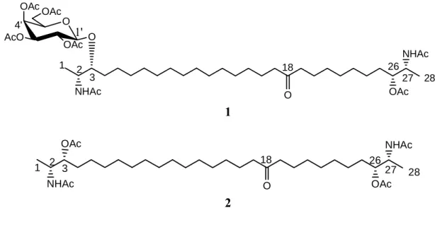
Figure 1. Structures of acetylated compounds 1 and 2.
NHAc O OAc AcO OAc O OAc 2 27 26 3 18 1 28 1' 4' O OAc NHAc 1 NHAc OAc 2 27 26 3 1 28 O 18 OAc NHAc 2 Results and Discussion
The structures of these compounds were determined on the basis of their 1D and 2D NMR spectra and by comparison with literature data [3]. The molecular formula of 1, isolated as pale yellow oil, was established as C48H82O15N2 on the basis of NMR and EI-MS data (m/z 926, M+, calcd 926.5715). The
analysis of the corresponding 1H- and 13C-NMR spectra (Table 1) revealed the presence of a signal characteristic of a lipid chain (envelope of CH2 units) at δH 1.23 and δC 29.1-29.9 ppm. Moreover,
signals at δH 4.48 and δC 100.4 ppm indicated the presence of an acetal function, suggesting the
presence of a sugar. This was confirmed by the chemical shifts of the other carbon atoms at δ 71.0, 69.2, 67.0 and 61.5 ppm along with the respective protons at δ 4.48, 5.04, 3.91, 5.16, 5.39, 4.19 and 4.09 ppm. By comparison with the literature [3, 7], the sugar was identified as a galactose moiety.
Moreover, the coupling constant for the anomeric proton signal H-1’ (δ 4.48, J = 7.9 Hz) indicated that the galactose had a β-configuration.
The analysis of 13C-NMR and DEPT spectra showed the presence of one methylene group bearing an oxygen (δ 61.3 ppm), two methine groups bearing a nitrogen (δ 47.8 ppm) and a carbonyl carbon (δ 211.7 ppm). HMBC correlation between H-1’ and C-3 (δ 82.5 ppm) and COSY correlation between H-1’ (δ 4.48 ppm) and H-3 (δ 3.48 ppm) indicated that the galactopyranosyl group was located at C-3. Moreover, the analysis of COSY and HMBC spectra showed also the presence of two NH-acetamides at δ 5.82 ppm (d, J = 8.8 Hz) and δ 5.54 ppm (d, J = 8.9 Hz), located at C-2 and C-27, respectively. Interpretation of the COSY and HMBC data revealed the presence of two aglycon terminal chains: -CH(O-)CH(NHAc)-CH3 and -CH(OAc)-CH(NHAc)-CH3. This was corroborated by the chemical
shifts of carbon atoms C-1 (CH3, δ 18.5), C-2 (CH-N, δ 46.83), C-3 (CH-O, δ 82.62), C-26 (CH-Oac, δ
76.43), C-27 (CH-N, δ 47.8) and C-28 (CH3, δ 18.5) ppm. The HMBC spectrum showed a correlation
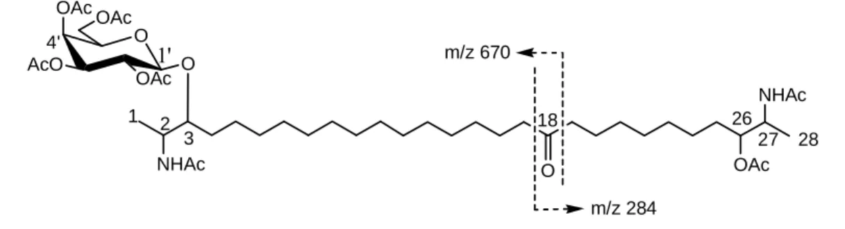
between the H-19 protons (δ 2.38) and the carbon atom at δ 211.5 ppm, suggesting the presence of a carbonyl group. The analysis of the mass spectrum confirmed the localization of the carbonyl group at C-18, as suggested by the fragmentations on both sides of the ketone function observed at m/z 670, corresponding to C34H56NO12, and at m/z 284, corresponding to C15H26NO4 [2, 3] (Figure 2).
Moreover, the chemical shifts of the two CH2 groups (δH 2.38, t, J = 7.2 Hz; δC 42.83 (C-19) and 42.74
ppm (C-17)) α to the ketone carbon were readily assigned by comparison with the literature data [3]. Thus compound 1 was identified as rhizochalin peracetate.
Figure 2. MS Fragmentation of rhizochalin peracetate 1.
NHAc O OAc AcO OAc O OAc 2 27 26 3 18 1 28 1' 4' O OAc NHAc m/z 670 m/z 284
The structure of compound 2, obtained as a brown oil, was established by comparison of the 1- and 2-D NMR (Table 2) spectra of 2 with literature data [3, 9] and those of rhizochalin peracetate (1). The analysis of the NMR spectrum showed clearly the absence of the sugar seen in compound 1. Aside from this difference, the NMR spectra are quite close. Again, the MS fragmentations at m/z 382 and m/z 284 indicated the presence of a carbonyl located at C-18. Thus, compound 2 corresponds to the peracetyl aglycone of 1, in agreement with the literature [3].
This is the first report of these compounds in the sponge Oceanapia ramsayi. These compounds have been found previously in other investigated Oceanapia spp. (or in its synonym Rhizochalina), such as O. incrustata [3], O. bartschi [4], O. philipensis [9], as well as an unidentified Oceanapia spp. [2, 5-7]. The common occurrence of these two compounds suggests that they might be good chemotaxonomic markers for this genus.
Sphingolipids seem to play an important role in cellular regulation. These compounds are found within the cell membranes of all living organisms. They have a protection role in the latter against
Molecules 2008, 13 775
infection [8]. Two-headed sphingolipids from marine sponges are striking because of their rare α,ω-bifunctionalized structures and high biological activity. Since the discovery of rhizochalin, the first member of this series, only two other compounds have been isolated, which are oceanapiside [9] and the calyxoside [10]. Moreover, each one of these compounds possesses a conserved number of carbon atoms (24) in the long chains between the functionalized termini. It suggests that these compounds are derived from a similar lipid carboxyl precursor.
Table 1. 1H- (500 MHz) and 13C- (100 MHz) NMR of rhizochalin peracete (1) in CDCl3 a.
Position δC (mult.) δH (mult., J in Hz)
1 18.45 (q) 1.19 (d, 6.7) 2 46.83 (d) 4.09 (m) 3 82.45 (d) 3.48 (m) 4 31.51 (t) 1.58 (m) 5 25.18 (t) 1.57 (m) 6-15 29.09-29.83 (t) 1.23 (brs) 16 23.88 (t) 1.58 (m) 17 42.72 (t) 2.38 (t, 7.2) 18 211.70 (s) – 19 42.83 (t) 2.38 (t, 7.2) 20 23.41 (t) 1.58 (m) 21-24 29.09-29.83 (t) 1.23 (brs) 25 31.54 (t) 1.58 (m) 26 76.43 (d) 4.85 (m) 27 46.83 (d) 4.21 (ddd, 9.0 ; 4.3; 6.9) 28 18.45 (q) 1.16 (d, 6.7) NH-C2 5.82 (d, 8.8) NH-C27 5.54 (d, 8.9) 1' 100.43 (d) 4.48 (d, 7.9) 2' 69.21 (d) 5.16 (dd, 10.5, 7.9) 3' 70.69 (d) 5.04 (dd, 10.1 ; 3.4) 4' 67.94 (d) 5.39 (brd, 3.5) 5' 70.69 (d) 3.91 (t, 6.7) 6'a 6'b 61.28 (t) 4.19 (m) 4.09 (m) a in CDCl 3; δ ppm. (For Ac, δH 2.00-2.20; δCO 169.5-170.4 ppm)
Table 2. 1H- (500 MHz) and 13C- (100 MHz) NMR of peracetylaglycone 2 a. Position δC (mult.) δH (mult., J in Hz)
1 19.73 (q) 0.96 (d, 6.7) 2 50.79 (d) 3.97 (m) 3 74.69 (d) 4.74 (m) 4 35.00 (t) 1.41 (m) 5 30.00 (t) 1.41 (m) 6-15 28.50-34.70 (t) 1.22 (brs) 16 28.33 (t) 1.41 (m) 17 46.79 (t) 2.36 (t, 7.1) 18 211.90 (s) 19 46.79 (t) 2.36 (t, 7.1) 20 28.33 (t) 1.41 (m) 21-24 28.50-34.70 (t) 1.22 (brs) 25 35.00 (t) 1.41 (m) 26 74.69 (d) 4.74 (m) 27 51.34 (d) 3.97 (m) 28 19.73 (q) 0.96 (d, 6.7) NH-C2 7.69 (d, 8.9) NH-C27 7.50 (d, 7.9)
a in MeOD; δ ppm. (For Ac, δ
H 2.00-2.20; δCO 169.5-170.4 ppm)
The absolute configurations of rhizochalin and its aglycone (threo, threo) differ from those obtained for calyxoside and oceanapiside (erythro, threo) as well as those obtained for the sphingolipids with methyl termini such as the fumonisins B1 and B2 described by Branham and Plattner [11]. These authors propose that at least two independent amino acid fatty acyl transferases, or homologous subunits in the same enzyme are operative in the biosynthesis of dimeric sphingolipids in marine sponge, one incorporating alanine with 2R,3R stereoselectivity and the second incorporating serine with 2S,3S stereoselectivity [12].
Experimental
General
1H, 13C, COSY, HSQC and HMBCNMRspectrawere recorded on a Bruker ARX-500 instrument
using standard Bruker pulse sequences.
Biological Material
A specimen of the sponge Oceanapia ramsayi (Lendelfeld, 1888; phylum Porifera, class Demospongiae, order Haplosclerida, family Phloeodictyidae) was collected at Itampolo (west coast of
Molecules 2008, 13 777
Madagascar Island) in 2005. It is a massive sponge, red in life, which displays numerous fistules. A voucher specimen is deposited in the Museum d’Histoire Naturelle de Marseille (MHNM, n°15830.0).
Extraction and Isolation
The crude extract (1.99 g), obtained by extraction of the sponge with CHCl3/MeOH (1:1) at room
temperature, was chromatographed over a Sephadex (LH-20) column, eluted with n-heptane/CHCl3/MeOH mixtures of increasing polarity. Among the five fractions recovered, fractions 2
and 3, obtained with n-heptane/CHCl3/MeOH (40:35:25), were acetylated in order to afford
compounds 1 (20 mg) and 2 (15 mg), respectively.
Acknowledgements
We are grateful to Prof. Y. Kashman and Dr. A. Rudi for the MS spectral data measurements.
References and Notes
1. Desqueyroux-Faùndez, R.; Valentine, C. Family Phloedictyidae Carter, 1882: In Systema
Porifera: A guide to the Classification of Sponges; Hooper J.N.A.; van Soest R.W.M., (eds.);
Kluwer Academic/Plenum Publishers: New York, 2002; pp. 893-905.
2. Makarieva, T.; Denisenko, V.; Dmitrenok, P.; Guzii, A.; Santalova, E.; Stonik, V.; MacMillan, J.; Molinski, T. Oceanalin A, a hybrid α, ω-bifunctionalized sphingoid tetrahydroisoquinoline β-glycoside from the marine sponge Oceanapia sp. Org. Lett. 2005, 7, 2897-2900.
3. Makarieva, T.N.; Denisenko,V.A.; Stonik, V.A. Rhizochalin, a novel secondary metabolite of mixed biosynthesis from the sponge Rhizochalina incrustata. Tetrahedron Lett. 1989, 30, 6581-6584.
4. Cafieri, F.; Fattorusso, E.; Mahajnah, Y.; Mangoni A. 6-Bromo-5-hydroxy-3-indole-carboxyaldehyde from the Caribbean sponge Oceanapia bartschi. Chem. Sci. 1993, 48, 1408-1410.
5. Boyd, K.G.; Harper, M.K.; Faulkner, D.J. Oceanapamine, a sesquiterpene alkaloid from the Philippine sponge Oceanapia sp. J. Nat. Prod. 1995, 58, 302-305.
6. Mancini, I.; Guella, G.; Debitus, C.; Pietra, F. Oceanapins A-F, unique branched ceramides isolated from the haplosclerid sponge Oceanapia cf. tenuis of the Coral Sea. Helv. Chim. Acta
1994, 77, 51-58.
7. Nicholas, G.M.; Newton, G.L.; Fahey, R.C.; Bewley, C.A. Novel bromotyrosine alkaloids: inhibitors of mycothiol S-conjugate amidase. Org. Lett. 2001, 3, 1543-1545.
8. Shier, W.T.; Shier, A.C. Sphingosine-and ceramide-analog toxins-an update. J. Toxicol. (Toxin
Rev.) 2000, 19, 189-246.
9. Nicholas, G.M.; Hong, T.W.; Molinski, T.F.; Lerch, M.L.; Cancilla, M.T.; Lebrilla, C.B. Oceanapiside, an antifungal bis-alpha,omega-amino alcohol glycoside from the marine sponge
10. Zhou, B.N.; Mattern, M.P.; Johnson, R.K.; Kingston, D.G.I. Structure and stereochemistry of a novel bioactive sphingolipid from a Calyx sp. Tetrahedron 2001, 57, 9549-9554.
11. Branham, B.E.; Plattner, R.D. Alanine is the precursor in the biosynthesis of fumonisin B1 by cultured Fusarium moniloforme. Mycopathologia 1993, 124, 99-104.
12. Molinski, T.F.; Makarieva, T.M.; Stonik, V.A. (-)-Rhizochalin is a dimeric enantiomorphic (2R)-sphingolipid: absolute configuration of pseudo-C2v-symmetric bis-2-amino-3-alkanols by CD.
Angew. Chem. Int. Ed. 2000, 39, 4076-4079.
Sample Availability: Samples of compounds 1 and 2 are available from the authors.