HAL Id: hal-01307224
https://hal-amu.archives-ouvertes.fr/hal-01307224
Submitted on 28 Apr 2016
HAL is a multi-disciplinary open access
archive for the deposit and dissemination of
sci-entific research documents, whether they are
pub-lished or not. The documents may come from
L’archive ouverte pluridisciplinaire HAL, est
destinée au dépôt et à la diffusion de documents
scientifiques de niveau recherche, publiés ou non,
émanant des établissements d’enseignement et de
Decreased Probability of Initial Pain Cessation in
Classic Trigeminal Neuralgia Treated With Gamma
Knife Surgery in Case of Previous Microvascular
Decompression: A Prospective Series of 45 Patients
With >1 Year of Follow-up.
Constantin Tuleasca, Romain Carron, Noémie Resseguier, Anne Donnet, P
Roussel, Jean Gaudart, Marc Levivier, Jean Régis
To cite this version:
Constantin Tuleasca, Romain Carron, Noémie Resseguier, Anne Donnet, P Roussel, et al..
De-creased Probability of Initial Pain Cessation in Classic Trigeminal Neuralgia Treated With Gamma
Knife Surgery in Case of Previous Microvascular Decompression:
A Prospective Series of 45
Patients With >1 Year of Follow-up..
Neurosurgery, Lippincott, Williams & Wilkins, 2015,
�10.1227/NEU.0000000000000739�. �hal-01307224�
Decreased Probability of Initial Pain Cessation in
Classic Trigeminal Neuralgia Treated With Gamma
Knife Surgery in Case of Previous Microvascular
Decompression: A Prospective Series of 45 Patients
With
.1 Year of Follow-up
BACKGROUND: Microvascular decompression (MVD) is the reference technique for pharmacoresistant trigeminal neuralgia (TN).
OBJECTIVE: To establish whether the safety and efficacy of Gamma Knife surgery for recurrent TN are influenced by prior MVD.
METHODS: Between July 1992 and November 2010, 54 of 737 patients (45 of 497 with .1 year of follow-up) had a history of MVD (approximately half also with previous ablative procedure) and were operated on with Gamma Knife surgery for TN in the Timone University Hospital. A single 4-mm isocenter was positioned in the cisternal portion of the trigeminal nerve at a median distance of 7.6 mm (range, 3.9-11.9 mm) anterior to the emergence of the nerve. A median maximum dose of 85 Gy (range, 70-90 Gy) was delivered.
RESULTS: The median follow-up time was 39.5 months (range, 14.1-144.6 months). Thirty-five patients (77.8%) were initially pain free in a median time of 14 days (range, 0-180 days), much lower compared with our global population of classic TN (P = .01). Their actuarial probabilities of remaining pain-free without medication at 3, 5, 7, and 10 years were 66.5%, 59.1%, 59.1%, and 44.3%. The hypoesthesia actuarial rate at 1 year was 9.1% and remained stable until 12 years (median, 8 months).
CONCLUSION: Patients with previous MVD showed a significantly lower probability of initial pain cessation compared with our global population with classic TN (P = .01). The toxicity was low (only 9.1% hypoesthesia); furthermore, no patient reported bothersome hypoesthesia. However, the probability of maintaining pain relief without medication was 44.3% at 10 years, similar to our global series of classic TN (P = .85).
KEY WORDS: Gamma knife surgery, Previous MVD, Recurrent trigeminal neuralgia
Neurosurgery 77:87–95, 2015 DOI: 10.1227/NEU.0000000000000739 www.neurosurgery-online.com
T
rigeminal neuralgia (TN) is a very disabling facial pain disorder. Several surgical meth-ods, including balloon microcompression, percutaneous radiofrequency, percutaneous glyc-erol rhizotomy, microvascular decompression (MVD), and Gamma Knife surgery (GKS), are the possible options to discuss when pharmaco-logical treatment fails.Several articles address the issue of the safety and efficacy of MVD after$1 GKS treatments.1,2To the best of our knowledge, there are no studies discussing the role of GKS as a retreatment for recurrent TN after a previous MVD.
GKS for TN was introduced by the Swedish neurosurgeon Lars Leksell3 and further refined by several others.4-12 GKS is currently consid-ered the least invasive treatment with the lowest rate of adverse effects and a similar rate of initial efficacy.11,13
MVD of the trigeminal nerve at the root entry zone was initially carried out on the basis of the
Constantin Tuleasca, MD*‡§¶k Romain Carron, MD, PhD* Noe´mie Resseguier, MD, MSc# Anne Donnet, MD** Philippe Roussel, MD** Jean Gaudart, MD, PhD# Marc Levivier, MD, PhD¶k Jean Re´gis, MD*
*Functional and Stereotactic Neurosurgery Unit, Centre Hospitalier Universitaire La Timone, Assistance Publique-Hopitaux de Marseille, Universite´ de la Me´diterrane´e, INSERM U 751, Marseille, France;‡Signal Processing Laboratory (LTS 5), Swiss Fed-eral Institute of Technology, Lausanne, Switzerland; §Medical Image Analysis Lab-oratory, Centre Hospitalier Universitaire Vaudois, Lausanne, Switzerland; ¶Centre Hospitalier Universitaire Vaudois, Depart-ment of Clinical Neurosciences, Neurosur-gery Service and Gamma Knife Center, Lausanne, Switzerland; kUniversity of Lausanne, Faculty of Biology and Medicine, Lausanne, Switzerland; #Department of Public Health and Medical Information, Centre Hospitalier Universitaire La Timone Assistance Publique-Hopitaux de Marseille, UMR 912 (INSERM-IRD-Universite´ de la Me´diterrane´e), Marseille, France; **Depart-ment of Neurology, Clinical Neuroscience Federation, Centre Hospitalier Universi-taire La Timone Assistance Publique-Hopitaux de Marseille, Marseille, France Correspondence:
Constantin Tuleasca, MD, MD-PHD Candidate, Lausanne University Hospital, Neurosurgery Service and Gamma Knife Center, Rue de Bugnon 44-46, BH-08, CH-1011, Lausanne, Switzerland. E-mail: constantin.tuleasca@gmail.com Received, September 23, 2014. Accepted, February 16, 2015. Published Online, March 23, 2015. Copyright© 2015 by the Congress of Neurological Surgeons.
ABBREVIATIONS: BNI,Barrow Neurological Institute;
CI,confidence interval;CTN,classic trigeminal neural-gia;GKS,Gamma Knife surgery;HR,hazard ratio;MVD,
microvascular decompression;TN,trigeminal neuralgia
observations made by Dandy14 with a technique developed by Gardner and Miklos and further perfected by Janetta.15Although frequently accepted as a first-line option, its failure rates vary between 15% and 35%, as reported in studies with long-term follow-up.16,17Additionally, MVD is sometimes attempted in the absence of a clear vascular compression and consequently without necessarily an objective compression of the trigeminal root.18
The purpose of the present study was to establish on a long-term follow-up whether the results in terms of safety and efficacy in patients treated with GKS for recurrent, classic TN (CTN) could be affected by prior MVD treatment. The population of recurrent TN patients with prior MVD was deemed especially relevant because MVD is regarded as the reference technique. Moreover, although the out-comes of MVD after previous GKS are well established,1,2,5,19-24 outcomes of GKS after previous MVD have not been reported so far.
METHODS
Type of Study
The study was designed as open, self-controlled, noncomparative, and prospective. For all patients, a case report form was created and prospectively completed. All patients were examined before treatment, and a magnetic resonance imaging (MRI) was done (the latest to exclude any secondary cases). Ethics committee (CPPRB1) permission was obtained for this study.
Diagnostic Criteria Using the International Headache Society Definition
All patients fulfilled the criteria of the International Headache Society.25The type of trigeminal pain was evaluated according to the
classification proposed by Eller et al,26comprising idiopathic TN1 and TN2. TN1 is described as typically sharp, shooting, and electric shock– like pain with pain-free intervals between the attacks that is present
.50% of the time; TN2 is described as an aching, throbbing, or burning pain that is present for .50% of the time and is constant in nature (constant background pain being the most significant attribute). Only patients fulfilling the criteria of the TN1 type were included.
Fifty-four patients (7.3%) from our global series of 737 patients had undergone a previous MVD. We further analyzed 45 patients with.1 year of follow-up. All patients with a history of multiple sclerosis27or megadolichobasilar artery compression28were excluded from the present analysis (reputed to have more variable results). The mean time of GKS after MVD was 67.7 months (range, 6-326 months).
Brief Description of GKS Technique
All patients underwent GKS. After application of the Leksell Model G stereotactic frame (Elekta Instruments AB, Stockholm, Sweden) under local anesthesia, all patients underwent stereotactic MRI (unless there were formal contraindications) and computed tomography for target definition. The MRI sequences used to identify the trigeminal nerve were T2-type constructive interference in steady state/fast imaging employing steady-state acquisition (Siemens; 0.5-mm thickness) without contrast and contrast-enhanced T1-weighted images. Bone computed tomogra-phy routinely supplements the neuroimaging investigation to correct any distortion errors on the MRIs.10,12
Between July 1992 and November 2010, models B, C, and 4C of the Gamma Knife (Elekta Instruments AB) were used successively.
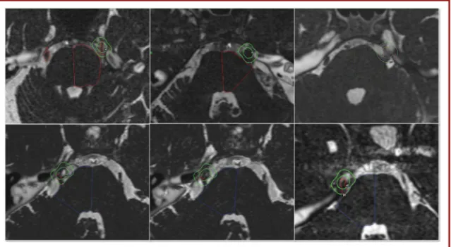
A single 4-mm isocenter was used for all 45 patients (100%) and was positioned in the cisternal portion of the trigeminal nerve at a median distance of 7.6 mm (range, 3.9-11.9 mm) anterior to the emergence of the nerve. This target had been used in our center since the beginning of GKS treatments for TN, as detailed in previous studies.10,11,29The placement of the target depended on the individual anatomy of the patient and on the local neurovascular conditions. Because these patients already had a previous MVD procedure, sometimes the trigeminal nerve turned out to be more difficult to identify (eg, the presence of the Teflon felt and postoperative changes; Figure 1).
FIGURE 1.Six different examples of targeting in previous microvascular decompression cases.
A median maximum dose of 85 Gy (range, 70-90 Gy) was delivered. The dose had been chosen according to the multicentric trial of Kondziolka et al30(which also included an important number of patients from our center), which recommended a minimal dose of 70 Gy for short- and long-term efficacy. According to the aforementioned findings, we initially prescribe a dose of 90 Gy at the 100% isodose. Beam channel blocking was used, depending on the dose received by 10 mm3of the brainstem. If this
dose was found to exceed 15 Gy, we scaled down the dose and then started plugging to avoid irradiating a longer length of the treated nerve, which has been previously shown to dramatically increase toxicity (the so-called Flickinger effect).7
All patients were operated on with GKS by the senior neurosurgeon (J.R.).
Follow-up Monitoring
Initial follow-up was based on clinical evaluation at regular intervals of 3 months, 6 months, and 1 year after treatment and on a yearly basis thereafter. All patients have been thoroughly examined by the authors for proper evaluation of safety and efficacy, including facial sensory testing, corneal reflex, and jaw motility. For long-term update of the follow-up, telephone interview was considered acceptable for patients unable to come to the outpatient clinic because of either distance or general health-related conditions.
The patients and referring physician were instructed to pursue the preoperative medication for at least 1 month after GKS and then were allowed to progressively taper the drug doses in cases of pain freedom.
Every clinical evaluation made by our medical team during the course of follow-up was reported prospectively in the database to have continuous and prospective up-to-date information.
The 15 items of essential data as given by Zakrzewska and Thomas31for the articles reporting outcomes of surgical treatment of TN were followed and are presented hereafter.
Explicit Definitions of Outcome Measures
Outcome measures included the existence of initial pain cessation, occurrence and timing of any sensory disturbance, recurrence, and recurrence without further surgery.
Efficacy was classified according to the Barrow Neurological Institute (BNI) scale (class I, no trigeminal pain, no medication; class II, occasional pain not requiring medication; class IIIa, no pain, continued medication; class IIIb, persistent pain controlled with medication; class IV, some pain not adequately controlled with medication; class V, severe pain/no pain relief; grade I-IIIa, significant pain relief; grade I-IIIb, adequate pain relief; and grade IV and V are failures).10 A successfully treated patient needed to be pain free without medication (BNI class 1).
The degree of hypoesthesia was reported with the use of the BNI facial hypoesthesia scale32 (class I, no facial numbness; class II, mild facial numbness, not bothersome; class III, facial numbness, somewhat bothersome; class IV, facial numbness, very bothersome). Corneal reflex was assessed for all patients. Additionally, the appearance or not of dysesthesias, paresthesias, anesthesia dolorosa, masseter muscle weakness, neurological complications outside the trigeminal nerve territory, systemic complications, or death was noted.
Recurrence was defined as change from class I to a lower outcome class during the follow-up.
The latency intervals to becoming pain free or developing a recurrence or a sensory disturbance, the date of medication changes, and the date of all surgical procedures were also carefully monitored.
Definition of Recurrence (Minor and Major)
A minor recurrence was defined as well tolerated by the patient (lower frequency and intensity of the pain) and not requiring a new surgical therapy.
A major recurrence was defined as requiring an additional surgical procedure. We use the term initial efficacy when the patient was pain free with or without medication in the first 6 months after the radiosurgery and had no recurrence in the year after the procedure.
Patient Satisfaction
Patient satisfaction was evaluated during the last follow-up. We used a semistructured interview conducted by the GKS team during the follow-up visits. Patients were asked if they regret undergoing radiosurgery and if they would agree to undergo this procedure again. The answers in our semistructured questionnaire included the following: a, no regret, I would undergo radiosurgery again with no hesitation; b, no opinion; and c, I regret having had radiosurgery (and would not have it again).
Details of Follow-up Period (Including Median and Range)
The median follow-up period was 39.5 months (range, 14.1-144.6 months).
Basic Demographic Data
The median age was 56.7 years (range, 28.1-82.4 years).
Pain was slightly predominant on the left side in 26 patients (57.8%) vs on the right side in 19 (42.2%). Only 2 patients (4.4%) had bilateral pain. Pain was mainly distributed in the V2 dermatoma in 17 patients (37.8%), followed by V2 and V3 territory in 12 patients (26.3%); V1, V2, and V3 in 3 patients (6.7%); and V1and V2 in 2 patients (4.4%; Table 1).
TABLE 1.Preoperative Assessment
Variable Patients, n (%) Sex Male 20 (44.4) Female 25 (55.6) Side of pain Right 19 (42.2) Left 26 (57.8) Bilateral 2 (4.4) Pain distribution V2 17 (37.8) V2 and V3 12 (26.3) V1, V2, and V3 3 (6.7) V1 and V2 2 (4.4) V1 0 (0) V3 0 (0) V1 and V3 0 (0) Dead 0 (0)
Preoperative magnetic resonance imaging showed persisting vascular conflict other than
megadolichobasilar compression
In 21 cases (46.7%), there was still a neurovascular conflict on the preoperative stereotactic MRI scan, apart from megadolichobasilar artery compression.
Data Regarding Previous Treatments (Including Number and Types of Previous Procedures, Resulting Sensory Deficits, or Constant Background Pain if the Case)
All patients had a past history of surgery, with at least 1 previous MVD (Table 2). Twenty-five patients (55.5%) had only 1 previous procedure, 8 patients (17.8%) had 2 previous surgeries, and 12 patients (26.7%) had$3 previous surgeries. The preoperative surgical technique used was MVD in 45 patients (100%), radiofrequency lesion in 16 patients (35.6%), balloon microcompression in 7 patients (15.6%), and glycerol rhizotomy in 1 patient (2.2%).
Data of Side Effects Related to Prior Surgical Interventions
Twenty-three patients (51.1%) had preoperative hypoesthesia, which was mild in 21 patients (46.7%) and severe in 2 patients (4.4%). In no patient was preoperative anesthesia present.
Statistical Analysis
All statistical analyses were performed with R software, version 2.12.0 (R Foundation for Statistical Computing, Vienna, Austria). The survival R package was used for survival analysis. For the evaluation of outcomes such as pain free, hypoesthesia, and recurrence, the time to event was estimated by use of the Kaplan-Meier method. A bivariate analysis was then performed to identify predictive factors among the collected variables. For qualitative variables, Kaplan-Meier curves were used to graphically represent survival among the different groups and were compared by use of the univariate log-rank test. For all variables, the effects were estimated and tested by fitting univariate Cox proportional
hazards regression models. Proportionality of hazards was assessed graphically by log cumulative hazard plots. For qualitative variables, the x2test was performed when valid; the Fischer exact test was performed
otherwise. For quantitative variables, the Mann-Whitney test was performed given the number of patients. All tests were 2 sided, and values of P,.05 were judged to be significant.
RESULTS
Initial Pain Cessation
Thirty-five patients (77.8%) experienced initial pain cessation in a median time of 14 days (range, 0-180 days). The initially pain-free (BNI classes I-IIIa) actuarial rate at 14 days and 1, 2, 3, 4, 5, and 6 months was 46.7%, 62.2%, 71.1%, 73.3%, 73.3%, 73.3%, and 77.8%, respectively, attaining at 6 months the flat part of the curve and remaining stable thereafter. Of note, the number of patients at risk at 0.5, 1, 2, 3, 4, 5, and 6 months was 24, 17, 13, 12, 12, 12, and 10, respectively, and the number of events was 21, 7, 4, 1, 0, 0, and 2, respectively.
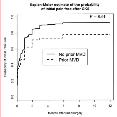
Patients with previous MVD had a significantly lower proba-bility of initial pain cessation compared with our global population of patients with CTN with no history of MVD (P = .01; hazard ratio [HR], 0.64). Figure 2 shows the probability of being pain free in patients with a previous MVD procedure compared with those from our series of 497 patients with CTN. Bilateral pain (P = .65), continuous pain (P = .19), atypical pain (P = .61), and the number of previous surgeries (P = .07 for 1 previous surgery,
TABLE 2.Previous Surgical Interventions and Related Side Effectsa
Variable Patients, n (%) No prior surgery 0 (0) Prior surgeries, n 45 (100) 1 25 (55.5) 2 8 (17.8) $3 12 (26.7)
Type of prior surgery
Radiofrequency lesion 16 (35.6)
Balloon microcompression 7 (15.6)
Microvascular decompression 45 (100)
Glycerol rhizotomy 1 (2.2)
Side effects from prior surgery 24 (53.3)
Facial sensibility before GKS
Normal 22 (48.9)
Slight hypoesthesia 21 (46.7)
Severe hypoesthesia 2 (4.4)
Anesthesia 0 (0)
a
GKS, Gamma Knife surgery.
FIGURE 2. The actuarial probability rate of initial pain cessation in previous microvascular decompression (MVD) cases compared with our classic trigeminal neuralgia global series without previous MVD.
P = .12 for 2 previous surgeries, P = .18 for$3 surgeries) were not shown to be statistical factors.
Patients with both previous MVD and thermocoagulation had a lower probability of pain cessation, but this was not statistically significant (HR, 0.53; 95% confidence interval [CI], 0.25-1.11; P = .08).
Age (P = .11), topography of pain (P = .46), and time lapse to treatment onset (P = .65) also showed no statistically significant correlation.
Postoperative Sensory Assessment and Details of Other Postoperative Complications if the Case
No patient developed an early complication after GKS. Two patients (4.4%) developed delayed facial sensory loss, which occurred in both patients within the first year after the GKS. Hypoesthesia actuarial rate at 1 year was 9.1% and remained stable until 12 years with a median delay of onset of 8 months. Figure 3 shows the probability of hypoesthesia onset after GKS in patients with previous MVD.
Postoperative hypoesthesia was also assessed regarding the BNI facial hypoesthesia scale: mild facial numbness was found in 2 patients (4.4%); facial numbness, somewhat bothersome, 0 pa-tients (0%); and facial numbness, very bothersome in 0 papa-tients (0%). Table 3 shows the postoperative assessment in terms of hypoesthesia, recurrence, and management of recurrence.
The side of the pain in patients with post-GKS hypoesthesia was not a statistically significant correlation (P = 1).
Bilateral pain (P = .04) and previous side effects (P = .04) were associated with a higher incidence of sensory disturbance.
The topography of the pain (P = .7), the number of previous surgeries (P = .3), the persistence of a preoperative neurovascular conflict (P = .2), and sex (P = 1) showed no statistically significant correlations.
No patient developed a trigeminal motor deficit or other cranial nerve deficits after GKS.
Management and Results of Recurrent Pain
Twelve patients (34.3%) from the initially pain-free 35 patients experienced a recurrence in a median time of 31.2 months (range, 3.4-89.9 months).
Additional surgical treatment was performed for 7 patients, with 1 further surgery in 5 patients (11.1%) and 2 additional procedures in 2 cases (4.4%);$3 surgeries were never performed (0%). In our department, the most frequently used was balloon microcompression in 3 patients (6.7%), followed by radio-frequency lesion in 3 patients (6.7%), second MVD in 1 patient (2.2%), second GKS in 1 patient (2.2%), and cortical stimulation and glycerol rhizotomy in 0 patients.
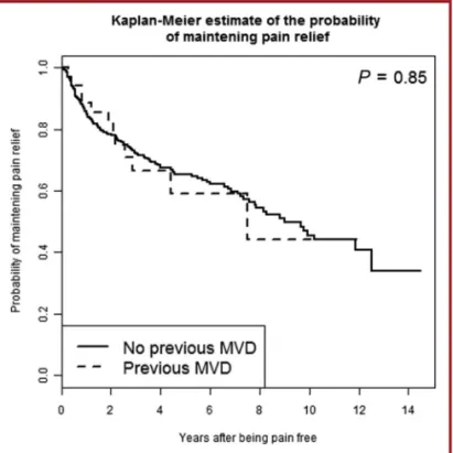
The actuarial probability rates of maintaining pain relief without medication were 88.6%, 66.5%, 59.1%, and 44.3% at 1, 3, 5, and 10 years, respectively; the flat part of the curve was reached at 10 years (Figure 4). Patients with previous MVD had a probability of maintaining pain relief similar to that of our global population of CTN without a previous MVD (P = .85; HR, 1.06).33
FIGURE 3. The actuarial probability of hypoesthesia onset in patients with previous microvascular decompression. GKS, Gamma Knife surgery.
TABLE 3.Postoperative Assessmenta
Variable Patients, n (%) Initially pain free 35 (77.8)
Post GKS sensory dysfunction 2 (4.4)
Mild 2 (100)
Severe 0 (0)
BNI facial hypoesthesia scale (GKS related)
No facial numbness 20 (44.4)
Mild facial numbness 2 (4.4)
Facial numbness, somewhat bothersome 0 (0) Facial numbness, very bothersome 0 (0)
Recurrence of pain 12 in 35 patients (34.3)
Median time to pain recurrence (range), mo 31.21 (range 3.4-89.9)
Additional treatments after GKS 7 (15.5)
1 5 (11.1) 2 2 (4.4) $3 0 (0) Balloon microcompression 3 (6.7) Radiofrequency lesion 3 (6.7) Microvascular decompression 1 (2.2) GKS 1 (2.2) Glycerol rhizotomy 0 (0) Cortical stimulation 0 (0) a
A preoperative pain distributed in the V1 dermatome was related to a much higher probability of recurrence (P = .03; HR, 7.16; 95% CI, 1.19-42.96).
Having$3 previous surgical interventions was associated with a higher probability of maintaining pain relief, but this was not statistically significant (P = .10).
The persistence of a neurovascular conflict despite the previous MVD at the moment of GKS was associated with a higher probability of recurrence (P = .03; HR, 3.97; 95% CI, 1.06-14.89).
The existence of postoperative GKS hypoesthesia onset was not statistically significant (P = .15).
The Probability of Recurrence Not Requiring Further Surgery
Among the 12 patients (34.3%) who experienced a post-GKS recurrence, 7 needed a further surgery. The actuarial probability of recurrence not requiring any further surgery at 1, 3, and 5 years was 94.3%, 80.3%, and 73%, respectively, and remained stable until 12 years.
Sex (P = .5), age (P = .6), post-GKS hypoesthesia onset (P = .9), side of preoperative pain (P = .19), bilateral pain (P = .6), atypical pain (P = .9), and continuous pain (P = .7) were not found to be statistically significant factors.
The presence of preoperative pain within the V1 dermatome was associated with a higher risk of recurrence with further surgery (P = .01; HR, 23.47; 95% CI, 2.11-260.52).
The number of territories involved in the preoperative pain was not statistically significant (P = .06, with the highest rate of recurrence with further surgery in 3 dermatomes involved; HR, 23.47; 95% CI, 2.11-260.52).
Previous MVD Cases vs Previous MVD Plus Another Procedure
Slightly fewer than half of our patients (approximately 45%) had also undergone another intervention. We looked for potential selection bias in this population. We compared mainly the outcomes for previous MVD as unique intervention and previous MVD plus another surgical procedure. The results were not statistically significant and were as follows: for initial pain freedom response, P = .65 (HR, 0.858; 95% CI, 0.43-1.67); for hypoesthesia, P = .27 (HR, 4.74; 95% CI, 0.29-75.9); and for maintaining pain relief, P = .28 (HR = 0.508; 95% CI, 0.14-1.73).
Furthermore, for the group with only previous MVD, 7 patients had preoperative hypoesthesia and 18 (28%) did not. For the group with previous MVD plus another surgical procedure, 16 patients had a preoperative hypoesthesia and 4 did not (80%). This difference is statistically significant (P , .001). However, this point is not relevant because the patients harboring a hypoesthesia before GKS were not analyzed in terms of facial sensory disturbance.
DISCUSSION
To date, several studies have addressed the alleged technical difficulties when MVD is performed after previous GKS.1,2,5,22,24,34,35 To the best of our knowledge, there is currently no study describing GKS after MVD or the dose-planning difficulties regarding the GKS trigeminal nerve target-ing after previous MVD. However, clinicians must cope with this particular situation.
It is known that pain relief tends to occur immediately after MVD and is delayed after GKS. This fact has to be balanced with the postoperative recovery after both MVD (eg, a few weeks) and GKS (eg, usually the next day) techniques. Previous studies have shown a positive association with pain freedom for both MVD and GKS, including the following: absence of multiple sclerosis,4,12,21 absence of a prior surgical procedure,36-38 absence of atypical features,10,11,36,38and blood vessel in contact with the trigeminal nerve on preoperative MRI.39 The rate of pain relief in our present study was 78%, less than reported for CTN general series (including ours)4,6,8-10,12 and less than in the opposite scenario (MVD after prior GKS), as reported by others.1
Hypoesthesia occurrence is similar in both surgical therapies, with GKS carrying a lower risk of the major complications of surgery, including infection, meningitis, cerebellar edema or hemorrhage, cerebrospinal fluid leak, and hearing loss.13,40
Recurrence rates in the long term are similar, with the risk of recurrence continuing as long as patients are followed up.6,8,12,13,16,17,41
FIGURE 4. The actuarial probability of maintaining pain relief in previous microvascular decompression (MVD) cases compared with our classic trigeminal neuralgia global series without previous MVD.
Visualizing the nerve after a prior MVD remains the most problematic issue for GKS. Postoperative displacement of the Teflon, the size of the trigeminal nerve (atrophy or not) and trigeminal cistern, the persistence or not of a neurovascular conflict, and other similar anatomic constraints make the targeting much more challenging than in the classic situations. In these cases, the postoperative result depends on proper targeting; this is a crucial issue for the clinical outcomes both in terms of safety and efficacy.
Limitations
The results presented should be interpreted carefully and may not be generalizable to all patients treated with GKS after previous MVD for recurrent TN. The personalized analysis of the patients, including duration of symptoms, number and type(s) of previous surgical procedure(s), time lapse since the previous surgery (or in particular between prior MVD and GKS), and typical or atypical pain, should also be carefully evaluated. Therefore, our study has several limitations: the different surgical techniques used during previous MVD, the image qualities when GKS was performed (with visualization of the treated nerve frequently being difficult and varying from one patient to another according to previous surgery), local anatomy (small vs large trigeminal cistern, presence of Teflon, etc), and targeting location as a factor affecting the initial efficacy of GKS, depending on individual anatomy. Additionally, other potential limitations reside in the lack of a control arm or the eventual use of a propensity score–matched control group. Some subgroup analysis in the Discussion might also have been underpowered because of the small number of patients and events taken into consideration. The data set might, in this context, not exclude the possibility that patients with 2 procedures (including, for instance, MVD and 1 ablative) could have a worse prognosis than those with prior MVD only.
The message of this study should not be misinterpreted. We are not advocating that GKS should not be performed on patients with previous MVD procedures because of an initial pain relief rate that is lower than in CTN cases. On the contrary, it may be that these patients harbored a particular type of disease that also was resistant to the first MVD procedure. Therefore, in these situations, we must be aware of the fact that these patients may be more difficult to manage surgically. Conversely, studies also showed a decrease in the initial efficacy of MVD after previous destructive procedures.19
The hypoesthesia rates in the present study were low with no bothersome cases. Additionally, the maintenance of pain relief rates over the long term were similar to those already published in our CTN general population.33
We think that patients could benefit more from an initial noninvasive procedure, leaving an open door for those who need further procedures, including MVD and percutaneous techniques. This has to be tailored, of course, to the individual patient and to the baseline assessment.
MVD remains the reference technique for the majority of the surgeons, even in the absence of a prospective randomized
comparative trial with a statistically significant number of patients and an adequate follow-up. No definitive answer can be given to the question of the superiority of one technique over the other. Furthermore, significant controversy exists concern-ing the“ideal” surgical procedure to be applied (initially or as a retreatment) for CTN. MVD is a complex surgical procedure that carries low but significant risks of dreadful complications. That there is usually immediate pain relief and that the procedure directly tackles the supposed cause of the pain by separating the offending vessel (if one exists) from the trigeminal nerve are advantages of MVD. In case of recurrence, however, few neurosurgeons are keen on the idea of going back into a previously operated field, rendering the surgery much more complicated. The present study shows that, in this case, GKS may be extremely helpful with very limited complication rates. However, the significantly lower efficacy rate compared with GKS for CTN without prior MVD has to be kept in mind and explained to the patient, along with the long-term results, which are as good as and comparable to those in our CTN global series.
The majority of authors (among those who have at their disposal the technical and human resources allowing them to do MVD, GKS, or percutaneous techniques) agree that the evidence for the long-term safety and efficacy of GKS is currently sufficient for proposing GKS as a first therapeutic option, following the policy “from less to more invasive.”6,10,42,43Advantages and
disadvan-tages of each technique must be exposed to the patient according to the data provided by modern peer-reviewed series published over the past 20 years.
CONCLUSION
In our experience, in patients with previous MVD, early pain cessation is lower than in our CTN series, which may be related to a more resistant entity of TN. However, long-term results are good and are comparable to those of our CTN global series, which makes this approach of particular interest in this subgroup of patients.
Disclosures
Funding was provided by the Timone University Hospital, Assistance-Publique, Hopitaux de Marseille, France. Being an officer on the board of several scientific societies (president of the European Gamma Knife Surgery Society, vice president of the European Stereotactic and Functional Neurosurgery Society, and past president of the International Radiosurgery Society), Dr Régis has been involved in the quest for sponsorship of congresses for these societies. The major sponsors of these congresses are Accuray, Brainlab, Elekta, Medtronic, and Radionic. This is at the benefit of the societies with no personal benefit. The authors have no personal, financial, or institutional interest in any of the drugs, materials, or devices described in this article.
REFERENCES
1. Huang CF, Chuang JC, Tu HT, Chou MC. Microsurgical outcomes after failed repeated Gamma Knife Surgery for refractory trigeminal neuralgia. J Neurosurg. 2006;105(suppl):117-119.
2. Shetter AG, Zabramski JM, Speiser BL. Microvascular decompression after Gamma Knife surgery for trigeminal neuralgia: intraoperative findings and treatment outcomes. J Neurosurg. 2005;102(suppl):259-261.
3. Leksell L. Stereotaxic radiosurgery in trigeminal neuralgia. Acta Chir Scand. 1971; 137(4):311-314.
4. Brisman R. Gamma Knife radiosurgery for primary management for trigeminal neuralgia. J Neurosurg. 2000;93(suppl 3):159-161.
5. Brisman R. Microvascular decompression vs. Gamma Knife radiosurgery for typical trigeminal neuralgia: preliminary findings. Stereotact Funct Neurosurg. 2007;85(2-3):94-98.
6. Dhople AA, Adams JR, Maggio WW, Naqvi SA, Regine WF, Kwok Y. Long-term outcomes of gamma knife radiosurgery for classic trigeminal neuralgia: implica-tions of treatment and critical review of the literature: clinical article. J Neurosurg. 2009;111(2):351-358.
7. Flickinger JC, Pollock BE, Kondziolka D, et al. Does increased nerve length within the treatment volume improve trigeminal neuralgia radiosurgery? A prospective double-blind, randomized study. Int J Radiat Oncol Biol Phys. 2001;51(2):449-454. 8. Kondziolka D, Zorro O, Lobato-Polo J, et al. Gamma Knife stereotactic radiosurgery
for idiopathic trigeminal neuralgia. J Neurosurg. 2010;112(4):758-765.
9. Massager N, Lorenzoni J, Devriendt D, Desmedt F, Brotchi J, Levivier M. Gamma Knife surgery for idiopathic trigeminal neuralgia performed using a far-anterior cisternal target and a high dose of radiation. J Neurosurg. 2004;100(4):597-605. 10. Regis J, Metellus P, Hayashi M, Roussel P, Donnet A, Bille-Turc F. Prospective
controlled trial of Gamma Knife surgery for essential trigeminal neuralgia. J Neurosurg. 2006;104(6):913-924.
11. Regis J, Tuleasca C. Fifteen years of Gamma Knife surgery for trigeminal neuralgia in the Journal of Neurosurgery: history of a revolution in functional neurosurgery. J Neurosurg. 2011;115(suppl):2-7.
12. Tuleasca C, Carron R, Resseguier N, et al. Patterns of pain-free response in 497 cases of classic trigeminal neuralgia treated with Gamma Knife surgery and followed up for least 1 year. J Neurosurg. 2012;117(suppl):181-188.
13. Cruccu G, Gronseth G, Alksne J, et al. AAN-EFNS guidelines on trigeminal neuralgia management. Eur J Neurol. 2008;15(10):1013-1028.
14. Dandy W. Concerning the cause of trigeminal neuralgia. Am J Surg. 1934:447-455. 15. Jannetta PJ. Microvascular Decompression of the Trigeminal Root Entry Zone: Theoretical Considerations, Operative Anatomy, Surgical Technique, and Results. Baltimore, MD: Williams & Wilkins; 1990.
16. Barker FG II, Jannetta PJ, Bissonette DJ, Larkins MV, Jho HD. The long-term outcome of microvascular decompression for trigeminal neuralgia. N Engl J Med. 1996;334(17):1077-1083.
17. Sindou M, Leston J, Howeidy T, Decullier E, Chapuis F. Micro-vascular decompression for primary trigeminal neuralgia (typical or atypical): long-term effectiveness on pain; prospective study with survival analysis in a consecutive series of 362 patients. Acta Neurochir (Wien). 2006;148(12):1235-1245; discussion 1245. 18. Love S, Coakham HB. Trigeminal neuralgia: pathology and pathogenesis. Brain.
2001;124(pt 12):2347-2360.
19. Barba D, Alksne JF. Success of microvascular decompression with and without prior surgical therapy for trigeminal neuralgia. J Neurosurg. 1984;60(1):104-107. 20. Brisman R. Trigeminal neuralgia: radiosurgery before microvascular
decompres-sion. World Neurosurg. 2012;78(1-2):69-70.
21. Broggi G, Ferroli P, Franzini A, et al. Operative findings and outcomes of microvascular decompression for trigeminal neuralgia in 35 patients affected by multiple sclerosis. Neurosurgery. 2004;55(4):830-838; discussion 838-839. 22. Chen JC. Microvascular decompression for trigeminal neuralgia in patients with and
without prior stereotactic radiosurgery. World Neurosurg. 2012;78(1-2):149-154. 23. Maher CO, Pollock BE. Radiation induced vascular injury after stereotactic
radiosurgery for trigeminal neuralgia: case report. Surg Neurol. 2000;54(2):189-193. 24. Sekula RF Jr, Frederickson AM, Jannetta PJ, Bhatia S, Quigley MR. Microvascular decompression after failed Gamma Knife surgery for trigeminal neuralgia: a safe and effective rescue therapy?J Neurosurg. 2010;113(1):45-52.
25. Society HCSotIH, Headache Classification Subcommittee of the International Headache Society. The International Classification of Headache Disorders. Vol 24(suppl 1). Blackwell Publishing, Cephalalgia, and the International Journal of Headache; 2004.
26. Eller JL, Raslan AM, Burchiel KJ. Trigeminal neuralgia: definition and classification. Neurosurg Focus. 2005;18(5):E3.
27. Tuleasca C, Carron R, Resseguier N, et al. Multiple sclerosis-related trigeminal neuralgia: a prospective series of 43 patients treated with Gamma Knife surgery
with more than one year of follow-up. Stereotact Funct Neurosurg. 2014;92(4): 203-210.
28. Tuleasca C, Carron R, Resseguier N, et al. Trigeminal neuralgia related to megadolichobasilar artery compression: a prospective series of twenty-nine patients treated with Gamma Knife surgery, with more than one year of follow-up. Stereotact Funct Neurosurg. 2014;92(3):170-177.
29. Regis J, Arkha Y, Yomo S, et al. Radiosurgery in trigeminal neuralgia: long-term results and influence of operative nuances [in French]. Neurochirurgie. 2009;55(2): 213-222.
30. Kondziolka D, Lunsford LD, Flickinger JC, et al. Stereotactic radiosurgery for trigeminal neuralgia: a multiinstitutional study using the gamma unit. J Neurosurg. 1996;84(6):940-945.
31. Zakrzewska JM, Thomas DG. Patient’s assessment of outcome after three surgical procedures for the management of trigeminal neuralgia. Acta Neurochir (Wien). 1993;122(3-4):225-230.
32. Rogers CL, Shetter AG, Fiedler JA, Smith KA, Han PP, Speiser BL. Gamma Knife radiosurgery for trigeminal neuralgia: the initial experience of the Barrow Neurological Institute. Int J Radiat Oncol Biol Phys. 2000;47(4):1013-1019. 33. Régis J, Carron R, Tuleasca C, Donnet A. Distal radiosurgical targeting for trigeminal
neuralgia. In: Sheehan J, Gerszten P, eds. Controversies in Stereotactic Radiosurgery: Best Evidence Recommendations. New York, NY: Thieme; 2014:120-130. 34. Linskey ME, Ratanatharathorn V, Penagaricano J. A prospective cohort study of
microvascular decompression and Gamma Knife surgery in patients with trigeminal neuralgia. J Neurosurg. 2008;109(suppl):160-172.
35. Sanchez-Mejia RO, Limbo M, Cheng JS, Camara J, Ward MM, Barbaro NM. Recurrent or refractory trigeminal neuralgia after microvascular decompression, radiofrequency ablation, or radiosurgery. Neurosurg Focus. 2005;18(5):e12. 36. Maesawa S, Salame C, Flickinger JC, Pirris S, Kondziolka D, Lunsford LD.
Clinical outcomes after stereotactic radiosurgery for idiopathic trigeminal neuralgia. J Neurosurg. 2001;94(1):14-20.
37. Pollock BE, Phuong LK, Gorman DA, Foote RL, Stafford SL. Stereotactic radiosurgery for idiopathic trigeminal neuralgia. J Neurosurg. 2002;97(2):347-353. 38. Young RF, Vermulen S, Posewitz A. Gamma Knife radiosurgery for the treatment of trigeminal neuralgia. Stereotact Funct Neurosurg. 1998;70(suppl 1):192-199. 39. Brisman R, Khandji AG, Mooij RB. Trigeminal nerve-blood vessel relationship as
revealed by high-resolution magnetic resonance imaging and its effect on pain relief after Gamma Knife radiosurgery for trigeminal neuralgia. Neurosurgery. 2002;50 (6):1261-1266; discussion 1266-1267.
40. Gronseth G, Cruccu G, Alksne J, et al. Practice parameter: the diagnostic evaluation and treatment of trigeminal neuralgia (an evidence-based review): report of the Quality Standards Subcommittee of the American Academy of Neurology and the European Federation of Neurological Societies. Neurology. 2008;71(15):1183-1190. 41. McLaughlin MR, Jannetta PJ, Clyde BL, Subach BR, Comey CH, Resnick DK. Microvascular decompression of cranial nerves: lessons learned after 4400 operations. J Neurosurg. 1999;90(1):1-8.
42. Kondziolka D, Lunsford L, Flickinger J. Gamma Knife radiosurgery as the first surgery for trigeminal neuralgia. Stereotact Funct Neurosurg. 1998;70(suppl 1):187-191. 43. Kondziolka D, Zorro O, Lobato-Polo J, et al. Gamma Knife stereotactic radiosurgery
for idiopathic trigeminal neuralgia. J Neurosurg. 2010;112(4):758-765.
COMMENTS
D
espite multiple safe and effective treatments for idiopathic trigeminal neuralgia (TN), the incidence of persistent or recurrent pain in a significant subset of patients after intervention remains a vexing prob-lem. The authors report their cohort of patients with TN who underwent Gamma Knife radiosurgery after having a previous microvascular decompression. After radiosurgery, 78% of patients experienced initial pain relief, and 44% maintained long-term pain relief without medica-tion. Although this is not a true case-control study, this patient group (45 patients with at least 1 year of follow-up) was retrospectively compared with their center’s large cohort of TN Gamma Knife patients (n = 497). Although the rate of initial pain relief was lower in this cohort than in their global population of TN Gamma Knife patients, the long-term outcomes are quite similar. They also found no difference in outcome between the microvascular decompression (MVD) patients who hadMVD only before Gamma Knife and those who had additional pro-cedures. Furthermore, Gamma Knife radiosurgery after MVD was well tolerated, with no increased incidence of complications.
The authors clearly state their own treatment bias, namely that less invasive techniques such as Gamma Knife radiosurgery should now be offered as first-line therapy. Although this article provides no evidence to bolster this claim, at the very least, it provides evidence that when pain persists or recurs after MVD, radiosurgery is a safe and appropriate treatment option.
Alon Y. Mogilner New York, New York
T
he authors have presented a case series of patients who underwent Gamma Knife radiosurgery for recurrent trigeminal neuralgia after microvascular decompression (and sometimes other ablative proce-dures as well). In general, their results are promising, with 78% ofpatients becoming pain free at some point. However, like with other studies of ablative procedures, 34% of patients here had recurrent pain at a median time of 31 months after Gamma Knife radiosurgery. The authors also note that their long-term outcome in this group was similar to that of patients undergoing Gamma Knife radiosurgery as primary treatment.
This series helps to reinforce that, for the most part, the choice of surgical procedure for trigeminal neuralgia often should be best left up to the patient when several options would suffice (such as Gamma Knife radiosurgery vs microvascular decompression vs radiofrequency ablation or pencil beam convolution). Trigeminal neuralgia continues to be a challenging clinical entity, and the ability to provide patients with more autonomy in treatment selection, knowing that long-term outcomes may eventually be similar, is important.
Joshua Rosenow Chicago, Illinois