HAL Id: hal-03095902
https://hal.archives-ouvertes.fr/hal-03095902
Submitted on 2 Feb 2021
HAL is a multi-disciplinary open access
archive for the deposit and dissemination of
sci-entific research documents, whether they are
pub-lished or not. The documents may come from
teaching and research institutions in France or
abroad, or from public or private research centers.
L’archive ouverte pluridisciplinaire HAL, est
destinée au dépôt et à la diffusion de documents
scientifiques de niveau recherche, publiés ou non,
émanant des établissements d’enseignement et de
recherche français ou étrangers, des laboratoires
publics ou privés.
Foxp3+ CD25+ regulatory T cells specific for a
neo-self-antigen develop at the double-positive thymic
stage
Julie Cabarrocas, Cécile Cassan, Fay Magnusson, Eliane Piaggio, Lennart
Mars, Jens Derbinski, Bruno Kyewski, David-Alexandre Gross, Benoit
Salomon, Khashayarsha Khazaie, et al.
To cite this version:
Julie Cabarrocas, Cécile Cassan, Fay Magnusson, Eliane Piaggio, Lennart Mars, et al.. Foxp3+
CD25+ regulatory T cells specific for a neo-self-antigen develop at the double-positive thymic stage.
Proceedings of the National Academy of Sciences of the United States of America , National Academy
of Sciences, 2006, 103 (22), pp.8453-8458. �10.1073/pnas.0603086103�. �hal-03095902�
Foxp3
ⴙ
CD25
ⴙ
regulatory T cells specific for a
neo-self-antigen develop at the double-positive
thymic stage
Julie Cabarrocas*†, Ce´cile Cassan*†, Fay Magnusson*‡, Eliane Piaggio*, Lennart Mars*, Jens Derbinski§, Bruno Kyewski§,
David-Alexandre Gross¶, Benoit L. Salomon储, Khashayarsha Khazaie‡, Abdelhadi Saoudi*, and Roland S. Liblau*,**
*Institut National de la Sante´ et de la Recherche Me´dicale U563, Purpan Hospital, 31000 Toulouse, France;‡Dana–Farber Cancer Institute, Harvard Medical
School, Boston, MA 02115;§Division of Developmental Immunology, German Research Center, D-69120 Heidelberg, Germany;¶Centre National de la
Recherche Scientifique Unite´ Mixte de Recherche 8115, Genethon, 91002 Evry, France; and储Centre National de la Recherche Scientifique Unite´ Mixte de Recherche 7087, Pitie´-Salpeˆtrie`re Hospital, 75013 Paris, France
Communicated by Hugh O. McDevitt, Stanford University School of Medicine, Stanford, CA, April 18, 2006 (received for review November 21, 2005)
Thymus-derived regulatory T cells (Tregs) expressing CD4, CD25, and the transcription factor Foxp3 play major roles in preventing autoimmunity. The Treg population is enriched in T cells expressing high-avidity self-reactive T cell receptors, and thymic epithelial cells expressing self-antigens (Ag) have been implicated in their induc-tion and兾or selection. However, the thymic selection events lead-ing to Treg lineage commitment remain unclear. We followed the thymic development of self-Ag-specific Tregs in double-transgenic mice coexpressing a neo-self-Ag, hemagglutinin (HA) under the control of a neural tissue-specific promoter, and a transgenic class II-restricted T cell antigen receptor specific for HA111-119. Our data show that the promiscuous expression of the HA transgene in thymic epithelial cells is involved in the selective induction and兾or
expansion of HA-specific Foxp3ⴙTreg thymic precursors as early as
the double-positive stage.
autoimmunity兩 immune tolerance 兩 nervous system
P
otentially autoaggressive T cells can develop because of the stochastic generation of diversity in the T cell repertoire. However, there are several central and peripheral mechanisms that maintain self-tolerance. These mechanisms include thymic clonal deletion, which is a major mechanism of tolerance induc-tion that results in thymocytes expressing high-affinity self-reactive T cell antigen receptors (TCRs) being physical elimi-nated. Specialized T cell subsets able to suppress immune responses are also generated in the thymus, where they acquire their phenotype and suppressive function. Among these sub-populations, a CD4 T cell subset constitutively expressing the IL-2 receptor␣ chain, CD25, plays a crucial role in preventing autoimmunity (1, 2). CD4⫹CD25⫹ regulatory T cells (Tregs) represent between 2% and 10% of the peripheral CD4 T cells in mice, rats, and humans (3). In vitro studies have shown that upon TCR stimulation, Tregs can inhibit the proliferative response of naive CD8 or CD4 T cells specific for the same or different MHC–peptide complexes (1, 2). Tregs also inhibit a wide variety of immune responses in vivo (2).Tregs constitutively express high levels of CTLA-4, GITR, and the transcription factor Foxp3 (4, 5). Foxp3 expression is essen-tial for Treg lineage specification and for Treg function. How-ever, the events leading to Foxp3 expression in developing thymocytes are largely unknown. Tregs express a diverse TCR␣ repertoire, and this cell population is enriched with cells ex-pressing self-reactive TCRs (2, 6). Recent data suggest that Treg differentiation is promoted by high-affinity interactions with thymic antigen-presenting cells (APCs) presenting self-peptide– MHC complexes (7, 8). Thymic epithelial cells (TECs) and, in particular, cortical TECs are involved in Treg selection, and Treg differentiation requires MHC class II expression (7-10). The array of self-antigens (Ags) expressed by thymic APCs, most notably by TECs, has been shown to include a variety of
tissue-restricted proteins (11). This promiscuous expression of tissue-restricted self-Ags by thymic APCs is, at least partly, controlled by transcription regulators such as AIRE (12) and has been shown to play a role in clonal deletion (13). An increase in the proportion of Ag-specific Tregs also has been observed in transgenic (Tg) mice coexpressing a Tg TCR and the specific Ag under the control of tissue-restricted promoters, promiscuously transcribed in radio-resistant thymic cells (14, 15). However, the events involved in the development of self-reactive Tregs remain largely unknown (16). Also, the nature and relative contribution of central mechanisms to tolerance toward neural self-Ags are not well understood (17-19). Here, we show that the promiscuous expression of hemagglutinin (HA), a neo-self-Ag under the control of a nervous system-specific promoter, by thymic APCs induces central tolerance mechanisms, including the differenti-ation of HA-specific thymocytes into CD4⫹CD25⫹Foxp3⫹ Tregs. Our data also strongly suggest that the development of Tregs is promoted by cognate interactions with the neo-self-Ag presented by radio-resistant thymic stromal cells as early as the double-positive (DP) stage.
Results
Immune Tolerance Develops in [Glial Fibrillary Acidic Protein (GFAP)-HAⴛ 6.5-TCR]F1Double-Tg (DTg) Mice.We assessed the mechanisms of CD4 T cell tolerance to a model neural self-Ag. For this purpose, we crossed GFAP-HA Tg mice, in which the GFAP promoter drives expression of the influenza virus HA in astro-cyte-like cells (20), with 6.5-TCR Tg mice, which express on 15-20% of CD4⫹T cells a TCR specific for the I-Ed–HA111-119
complex (21). None of (GFAP-HA⫻ 6.5-TCR)F1DTg mice,
followed for up to 12 months, developed clinical (n⫽ 87) or histological (n⫽ 6; data not shown) lesions of autoimmunity, particularly at HA expression sites, such as the CNS and gut (20). These results are in sharp contrast to the fulminant autoimmune disease that develops when GFAP-HA mice are crossed with CL4-TCR Tg mice, which express an HA-specific TCR on⬇95% of their CD8⫹T cells (20).
We compared the HA-specific CD4 T cells from these (GFAP-HA ⫻ 6.5-TCR)F1 DTg mice with those of 6.5-TCR
single-Tg (STg) littermates to understand the tolerogenic mech-anisms taking place. Using the 6.5 mAb, we found that the
Conflict of interest statement: No conflicts declared.
Abbreviations: Ag, antigen; APC, antigen-presenting cell; BM, bone marrow; DP, double-positive; DTg, (GFAP-HA⫻ 6.5-TCR)F1double-transgenic; GFAP, glial fibrillary acidic
pro-tein; HA, hemagglutinin; rAAV-HA, adeno-associated virus recombinant for HA; SP, CD4⫹
CD8⫺thymocytes; STg, 6.5-TCR single-transgenic; TCR, T cell antigen receptor; TECs, thymic epithelial cells; mTECs, medullary TECs; Tg, transgenic; Tregs, regulatory T cells.
†J.C. and C.C. contributed equally to the work.
**To whom correspondence should be addressed. E-mail: [email protected]. © 2006 by The National Academy of Sciences of the USA
proportion of HA-specific (6.5⫹) cells among peripheral CD4 T cells was much lower in DTg animals (4.2⫾ 0.5% vs. 16.1 ⫾ 1.1% in STg mice; Fig. 1A). In addition, the level of Tg TCR expression was lower on the remaining 6.5⫹CD4⫹T cells (data not shown). Splenic CD4 T cells from DTg mice proliferated much less than CD4 T cells from STg littermates in response to stimulation with the HA111-119 peptide (Fig. 1B), even after normalization for the number of 6.5⫹ cells present in the culture. These results indicate that there are several tolerogenic mechanisms that protect DTg mice from destructive autoimmunity.
HA-Specific CD4ⴙCD25ⴙT Cells with Regulatory Properties Develop in DTg Mice.A large proportion of 6.5⫹CD4⫹splenocytes from DTg mice expressed CD25 (44.4 ⫾ 3.5% vs. 11.9 ⫾ 1.1% in STg littermates; Fig. 1C). Therefore, the absolute numbers of 6.5⫹CD4⫹CD25⫹splenocytes were similar in both DTg and STg mice (0.49⫾ 0.07 ⫻ 106vs. 0.64⫾ 0.14 ⫻ 106cells) despite there
being a large reduction in 6.5⫹T cells in DTg mice. Activation markers such as CD69 (30.0⫾ 4.3% vs. 18.0 ⫾ 2.0%; n ⫽ 5 per group; P⫽ 0.009), and CD44high(52.1⫾ 6.9% vs. 32.1 ⫾ 5.4%; n ⫽
5; P ⫽ 0.059) also were expressed at higher levels on 6.5⫹CD4⫹CD25⫹splenocytes from DTg than on 6.5⫹CD4⫹CD25⫹ splenocytes from STg mice. Lymph node 6.5⫹CD4⫹T cells from DTg mice displayed the same ‘activated’ phenotype (data not shown). In sharp contrast, we observed no differences between DTg and STg mice in the levels of CD25, CD69, and CD44 expressed on 6.5⫺CD4⫹peripheral T cells (Fig. 1C and data not shown). The 6.5⫹CD4⫹CD25⫹ T cells that develop in DTg animals clearly express GITR (Fig. 1D) and intracellular Foxp3 (Fig. 1E), strongly suggesting that they belong to the Treg subset. This surface phenotype is shared by a large subpopulation of Tregs from BALB兾c mice (22) and suggests recent in vivo activation by their cognate Ag.
We observed greater proliferative responses in CD4⫹CD25⫺ T cells from DTg animals than in unfractionated CD4⫹T cells, suggesting that the CD4⫹CD25⫹ fraction exerts suppressive functions (data not shown). Therefore, we carried out coculture experiments to compare the inhibitor y properties of
CD4⫹CD25⫹T cells from DTg mice and non-Tg BALB兾c mice. CD4⫹CD25⫹ cells from both types of donor mice strongly inhibited the anti-CD3 mAb-induced proliferation of naive CD4⫹CD25⫺T cells purified from 6.5-TCR STg mice (Fig. 2A). However, only CD4⫹CD25⫹T cells isolated from DTg donors were able to suppress the proliferation of HA-specific naive T cells upon stimulation with the HA111-119 peptide.
The regulatory potential of HA-specific CD4⫹CD25⫹T cells from DTg mice also was studied in vivo in a model of CD8-mediated rejection of HA gene transfer (23). BALB兾c mice were immunized with the adeno-associated virus recombinant for HA (rAAV-HA), which induces a potent anti-HA immune response. These mice also received 2⫻ 105CD4⫹CD25⫹T cells from DTg,
GFAP-HA Tg, or BALB兾c mice. Only Tregs from DTg mice were able to inhibit IFN-␥ secretion by anti-HA CD8 T cells (Fig. 2B), resulting in a strongly inhibited infiltration of inflammatory cells in muscle and in a sustained local expression of HA (Fig. 2C). Tregs from BALB兾c or GFAP-HA mice were unable to inhibit the immune response against the transduced muscle cells. Collectively, these data show that the 6.5⫹CD4⫹CD25⫹T cells that develop in DTg mice are HA-specific T cells that have regulatory abilities both in vitro and in vivo.
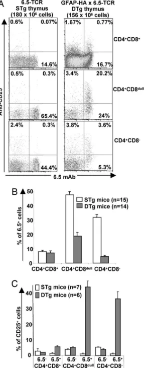
HA-Specific Foxp3ⴙCD25ⴙTregs Develop from the DP Stage Onward only in the Presence of the Agonist Ligand and Independently of Endogenous TCR Rearrangement. GFAP-HA mice express low levels of HA mRNA in the thymus (24). Studies with class II-restricted TCR-Tg mice coexpressing the specific Ag in the thymus have shown that both clonal deletion and Treg differ-entiation of self-reactive thymocytes can occur in the same DTg model (7, 8, 14, 15, 25). However, it is still not known when self-reactive T cells commit to the Treg lineage during thymic ontogeny. Therefore, we compared 6.5⫹thymocytes from DTg and STg littermates at different developmental stages. As shown in Fig. 3 A and B, HA-specific thymocytes were deleted from the CD4⫹CD8dulltransitional stage onward in DTg mice, with the
remaining HA-specific CD4⫹CD8⫺ thymocytes expressing lower levels of the Tg TCR (on average, a 57% reduction versus Fig. 1. Analysis of HA-specific peripheral CD4 T cells from DTg mice. (A) The percentage of splenic CD4⫹T cells from STg and DTg mice expressing the 6.5-TCR was determined by FACS and revealed the deletion of HA-specific CD4 T cells in DTg mice (P⬍ 10⫺7; two-tailed paired Student’s t test). (B) Purified CD4⫹T cells
from STg or DTg mice were stimulated with increasing concentrations of the HA111-119 peptide or anti-CD3 mAb. Results are representative of 10 independent experiments. (C) Percentages of CD25⫹cells among 6.5⫺CD4⫹or 6.5⫹CD4⫹splenocytes from STg and DTg mice. The proportion of 6.5⫹CD4⫹splenocytes expressing CD25 is greatly increased in DTg mice (P⫽ 5 ⫻ 10⫺7). (D and E) Expression of CD25, GITR (D), and intracellular Foxp3 (E) on gated 6.5⫹CD4⫹splenic T cells from mice representative of three STg and five DTg mice.
their STg counterparts). In addition, the proportion of 6.5⫹ thymocytes expressing CD25 was increased at all stages of thymic development, including the early CD4⫹CD8⫹DP stage (Fig. 3 A and C). The absolute numbers of 6.5⫹CD25⫹thymocytes also were significantly higher in DTg mice (n⫽ 5) than in STg mice (n⫽ 6) at the DP stage (392 ⫾ 121 ⫻ 103vs. 64⫾ 25 ⫻ 103cells
per thymus; P⫽ 0.009) but not at the single-positive (SP) stage (202⫾ 72 ⫻ 103vs. 91⫾ 24 ⫻ 103cells; P⫽ 0.18). By contrast,
we observed no differences between DTg and STg mice for the
proportion or absolute numbers of 6.5-negative CD25⫹ thymo-cytes, at any developmental stage.
Previous data have shown that the development of Tregs in TCR-Tg mice may depend on the expression of endogenous TCR␣ chains (26). Therefore, we generated RAG⫺/⫺STg and DTg mice to further characterize the selection of 6.5⫹CD25⫹ thymocytes. A large clonal deletion of 6.5⫹SP thymocytes was detected in RAG⫺/⫺DTg mice versus RAG⫺/⫺STg littermates Fig. 2. Regulatory properties of CD4⫹CD25⫹T cells from DTg mice. (A)
CD4⫹CD25⫺T cells (responder cells) from STg mice were stimulated with HA111-119 or with anti-CD3 mAb, either alone or in the presence of CD4⫹CD25⫹T cells from BALB兾c or DTg mice at a 1:1 ratio. Results are expressed as percentages of the maximum response generated by the re-sponder T cells alone (46,921 ⫾ 6,527 cpm for HA-stimulated wells and 17,430⫾ 3,031 cpm for anti-CD3-stimulated wells). These results are repre-sentative of three independent experiments. (B) BALB兾c mice were immu-nized with rAAV-HA and received 2⫻ 105CD4⫹CD25⫹T cells from BALB兾c,
GFAP-HA, or DTg mice. On day 14, spleen cells were tested in an IFN-␥ enzyme-linked immunospot assay against the HA512-520 peptide. The graph shows the number of HA-specific spot-forming units (mean⫾ SEM SFU) for a total of seven mice per group from two independent experiments.*, The number of SFU is lower in recipients of DTg CD4⫹CD25⫹cells than in both the GFAP-HA (P⫽ 0.035; two-tailed Mann–Whitney U test) and the BALB兾c (P ⫽ 0.018) groups. (C) Fourteen days after injection of rAAV-HA, HA expression in muscles was revealed by immunohistochemistry (brown), and sections were counterstained with hematoxylin. Muscle sections of one representative mouse per group are shown. (C Left) Transfer of BALB兾c CD4⫹CD25⫹cells. (C
Center) Transfer of GFAP-HA CD4⫹CD25⫹cells. (C Right) Transfer of DTg CD4⫹CD25⫹cells.
Fig. 3. Expression of CD25 on, and deletion of, autoreactive 6.5⫹thymocytes in DTg mice. (A) Expression of 6.5 and CD25 on CD4⫹CD8⫹, CD4⫹CD8dull, and
CD4⫹CD8⫺thymocytes from STg and DTg mice as determined by four-color FACS analyses. (B) The percentage of cells expressing the HA-specific TCR was lower in DTg mice than in STg mice for CD4⫹CD8dullthymocytes (P⬍ 10⫺4; two-tailed Mann–Whitney U test) and CD4⫹CD8⫺SP thymocytes (P⬍ 10⫺4) but not for
CD4⫹CD8⫹DP thymocytes (P⫽ 0.47). (C) The percentage of CD25⫹cells among 6.5⫹thymocytes was higher in DP (P⫽ 0.005), CD4⫹CD8dull(P⫽ 10⫺3), and SP (P⫽
10⫺3) thymocytes from six DTg mice than in those from seven STg mice.
(1.19 ⫾ 0.2 ⫻ 106 vs. 22.7 ⫾ 3.3 ⫻ 106 cells per thymus,
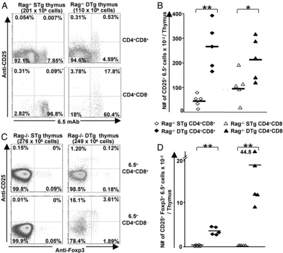
respectively). We also observed a moderate, but significant, increase in the absolute numbers of 6.5⫹CD25⫹SP thymocytes (2-fold) in DTg versus STg mice (Fig. 4 A and B), consistent with previous studies on the absolute numbers of TCR-Tg⫹ SP thymocytes (14, 15, 16, 25). Moreover, the absolute numbers of 6.5⫹CD25⫹DP thymocytes present in RAG⫺/⫺DTg were clearly higher (almost 6-fold) than in RAG⫺/⫺6.5-TCR mice (Fig. 4 A and B), similar to that described for RAG⫹animals. At this DP stage, there was no clonal deletion of 6.5⫹thymocytes in RAG⫹ DTg mice (Fig. 3B), whereas clonal deletion was present, yet not prominent, in RAG⫺/⫺ DTg mice (5.3 ⫾ 0.9 ⫻ 106 cells per
thymus vs. 17.4⫾ 2.3 ⫻ 106in RAG⫺/⫺STg mice).
We next evaluated Foxp3 expression in thymocytes to deter-mine whether the increase in frequency and absolute numbers of 6.5⫹CD25⫹ thymocytes was due to a Treg commitment. As shown in Fig. 4C, 6.5⫹CD25⫹Foxp3⫹ thymocytes were clearly present in DTg, but not in STg, mice. Indeed, the number of 6.5⫹CD25⫹Foxp3⫹thymocytes in DTg mice was 3.4⫾ 0.3 ⫻ 103
in the DP compartment and 19.1 ⫾ 6.6 ⫻ 103 in the SP
compartment (Fig. 4D). Only 13% of 6.5⫹CD25⫹DP and 19% of 6.5⫹CD25⫹ SP thymocytes were on average Foxp3⫹, as determined by FACS. It is unclear whether this observation is due to a limited sensitivity of Foxp3 detection or whether the 6.5⫹CD25⫹Foxp3⫺thymocytes are undergoing specific thymic selection processes, such as negative selection. Of interest,
although the MHC class II-restricted 6.5-TCR can positively select CD8⫹CD4⫺thymocytes (21), virtually none of these cells expressed CD25 and Foxp3 in both STg (range 0 to 0.07⫻ 103
per thymus; mean 0.02⫻ 103) and DTg mice (range 0 to 0.4⫻
103per thymus; mean 0.08⫻ 103), suggesting that Foxp3⫹CD8
T cells follow a different developmental program than Foxp3⫹ CD4 T cells.
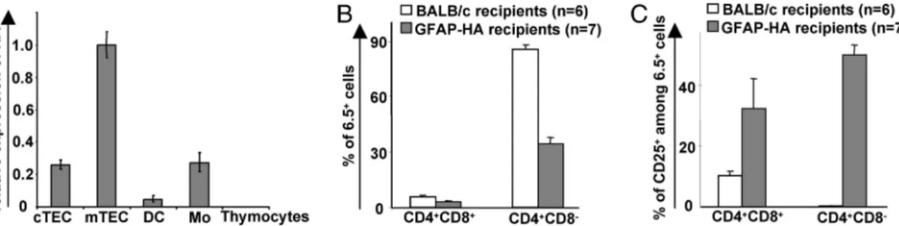
Promiscuous Expression of HA by Thymic Stroma Cells Promotes Development of HA-Specific CD25ⴙ Tregs from the DP Stage. To define precisely the thymic APC subsets expressing HA in GFAP-HA, we purified cortical TECs (cTECs), medullary TECs (mTECs), thymic dendritic cells, and macrophages and measured their HA mRNA content by using real-time RT-PCR. A clear expression of HA was detected in cTECs, mTECs, and macrophages (Fig. 5A). The gfap gene showed a very similar promiscuous transcription pattern in thymic stromal cells (Fig. 6, which is published as supporting information on the PNAS web site). Although we did not detect endogenous GFAP expression in thymic macrophages, this finding suggested that the GFAP promoter fragment used to generate the GFAP-HA Tg mice follows the physiological expression pattern of the endogenous promoter.
We then generated bone marrow (BM) chimeras by using RAG⫺/⫺6.5-TCR mice as BM donors to determine better in our system which HA-expressing thymic APCs were necessary for Fig. 4. Autoreactive 6.5⫹thymocytes from RAG⫺/⫺DTg mice express CD25 and Foxp3. (A) Expression of the 6.5-TCR and CD25 on DP and SP thymocytes from RAG⫺/⫺STg or DTg mice was determined by FACS. (B) Absolute numbers of DP and SP thymocytes expressing both 6.5 and CD25 were calculated in five RAG⫺/⫺ STg mice and five RAG⫺/⫺DTg mice. (C) The frequency of cells expressing CD25 and Foxp3 was determined on 6.5⫹DP and 6.5⫹SP thymocytes from RAG⫺/⫺STg or DTg mice by five-color FACS analyses. (D) Absolute numbers of 6.5⫹DP and 6.5⫹SP thymocytes expressing both CD25 and Foxp3 were calculated in 5 RAG⫺/⫺ DTg mice and 5 RAG⫺/⫺STg mice. The Mann–Whitney U test was used to compare groups:*, P⫽ 0.03;**, P⫽ 0.008.
Treg development. We observed a clear compartmentalization of thymic selection processes in GFAP-HA recipients of RAG⫺/⫺6.5-TCR BM, most likely because of the lack of HA expression in BM-derived APCs. Indeed, thymic negative selec-tion barely was detectable at the DP stage, although it became pronounced during the later CD4⫹CD8dull(data not shown) and
CD4⫹ SP (Fig. 5B) stages. However, the induction of CD25 expression on a large fraction of 6.5⫹thymocytes occurred at the early DP stage in GFAP-HA recipients but not in BALB兾c recipients (Fig. 5C). This result indicates that the expression of HA by radio-resistant thymic stromal cells, such as cTECs and mTECs, is responsible for CD25 expression on 6.5⫹thymocytes. In the BM chimeras, acquisition of the CD25 marker on devel-oping 6.5⫹thymocytes occurred earlier than negative selection and can be clearly dissociated from it.
Discussion
Our results show that the promiscuous expression of a nervous system-associated self-Ag by radio-resistant thymic stromal cells, most likely TECs, promotes the development of specific Tregs. We clearly observed an increase in both the relative and absolute number of specific Treg precursors at an early stage of thymo-cytes development in the presence of the cognate Ag. In this model, the expression of endogenous TCR chains was not required for generating TCR-Tg Tregs.
Previous observations in several TCR-Tg mouse autoreactiv-ity models suggest that high-avidautoreactiv-ity interactions between auto-reactive thymocytes and thymic radio-resistant APCs expressing the agonist ligand can result in both deletion of specific thymo-cytes and increase in the proportion of Tregs expressing the TCR-Tg (7, 8, 14). Further data show that MHC class II⫹TECs expressing the target self-Ag are involved in the differentiation of these cells (9). By contrast, it has recently been shown by using a TCR-Tg mouse model with controllable levels of the specific self-Ag in the thymus that the frequency, but not the absolute number, of TCR-Tg⫹CD25⫹SP thymocytes was increased upon moderate self-Ag expression (16). Moreover, a parallel increase in the number of clonotype-negative CD25⫹SP thymocytes was observed. It therefore was suggested that Tregs are not induced by interactions with self-Ag but are induced by as-yet-unidentified factors. The enrichment of Tregs in certain exper-imental situations, such as DTg mice, was associated with their relative resistance to negative selection (27) and with their expansion in a relatively empty thymic environment. However, in our model, we observed a clear increase in the absolute number of DP Treg precursors in the presence of the specific Ag. Moreover, the developing 6.5⫹ thymocytes could acquire the Treg markers CD25 and Foxp3 before negative selection.
Fi-nally, we observed no differences between DTg and STg mice for the proportions and absolute number of CD25⫹6.5-negative thymocytes. These results are consistent with earlier studies (14, 15, 25) and suggest that recognition of the neo-self-Ag promotes selection and兾or expansion of specific Treg-thymic precursors. We therefore suggest an inductive, rather than a selective, Treg differentiation model. This model is supported by the complete absence of CD25⫹6.5⫹cells expressing Foxp3 in the thymus of RAG-deficient STg mice that is in the absence of HA expression. The early expression of a rearranged transgenic TCR can affect thymocyte development. Therefore, Foxp3⫹cells may emerge artificially early in thymic ontogeny in our DTg model. However, Foxp3⫹ DP thymocytes have been described recently among polyclonal thymocytes and show an absolute dependence on MHC expression (10). Although the absolute number of DP CD25⫹6.5⫹ thymocytes in DTg mice were higher than in STg littermates, this difference was much less at the later SP stage. These data suggest that developing HA-specific Tregs may be selected in the thymic cortex of DTg mice but may undergo partial negative selection upon further differentiation in the thymus (16).
Previous studies have investigated the effect on T cell tolerance of the thymic expression of neural self-Ags. Developing thymocytes specific for myelin basic protein (MBP) peptides that are shared between the thymically expressed golli-MBP isoforms and the classic MBP isoforms are negatively selected (19). However, patho-genic proteolipid protein (PLP) 139–151-specific CD4 T cells escape thymic tolerance induction in SJL mice, because the plp splice variant DM-20, which lacks residues 116-150, is preferentially expressed in the thymus (18, 28). Similarly, the very low level of myelin兾oligodendrocyte glycoprotein (MOG) expression in C57BL兾6 thymus was not associated with a detectable tolerance to MOG (29). Our finding that Tregs specific for neural neo-self-Ags are naturally generated in the thymus is potentially interesting because these cells exert potent Ag-specific suppressive activities both in vitro and in vivo. GFAP and GFAP promoter-driven transgenes are not absolutely restricted to neural cells and may have slightly different expression patterns. However, GFAP is probably representative of a class of self-Ags that are thymically expressed and can elicit both thymic clonal deletion and Treg development. Therefore, in a polyclonal situation, Tregs against a variety of thymically expressed neural Ags are most likely also generated. Interestingly, CD4⫹CD25⫹Tregs contribute to resistance to CNS autoimmunity (30), and they and have been successfully used to prevent or treat animal models of multiple sclerosis (31). Although Treg differentiation does not occur only in the thymus (32), it may be possible to take advantage of this physiological phenomenon to design new therapeutic strategies for CNS inflammatory diseases. Fig. 5. Expression of HA in TECs correlates with Treg development. (A) Relative expression of HA mRNA determined by real-time RT-PCR in purified thymic APCs from GFAP-HA mice (cTEC, cortical TECs; mTECs, medullary TECs; DC, dendritic cells; Mo, macrophages). HA expression values in mTECs was defined as 1. Results are from one of two experiments conducted on a pool of 18 mice. (B) Thymic development in irradiated GFAP-HA or BALB兾c mice reconstituted with BM cells from RAG⫺/⫺6.5-TCR mice. The percentage of SP thymocytes expressing the Tg TCR was lower in seven GFAP-HA recipients than in six BALB兾c recipients (P ⫽ 2⫻ 10⫺7; two-tailed Student’s t test). There were only minor differences among DP thymocytes (P⫽ 0.06). (C) The percentage of CD25⫹cells among 6.5⫹
thymocytes was assessed in the BM chimeras and was higher in GFAP-HA recipients than in BALB兾c recipients at the DP (P ⫽ 0.0023) and SP (P ⫽ 0.0012) developmental stages.
These strategies could be based on either autologous cell therapy or in vivo amplification of self-reactive Tregs and should enrich our approaches to selective immunotherapy of human autoimmune diseases (33).
Materials and Methods
Mice. GFAP-HA Tg and 6.5-TCR Tg mice lines have been described in refs. 20 and 21. Mice were backcrossed⬎10 times onto the BALB兾c background. Animal experiments were carried out in accordance with the European Union guidelines and had local committee approval.
Flow Cytometry.The Abs used in this study were as follows: 6.5 anti-clonotypic mAb (21), anti-CD4 CD4), anti-CD8 (CT-CD8), anti-Foxp3 (FJK-16s), anti-CD25 (PC61), anti-CD25 (7D4), anti-CD69 (H1.2F3), anti-CD44 (IM7), anti-CD62L (MEL-14), and anti-GITR (goat polylonal Ab). Analysis was carried out by using either an EPICS Elite (Beckman Coulter), a FACScan, a FACScalibur or a FACSAria (BD Biosciences). Data were analyzed by using CELLQUEST(BD Biosciences) or FLOWJO(Tree Star, Ashland, OR). Cell numbers and percent-ages are the mean⫾ SEM.
Cell Purification.For the selection of CD4⫹ T cells, spleen and lymph node cells were incubated with rat anti-mouse CD8␣, B220, and Mac1 mAbs and then with goat anti-rat IgG-coated microbeads. For the purification of CD25⫹and CD25⫺T cells, the CD4 T cell-enriched negative fraction was incubated with biotinylated anti-CD25 mAb (7D4) and then with streptavidin microbeads. Cells were sorted by using MS columns (Miltenyi Biotec). The purity of the resulting cell populations was 93-96% for CD4⫹CD25⫺T cells and 75-97% for CD4⫹CD25⫹T cells.
Proliferation and Coculture Assays. CD4⫹, CD4⫹CD25⫺, or CD4⫹CD25⫹T cells (2⫻ 105cells per well) were stimulated by
incubation for 64 h in complete culture medium with increasing concentrations of HA111-119 peptide or anti-CD3 mAb (145-2C11) in the presence of either 5 or 20⫻ 105irradiated syngeneic
splenocytes. In coculture experiments, CD4⫹CD25⫺T cells from
6.5-TCR Tg mice were cultured in triplicate for 96 h with or without CD4⫹CD25⫹cells from BALB兾c or DTg mice. [3
H]thy-midine (1Ci per well; 1 Ci ⫽ 37 GBq) was added for the last 16 h. SEMs were consistently⬍15% of the mean.
FACS Cell Sorting and RT-PCR.Thymic APCs were purified from the thymus of GFAP-HA female mice by using a combination of density fractionation and cell sorting, as described in ref. 11. Real-time PCR was carried out in a final volume of 25l with optimal concentrations of primers (-actin:
5⬘-ACGGCCAG-GTCATCACTATTG-3⬘ and
5⬘-AGGATTCCATACCCAA-GAAGGAA-3⬘; HA: 5⬘-GCCATTGCCGGTTTTATTGA-3⬘
and 5⬘-TCCGCTGCATAGCCTGATC-3⬘) by using the qPCR Core Kit for SybrGreen I (Eurogentec, Brussels). Reactions were run on a GeneAmp 5700 Sequence Detection System (Applied Biosystems) in triplicate, and expression values were normalized to-actin expression by using the comparative CT method.
rAAV-HA Injections, Histology, and IFN-␥ Enzyme-Linked Immunospot
(ELISPOT) Assays.Intramuscular immunization of BALB兾c mice by using rAAV-HA, assessment of the inhibition of immune-mediated transgene rejection by CD4⫹CD25⫹T cells, and the IFN-␥ enzyme-linked immunospot assay against the immuno-dominant Kd-binding HA512-520 peptide were carried out as
described in ref. 23.
Radiation BM Chimeras.Two- to three-month-old GFAP-HA and BALB兾c mice were ␥-irradiated (650 rads) and reconstituted 24 h later with 107 BM cells from RAG2⫺/⫺ 6.5-TCR mice.
Recipient mice were kept on antibiotics (bactrim) and analyzed 8-10 weeks after reconstitution.
We thank Maryline Calisse (IFR30 animal facility) for animal care, Dr. Jan Bauer for histological analyses, and Dr. Daniel Dunia and Jean Davoust for a critical review of the manuscript. This work was supported by grants from the Institut National de la Sante´ et de la Recherche Me´dicale, the European Union, the Midi-Pyre´ne´es region, and the French MS society (Association pour la Recherche sur la Scle´rose en Plaques).
1. Shevach, E. M. (2002) Nat. Rev. Immunol. 2, 389–400. 2. Sakaguchi, S. (2004) Annu. Rev. Immunol. 22, 531–562.
3. Baecher-Allan, C., Brown, J. A., Freeman, G. J. & Hafler, D. A. (2001)
J. Immunol. 167, 1245–1253.
4. Fontenot, J. D., Gavin, M. A. & Rudensky, A. Y. (2003) Nat. Immunol. 4, 330–336. 5. Hori, S., Nomura, T. & Sakaguchi, S. (2003) Science 299, 1057–1061. 6. Hsieh, C. S., Liang, Y., Tyznik, A. J., Self, S. G., Liggitt, D. & Rudensky, A. Y.
(2004) Immunity 21, 267–277.
7. Jordan, M. S., Boesteanu, A., Reed, A. J., Petrone, A. L., Holenbeck, A. E., Lerman, M. A., Naji, A. & Caton, A. J. (2001) Nat. Immunol. 2, 301–306. 8. Apostolou, I., Sarukhan, A., Klein, L. & von Boehmer, H. (2002) Nat. Immunol.
3,756–763.
9. Bensinger, S. J., Bandeira, A., Jordan, M. S., Caton, A. J. & Laufer, T. M. (2001) J. Exp. Med. 194, 427–438.
10. Fontenot, J. D., Rasmussen, J. P., Williams, L. M., Dooley, J. L., Farr, A. G. & Rudensky, A. Y. (2005) Immunity 22, 329–341.
11. Derbinski, J., Schulte, A., Kyewski, B. & Klein, L. (2001) Nat. Immunol. 2, 1032–1039.
12. Anderson, M. S., Venanzi, E. S., Klein, L., Chen, Z., Berzins, S. P., Turley, S. J., von Boehmer, H., Bronson, R., Dierich, A., Benoist, C. & Mathis, D. (2002)
Science 298, 1395–1401.
13. Liston, A., Lesage, S., Wilson, J., Peltonen, L. & Goodnow, C. C. (2003) Nat.
Immunol. 4, 350–354.
14. Lerman, M. A., Larkin, J., 3rd, Cozzo, C., Jordan, M. S. & Caton, A. J. (2004)
J. Immunol. 173, 236–244.
15. Walker, L. S., Chodos, A., Eggena, M., Dooms, H. & Abbas, A. K. (2003) J. Exp.
Med. 198, 249–258.
16. van Santen, H. M., Benoist, C. & Mathis, D. (2004) J. Exp. Med. 200, 1221–1230. 17. Kojima, K., Reindl, M., Lassmann, H., Wekerle, H. & Linington, C. (1997) Int.
Immunol. 9, 897–904.
18. Klein, L., Klugmann, M., Nave, K. A., Tuohy, V. K. & Kyewski, B. (2000) Nat.
Med. 6, 56–61.
19. Perchellet, A., Stromnes, I., Pang, J. M. & Goverman, J. (2004) Nat. Immunol.
5,606–614.
20. Cornet, A., Savidge, T. C., Cabarrocas, J., Deng, W.-L., Colombel, J.-F., Lassmann, H., Desreumaux, P. & Liblau, R. S, (2001) Proc. Natl. Acad. Sci.
USA 98, 13306–13311.
21. Kirberg, J., Baron, A., Jakob, S., Rolink, A., Karjalainen, K. & von Boehmer, H. (1994) J. Exp. Med. 180, 25–34.
22. Fisson, S., Darrasse-Jeze, G., Litvinova, E., Septier, F., Klatzmann, D., Liblau, R. & Salomon, B. L. (2003) J. Exp. Med. 198, 737–746.
23. Gross, D. A., Leboeuf, M., Gjata, B., Danos, O. & Davoust, J. (2003) Blood 102, 4326–4328.
24. Cabarrocas, J., Piaggio, E., Zappulla, J. P., Desbois, S., Mars, L. T., Lassmann, H. & Liblau, R. S. (2004) J. Autoimmun. 22, 179–189.
25. Kawahata, K., Misaki, Y., Yamauchi, M., Tsunekawa, S., Setoguchi, K., Miyazaki, J. & Yamamoto, K. (2002) J. Immunol. 168, 4399–4405. 26. Hori, S., Haury, M., Coutinho, A. & Demengeot, J. (2002) Proc. Natl. Acad.
Sci. USA 99, 8213–8218.
27. Papiernik, M., de Moraes, M. L., Pontoux, C., Vasseur, F. & Pe´nit, C. (1998)
Int. Immunol. 10, 371–378.
28. Anderson, A. C., Nicholson, L. B., Legge, K. L., Turchin, V., Zaghouani, H. & Kuchroo, V. K. (2000) J. Exp. Med. 191, 761–770.
29. Delarasse, C., Daubas, P., Mars, L. T., Vizler, C., Litzenburger, T., Iglesias, A., Bauer, J., Della Gaspera, B., Schubart, A., Decker, L., et al. (2003) J. Clin.
Invest. 112, 544–553.
30. Reddy, J., Illes, Z., Zhang, X., Encinas, J., Pyrdol, J., Nicholson, L., Sobel, R. A., Wucherpfennig, K. W. & Kuchroo, V. K. (2004) Proc. Natl. Acad. Sci.
USA 101, 15434–15439.
31. Kohm, A. P., Carpentier, P. A., Anger, H. A. & Miller, S. D. (2002) J. Immunol.
169,4712–4716.
32. von Boehmer, H. (2005) Scand. J. Immunol. 62, Suppl. 1, 49–54. 33. Feldmann, M. & Steinman, L. (2005) Nature 435, 612–619.