HAL Id: hal-02877729
https://hal.archives-ouvertes.fr/hal-02877729
Submitted on 4 Nov 2020HAL is a multi-disciplinary open access archive for the deposit and dissemination of sci-entific research documents, whether they are pub-lished or not. The documents may come from teaching and research institutions in France or abroad, or from public or private research centers.
L’archive ouverte pluridisciplinaire HAL, est destinée au dépôt et à la diffusion de documents scientifiques de niveau recherche, publiés ou non, émanant des établissements d’enseignement et de recherche français ou étrangers, des laboratoires publics ou privés.
Revisiting Cationic Phosphorus Dendrimers as a
Nonviral Vector for Optimized Gene Delivery Toward
Cancer Therapy Applications
Liang Chen, Jin Li, Yu Fan, Jieru Qiu, Liu Cao, Regis Laurent, Serge
Mignani, Anne-Marie Caminade, Jean Pierre Majoral, Xiangyang Shi
To cite this version:
Liang Chen, Jin Li, Yu Fan, Jieru Qiu, Liu Cao, et al.. Revisiting Cationic Phosphorus Dendrimers as a Nonviral Vector for Optimized Gene Delivery Toward Cancer Therapy Applications. Biomacro-molecules, American Chemical Society, 2020, 21 (6), pp.2502-2511. �10.1021/acs.biomac.0c00458�. �hal-02877729�
1
Revisiting Cationic Phosphorus Dendrimers as a Nonviral Vector for
Optimized Gene Delivery Towards Cancer Therapy Applications
Liang Chena,b,c#, Jin Lia#,Yu Fana,Jieru Qiub,c, Liu Caoa, Régis Laurentb,c,Serge Mignani d,e*, Anne-Marie Caminade b,c, Jean-Pierre Majoralb,c* and Xiangyang Shia,e*
a
State Key Laboratory for Modification of Chemical Fibers and Polymer Materials, College of
Chemistry, Chemical Engineering and Biotechnology, Donghua University, Shanghai 201620, People’s Republic of China
b
Laboratoire de Chimie de Coordination du CNRS, 205 Route de Narbonne, BP 44099, 31077
Toulouse CEDEX 4, France
c
Université de Toulouse, UPS, INPT, 31077 Toulouse CEDEX 4, France
d ni e si Pa is esca es PR o bonne Pa is Ci , CNRS UMR 860, Laboratoi e e Chi ie e
e iochi ie Pha acolo i ues e o icolo i ue ue es ain s P es, 75006 Paris, France
e
CQM - Centro de Química da Madeira, MMRG, Universidade da Madeira, Campus da Penteada,
9020-105 Funchal, Portugal
KEYWORDS: cationic phosphorus dendrimers; gene delivery; cancer therapy; surface modification
________________________________
#
These authors contributed equally to this work.
* Corresponding author. E-mail: serge_mignani@orange.fr (S. Mignani), majoral@lcc-toulouse.fr (J. P. Majoral), and xshi@dhu.edu.cn (X. Shi)
2
ABSTRACT
Gene delivery, one important cancer therapy mode, still remains to be challenging due to the
shortage of highly efficient and non-toxic non-viral vectors. Here, we revisit the development of
cationic phosphorus dendrimers by synthesizing them with different generations (G1-3) and surface
ligands (1-(2-aminoethyl) pyrrolidine, 1-(3-aminopropyl) piperidine, or 1-(2-aminoethyl) piperidine)
for optimized gene delivery toward cancer gene therapy applications. First, the synthesized
dendrimer derivatives were used to compact plasmid DNA (pDNA) encoding enhanced green
fluorescent protein (EGFP) to optimize their gene delivery efficiency by varying the dendrimer
generations and surface polycationic ligands. We show that all dendrimer/pDNA polyplexes display
good cytocompatibility, and the 1-(2-aminoethyl) pyrrolidine-modified protonated G1 dendrimers
(1-G1) display the best gene delivery efficiency to HeLa cells under the same conditions through
flow cytometry and fluorescence microscopic imaging analyses. Hence, 1-G1 dendrimers were then
used as a vector to transfect pDNA encoding both EGFP and p53 protein for cancer gene therapy
applications. Our results reveal that under the optimized conditions, the transfection of pDNA
induces the significant p53 protein expression as verified through the resulted cell cycle arrest
(regulation of p21 and Cdk4/Cyclin-D1 expression) and Western blotting. The cancer gene therapy
potential of the polyplexes was finally validated through therapy of a xenografted tumor model after
intratumoral injection without systemic toxicity. The developed cationic 1-G1 dendrimers may hold
a great promise to be used as a powerful vector for cancer gene therapy applications, as well as for
3
INTRODUCTION
Successful gene delivery relies on the highly efficient and non-toxic vectors. Viral-based gene
delivery is currently still one of the most efficient methods to introduce exogenous genes into
cells.1-2 However, concerns about immunogenicity or long-term oncologic effects of the viral vectors
have prompted the development of synthetic non-viral gene delivery systems.3-4 In general, non-viral
delivery systems consist of linear cationic polymers, such as polylysine and their conjugates,5-6
branched polyethylenimine,7 poly(amidoamine) (PAMAM) or poly(propyleneimine) dendrimers,8-12
cationic lipids,13-14 or liposomes.15-16 These vector systems can electrostatically compact negatively
charged DNA to form polyplexes for effective cellular uptake to implement the gene expression.
The major advantages of these non-viral delivery systems over viral vectors can be summarized
as simplicity to prepare, stability, easiness to be modified, and biosafety. However, unlike viral
vectors that have evolved means to overcome cellular barriers and immune defense mechanisms,
non-viral gene carriers consistently exhibit limited transfection efficiency due to numerous extra-
and intracellular obstacles.17 Further, some key properties of non-viral gene carriers such as
dendrimers with defined architecture, tunable biocompatibility and a high ratio of multivalent
surface moieties to molecular volume also make them highly promising to be developed as synthetic
(non-viral) vectors for delivery therapeutic nucleic acids.18-20 Among many types of dendrimers,
phosphorus dendrimers possessing biocompatible backbone have been employed as anti-prion
agents,21-22 anti-HIV agents,23 and stimulator of immune blood cells24-25 and human natural killer
(NK) cells26 depending on the type of their terminal groups.
Cationic phosphorus dendrimers have also been used as promising gene delivery platforms for
numerous biomedical applications.27-29 The most widely investigated cationic phosphorus
dendrimers bear diethylamine termini with cyclotriphosphazene (CTP) core. They have been used as
carriers of plasmids,12, 30-31 oligodeoxynucleotides32 and siRNAs.33-35 For instance, Loup et al.12
disclosed the better transfection efficiency of dendrimers in the presence of serum than in the
absence of serum for plasmid transfection. As demonstrated by Padié et al.,36 the surface of
4
morpholine, methyl piperazine and phenyl piperazine. Hence, cyclic amine-modified phosphorous
dendrimers have been used for gene delivery applications. For instance, pyrrolidine-terminated
dendrimers were found to be low-toxic, efficient to form dendriplexes among other surface cyclic
amine modifications for highly efficient gene transfection.36 Dzmitruk et al.35 reported the use of
piperidine-terminated phosphorus dendrimers to compact siRNA for possible cancer cell gene
silencing. However, the molecular mechanism of antiproliferation and in vivo tumor gene silencing
were not accomplished. As a whole, there is a lack of systematic and in-depth exploration of cationic
phosphorus dendrimers with different cyclic amine groups for optimized gene delivery and also for
cancer gene therapy applications.
Figure 1. Synthesis of cationic phosphorous dendrimers with varying generations and cyclic amine
group.
5
cyclic amine groups as a non-viral vector for optimized gene delivery towards cancer therapy
applications. We first synthesized and characterized the first, second and third generations of
phosphorus dendrimers ended with protonated cyclic amine groups (Figure S1 and Figure 1). Then,
plasmid DNA (pDNA) encoding enhanced green fluorescent protein (EGFP) was used as a model to
test the gene compaction ability of the dendrimers, the cytotoxicity of the dendrimer/pDNA
polyplexes, and gene delivery efficiency. After screening, the optimized dendrimer derivative was
used as a vector to condense pDNA encoding both EGFP and p53 protein for cancer gene therapy
applications through investigation of p53 protein expression validated from cell cycle arrest
(regulation of p21 and Cdk4/Cyclin-D1 expression) and Western blotting. Finally, we verified the
potential to the polyplexes for gene therapy of a xenografted tumor model after intratumoral
injection. According to our thorough literature investigation, this work is a systematic and in-depth
exploration of cationic phosphorus dendrimers in gene delivery towards cancer therapy applications.
Table 1. The prepared cationic phosphorus dendrimers.
Generation Numbers of terminal groups per dendrimers Series 1 12 1-G`1 1-G1 1 2 24 1-G`2 1-G2 3 48 1-G`3 1-G3 2 1 12 3-G`1 3-G1 3 1 12 2-G`1 2-G1
6
dendrimer derivatives.
RESULTS AND DISCUSSION
Synthesis and Characterization of Cationic Dendrimer Derivatives. As shown in Table 1
and Figure 1, we prepared five different cationic phosphorus dendrimers with varying generations
and cyclic amine groups. First, generation 1-3 (G1-3) dendrimers were prepared and characterized
according to the literature.37 Then, the dendrimers were reacted with 1-(2-aminoethyl) pyrrolidine
(1-a), 1-(3-aminopropyl) piperidine (2-a), or 1-(2-aminoethyl) piperidine (3-a) to form 1-G`1, 1-G`2,
1-G`3, 3-G`1, or 2-G`1, and protonated to create the final cationic 1-G1, 1-G2, and 1-G3 dendrimers.
These dendrimers were fully characterized through different NMR techniques (Figure S2-S6, see
details in Supporting Information).
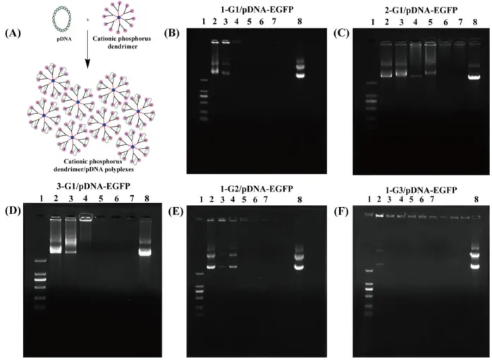
Formation of Dendrimer/pDNA Polyplexes for Optimized Gene Transfection. pDNA
encoding EGFP (pDNA-EGFP) was employed to evaluate the gene transfection efficiency of the
designed vectors in vitro. After formation of dendrimer/pDNA polyplexes under different N/P ratios
(the molar ratio of positive charge of the dendrimers to phosphates in the pDNA backbone) through
electrostatic interaction (Figure 2A), agarose gel retardation assay was utilized to check the DNA
compaction ability of the positively charged phosphorous dendrimers (Figure 2B-F). It is clear that
the pDNA-EGFPis able to be completely compressed by the cationic phosphorous dendrimers under
different N/P ratios. For the n-G1 (n=1, 2 and 3) dendrimers that possess the same number of
positive charges, the DNA condensation ability follows the order of 1G1 (N/P = 1 or above) > 3G1
(N/P = 2 or above) > 2G1 (N/P = 4 or above), imply that the DNA condensation ability is dependent
on the type of cyclic amine groups (Figure 2B-D). This could be due to the distinct cyclic amine
termini that have different dendrimer spatial structures to have different electrostatic compactions of
DNA. Namely, the dendrimers with terminal pyrrolidine rings may render them with higher charge
densities (compared to mass) than those with the terminal piperidine rings due to the smaller Mw
7
more methylene group, hence 2G1 dendrimers have slightly decreased molecular rigidity and
charge/mass ratio as compared to 3G1 dendrimers, thus having less DNA compaction ability than
3G1. Therefore, for optimized gene compaction, 1a-modified dendrimers should be better than 2a-
and 3a-modified dendrimers with the same generation.
Figure 2. (A) Preparation of the dendrimer/pDNA polyplexes for gene delivery; (B-F) Gel
retardation assay of pDNA-EGFP complexed with 1-G1, 2-G1, 3-G1, 1-G2 and 1-G3 at various N/P
ratios. Lane 1: DNA marker, lane 2: N/P = 0.25:1, lane 3: N/P = 0.5:1, lane 4: N/P = 1:1, lane 5: N/P
= 2:1, lane 6: N/P = 4:1, lane 7: N/P = 6:1, and lane 8: free pDNA-EGFP.
To check the dependence of the dendrimer generation, we investigated the DNA compaction
ability of the 1a-modified G1-3 dendrimers (Figure 2B, E-F). It can be seen that the DNA
compaction ability is in the order of 1G3 (N/P = 0.5 or above) > 1G1 (N/P = 1 or above) > 1G2 (N/P
= 2 or above). It seems that unlike the generation dependence of gene delivery efficiency of
8
terminal structure has no clear dendrimer generation dependence due to their intrinsic rigid
molecular backbones. In this context, one has to comprehensively consider all facts to optimize their
gene delivery efficiency.
The hydrodynamic size and surface potential of the polyplexes have been known to be
important factors in influencing the cytotoxicity, intracellular uptake, and release of the gene
cargos.39-41 The selection of N/P ratios at 10-30 was based on the fact that a large N/P ratio would
lead to a compacted structure and a relatively high positive surface potential of the polyplexes,
which facilitates enhanced cellular uptake of the polyplexes. As shown in Figure 3A,
1-Gn/pDNA-EGFP (n = 1, 2 and 3) polyplexes are all in the size range of ~250 nm. Nevertheless,
the size of the 2-G1 and 3-G1 are around ~ 400 nm. It appears that the 1-a-modified dendrimers have
better gene compaction ability than 2-a- and 3-a-modified 2-G1 and 3G1 dendrimers, in agreement
with the gel retardation assay data. In addition, under the same 1-a modification, there is no distinct
generation-dependent difference in the hydrodynamic size of the dendrimer/pDNA polyplexes for
1G1-3 dendrimers. On the other hand, with the increase of N/P ratio, more dendrimers participate in
the compaction of pDNA, thereby leading to decreased size of the polyplexes until reach saturation
to have a slightly increased size.
The surface potentials of the dendrimer/pDNA-EGFP polyplexes were then measured (Figure
3B). Clearly, 1-Gn/pDNA-EGFP (n = 1, 2 and 3) polyplexes under various N/P ratios show a surface
potential of approximately 20-30 mV. However, the surface potentials of 2-G1 and 3-G1 dendrimers
complexed with pDNA-EGFP under different N/P ratios are all around ~10 mV. This suggests that
polyplexes of dendrimers with the pyrrole ammonium group possess much more positive surface
potential than those with the piperidine ammonium group, presumably due to the adaptability of the
pyrrole tertiary ammonium group to pH.36 It should be noted that with the continuous increase of
N/P ratio, the interaction between dendrimers and pDNA could reach saturation and the surface
potentials of polyplexes do not have a significant change since only the dendrimers covered onto the
surface of the polyplexes contribute to the surface potential of the polyplexes. According to the
9
20-30 mM are suitable for gene delivery. Hence, the N/P ratio of 20 was selected for the subsequent
experiments.
Figure 3. (A) Mean hydrodynamic size and (B) zeta-potential of pDNA-EGFP complexed with
1-G1, 2-G1, 3-G1, 1-G2 and 1-G3 at different N/P ratios; (C-F) CCK8 assay of HeLa cells treated
with dendrimers and dendrimer/pDNA-EGFP polyplexes at different dendrimer concentrations for
24 h. Data are presented as mean ± SD (n = 6); (H) Schematic diagram of the gene transfection of
dendrimer/pDNA-EGFP polyplexes to express EGFP protein; (I) Relative EGFP protein expression
in the HeLa cells after transfected with the dendrimer/pDNA-EGFP polyplexes at different N/P
10
Further, the cytotoxicity of the dendrimer/pDNA-EGFP was tested via CCK8 viability assay of
HeLa cells (a human cervical carcinoma cell line, Figure 3C-F). The cell viability displays a
concentration-dependent decreasing trend. At the highest concentration of dendrimers and
dendrimer/pDNA (3000 nM, at the N/P ratio of 20), cells display a viability around > 50%. In any
case, at the same dendrimer concentration, the dendrimer/pDNA polyplexes show a lower
cytotoxicity than the dendrimers, possibly due to the complexation of pDNA that results in reduced
positive charges of the dendrimers, in agreement with the literature.43
Next, we evaluated the transfection efficiency of the polyplexes to express EGFP in HeLa cells
(Figure 3H-I) through fluorescence microscopic imaging and flow cytometry assay. Since the
change of N/P ratio has been reported to have an effect on the gene transfection efficiency in cancer
cells,44 polyplexes with the N/P ratios at 10-30 were tested. Flow cytometry data reveal that cells
transfected with polyplexes formed using dendrimers with pyrrolidine amine termini display much
more EGFP expression than those with piperidine amine termini at the same N/P ratios, with the best
gene transfection efficiency achieved at an N/P ratio of 20 (Figure 3I). Fluorescence microscopic
images qualitatively show the similar results (Figure S7). Therefore, the 1-G1 dendrimers were
selected as a vector to transfect the tumor suppressor p53 gene for the subsequent study.
Formation of Dendrimer/pDNA Polyplexes for Gene Therapy of Cancer Cells. The primary
objective was to investigate the gene therapy effect in cancer cells. The tumor suppressor p53 pDNA
(p53, encoding both EGFP and p53 protein) was complexed with 1-G1 dendrimers to form
polyplexes for therapeutics of cancer cells (Figure 4A). Firstly, the ability of 1-G1 to compact
pDNA-p53 was evaluated using gel retardation assay (Figure 4B). Apparently, the pDNA-p53 is
able to be completely compressed by 1-G1 dendrimers at an N/P ratio of 1 or greater. Secondly, the
hydrodynamic size and surface potential of the polyplexes were tested. The surface potential and
hydrodynamic size of the polyplexes are ~30 mV and ~200 nm under various N/P ratios (Figure
4C-D), which are suitable for further gene delivery applications. Since the p53 plasmid contains
gene segment decoding EGFP, the EGFP gene was used as a reporter gene to first evaluate the
11
that the best gene transfection efficiency can be achieved at an N/P ratio of 20 (Figure 4E), which
can be further confirmed through qualitative fluorescence microscopic imaging of HeLa cells
expressing the EGFP (Figure S8). Therefore, the N/P ratio of 20 was selected for the subsequent
experiments. Cytotoxicity assay results reveal that more than the cells possess a viability of 60%
after treated with the polyplexes at the highest dendrimer concentration (3000 nM) at the N/P ratio of
20 (Figure 4F), implying the good cytocompatibility of the polyplexes.
Figure 4. (A) Schematic diagram of p53 protein expression-mediated G0/G1 phase arrest; (B) Gel
retardation assay of p53 plasmid DNA complexed with 1-G1 at various N/P ratios (Lane 1: DNA
marker, lane 2: N/P = 0.25:1, lane 3: N/P = 0.5:1, lane 4: N/P = 1:1, lane 5: N/P = 2:1, lane 6: N/P =
4:1, lane 7: N/P = 6:1, and lane 8: P53 plasmid DNA); (C) Zeta potential and (D) mean
hydrodynamic size of p53 plasmid DNA complexed with 1-G1 at various N/P ratios; (E) Relative
EGFP protein expression in HeLa cells transfected with the 1-G1/pDNA polyplexes at different N/P
ratios as measured by flow cytometry analysis; (F) CCK8 assay of HeLa cells treated with the
polyplexes at different dendrimer concentrations for 24 h.
12
cells, we investigated the block of cell cycle progression in G1 stage in response to DNA damage or
other stresses (Figure 4A).45-47 Flow cytometric analysis of the cell cycle distribution was performed
(Figure 5A-B). In the PBS group, cells contain 2n chromosomes (phase G0/G1, ~40%) and few cells
are in an active DNA synthesis stage (phase S, ~25%) or already engage in the mitosis process
(phase G2/M, ~ 35%). Meanwhile, for the 1-G1 and free pDNA-p53, cells are in the phase S (~25%)
without a substantial increase of the G0/G1 phase (50%) after 48 h transfection. However, in the
1-G1/pDNA-p53 polyplex group, there are more potent promoters of G1/S cell cycle arrest than
other groups (phase G0/G1, ~ 65%; phase G2/M, ~ 5%; and phase S, ~ 30%). The cells in the
mitosis process (phase G2/M, ~ 35%) have a substantial decrease after 48 h gene transfection.
To elucidate the molecular mechanism of the cell cycle arrest, the cell cycle-related mRNAs
were inspected using quantitative real time-polymerase chain reaction (rt-qPCR) assay. HeLa cells
treated with PBS were used as negative control. It is well known that p21 gene was among the first
p53 protein target genes to be identified and is now recognized as an encoder for a major cell cycle
checkpoint control protein.48-55 The p21 protein functions primarily by binding to and inhibiting
Cyclin-D1/Cdk-4 complexes.56-57 Progression of the mammalian cell cycle is regulated by Cdks and
regulatory subunits cyclins, cell cycle progression is triggered by partial phosphorylation of Rb by
Cyclin-D1/Cdk-4 complexes, and p21 protein disrupts this interactions and inhibits cell cycle
progression.58 So the relative mRNA level of p53, p21, Cyclin-D1 and Cdk-4 were examined. As
shown in Figure 5C, around 6.5-fold and 3.8-time increase in mRNA of p53 and p21, respectively
can be observed in cells treated with polyplexes, which is much higher than other groups.
Meanwhile, the mRNA levels of Cdk-4 and Cyclin-D1 decrease to 50-60% in the polyplex group
when compared to other groups. Rt-qPCR evaluation implies that pDNA-p53 can be transfected by
1-G1 dendrimers to express p53 gene, consistent with the results of confocal microscopy and flow
cytometry.
For further elucidation of the molecular mechanism of the cell cycle arrest, the cell
cycle-related proteins and p53 were inspected using Western blotting in vitro (Figure 5D-E). Around
13
treated with the polyplexes, which is higher than all other groups. The G1-phase arresting effect of
HeLa cells treated with the polyplexes is related to the down-stream expression level of
Cdk-4/Cyclin-D1 that is regulated by kinase inhibitor p21 protein. In order to prove the progression
of cell cycle is regulated by Cdks and regulatory subunits cyclins, the expression level of Cdk-4 and
Cyclin-D1 proteins was also tested. Clearly, the expression of both Cdk-4 and Cyclin-D1 distinctly
decreases in HeLa cells after treated with the polyplexes. In contrast, 1-G and free p53 do not cause
significant changes in Cdk-4 and Cyclin-D1 level in HeLa cells. These results demonstrate the
excellent gene therapy of cancer cells through the transfection of dendrimer/pDNA polyplexes.
Figure 5. (A) Cell cycle analysis of HeLa cells treated with PBS, 1-G1, free pDNA-p53 and
1-G1/pDNA-p53 polyplexes at an N/P ratio of 20, respectively for 48 h. (B) The percentages of the
cellular distribution in different cell cycle phases of the HeLa cells. (C) Validation of relative
expression level of mRNA related to G0/G1 phase in HeLa cells after incubation with PBS, 1-G1,
free pDNA-p53 and 1-G1/pDNA-p53 polyplexes, respectively for 48 h. (D) Western blot assay of
the expression of proteins related to G0/G1 phase in HeLa cells transfected with 1-G1/pDNA-p53
polyplexes (at an N/P ratio of 20). PBS and free pDNA-p53 were used as controls. The GAPDH
protein was used as an internal control. (E) Quantitative analysis of proteins related to G0/G1 phase
14
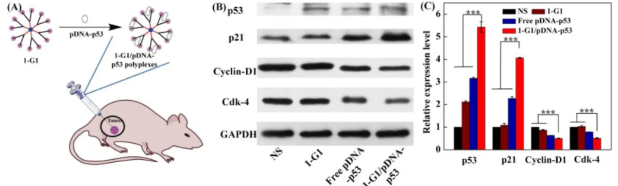
Dendrimer/pDNA Polyplexes for Gene Therapy of a Tumor Model in Vivo. Next, the
polyplex transfection-mediated cancer gene therapy was investigated using a xenografted tumor
model (Figure 6A). Normal saline (NS) was used as control, besides the free pDNA-p53 and 1G1
controls. After transfection for 3 days, the tumor tissue was processed to isolate the tumor cells for
Western blot assay of the protein expression (Figure 6B-C). Obviously, the tumor cells treated with
the 1-G1/pDNA-p53 polyplexes have higher p53 and p21 protein expression level than all other
groups (p < 0.001), and lower Cdk-4 and Cyclin-D1 expression level than other groups, in
agreement with the in vitro transfection data (Figure 5D-E). These results suggest that the developed
phosphorus dendrimers with the optimized surface cyclic amine termini and generation are able to
condense pDNA from degradation in vivo to implement effective tumor gene transfection in vivo.
Figure 6. (A) Preparation of the dendrimer/pDNA polyplexes for gene delivery in vivo; (B) Western
blot assay of the expression of the proteins related to G0/G1 phase in xenografted tumor cells at 4
days post treatment of NS, 1-G1, free pDNA-p53 and 1-G1/pDNA-p53 polyplexes (20 μ
pDNA/mouse for pDNA groups). The GAPDH protein was used as an internal control. (C)
Quantitative analysis of the G0/G1 phase-related protein expression level from the Western blot data
in vivo.
We further used H&E staining to evaluate the in vivo biocompatibility of the 1-G1/pDNA-p53
polyplexes (Figure S9). Similar to the control group injected with NS, no significant organ
15
lung, and kidney, declaring that the treatment of the 1-G1/pDNA-p53 polyplexes is quite safe and
does not induce any toxic effects. Our results suggest that the designed 1-G1 can be used for safe
gene delivery applications.
Conclusions
In conclusion, we revisited the development of cationic phosphorous dendrimers for optimized
gene delivery toward cancer gene therapy applications. Cationic phosphorous dendrimers with
different generations and surface cyclic amine groups were synthesized, characterized, and used to
first compact pDNA-EGFP to form polyplexes and then deliver the polyplexes to cancer cells in
vitro in order to screen those with the optimized gene delivery efficiency. 1-G1 dendrimers with
pyrrolidine ammonium termini were proven to be the most promising vector that was further used to
transfect pDNA-p53 to induce the cell cycle arrest through the regulation of p21 and
Cdk4/Cyclin-D1 expression and p53 protein expression in vitro and in vivo. The developed cationic
pyrrolidine ammonium-terminated 1-G1 dendrimers may be used for highly efficient gene delivery
to different biosystems for gene therapy of various diseases.
Experimental Section
Synthesis of 1-(2-aminoethyl) pyrrolidine-modified G1 (1-G`1). All reactions were carried
out in organic solvents using standard high vacuum and dry-argon techniques. 1-(2-Aminoethyl)
pyrrolidine (1-a, 27.12 mmol, 3.096 g) and N, N-diisopropylethylamine (DIPEA, 27.12 mmol, 3.505 g) were added to solution of G1 (2.26 mmol, 0.786 g, in 40 mL tetrahydrofuran (THF)) at 0 ℃. The reaction mixture was stirred overnight at room temperature and then concentrated under reduced
pressure. Then, 100 mL of pentane was added to the residue and the resulting precipitate was filtered
off and dried under reduced pressure to afford 1-G`1 as a powder in 95.7% yield.
Similarly, 1-(3-aminopropyl) piperidine (2-a)-modified G1 (2-G`1) and 1-(2-aminoethyl)
16
the above conditions with a starting G1 amount of 0.786 g. Likewise, 1-(2-aminoethyl) pyrrolidine
(1-a)-modified G2 (1-G`2) and G3 (1-G`3) were also synthesized under the same conditions with
starting G2 and G3 amount of 2.162 g and 2.419 g, respectively. The reaction conditions for all
dendrimer derivatives are listed in Table S1.
Synthesis of cationic phosphorus dendrimers. Hydrogen chloride solution (1 M in diethyl
ether, 1.2 mmol, 1 mL) was added to a solution of dendrimers of different generations (1-G`1, 0.1
mmol; 2-G`1, 0.1 mmol; 3-G`1, 0.1 mmol; 1-G`2, 0.05 mmol; and 1-G`3, 0.025 mmol) in 15 mL of THF at 0 ℃. The solution was stirred for 4 h and then dried under reduced pressure to afford cationic phosphorus dendrimers as a white powder in around 95% yield for all products. All animal
experiments were carried out after approval by the ethical committee for animal care of Donghua
University and according to the policy of the National Ministry of Health. See more experimental
details in the Supporting Information.
ASSOCIATED CONTENT Supporting Information
Part of experimental details and additional experimental data.
AUTHOR INFORMATION Corresponding Authors
*E-mail for Serge Mignani: serge.mignani@staff.uma.pt;
*E-mail for Jean-Pierre Majoral: jean-pierre.majoral@lcc-toulouse.fr;
*E-mail for Xiangyang Shi: xshi@dhu.edu.cn.
Notes
The authors declare no competing financial interest.
17
This research has been financially supported by the National Natural Science Foundation of China
(21911530230, 81761148028 and 21773026), the Science and Technology Commission of Shanghai
Municipality (19XD1400100 and 18520750400), the National Key R&D Program
(2017YFE0196200), and the Sino-French Cai Yuanpei Programme. J.P.M, S.M, A.M.C and R.L.
thank the collaborative NSFC-CNRS grant (from the France part) for financial support.
REFERENCES
1. Briand, P.; Kahn, A., Interests and limits of adenoviruses vectors for gene-transfer in-vivo.
Pathol. Biol. 1993, 41 (8), 663-671.
2. Roessler, B. J.; Hartman, J. W.; Vallance, D. K.; Latta, J. M.; Janich, S. L.; Davidson, B. L., Inhibition of interleukin-1-induced effects in synoviocytes transduced with the human il-1 receptor antagonist cdna using an adenoviral vector. Hum. Gene Ther. 1995, 6 (3), 307-316.
3. Cotten, M.; Wagner, E., Non-viral approaches to gene therapy. Curr. Opin. Biotechnol. 1993, 4 (6), 705-10.
4. Curiel, D. T., High-efficiency gene-transfer mediated by adenovirus-polylysine-dna complexes. In Gene Therapy for Neoplastic Diseases, Huber, B. E.; Lazo, J. S., Eds. 1994; Vol. 716, pp 36-58. 5. Wagner, E.; Cotten, M.; Foisner, R.; Birnstiel, M. L., Ttransferrin polycation dna complexes - the effect of polycations on the structure of the complex and dna delivery to cells. Proc. Natl. Acad.
Sci. U. S. A. 1991, 88 (10), 4255-4259.
6. Stankovics, J.; Crane, A. M.; Andrews, E.; Wu, C. H.; Wu, G. Y.; Ledley, F. D., Overexpression of human methylmalonyl coa mutase in mice after in-vivo gene-transfer with asialoglycoprotein polylysine dna complexes. Hum. Gene Ther. 1994, 5 (9), 1095-1104.
7. Boussif, O.; Lezoualch, F.; Zanta, M. A.; Mergny, M. D.; Scherman, D.; Demeneix, B.; Behr, J. P., A versatile vector for gene and oligonucleotide transfer into cells in culture and in vivo: polyethylenimine. Proc. Natl. Acad. Sci. U. S. A. 1995, 92 (16), 7297-7301.
8. Haensler, J.; Szoka, F. C., Polyamidoamine cascade polymers mediate efficient transfection of cells in culture. Bioconjugate Chem. 1993, 4 (5), 372-379.
9. Kang, S. H.; Zirbes, E. L.; Kole, R., Delivery of antisense oligonucleotides and plasmid DNA with various carrier agents. Antisense Nucleic Acid Drug Dev. 1999, 9 (6), 497-505.
10. Bielinska, A.; KukowskaLatallo, J. F.; Johnson, J.; Tomalia, D. A.; Baker, J. R., Regulation of in vitro gene expression using antisense oligonucleotides or antisense expression plasmids transfected using starburst PAMAM dendrimers. Nucleic Acids Res. 1996, 24 (11), 2176-2182.
11. Tang, M. X.; Redemann, C. T.; Szoka, F. C., In vitro gene delivery by degraded polyamidoamine dendrimers. Bioconjugate Chem. 1996, 7 (6), 703-714.
12. Loup, C.; Zanta, M.-A.; Caminade, A.-M.; Majoral, J.-P.; Meunier, B., Preparation of Water-Soluble Cationic Phosphorus-Containing Dendrimers as DNA Transfecting Agents. Chem. -
Eur. J. 1999, 5 (12), 3644-3650.
13. Byk, G.; Dubertret, C.; Escriou, V.; Frederic, M.; Jaslin, G.; Rangara, R.; Pitard, B.; Crouzet, J.; Wils, P.; Schwartz, B.; Scherman, D., Synthesis, activity, and structure-activity relationship studies of novel cationic lipids for DNA transfer. J. Med. Chem. 1998, 41 (2), 224-235.
18
14. De Jong, G.; Telenius, A.; Vanderbyl, S.; Meitz, A.; Drayer, J., Efficient in-vitro transfer of a 60-Mb mammalian artificial chromosome into murine and hamster cells using cationic lipids and dendrimers. Chromosome Res. 2001, 9 (6), 475-485.
15. Hara, T.; Tan, Y.; Huang, L., In vivo gene delivery to the liver using reconstituted chylomicron remnants as a novel nonviral vector. Proc. Natl. Acad. Sci. U. S. A. 1997, 94 (26), 14547-14552. 16. Koltover, I.; Salditt, T.; Radler, J. O.; Safinya, C. R., An inverted hexagonal phase of cationic liposome-DNA complexes related to DNA release and delivery. Science 1998, 281 (5373), 78-81. 17. Mintzer, M. A.; Simanek, E. E., Nonviral Vectors for Gene Delivery. Chem. Rev. 2009, 109 (2), 259-302.
18. Dufes, C.; Uchegbu, I. F.; Schatzlein, A. G., Dendrimers in gene delivery. Adv. Drug Delivery
Rev. 2005, 57 (15), 2177-2202.
19. Hou, W. X.; Wei, P.; Kong, L. D.; Guo, R.; Wang, S. G.; Shi, X. Y., Partially PEGylated dendrimer-entrapped gold nanoparticles: a promising nanoplatform for highly efficient DNA and siRNA delivery. J. Mater. Chem. B 2016, 4 (17), 2933-2943.
20. on l es C ou iu h al , H.; Tomás, H.; Shi, X. Y., RGD peptide-modified dendrimer-entrapped gold nanoparticles enable highly efficient and specific gene delivery to stem cells. ACS Appl. Mater. Interfaces 2015, 7 (8), 4833-4843.
21. Solassol, J.; Crozet, C.; Perrier, V.; Leclaire, J.; Beranger, F.; Caminade, A. M.; Meunier, B.; Dormont, D.; Majoral, J. P.; Lehmann, S., Cationic phosphorus-contain ing dendrimers reduce prion replication both in cell culture and in mice infected with scrapie. J. Gen. Virol. 2004, 85, 1791-1799. 22. Klajnert, B.; Cangiotti, M.; Calici, S.; Ionov, M.; Majoral, J. P.; Caminade, A.-M.; Cladera, J.; Bryszewska, M.; Ottaviani, M. F., Interactions between dendrimers and heparin and their implications for the anti-prion activity of dendrimers. New J. Chem. 2009, 33 (5), 1087-1093.
23. Blanzat, M.; Turrin, C. O.; Aubertin, A. M.; Couturier-Vidal, C.; Caminade, A. M.; Majoral, J. P.; Rico-Lattes, I.; Lattes, A., Dendritic catanionic assemblies: In vitro anti-HIV activity of phosphorus-containing dendrimers bearing Gal beta(1)cer analogues. Chembiochem 2005, 6 (12), 2207-2213.
24. Poupot, M.; Griffe, L.; Marchand, P.; Maraval, A.; Rolland, O.; Martinet, L.; L'Faqihi-Olive, F. E.; Turrin, C. O.; Caminade, A. M.; Fournie, J. J.; Majoral, J. P.; Poupot, R., Design of phosphorylated dendritic architectures to promote human monocyte activation. FASEB J. 2006, 20 (13), 2339-2351.
25. Rolland, O.; Griffe, L.; Poupot, M.; Maraval, A.; Ouali, A.; Coppel, Y.; Fournie, J.-J.; Bacquet, G.; Turrin, C.-O.; Caminade, A.-M.; Majoral, J.-P.; Poupot, R., Tailored control and optimisation of the number of phosphonic acid termini on phosphorus-containing dendrimers for the ex-vivo activation of human monocytes. Chem. - Eur. J. 2008, 14 (16), 4836-4850.
26. Griffe, L.; Poupot, M.; Marchand, P.; Maraval, A.; Turrin, C.-O.; Rolland, O.; Metivier, P.; Bacquet, G.; Fournie, J.-J.; Caminade, A.-M.; Poupot, R.; Majoral, J.-P., Multiplication of human natural killer cells by nanosized phosphonate-capped dendrimers. Angew. Chem., Int. Ed. Engl. 2007,
46 (14), 2523-2526.
27. Caminade, A. M.; Turrin, C.-O.; Majoral, J.-P., Biological properties of phosphorus dendrimers.
New J. Chem. 34 (8), 1512.
28. Caminade, A. M.; Majoral, J. P., Positively charged phosphorus dendrimers. An overview of their properties. New J. Chem. 2013, 37, 3358-3373.
29. Klajnert-Maculewicz; Barbara; Zablocka; Maria; Marcinkowska; Monika; Majoral; Jean-Pierre; Wasiak; Tomasz, Cationic phosphorus dendrimers and therapy for Alzheimer's disease. New J. Chem.
2015, 39, 4852-4859.
19
J.-P., Water-soluble polycationic dendrimers with a phosphoramidothioate backbone: preliminary studies of cytotoxicity and oligonucleotide/plasmid delivery in human cell culture. Oligonucleotides
2003, 13 (4), 193-205.
31. Shcharbin, D.; Dzmitruk, V.; Shakhbazau, A.; Goncharova, N.; Seviaryn, I.; Kosmacheva, S.; Potapnev, M.; Pedziwiatr-Werbicka, E.; Bryszewska, M.; Talabaev, M., Fourth Generation Phosphorus-Containing Dendrimers: Prospective Drug and Gene Delivery Carrier. Pharmaceutics
2011, 3 (4), 458-473.
32. Munoz-Fernandez, M. A.; Majoral, J. P.; Caminade, A. M.; Hameau, A.; Madrid, R.; Serramia, M. J.; Briz, V., Validation of a Generation 4 Phosphorus-Containing Polycationic Dendrimer for Gene Delivery Against HIV-1. Curr. Med. Chem 2012, 19 (29), 5044-5051.
33. Ionov, M.; Lazniewska, J.; Dzmitruk, V.; Halets, I.; Loznikova, S.; Novopashina, D.; Apartsin, E.; Krasheninina, O.; Venyaminova, A.; Milowska, K., Anticancer siRNA cocktails as a novel tool to treat cancer cells. Part (A). Mechanisms of interaction. International Journal of Pharmaceutics 485 (1-2), 261-269.
34. Dzmitruk, V.; Szulc, A.; Shcharbin, D.; Janaszewska, A.; Shcharbina, N.; Lazniewska, J.; Novopashina, D.; Buyanova, M.; Ionov, M.; Klajnert-Maculewicz, B., Anticancer siRNA cocktails as a novel tool to treat cancer cells. Part (B). Efficiency of pharmacological action. Int J Pharm 485 (1-2), 288-294.
35. Ihnatsyeu-Kachan, A.; Dzmitruk, V.; Apartsin, E.; Krasheninina, O.; Bryszewska, M., Multi-Target Inhibition of Cancer Cell Growth by SiRNA Cocktails and 5-Fluorouracil Using Effective Piperidine-Terminated Phosphorus Dendrimers. Colloids Interfaces 2017, 1 (6).
36. Padie, C.; Maszewska, M.; Majchrzak, K.; Nawrot, B.; Caminade, A.-M.; Majoral, J.-P., Polycationic phosphorus dendrimers: synthesis, characterization, study of cytotoxicity, complexation of DNA, and transfection experiments. New J. Chem. 2009, 33 (2), 318-326.
37. El Kazzouli, S.; Mignani, S.; Bousmina, M.; Majoral, J.-P., Dendrimer therapeutics: covalent and ionic attachments. New J. Chem. 2012, 36 (2), 227-240.
38. El-Sayed, M.; Ginski, M.; Rhodes, C.; Ghandehari, H., Transepithelial transport of poly(amidoamine) dendrimers across Caco-2 cell monolayers. J. Controlled Release 2002, 81 (3), 355-365.
39. Xiao, T.; Cao, X.; Hou, W.; Peng, C.; Qiu, J.; Shi, X., Poly(amidoamine) Dendrimers Modified with 1,2-Epoxyhexane or 1,2-Epoxydodecane for Enhanced Gene Delivery Applications. J. Nanosci.
Nanotechnol. 2015, 15 (12), 10134-10140.
40. Putnam, D., Polymers for gene delivery across length scales. Nat. Mater. 2006, 5 (6), 439-451. 41. Gratton, S. E. A.; Ropp, P. A.; Pohlhaus, P. D.; Luft, J. C.; Madden, V. J.; Napier, M. E.; DeSimone, J. M., The effect of particle design on cellular internalization pathways. Proc. Natl. Acad.
Sci. U. S. A. 2008, 105 (33), 11613-11618.
42. Conner, S. D.; Schmid, S. L., Regulated portals of entry into the cell. Nature 2003, 422 (6927), 37-44.
43. Song, C.; Xiao, Y.; Ouyang, Z.; Shen, M.; Shi, X., Efficient co-delivery of microRNA 21 inhibitor and doxorubicin to cancer cells using core-shell tecto dendrimers formed via supramolecular host-guest assembly. J. Mater. Chem. B 2020, DOI: 10.1039/d0tb00346h.
44. Shan, Y.; Luo, T.; Peng, C.; Sheng, R.; Cao, A.; Cao, X.; Shen, M.; Guo, R.; Tomas, H.; Shi, X., Gene delivery using dendrimer-entrapped gold nanoparticles as nonviral vectors. Biomaterials 2012,
33 (10), 3025-3035.
45. Kastan, M. B.; Onyekwere, O.; Sidransky, D.; Vogelstein, B.; Craig, R. W., Participation of p53 protein in the cellular-response to dna damage. Cancer Res. 1991, 51 (23), 6304-6311.
20
wild-type p53 protein blocks cells prior to or near the restriction point in late G1 phase. Proc. Natl.
Acad. Sci. U. S. A. 1992, 89 (19), 9210-9214.
47. Agarwal, M. L.; Agarwal, A.; Taylor, W. R.; Stark, G. R., P53 controls both the G2/M and the G1 cell-cycle checkpoints and mediates reversible growth arrest in human fibroblasts. Proc. Natl.
Acad. Sci. U. S. A. 1995, 92 (18), 8493-8497.
48. Eldeiry, W. S.; Tokino, T.; Velculescu, V. E.; Levy, D. B.; Parsons, R.; Trent, J. M.; Lin, D.; Mercer, W. E.; Kinzler, K. W.; Vogelstein, B., WAF1, a potential mediator of p53 tumor suppression.
Cell 1993, 75 (4), 817-825.
49. Eldeiry, W. S.; Tokino, T.; Waldman, T.; Oliner, J. D.; Velculescu, V. E.; Burrell, M.; Hill, D. E.; Healy, E.; Rees, J. L.; Hamilton, S. R.; Kinzler, K. W.; Vogelstein, B., Topological control of p21(WAF1/CIP1) expression in normal and neoplastic tissues. Cancer Res. 1995, 55 (13), 2910-2919.
50. Abbas, T.; Dutta, A., P21 in cancer: intricate networks and multiple activities. Nat. Rev. Cancer
2009, 9 (6), 400-414.
51. Waldman, T.; Kinzler, K. W.; Vogelstein, B., P21 is necessary for the p53-mediated G(1) arrest in human cancer cells. Cancer Res. 1995, 55 (22), 5187-5190.
52. Brugarolas, J.; Chandrasekaran, C.; Gordon, J. I.; Beach, D.; Jacks, T.; Hannon, G. J., Radiation-induced cell cycle arrest compromised by p21 deficiency. Nature 1995, 377 (6549), 552-557.
53. Deng, C. X.; Zhang, P. M.; Harper, J. W.; Elledge, S. J.; Leder, P., Mice lacking p21 (CIP1/WAF1) undergo normal development, but are defective in G1 checkpoint control. Cell 1995,
82 (4), 675-684.
54. Bunz, F.; Dutriaux, A.; Lengauer, C.; Waldman, T.; Zhou, S.; Brown, J. P.; Sedivy, J. M.; Kinzler, K. W.; Vogelstein, B., Requirement for p53 and p21 to sustain G(2) arrest after DNA damage.
Science 1998, 282 (5393), 1497-1501.
55. Holland, T. A.; Elder, J.; McCloud, J. M.; Hall, C.; Deakin, M.; Fryer, A. A.; Elder, J. B.; Hoban, P. R., Subcellular localisation of cyclin D1 protein in colorectal tumours is associated with p21(WAF1/CIP1) expression and correlates with patient survival. Int. J. Cancer 2001, 95 (5), 302-306.
56. Luo, Y.; Hurwitz, J.; Massague, J., Cell-cycle inhibition by independent CDK and PCNA binding domains in p21(Cip1). Nature 1995, 375 (6527), 159-161.
57. Chen, J. J.; Jackson, P. K.; Kirschner, M. W.; Dutta, A., Separate domains of p21 involved in the inhibition of Cdk kinase and PCNA. Nature 1995, 374 (6520), 386-388.
58. Wang, Y.; Fisher, J. C.; Mathew, R.; Ou, L.; Otieno, S.; Sublet, J.; Xiao, L.; Chen, J.; Roussel, M. F.; Kriwacki, R. W., Intrinsic disorder mediates the diverse regulatory functions of the Cdk inhibitor p21. Nat. Chem. Biol. 2011, 7 (4), 214-221.
21