HAL Id: hal-02499180
https://hal.umontpellier.fr/hal-02499180
Submitted on 5 Mar 2020
HAL is a multi-disciplinary open access
archive for the deposit and dissemination of sci-entific research documents, whether they are pub-lished or not. The documents may come from teaching and research institutions in France or abroad, or from public or private research centers.
L’archive ouverte pluridisciplinaire HAL, est destinée au dépôt et à la diffusion de documents scientifiques de niveau recherche, publiés ou non, émanant des établissements d’enseignement et de recherche français ou étrangers, des laboratoires publics ou privés.
FISH analysis reveals aneuploidy and continual
generation of chromosomal mosaicism in Leishmania
major
Yvon Sterkers, Laurence Lachaud, Lucien Crobu, Patrick Bastien, Michel
Pagès
To cite this version:
Yvon Sterkers, Laurence Lachaud, Lucien Crobu, Patrick Bastien, Michel Pagès. FISH analysis re-veals aneuploidy and continual generation of chromosomal mosaicism in Leishmania major. Cellular Microbiology, Wiley, 2011, 13 (2), pp.274-283. �10.1111/j.1462-5822.2010.01534.x�. �hal-02499180�
FISH analysis reveals aneuploidy and continual generation of
chromosomal mosaicism in Leishmania major
Yvon Sterkers123*, Laurence Lachaud12*, Lucien Crobu2, Patrick Bastien123, Michel Pagès124
Running: Constitutive chromosomal mosaicism in Leishmania
1
Université Montpellier 1, UFR Médecine, Laboratoire de Parasitologie-Mycologie,
2
CNRS "Génétique et Evolution des Maladies Infectieuses" UMR2724 (CNRS-IRD-Université Montpellier 1),
3
Centre Hospitalier Universitaire de Montpellier, Montpellier, France.
4
To whom correspondence should be addressed. Postal address : Laboratoire de Parasitologie-Mycologie, 39, avenue Charles Flahault, 34295 Montpellier cedex 5, France.
E-mail: gpp@univ-montp1.fr, yvon.sterkers@umontpellier.fr * Both authors contributed equally to this work
This manuscript has published, please cite: Sterkers Y, Lachaud L, Crobu L, Bastien P, Pagès
M. FISH analysis reveals aneuploidy and continual generation of chromosomal mosaicism in
Leishmania major. Cell Microbiol. 2011 Feb;13(2):274-83. doi: 10.1111/j.1462-5822.2010.01534.x.
Summary
The protozoan parasite Leishmania is generally considered to be diploid, although a few chromosomes have been described as aneuploid. Using Fluorescent In Situ Hybridization (FISH), we determined the number of homologous chromosomes per individual cell in L. major, (i) during interphase, and (ii) during mitosis. We show that, in Leishmania, aneuploidy appears to be the rule, as it affects all the chromosomes that we studied. Moreover, every chromosome was observed in at least two ploidy states, among monosomic, disomic or trisomic, in the cell population. This variable chromosomal ploidy among individual cells generates intra-strain heterogeneity, here precisely chromosomal mosaicism. Finally, when we examined dividing nuclei, we found a surprisingly high rate of asymmetric chromosome allotments, showing that the transmission of genetic material during mitosis is highly unstable in this 'divergent' eukaryote: this leads to continual generation of chromosomal mosaicism. Using these results, we propose a model for the occurrence and persistence of this mosaicism. We discuss the implications of this additional unique feature of Leishmania for its biology and genetics, in particular as a novel genetic mechanism to generate phenotypic variability from genomic plasticity.
Introduction
One paradigm in biology is that all living organisms reproduce as identical or similar organisms. To do so, and in most biological models, the golden rule is the extreme accuracy of the mechanisms involved in the transmission of the genetic material. During evolution, two major ploidy modes have appeared : haploidy (where there is a single complete set of genetic information) and diploidy (where this is duplicated). These two modes can be shared in the same organism (Otto et al., 2008). Alternatively, aneuploidy, an aberrant number of chromosomes, is generally considered as a dysfunction of chromosomal segregation (Gisselsson et al., 2008) and, except in rare cases (Jallepalli et
al., 2007, Selmecki et al., 2006, Polakova et al., 2009, Huettel et al., 2008), its
consequences are deleterious (Torres et al., 2007, Iourov et al., 2008). In particular, aneuploidy is the most common cause of birth defects in humans and a prominent hallmark of cancer (Duesberg et al., 2005). In pathogens, aneuploidy may be associated with drug resistance and may appear in vitro under drug pressure (Polakova et al., 2009).
Leishmania is an important flagellated
protozoan parasite of the family of Trypanosomatids. It is responsible for a wide spectrum of human diseases over four continents, and is recognized as a major public health problem by the WHO. The biology of this ancient or 'divergent' unicellular eukaryote (Baldauf, 2003) is characterized by markedly original molecular features, in particular two independent though coordinated (nuclear and mitochondrial) cell cycles (Ploubidou et al., 1999), trans-splicing of mRNAs (Curotto de Lafaille et al., 1992), extensive mRNA editing (Shaw et al., 1988), as well as unique arrangements of genes in large directional single-strand clusters (Ivens et al., 2005), and the near absence of promoter for RNA polymerase II–directed transcription (Clayton, 2002). The L. major genome comprises 36 chromosomes (Wincker et al., 1996) and is generally considered as diploid. However, a few examples of aneuploidy have been reported or suspected in the literature (Cruz et al., 1993, Ravel et al., 1998, Dubessay et al., 2002, Martinez-Calvillo et
al., 2005), in particular for chromosome 1 of
strain L. major 'Friedlin' (LmjF) (Ivens et al., 2005), which is known to be trisomic (Sunkin
et al., 2000). Despite large amounts of
information regarding the biology of this protozoan parasite (including the complete sequence of its genome), core processes such as DNA replication and chromosome segregation remain largely unexplored; and even the presence of genetic exchange has been controversial until recently (Bastien et
al., 1992, Akopyants et al., 2009, Rougeron et al., 2009). We are interested in the
capacity of Leishmania to utilize genomic plasticity to modulate phenotypes, for example to increase its survival abilities within a host. Here, we explored the chromosome ploidy in individual Leishmania cells during interphase and during mitosis using Fluorescence In Situ Hybridization (FISH). Indeed, FISH is the only method allowing the examination of ploidy at the single cell level and the determination of the actual number of chromosome homologues in a cell. All studied chromosomes were observed in at least two ploidy states among monosomic, disomic or trisomic, generating an intra-strain heterogeneity of ploidy, so called chromosomal mosaicism (Iourov et al., 2008). Moreover, we found that asymmetrical chromosome allotments during mitosis are responsible for the high level of aneuploidy and for continual generation of chromosomal mosaicism. To our knowledge, this is the first report of chromosomal mosaicism as a constitutive feature in a protozoan parasite. The intra-strain heterogeneity thus generated might be crucial for the survival of these parasites in their hosts.
Results
We hybridized chromosome-specific FISH probes onto slides of L. major cells and performed systematic 3D analysis of Z-stack acquisitions of their nuclei. This permitted us to determine precisely the number of
homologous chromosomes per individual interphasic cell in a given strain cell population (Fig. 1A).
Reliability of the method for counting chromosomes in Trypanosomatids
FISH being the only available technique that enables observing ploidy at the individual cell level, we took great caution to rule out possible biases due to artefacts of this powerful technique. The reliability of both the FISH and the chromosome counting methods was thoroughly controlled (see Materials and Methods). This included two-color labeling of the same chromosome (Fig. 2). One of the most delicate problems was to evaluate the margin of error of the technique in determining the ploidy of a given chromosome in a given strain. In order to estimate this, we validated our technique using the related Trypanosomatid
Trypanosoma brucei, so far considered as
disomic for chromosome 1 (Bessat et al., 2009). In our hands, this chromosome was observed as disomic in 95% of the cells (Supplemental Data, Fig. S1 and Table S1). This actually allowed experimentally estimating the overall error margins inherent to the technique in these organisms, at around 5%.
Aneuploidy and mosaicism in Leishmania major
Seven chromosomes (chr. # 1, 2, 5, 12, 17, 27 and 36), whose size varies from 0.3 to 3 Mb, were first analyzed by FISH in the LmjF strain. Each of these chromosomes was observed in at least two ploidy states among monosomic, disomic or trisomic, in the cell population (Fig. 1A-C, Supplemental Table S1, Supplemental Fig. S2 and Supplemental Movies M1-3). Moreover, we show that the chromosomal contents vary among cells and, therefore, that LmjF displays a strongly mosaic structure. Indeed, chr. # 12, 27 and 36 appeared predominantly albeit non-exclusively disomic in this population; but
chr. # 1, 2, 5 and 17 were found either mainly trisomic (chr. # 1, 5 and 17) or mainly monosomic (chr. 2). For example, chr. 1 was observed trisomic and disomic in 62% and 37% of cells, respectively, and chr. 2 was monosomic and disomic in 55% and 41% of cells, respectively (Fig.1C, and Supplemental Table S1).
The subsequent study of the distribution of these seven chromosomes in two other strains of L. major (LEM62 and LEM265) showed that both aneuploidy and mosaicism are not restricted to the LmjF strain (Fig. 1D-E, and Supplemental Table S1). However, LEM62 and LEM265 display a more diploid pattern than LmjF.
Transmission of mosaicism to strain-derived clones
We isolated two cell clones and four non-related subclones from the LmjF strain, as well as one clone each from LEM62 and LEM265 (see Material & Methods). We then analyzed the ploidy distribution of the same seven chromosomes in these eight clones. Surprisingly, all clones and subclones displayed mosaicism. In most cases, the chromosome ploidy pattern in clones was similar to that of the parental strain. The two clones and four subclones of LmjF were predominantly trisomic for chr. 1: depending on the clone considered, this trisomy was found in 61-72% of cells, a proportion similar to the 62% observed in the parent strain (Supplemental Table S1). Similarly, the predominant trisomy and disomy established in LmjF for chr. 5, and for chr. # 12, 27 and 36, respectively, were also observed in the clones (Fig. 3A-B and Supplemental Table S1). However, in some cases, these percentages were observed to diverge between parental strains and clones, for example, (i) in clone D5H5, the proportion of di/trisomic cells for chr. 5 is 9%/89% instead of the 34%/63% observed in LmjF; (ii) for chr. 2 which was monosomic in 55% of cells in LmjF, the pattern of ploidy was strikingly
different in two clones, with 15% and 21% of monosomic cells (Fig. 3C and Supplemental Table S1). Similar sorts of data were found in derived clones from both other L. major strains (LEM62 and LEM265) (Supplemental Table S1). Thus, the intra-strain ploidy heterogeneity observed in parental strains was maintained after cloning.
Asymmetric chromosomal allotments in Leishmania major
In order to get an insight into the mechanisms which might generate and maintain such a mosaicism, we decided to examine the segregation of these chromosomes into dividing nuclei at the anaphase stage. This work was performed in LmjF for three chromosomes representative of predominant monosomy (chr. 2), disomy (chr. 27) and trisomy (chr. 5). With respect to chr. 5, we had observed 34% of disomic cells (Fig. 1C): unexpectedly, only 16% of dividing nuclei presented a symmetric ‘2+2’ distribution of chromosomes into the two daughter nuclei. Similarly, while 63% of interphasic cells were trisomic for the same chromosome, we found only 47% of symmetric ‘3+3’ chromosomal allotments. It may be inferred from these data that a significant proportion of disomic and trisomic cells give rise to asymmetric nuclear divisions. This was verified since we observed 34% of dividing cells displaying an asymmetric ‘2+3’ chromosome allotment (Fig. 4 and Supplemental Table S2). Remarkably, the rate of aneusomy in daughter cells, as inferred from the observation of dividing nuclei, is consistent with the observed distribution of di- and trisomic interphasic cells (Fig. 4B). For example, the proportions of di/trisomic chr. 5 observed here in interphasic cells (34%/63%) is almost identical to that inferred for daughter cells from our study of dividing nuclei (33%/64%). Similar results were obtained for chr. 2 and chr. 27 (Fig. 5, Supplemental Fig. S3 and Table S2). Finally, it is noteworthy
that, in all asymmetric chromosome allotments, the total number of copies of the chromosome studied was always an odd number (e.g. ‘1+2’ for chr. 2, ‘2+3’ for chr. 5, or ‘1+2’ and ‘2+3’ for chr. 27). We propose a model integrating the observed data and illustrating how mosaicism may arise as well as be maintained (Fig. 4B, Fig. 5B, Supplemental Fig.S3B).
Discussion
Using FISH, we have established that aneuploidy, which was suspected in previous studies (Cruz et al., 1993, Ravel et al., 1998, Dubessay et al., 2002, Martinez-Calvillo et
al., 2005, Sunkin et al., 2000), is the rule for
the protozoan parasite L. major. Moreover, we observed that the population of cells within a strain or a clone is mosaic, in that sense that the population is composed of mono-, di- and trisomic cells, in proportions that vary from one chromosome to another and from a strain to another, as well as between parental strain and derived clones. Finally, the observation of asymmetric chromosome allotments in dividing nuclei gave us an insight into the mechanisms involved and allowed us to infer that aneuploidy and mosaicism are constitutive features for this parasite.
Reliability of the method
First of all, we have carefully endeavored to rule out any technical bias or artifact. (i) All countings were performed blinded by at least two operators (see Materials & Methods). (ii) The efficiency of chromosome labeling by FISH was verified by counting the numbers of non-labeled nuclei: almost 100% of the nuclei were labeled (Supplemental Table S1). (iii) For one chromosome, two probes were available and the counts obtained by two-color FISH or from two independent FISH experiments were not found statistically different (see Materials and Methods and Supplemental Table S1) (iv) We validated
our technique using T. brucei and estimated the error margin to be around 5%. This figure was higher than, but not largely different from, the probability of confusion of two independent fluorescent dots in the nucleus, which we calculated as around 3% (Supplemental Data, Text S1). All in all, we consider that, according to our methodology, the numbers of homologues observed here in
Leishmania cells are reliable (within the error
margins given above). Of note is the observation of scarce tetrasomic cells. It is likely that these represent brief cell stages where chromosomal replication had been initiated but where the bright field and DAPI images did not yet allow detecting entry into mitosis. Indeed, a mis-synchronization between the kinetoplast and nuclear cell cycles exists in a low percentage of cells in
Leishmania like in other Trypanosomatids
(Ploubidou et al., 1999). However, we cannot rule out that these are true tetrasomic cells.
Chromosomal mosaicism is constitutive in L. major
The strain LmjF was primarily studied because it was the reference strain of the genome sequencing project, and because BAC and cosmid libraries were available for this strain. We also studied two other strains (LEM 62 and LEM 265) that were extracted from clinical specimens (human and dog) and cryopreserved in the International Cryobank of Leishmania in Montpellier
(LeishCryoBank WDCM879,
http://www.parasitologie.univ-montp1.fr/catalogue_list.asp), so as to prevent a core concern that the findings may be an artifactual genetic drift due to long-term laboratory cultivation. Of note is the fact that no drug selection was ever applied to the cultures, therefore that the ploidy modifications observed do arise spontaneously. Considering the whole of these data, it seems likely that aneuploidy and mosaicism can be extended to the whole of L.
eukaryotic microorganism exhibiting constitutive chromosomal mosaicism. Moreover, this phenomenon appears to occur rapidly as shown by the observation of mosaicism in all derived clones in less than 100 generations.
Mechanisms for the occurrence and continual generation of chromosomal mosaicism.
This mosaicism appears related to a high rate of asymmetric chromosomal allotments in dividing nuclei. Several hypotheses may explain these asymmetric allotments: (i) The most straightforward hypothesis is that of a chromosomal segregation defect (Gisselsson, 2008). A recent publication has shown that a dysfunction of chromosomal segregation
stricto sensu in T. brucei yields asymmetric
chromosomal allotments of the type '1+3' or '0+4', hence an even total number of chromosomes in the dividing nuclei: both homologues have replicated, yielding four copies as expected (Bessat et al., 2009). However, in our study, the total number of copies of the chromosome studied in asymmetric chromosome allotments was always an odd number (e.g. '1+2' or '2+3'), ruling out this hypothesis. (ii) The hypothesis of a chromosome loss during mitosis can also be ruled out, because it may explain that a trisomic cell leads to a '2+3' asymmetric division, but it cannot explain that a disomic cell yields the same '2+3' pattern (Fig. 4B). (iii) The hypothesis of a non-disjunction of sister chromatids (Barbero, 2009) may be eliminated by following the same reasoning. Moreover, if this hypothesis may at first sight explain an apparent monosomy, it seems highly unlikely that sister chromatids would remain coalesced during the whole cell cycle, which renders inconceivable a percentage of monosomy as high as 55 % (Chr.2 in LmjF). (iv) The hypothesis we favor is that of a chromosomal replication defect. A deficiency in the control of chromosomal replication (consequently followed by asymmetric
segregation) would indeed explain all our observations, principally the fact that, in asymmetric chromosome allotments, the total number of copies of the chromosome studied was always an odd number. This hypothesis obviously needs to be supported by further experimental evidence.
In any case, asymmetric chromosomal allotments at every mitosis induce a continual generation of aneuploidy and mosaicism. Moreover, in our data, the correspondance between the figures inferred from mitotic divisions and those observed in interphasic cells, implies that the population, here with respect to the chromosomal mosaic state, is at equilibrium.
Biological significance of mosaicism
In Leishmania, there is a minimal control at the level of transcription initiation, compensated by post-transcriptional and post-translational regulations (Clayton, 2002). These unusual features may explain how the parasite tolerates gene dosage fluctuations induced by the mosaicism. By contrast, several studies have highlighted that, in the presence of drug pressure, karyotype modifications occur consisting in supernumerary chromosome and/or chromosome loss, accompanied by a modulation of the transcriptional profile (Ubeda et al., 2008, Leprohon et al., 2009): this suggests that the parasite is also able to take advantage of this gene dosage fluctuation. Therefore, mosaicism might be considered as part of the strategy used by the parasite in its adaptation to different environments during the life cycle as well as in the emergence of drug resistance. To go further, studying field isolates of Leishmania from the mammalian and insect hosts will be of great interest. In the end, what we describe here may be considered as a new mechanism for generating phenotypic diversity driven by genomic plasticity.
Experimental procedures
Parasites and in vitro cultivation.
Three strains of L. major were analyzed : the
Leishmania genome reference strain 'Friedlin'
(MHOM/IL/81/Friedlin), LEM62 (MHOM/YEM/76/LEM62) and LEM265 (MHOM/MA/81/LEM265); as well as eight clones derived from them. Six cell clones were derived from the 'Friedlin' strain by limit dilution : A3 and B6 were cloned once, and D5H5, E8E11, G9G6, and H3B7 were cloned twice sequentially. Two clones (termed LEM1958 and LEM1479) were derived from LEM62 and LEM265, respectively. For these two clones, the cloning method has been described elsewhere (Bastien et al., 1989): briefly, a single parasite was isolated from a limiting dilution into a micro-droplet (0.2 µL) and then grown
in vitro after thorough microscopic examination of the droplet by two examiners.
L. major promastigotes were grown in
supplemented RPMI1640 medium as described (Dubessay et al., 2004). The
Trypanosoma brucei strain Tb427 were also
analyzed and grown in SDM as described (Brun et al., 1979). Parasites were maintained in log phase by splitting the culture every second day. Then, for FISH analysis, in order to minimize the number of dividing cells in the population, they were grown to late log phase and harvested. For studies of dividing cells, early log phase cells were collected.
DNA probes
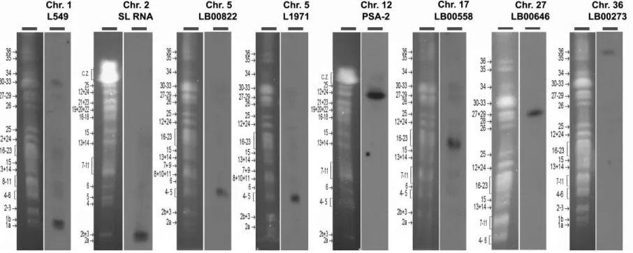
Different sources of chromosome-specific probes were used. The spliced leader RNA gene probe, specific for chr. 2, and the PSA-2 probe, specific for chr. 12 (Wincker et al., 1996), were early gifts from S.M. Beverley (Washington University School of Medicine, St. Louis) and T.W. Spithill (Charles Sturt University, Waga Waga, Australia), respectively. The other probes were BAC (LB00273, LB00558, LB00646, LB00822)
and cosmid (L549, L1971) clones that were kindly provided by Peter Myler (Seattle Biomedical Research Institute) and Christiane Hertz-Fowler (Sanger Centre). The definitive chromosomal location of these probes was made possible from the analysis of a spreadsheet file provided by Al Ivens (Sanger Centre) (Table S3) and confirmed by Southern analysis of the molecular karyotype of the 'Friedlin' strain (Ravel et al., 1998) (see Figure S4). The DNA from BAC and cosmid clones was prepared using Qiagen® Large-Construct Kit. For T. brucei, two DNA probes, corresponding to the alpha- and beta-tubulin genes respectively, were PCR-amplified. Both were used simultaneously in FISH experiments as described elsewhere (Bessat et al., 2009).
Fluorescent In Situ Hybridization.
All probes were labeled with tetramethyl-rhodamine-5-dUTP (Roche Applied Sciences®) by using the Nick Translation Mix (Roche Applied Sciences®). For two-color FISH, the chr. 5-specific BAC clone LB00822 was FITC-labeled using the High Prime labeling kit (Roche Applied Sciences®) according to the manufacturer’s instructions. Cells were fixed in 4% paraformaldehyde and 4% acetic acid, then air-dried on microscope immunofluorescence slides and dehydrated in serial ethanol baths (50-100%). Slides were then hybridized with a heat-denatured DNA probe under a sealed rubber frame at 94°C for 2 min and then overnight at 37°C. The hybridization solution contained 50% formamide, 10% dextran sulphate, 2X SSPE, 250 µg/ml salmon sperm DNA and ≈100 ng of labeled dsDNA probe After hybridization, parasites were sequentially washed in 50% formamide/2X SSC at 37°C for 30 min, 2X SSC at 50°C for 10 min, 2X SSC at 60°C for 10 min, 4X SSC at room temperature. Slides were then mounted in Vectashield (Vector Laboratories®) with DAPI and
microscopically examined (Ersfeld et al., 1997, Freitas-Junior et al., 2000).
Microscopy and fluorescence imaging. Leishmania cells were viewed by phase
contrast, and fluorescence was visualized using appropriate filters on a Zeiss® Axioplan 2 microscope with a 100x objective. Digital images were captured using a Photometrics CoolSnap CDD camera (Roper Scientific®) and processed with MetaView (Universal Imaging®). Z-Stack image acquisitions (15 planes of 0.25 µm) were systematically performed for each cell analyzed using a Piezo controller, allowing to view the nucleus in all planes and to count the total number of labeled chromosomes. In order to generate 3D representations of the complete cell, as well as movies, Z-Stack acquisitions were realized on 35 planes (Supplemental Data, Fig. S2 and Movies M1-3). Movies were made from stack overlays obtained from Metamorph (Universal Imaging®) using Image J 1.37v (National
Institute of Health, USA,
http://rsb.info.nih.gov/ij/).
Data analysis, statistics, controls and precautions.
The ploidy was estimated on a mean of 281 labeled cells for each probe and strain/clone. A total of 21880 cells were examined. Each chromosome was then defined as mono-, di-, tri- or tetrasomic in the cell analyzed, and the ploidy / aneuploidy of the chromosome in the strain expressed as proportions. Examination of the phase contrast and DAPI acquisitions allowed to identify the cells to be counted.
Mitotic cells were excluded from the first analysis. These were either cells with a large or a double kinetoplast and/or with two nuclei; or cells in the early mitotic stage, which were identifiable from the presence of a nascent second flagellum (Ploubidou et al., 1999). For the study of dividing cells, only cells with two clearly individualized nuclei and two flagella were analyzed. Thorough examination, plane by plane, of a numerically magnified view of each cell allowed the determination of the number of labeled chromosomes per individual cell. The efficiency of chromosome labeling by FISH was verified by counting the numbers of non-labeled nuclei (see Table S1). The reliability of the counting method was verified in a number of ways. (i) Counting was performed blinded, i.e. slides were anonymized and neither the strain nor the probe were known by the examiner. (ii) Moreover, the FISH labeling for the seven chromosomes of LmjF was examined twice independently by two researchers, and no statistically significant differences were found between both counts (chi-square test, p value range from 0.06 to 0.74). Similarly, seven other randomly chosen slides were counted twice and no statistical differences were found. (iii) For chr. 5, two probes were available and the counts obtained from two independent FISH experiments were compared (Table S1): again, no statistically significant difference was found between both (chi-square test, p = 0.104); (iv) these two probes were also used in two-color FISH, and the counts obtained for both colors were not statistically different (chi-square test, p = 0.596; see Fig. 2).
Acknowledgments
We wish to thank Peter Myler (Seattle Biomedical Research Institute, USA) and Christiane Hertz-Fowler and David Harris (Sanger Centre, Hinxton, Cambridge, UK) for providing BAC and cosmid clones, and Al Ivens (Sanger Centre, Hinxton, Cambridge, UK) for chromosomal mapping data. We also thank Stephen M. Beverley (Washington University School of Medicine, St. Louis, USA) and Terry W. Spithill (School of Animal and Veterinary Sciences, Charles Sturt University,
Waga Waga, NSW, Australia), for their gift of the spliced leader RNA gene and PSA-2 plasmids, respectively. We are also grateful to Etienne Schwob for critical reading of the manuscript. Finally, we gratefully acknowledge Pierre Travo (RIO Imaging Platform in Montpellier) and Laurence Berry (University Montpellier 2 / CNRS, UMR 5539) for expert advice and assistance in fluorescence microscopy, as well as Sharon Wein (University Montpellier 2 / CNRS, UMR 5539) for her fruitful help at a critical step.
References
Akopyants, N.S., Kimblin, N., Secundino, N., Patrick, R., Peters, N., Lawyer, P., et al. (2009). Demonstration of genetic exchange during cyclical development of Leishmania in the sand fly vector. Science 324, 265-268.
Baldauf, S.L. (2003). The deep roots of eukaryotes. Science 300, 1703-1706.
Barbero, J.L. (2009). Cohesins: chromatin architects in chromosome segregation, control of gene expression and much more. Cell Mol Life Sci 66, 2025-2035.
Bastien, P., Blaineau, C. and Pages, M. (1992). Leishmania: sex, lies and karyotype. Parasitol Today 8, 174-177. Bastien, P. and Wahba, M. (1989). A simplification of the technique for cloning Leishmania. Ann Trop Med Parasitol
83, 435-437.
Bessat, M. and Ersfeld, K. (2009). Functional characterization of cohesin SMC3 and segregation of large and minichromosomes in Trypanosoma brucei. Mol Microbiol. 71, 1371-1385.
Brun, R. and Schönenberger (1979). Cultivation and in vitro cloning or procyclic culture forms of Trypanosoma
brucei in a semi-defined medium. Short communication. Acta Trop 36, 289-292.
Clayton, C.E. (2002). Life without transcriptional control? From fly to man and back again. EMBO J 21, 1881-1888. Cruz, A.K., Titus, R. and Beverley, S.M. (1993). Plasticity in chromosome number and testing of essential genes in
Leishmania by targeting. Proc Natl Acad Sci U S A 90, 1599-1603.
Curotto de Lafaille, M.A., Laban, A. and Wirth, D.F. (1992). Gene expression in Leishmania: analysis of essential 5' DNA sequences. Proc Natl Acad Sci U S A 89, 2703-2707.
Dubessay, P., Blaineau, C., Bastien, P. and Pagès, M. (2004). Chromosome fragmentation in Leishmania. Methods
Mol Biol 270, 353-378.
Dubessay, P., Ravel, C., Bastien, P., Stuart, K., Dedet, J.P., Blaineau, C. and Pages, M. (2002). Mitotic stability of a coding DNA sequence-free version of Leishmania major chromosome 1 generated by targeted chromosome fragmentation. Gene 289, 151-159.
Duesberg, P., Li, R., Fabarius, A. and Hehlmann, R. (2005). The chromosomal basis of cancer. Cell Oncol 27, 293-318.
Ersfeld, K. and Gull, K. (1997). Partitioning of large and minichromosomes in Trypanosoma brucei. Science 276, 611-614.
Freitas-Junior, L.H., Bottius, E., Pirrit, L.A., Deitsch, K.W., Scheidig, C., Guinet, F., et al. (2000). Frequent ectopic recombination of virulence factor genes in telomeric chromosome clusters of P. falciparum. Nature 407, 1018-1022. Gisselsson, D. (2008). Classification of chromosome segregation errors in cancer. Chromosoma 117, 511-519.
Gisselsson, D., Hakanson, U., Stoller, P., Marti, D., Jin, Y., Rosengren, A.H., et al. (2008). When the genome plays dice: circumvention of the spindle assembly checkpoint and near-random chromosome segregation in multipolar cancer cell mitoses. PLoS One 3, e1871.
Huettel, B., Kreil, D., Matzke, M. and Matzke, A. (2008). Effects of aneuploidy on genome structure, expression, and interphase organization in Arabidopsis thaliana. PLoS Genet 4, e1000226.
Ivens, A.C., Peacock, C.S., Worthey, E.A., Murphy, L., Aggarwal, G., Berriman, M., et al. (2005). The genome of the kinetoplastid parasite, Leishmania major. Science 309, 436-442.
Jallepalli, P.V. and Pellman, D. (2007). Cell biology. Aneuploidy in the balance. Science 317, 904-905.
Leprohon, P., Legare, D., Raymond, F., Madore, E., Hardiman, G., Corbeil, J. and Ouellette, M. (2009). Gene expression modulation is associated with gene amplification, supernumerary chromosomes and chromosome loss in antimony-resistant Leishmania infantum. Nucleic Acids Res 37, 1387-1399.
Martinez-Calvillo, S., Stuart, K. and Myler, P.J. (2005). Ploidy changes associated with disruption of two adjacent genes on Leishmania major chromosome 1. Int J Parasitol 35, 419-429.
Otto, S.P. and Gerstein, A.C. (2008). The evolution of haploidy and diploidy. Curr Biol 18, R1121-1124.
Ploubidou, A., Robinson, D.R., Docherty, R.C., Ogbadoyi, E.O. and Gull, K. (1999). Evidence for novel cell cycle checkpoints in trypanosomes: kinetoplast segregation and cytokinesis in the absence of mitosis. J Cell Sci 112 ( Pt 24), 4641-4650.
Polakova, S., Blume, C., Zarate, J.A., Mentel, M., Jorck-Ramberg, D., Stenderup, J. and Piskur, J. (2009). Formation of new chromosomes as a virulence mechanism in yeast Candida glabrata. Proc Natl Acad Sci U S A 106, 2688-2693. Ravel, C., Dubessay, P., Bastien, P., Blackwell, J. and Ivens, A. (1998). The Complete Chromosomal Organization of the Reference Strain of the Leishmania Genome Project, L. major ;Friedlin'. Parasitol Today 14, 301-303.
Rougeron, V., De Meeus, T., Hide, M., Waleckx, E., Bermudez, H., Arevalo, J., et al. (2009). Extreme inbreeding in
Leishmania braziliensis. Proc Natl Acad Sci U S A 106, 10224-10229.
Selmecki, A., Forche, A. and Berman, J. (2006). Aneuploidy and isochromosome formation in drug-resistant Candida
albicans. Science 313, 367-370.
Shaw, J.M., Feagin, J.E., Stuart, K. and Simpson, L. (1988). Editing of kinetoplastid mitochondrial mRNAs by uridine addition and deletion generates conserved amino acid sequences and AUG initiation codons. Cell 53, 401-411.
Sunkin, S.M., Kiser, P., Myler, P.J. and Stuart, K. (2000). The size difference between Leishmania major friedlin chromosome one homologues is localized to sub-telomeric repeats at one chromosomal end. Mol Biochem Parasitol
109, 1-15.
Torres, E.M., Sokolsky, T., Tucker, C.M., Chan, L.Y., Boselli, M., Dunham, M.J. and Amon, A. (2007). Effects of aneuploidy on cellular physiology and cell division in haploid yeast. Science 317, 916-924.
Ubeda, J.M., Legare, D., Raymond, F., Ouameur, A.A., Boisvert, S., Rigault, P., et al. (2008). Modulation of gene expression in drug resistant Leishmania is associated with gene amplification, gene deletion and chromosome aneuploidy. Genome Biol 9, R115.
Wincker, P., Ravel, C., Blaineau, C., Pages, M., Jauffret, Y., Dedet, J. and Bastien, P. (1996). The Leishmania genome comprises 36 chromosomes conserved across widely divergent human pathogenic species. Nucleic Acids Res 24, 1688-1694.
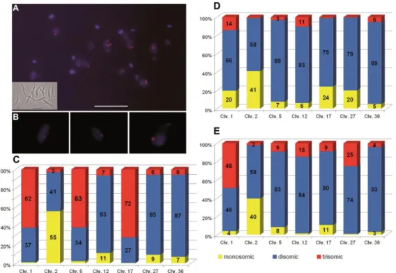
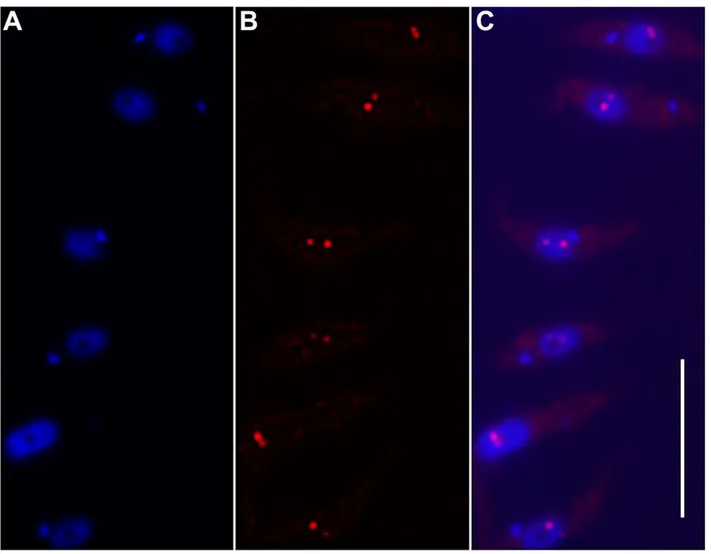
Figure 1. Aneuploidy and mosaicism in Leishmania major 'Friedlin' strain
A. The mosaic structure of L. major 'Friedlin' strain (LmjF) shown by Fluorescence In Situ Hybridization. Chr. 5 of LmjF probed with the rhodamine-labeled BAC clone LB00822 (red) is
shown as an example on this focused plane. The nucleus and the intensely staining kinetoplast, which is the single mitochondrial DNA, are stained with DAPI (blue). Insert shows a view of the same promastigote L. major cells in log-phase growth in phase-contrast microscopy. Cells with different numbers of fluorescent dots (corresponding to copies of chr. 5) can be seen. However, only Z-stack acquisitions (allowing 3D analysis) allow determining exactly the number of labeled chromosomes per individual nucleus (see B). Variable ploidy among individual cells generates intra-strain heterogeneity, i.e. mosaicism. Bar = 10 µm. B. Enlarged 2-D projection of Z-series
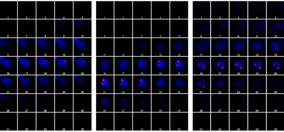
of individual L. major cells, showing one (left image), two (centre) or three (right) copies of the
labeled chromosome. The images are extracted from the 3-D reconstructions shown in Supplemental Data (Fig. S2 and Movies M1-3). C. Variable ploidy for seven chromosomes in
strain LmjF. The graph shows the proportions (percentages in ordinate) of mono- (yellow), di-
(blue) and trisomic (red) chromosomes in LmjF. Each bar represents one of the seven chromosomes studied (abscissa). The ploidy was estimated from the examination of 3-D views of a mean of 338 labeled cells per each chromosome. Four chromosomes appear strongly aneuploid. Chr. # 1, 5 and 17 are predominantly trisomic, chr. 2 predominantly monosomic, and chr. # 12, 27 and 36 predominantly disomic. Remarkably, mosaicism was observed for all the chromosomes analyzed; nevertheless, the proportions of mono-, di-, or trisomic chromosomes vary depending on the chromosome studied. D-E. Mosaicism extends to other L. major strains. The seven chromosomes studied in strain LmjF were analyzed in strain LEM62 (D) and in LEM265 (E). Same color code as in C. Aneuploidy and mosaicism were observed in both strains for all chromosomes analyzed. Moreover, mono-, di- or trisomy does not appear as a specific feature of a given chromosome in L. major (see text).
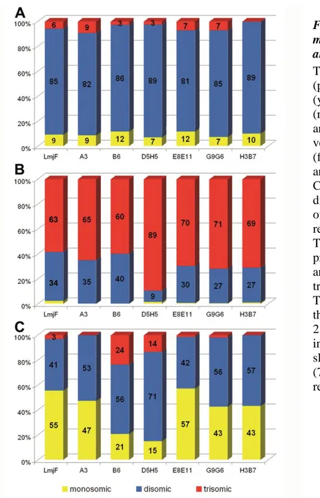
Figure 2. Two-color FISH on the same chromosome.
Chr. 5 in strain LmjF was studied in two-color FISH using the specific BAC clone LB00822 and cosmid clone L1971 as DNA probes. The distance between both probes on the chromosome was 76kb. A focused plane of three individual nuclei, monosomic (A), disomic (B) and trisomic (C) for chr. 5, is shown as an example. First column: DAPI-stained nucleus (and kinetoplast in the bottom row). Chr.5 was simultaneously probed with the FITC-labeled clone LB00822 (green) and the tetramethyl-rhodamine-5-dUTP-labeled clone L1971 (red). Right-hand side columns: merged images of both probes and of both probes with DAPI (blue). Both hybridizations gave identical results. Bar = 2 µm
Figure 3. Variable transmission of mosaicism between parental strain and derived clones.
The graph shows the proportions (percentages in ordinate) of mono- (yellow), di- (blue) and trisomic (red) chromosomes in strain LmjF and derived cell clones. The vertical bars represent strain LmjF (far left) and the different clones analyzed (abscissa). A: Chr. 27; B: Chr. 5; C: Chr. 2. The predominant disomy of chr. 27 (A) and trisomy of chr. 5 (B) in strain LmjF were retained in all of its derived clones. The same was observed for the predominant disomy of chr. # 12 and 27 and for the predominant trisomy of chr. # 1 and 17 (see Table S1 for details). By contrast, the proportion of monosomy of chr. 2 in LmjF (C) was conserved only in clone E8E11, while disomy was slightly (53%-57%) or largely (71%) predominant in the remaining clones.
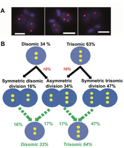
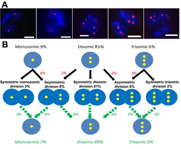
Figure 4. FISH analysis of mitotic cells revealed a high proportion of asymmetric chromosomal allotments, leading to constitutive mosaicism.
A. Chr. 5-specific probe LB00822 showed different chromosomal allotment patterns. As
expected in view of the di- and trisomic nature of chr. 5 in LmjF, 2+2 (left) and 3+3 (right) division patterns were seen. But asymmetric 2+3 chromosomal allotments were also observed (center). Bar: 2 µm. B. Proposed model for explaining the presence of 'constitutive' mosaicism
in L. major. The predominantly trisomic chr. 5 is taken as an example. Disomic and trisomic nuclei are figured by double and triple yellow dots in blue circles. The proportions observed while studying interphasic cells (top line) and mitotic cells (middle line) are in black. Figures that are inferred from the latter are in color (red or green). We observed 34% of disomic interphasic cells (top line) but only 16% of symmetric ‘2+2’ chromosomal allotments (middle line). Therefore, we inferred that 18% of disomic cells divide asymmetrically (‘2+3’ patterns). Following the same reasoning, 16% of trisomic cells must divide asymmetrically (‘2+3’). We then calculated the inferred proportions of di-and trisomic nuclei in the population of daughter cells (green, bottom line) from the observed proportions of symmetric (‘2+2’ and ‘3+3’) and asymmetric (‘2+3’) divisions (green arrows). Remarkably, these figures are almost identical to those observed in interphasic cells (33%/64% and 34%/63%, respectively). This leads to a stability of the mosaic state. Details of the observed chromosomal allotments are given in Table S2. It is noteworthy that the patterns ‘1+2’ and ‘3+4’ were exceptionally found for this chromosome but not for other chromosomes (see Fig. 5, Supplemental Fig. S3 and Table S2).
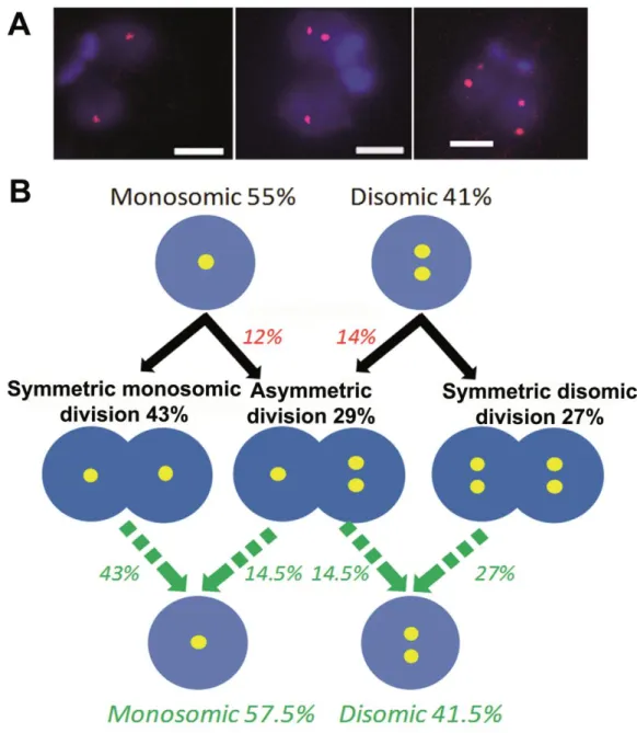
Figure 5. Proposed model for explaining the presence of 'constitutive' mosaicism in L. major for chromosome 2.
A. The spliced leader RNA gene probe, specific for the predominantly monosomic chr. 2, revealed
several chromosomal allotment patterns, some of them being different from those observed for chr. 5: indeed, on top of symmetric chromosomal allotments 1+1 and 2+2 (left and right), asymmetric ones of the 1+2 type were observed (center). Bar = 2 µm.
B. Proposed model for explaining 'constitutive' mosaicism for a predominantly monosomic
chromosome (chr. 2 in LmjF). The legend is similar to that of Fig.4. The model proposed for chr. 5 (Fig. 4B) also fits for a chromosome which is mainly monosomic.
Supporting information
Supplemental Text S1.
Calculation of the probability of confusion of two independent fluorescent dots in a
Leishmania nucleus.
We calculated the probability that two independent fluorescent dots be confused in (a 3D- reconstitution of) a Leishmania nucleus, taking into account the uncertainty on the position of such a dot in light microscopy in both the horizontal and vertical planes.
In conventional wide field fluorescence microscopy, the lateral resolution is generally considered to be about 0.2 µm and the axial resolution is roughly 0.6-0.8 µm (Petty et al. 2007, Microsc. Res.
Tech.70:687-709), this being in controlled experimental conditions (refraction index of the thin
slide and of the mounting medium, objective x100, high camera resolution). Consequently, a fluorescent dot forms an ellipsoid of which the volume can be calculated using the following formula: V= 4/3 q2p, with q (= 0.2 µm) and p (= 0.6-0.8 µm) corresponding to polar and equatorial radii respectively.
The probability that two distinct dots that are located in the nucleus appear as a single dot is given by the ratio between the volume of the ellipsoid and that of the nucleus :
P = 4/3 q2p / 4/3 R3 = q2p/ R3
with R being the radius of the nucleus (estimated at 1 µm).
In our study, therefore, the probability that two chromosomes be confused as one is 2.4 to 3.2 %. This probability appears actually quite small and rules out that this may be the cause of a severe bias in our study. This calculation is concordant with the margin of errors observed when analyzing the disomic Chr.1 of T. brucei (Supplemental Table S1 and Fig. S1).
Figure S1. FISH analysis of chromosome 1 in Trypanosoma brucei strain Tb427.
Chr. 1 of strain Tb427, probed with the rhodamine-labeled alpha-beta tubulin DNA probe (red), is shown on this focused plane. Left: DAPI-stained DNA. Right: merged image. A large majority (95%) of the cells was found disomic for this chromosome. Bar = 10 µm.
Figure S2. Gallery view of Z-stack images.
Thirty-five consecutive Z-stack acquisitions were used to generate 3D views and movies of the cell and to count the total number of labeled chromosomes. An example is given for mono-, di- and trisomic cells (left, central and right panel respectively). The DAPI-stained nucleus and the kinetoplast (when the latter is visible, left panel) are colored in blue and the rhodamine-labeled chromosomes are revealed as red dots. Typically, in these acquisitions, a dot is observable on five to seven consecutive planes.
Figure S3. Proposed model for explaining the presence of constitutive mosaicism in L. major for chromosome 27.
A. The LB00646 BAC DNA probe, specific for chr. 27, revealed several chromosome allotment
patterns: according to its being mainly disomic but also mono-and trisomic in some cells, a majority of symmetric chromosomal allotments 2+2 (middle) was found, but both other symmetric patterns (1+1 and 3+3, left and right respectively) were also seen, as well as asymmetric ones (1+2 and 2+3). Bar = 2 µm.
B-C. Proposed model for explaining the constitutive mosaicism for a predominantly disomic
chromosome. The legend is similar to that of Fig.4 and 5. The model proposed from chr. 5 (Fig. 4B) and 2 (Fig. 5B) applies to this chromosome as accurately as for chr. 5, even when the ploidy distribution is more complex.
Figure S4. Localization of the DNA probes used for this study onto chromosomes of the L. major Friedlin strain (LmjF) by PFGE Southern analysis.
The figure shows, for each probe, the ethidium bromide-stained PFGE gel of the molecular karyotype (on the left) and the autoradiogram of the probe hybridization on the corresponding Southern blot (on the right). Each probe (top line) was specific for a given chromosome (bottom line), confirming end-sequence data. SL RNA = spliced leader RNA (or mini-exon). PSA-2 = promastigote surface antigen 2. The position and number of the LmjF chromosomes are shown on the left of each karyotype (Ravel et al., 1998,
Parasitol Today. 14(8):301-303). C.Z., compression zone.
PFGE was run on a home-made device (Bastien, P. et al., 1992, in Intracellular Parasites (Avila, J.L. and Harris, R., eds), Subcellular
Biochemistry 18:131–187, Plenum Press) using the following migrating conditions: pulse times of 300s/130s/90s during 24h each
(probes L549 and LB00273), 300s/130s/90s during 30h each (probe LB00646), 270s/100s/70s during 24h each (probes LB00822, L1971 and LB00558), and 90s during 30h, 80s during 36h and 60s during 24h (probes SL RNA and PSA-2). Blottings and hybridizations were carried out according to current procedures (Sambrook et al., 1989, Molecular cloning: A laboratory manual (Cold Sprong Harbor Laboratory Press, Cold Spring Harbor, NY, USA).
Movies M1, M2 and M3.
Three-dimensional reconstructions of L. major nuclei showing the total number of chromosome homologues in one nucleus for a monosomic (M1), disomic (M2) and trisomic (M3) chromosome, respectively.
The DAPI-stained nucleus and kinetoplast (when the latter is visible) are coloured in blue and the rhodamine-labeled chromosomes in red. The 35 consecutive planes acquired (see gallery view in Fig. S2) were processed in Metaview®, Metamorph® and ImageJ.