S205
Pathogenesis and Potential Strategies for Prevention and Treatment of Septic
Shock: An Update
M. P. Glauser, D. Heumann,
J.
D. Baumgartner, andJ.
CohenFrom the Division of Infectious Diseases, Department of Internal Medicine, Centre Hospitalier Universitaire Vaudois, Lausanne. Switzerland; and the Departments of Infectious Diseases and o]
Bacteriology and Medicine, Hammersmith Hospital and Royal Postgraduate Medical School, London, United Kingdom Septic shock is mediated by complex interactions of cells, cytokines, and humoral pathways.
Clinical therapeutic strategies aimed at inhibiting selected pathways have been efficacious in subsets of patients. Experimental studies focusing on the activities of single cytokines have contributed to the understanding of the complex pathophysiology of septic shock. More precise delineation of the roles of each mechanism contributing to pathogenesis will permit the identifi-cation of subsets of patients who might benefit from particular therapeutic strategies and will guide the development of additional approaches to prevention and treatment.
Septic shock is a clinical syndrome that has become in-creasingly important in the last 40 years. The condition is most common among hospitalized patients, particularly those with underlying diseases. Although patients with dis-eases caused by "classic" gram-negative pathogens (such as plague or typhoid fever) may present with the clinical picture of septic shock, it is only since the I 950s-with the increas-ing incidence of disease caused by gram-negative bacilli of the normal host flora-that the sepsis/septic shock syn-dromes have been defined. We believe that these definitions (table I) [I, 2] are satisfactory: the identical incidences of shock and death in the various clinical studies using these criteria indicate that similar groups of patients are being enrolled. However, the definitions need to be improved to take into account various factors that are important for pre-dicting outcome, including underlying diseases and appropri-ateness ofantibiotic and medical/surgical treatment. A modi-fied scheme for classification of these syndromes-known as systemic inflammatory response syndrome (SIRS) and multi-ple organ dysfunction syndrome (MODS) [3]-is currently being discussed, but no consensus has yet been reached.
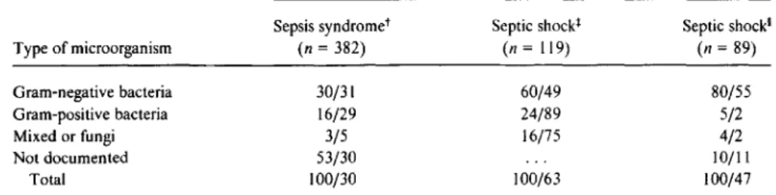
Septic shock has traditionally been recognized as a conse-quence of gram-negative bacterial infection, but it may also be caused by gram-positive organisms and fungi and proba-bly by viruses and parasites as well. Table 2 summarizes the organisms isolated and the mortality documented in three recent studies of sepsis syndrome/septic shock [2, 4, 5].
This article is adapted with permission from two previously published articles: Glauser MP, Zanetti G, Baumgartner JD, Cohen J. Septic shock: pathogenesis. Lancet 1991;338:732-6; and Cohen J, Glauser MP. Septic shock: treatment. Lancet 1991;338:736-9.
Reprints or correspondence: Prof. M. P. Glauser, Division of Infectious Diseases, Department of Internal Medicine, Centre Hospitalier Universi-taire Vaudois, 101I Lausanne, Switzerland.
Clinical Infectious Diseases 1994;18(8uppI2):8205-16 © 1994 by The University of Chicago. All rights reserved. 1058-4838/94/1802-0036$02.00
Gram-negative bacteria were isolated in 30%-80% of cases and gram-positive bacteria in 5%-24%. In one prospective study of sepsis syndrome [2], no etiologic agent was identi-fied in more than half of all cases. Notably, the severity of septic shock, as reflected by mortality, did not depend on the type of organism responsible.
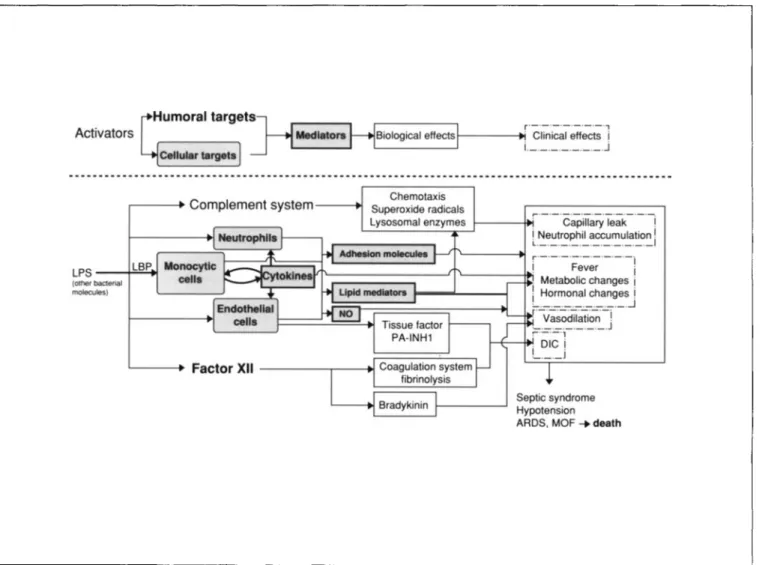
Initial studies of the pathophysiological features of septic shock concentrated on the interactions oflipopolysaccharide (LPS) from the gram-negative bacterial cell wall with various humoral pathways. However, attention is now focused on the central role of macrophages, endothelium, and cytokines that are released upon stimulation by most if not all of the recognized agents of septic shock (figure I). In this review, we address the known humoral pathways that are activated during septic shock, and we discuss the role of cytokines, particularly tumor necrosis factor (TNF) and interleukin I (IL-I ). Although the current emphasis is on the activation of macrophages and cytokine production, we think that evi-dence for direct activation of humoral pathways by microbial constituents remains relevant. We also review the mecha-nisms by which LPS interacts with macrophages, citing exper-imental and clinical studies evaluating the potential of anti-LPS and anticytokine agents in therapy for septic shock.
Bacterial Cell-Wall Components and Septic Shock The exotoxins produced by some bacteria (e.g., exotoxin A produced by
Pseudomonas aeruginosa
or toxic shock syn-drome toxin produced by some strains ofStaphylococcus
aur-eus)
can initiate septic shock. However, the bacteria them-selves-and in particular their cell wall components-are primarily responsible for the development of septic shock. These components are potent activators of numerous hu-moral pathways and also of macrophages and other cells in-volved in inflammatory processes.The prime initiator of gram-negative bacterial septic shock is endotoxin, an LPS component of the bacterial outer
mem-S206 Glauser et al. CID 1994; 18 (Suppl 2)
Table I. Definitions of sepsis syndrome and septic shock. Sepsis syndrome
Clinical evidence of infection Tachypnea*
Tachycardia"
Hyperthermia or hypothermia! Evidence of inadequate organ perfusion.
including one or more of the following:
Hypoxemia'
Elevated plasma lactate concentration'' Oliguria"
Septic shock Sepsis syndrome with
hypotension**
bacteria. Internal to the a side chains are the core oligosac-charides, which have similar structures in common gram-negative bacteria. Lipid A, which is bound to the core oligo-saccharide, has a highly conserved structure and is responsible for most of the toxicity of endotoxin. However, some types of natural lipid A and synthetic lipid A analogues that have different sugar and acyl residues are less-or not at all-toxic both in vitro and in vivo. This observation has led to the development of lipid A analogues that can block the toxic effects of endotoxin or act as endotoxin antagonists [8, 9].
* Respirations. >20/min; if mechanically ventilated. > 10 L/min. tPulse. >90/min.
*
Core or rectal temperature.>38.3°Cor<35.6°C., Pao2/FI02.~280(without other pulmonary or cardiovascular disease as the cause).
IIExceeding upper limits of normal for the laboratory.
# Documented urine output. <0.5 mL/kg of body weight for at least I
hour (in patients with catheters).
**Sustained decrease in systolic blood pressure to <90 mm Hg or drop by >40 mm Hg for at least I hour when volume replacement is adequate. the patient is taking no antihypertensive medication. and other causes of shock (such as hypovolemia. myocardial infarction. and pulmonary embolism) are absent.
brane. Endotoxin circulating in the blood appears to be a predictor of poor outcome in some clinical settings (e.g., meningococcemia [6]), but the levels of endotoxin required to trigger the cascade of events in septic shock may vary greatly. Indeed, it has been observed that bacterial products other than LPS may profoundly increase the host's sensitivity to endotoxin, thereby rendering toxic otherwise-harmless lev-els [7]. Hence, the measurement of endotoxin has not yet become standard clinical practice.
The outermost part of the endotoxin molecule consists of a series of structurally and antigenically diverse oligosaccha-rides that are responsible for the a serotype ofgram-negative
Antibodies to Endotoxin
The a-specific oligosaccharide side chains of endotoxin are highly immunogenic. Antibodies to these side chains in-hibit the effects of endotoxin and, by virtue of their opsono-phagocytic properties, eradicate the endotoxin-producing or-ganism. However, because these antibodies are specific for a particular 0 serotype, their clinical application is limited. An alternative approach has been to develop antibodies to the structurally conserved core glycolipid of endotoxin or to lipid A in the hope that these antibodies will offer cross-reac-tivity or cross-protection against the toxic component of all gram-negative bacteria. In initial clinical trials conducted10
years ago, antisera or preparations of hyperimmune polyclo-nal intravenous immunoglobulin were used(l0-13]. While the results of these trials suggested a benefit and stimulated subsequent trials of monoclonal antibodies, they did not spe-cifically demonstrate that antibodies to lipid A were responsi-ble for the protection observed.
Recently, clinical trials of two monoclonal antibodies to the core glycolipid of endotoxin have received considerable attention [14, 15]. In a prospective, randomized, placebo-controlled trial ofE5 (a murine monoclonal IgM antibody to lipid A), 486 patients with suspected gram-negative sepsis
Table2. Frequency of isolation of various types of microorganisms and corresponding mortality in three recent studies of sepsis syndrome/septic shock.
Frequency of isolation/mortality among patients with indicated condition*
Type of microorganism Gram-negative bacteria Gram-positive bacteria Mixed or fungi Not documented Total
* Figures are percentages. tData are from [2].
*
Data are from [4]. 'Data are from [5].Sepsis syndromet (n= 382) 30/31 16/29 3/5 53/30 100/30 Septicshockl (n=119) 60/49 24/89 16/75 100/63 Septic shock' (n=89) 80/55 5/2 4/2 10/11 100/47
CID 1994; 18 (Suppl 2) Pathogenesis and Treatment of Septic Shock S207
Activators Biological effects1 - - - . Clinical effectsr'-'-'-'-'-'-'-"i i.__.__._.__.J
Septic syndrome Hypotension ARDS, MOF ....death
1---:-·-·c~iil~~I~;k-·-·-1 iLNeutrophil accumulation. . ._._II r--'-'---'-'-'-'-'-'I '--- ...! Fever i Metabolic changesi : Hormonal changesi '-.__. . . .-.J ['-v;~~dii~ti~;;;-i - ._._. .J ;._._., . DICi I -._._._J Chemotaxis Superoxide radicals Lysosomal enzymes LBP L---t> Factor XII- - - . - - - - + 1 ,.---. Complement system- - - + I
Figure 1. Interaction of humoral factors and cytokinesin the pathogenesis of septicshock. LPS= lipopolysaccharide; LBP= LPS-binding protein; PA-INHI = plasmin activator-inhibitor I; DIe
=
disseminated intravascular coagulation; ARDS=
adult respiratory distress syndrome; NO= nitric oxide; and MOF= multiple organ failure.received either placebo or two intravenous doses of E5 (2 mg/kg) 24 hours apart [14]. The two groups of patients were reasonably well matched, although the unavailability of scores on the Acute Physiologic and Chronic Health Exami-nation (APACHE) II for 203 patients represented a poten-tially important source of bias. Of the 468 evaluable patients, 316 had a documented gram-negative infection; 179 of these 316 patients presented with shock. Among the 137 patients who did not present with shock, treatment with E5 was asso-ciated with significantly lower 30-day mortality(P = .03) and with significantly more frequent resolution of major morbidities (i.e., complications of shock such as dissemi-nated intravascular coagulation [DIC] and acute renal fail-ure)(P
=
.04) than was placebo administration. However, no differences in mortality were found between E5-treated pa-tients and placebo recipients who presented with shock or between E5-treated patients and placebo recipients who did not have gram-negative sepsis. Administration of E5 was found to be safe; it did cause an increase in the level ofanti-body to murine immunoglobulin in approximately one-half of patients, but the titer was usually low and of no clinical importance.
Since this study suggested that E5 was effective in a sub-group of patients defined retrospectively, a second multi-center study was conducted to verify the finding prospec-tively. A total of 847 patients without shock were enrolled. The favorable results of the first study were not confirmed: treatment with E5 did not affect survival among the 530 patients with documented gram-negative sepsis. However, a favorable-albeit statistically insignificant-trend was noted in the subgroup of patients in whom gram-negative sepsis was associated with major morbidities, such as DIe and renal failure [16]. A third trial of E5 is under way.
A clinical trial was also performed with HA-I A, a human monoclonal antibody to lipid A. In a study similar in design to that described for E5, patients with suspected gram-nega-tive infection were randomized to receive either an albumin placebo or a single 100-mg intravenous dose ofHA-1 A [15].
S208 Glauser et al. CIO 1994; 18 (Suppl 2)
Of 543 patients enrolled in the study, 317 had microbiologi-cally documented gram-negative infections; 117 of these 317 patients had had sterile blood cultures at randomization. HA-l A did not reduce mortality either in the overall study population or in the 117 patients with nonbacteremic gram-negative infections. However, mortality did decrease with treatment among the 200 patients with gram-negative bacter-emia(P
=
.014). While this difference in mortality was also noted among the 101 patients with gram-negative bacter-emia who presented with shock at enrollment, no such differ-ence was found among the 99 patients with gram-negative bacteremia who did not present with shock. These data con-trast with the findings from the first study of E5, in which patients appeared to be protected whether or not they were bacteremic but only when they were not in shock.This report of reduced mortality in a subgroup of patients given HA-l A led to the licensure of the product in some European countries. However, in the United States, the Food and Drug Administration did not approve this product for several reasons, including (1) changes in the protocol during the clinical trial; (2) the documentation of significant differences only in subgroups of patients; and (3) possible imbalances favoring the test drug at randomization of the patients [17]. Therefore, a second study that focused on pa-tients with gram-negative bacteremia and shock was initi-ated. During this trial the manufacturer voluntarily withdrew HA-l A from the market in Europe because of excess mortal-ity in patients without gram-negative bacteremia [18].
These disappointing results with the two monoclonal anti-bodies to lipid A are, perhaps, not totally unexpected in light of the studies that led to their development. First, in the original experiments testing the hypothesis that cross-protec-tion could be provided by antibodies to core glycolipid of endotoxin, killed bacteria from a rough-cell-wall mutant strain ofEscherichia coli
0
III (strain 15) were used for the immunization of animals or humans in order to obtain poly-clonal antisera. Although some studies showed protection with polyclonal antisera to 15, it was impossible to show definitively that protection was attributable to cross-protec-tive antibodies. Indeed, a favorable outcome could not be correlated with antibody titers in either of the two clinical studies done with human polyclonal antisera to 15 ([ 10, 11] and 1. D. Baumgartner, unpublished data). By solubilization of the core glycolipid in a physiological manner to circum-vent nonspecific binding [19], the 15 antisera used in one study were later shown to contain IgG and IgM antibody to 15 at a titer only threefold higher than that in control (preim-mune) serum; furthermore, these antisera contained no more antibodies to lipid A than did the control serum [20]. More-over, it now appears that antibodies to 15 are highly specific forE.coli15 and that they do not cross-react with endotoxin from other bacteria. Hence, the mechanisms of protection by antisera to 15 remain unknown. Nevertheless, this approach with polyclonal sera raised against rough structures of LPSlaid the groundwork for the development of therapeutic monoclonal antibodies to core glycolipid.
Second, it now appears that clinical trials with these mono-clonal antibodies to lipid A were initiated before the immu-nologic reactivity of these antibodies for LPS substructures was recognized. Since the first disclosure of the possible effi-cacy ofE5 and HA-l A in patients with septic shock [14, 15], many studies have been undertaken to define the reactivity of these antibodies for LPS. In studies by Fujihara et al. [21] and Mascelli et al. [22], HA-l A reportedly bound to lipid A, rough LPS structures, and some preparations of smooth LPS; however, Baumgartner could not confirm such reactivity [23]. Thus the specificity of these antibodies for lipid A is uncertain, in part because immunoglobulins-especially those of the IgM isotype-tend to bind nonspecifically to both the highly amphophilic core oligosaccharide ofLPS and lipid A molecules.
It has also been suggested that HA-IA lowers levels of endotoxin by mediating the clearance of LPS via comple-ment-dependent binding ofLPS to complement receptor ex-pressed on erythrocytes and neutrophils [24, 25]. Similarly, E5 was shown to bind to lipid A, to rough LPS, and to 15 preparations of smooth LPS isolated from various strains of gram-negative bacteria [26, 27]. However, neither HA-lA nor E5 was able to neutralize LPS in vitro, as assessed by the limulus lysate test, by a mitogenic assay for murine spleno-cytes, or by measurement of the production of cytokines in human whole blood [28].
Studies with HA-l A in vivo have yielded inconclusive re-sults. Initially, HA-lA was reported to be protective in mice when used unpurified (as hybridoma fluid) [29]; however, a purified monoclonal antibody obtained from the same clone was not protective in similar experiments and did not sup-press endotoxin-induced production of TNF in vivo [30]. A similar failure of HA-lA to protect mice from LPS-induced death was recently reported [31]. More important, in recent experiments with dogs, HA-lA did not alter levels of'bacter-emia or endotoxof'bacter-emia and was actually associated with a de-creased rate of survival [32]. E5, initially studied in vivo by Young et al. [33], was subsequently shown to be beneficial in a model of pseudomonas sepsis in neutropenic mice [34].
The discrepancies in these results of in vitro and in vivo studies stressed the need for extensive characterization of antibodies to core glycolipid before the initiation of clinical trials.
Activation of Pathways Other Than Cytokines by Cell
Wall Components
Numerous humoral mediators of sepsis have been identi-fied so far, and it is likely that more will be discovered. These mediators act through complex synergistic and antagonistic interactions. LPS in the blood activates the coagulation and complement cascades and induces a broad array ofmediators
CID 1994; 18 (Suppl 2) Pathogenesis and Treatment of Septic Shock S209
from macrophages and other cells, including endothelial cells.
The alternative complement pathway can be activated ex-perimentally by LPS and gram-positive bacterial cell-wall components. The classic pathway is activated mainly by complexes of cell wall components and antibodies. The ana-phylatoxins C3a and C5a, which are produced as a result of activation of these pathways, are responsible for a series of inflammatory events that have been implicated in the patho-physiology of septic shock. C5a has been directly associated with TNF and LPS in hemorrhagic necrosis [35]. Comple-ment components induce vasodilation and increased vascu-lar permeability, which can result in hemodynamic changes, aggregation of platelets, and aggregation and activation of neutrophils-all processes that have been implicated in the pathogenesis of the adult respiratory distress syndrome [36]. The subsequent release of arachidonic acid derivatives, cyto-toxic products of molecular oxygen, and lysosomal enzymes exerts additional local vasoactive effects on the microvascula-ture and causes endothelial cell cytotoxicity, which results in capillary leakage. An increased concentration of activated complement has been associated with a fatal outcome in septic shock of both gram-positive and gram-negative origin [37J.
Itis well known that arachidonic acid metabolites cause vasodilation, platelet aggregation, and neutrophil activation, which may contribute to the pathogenesis of septic shock. These substances are found in increased concentrations after experimental challenge with endotoxin and during septic shock in patients [38J. The role of inhibitor/antagonists of the pathways of arachidonic acid metabolism in the treat-ment of septic shock is being investigated.
Activated neutrophils, a key element in the inflammatory response, probably play an important part in the pathogene-sis of septic shock by contributing to vascular and tissue inju-ries. Strong evidence indicates that neutrophils are activated either directly by LPS or indirectly through the action of cytokines [39]. As a result, activated neutrophils may dam-age tissues by releasing oxygen metabolites and lysosomal enzymes, or they may cause microemboli after aggregation. Activated leukocytes adhere to one another, to endothelial cells, and to tissues through interactions of receptors (on en-dothelial cells) and ligands (on inflammatory cells) that are mediated by specific adhesion molecules (figure 2). The ad-hesion process is essential to most functions of leukocytes (including chemotaxis, phagocytosis, and cytotoxicity [40]), and blocking ofthe adhesion process by monoclonal antibod-ies prevents tissue injury and improves the survival rate in animal models of septic shock [41 ].
Two humoral factors are currently being evaluated for their role as mediators ofseptic shock: platelet-activating fac-tor (PAF) and nitric oxide (NO). The infusion of endotoxin induces the release of PAF, a potent phospholipid mediator that leads to autocatalytic amplification of cytokine release.
PAF is a mediator of inflammation caused by macro phages, neutrophils, platelets, and endothelial cells in response to injury (figure 2). Elevated levels ofPAF have been found in models of induced hypotension and endotoxin-induced lung injury in rats [42, 43]. Many antagonists to PAF exist [44]. The first data from a randomized clinical trial of the efficacy of a PAF antagonist in severe sepsis have re-cently been reported [45]. In this study 262 patients received either placebo or the PAF antagonist. Mortality decreased by 42% with PAF antagonist treatment in a subset of 119 pa-tients with documented gram-negative sepsis (57% vs. 33%;
P
=
.011). A confirmatory study focusing on gram-negative sepsis is now in progress.Hypotension during septic shock may reflect increased syn-thesis of the potent vasodilator NO. Considerable informa-tion is now available on the activity of NO in vitro [46]. Upon LPS challenge, NO is produced mainly by macro-phages, endothelial cells, smooth muscle cells, and the liver. Although LPS-induced release of NO by macrophages ap-pears to take several hours, endothelial cells react within min-utes-a phenomenon that may contribute to the rapid de-crease in blood pressure associated with endotoxic shock [47]. An increasing number of reports deal with the inhibi-tion of this pathway in animal models, but the precise role of NO in septic shock remains unclear [48]. Activity of NO has been detected in experimental gram-negative sepsis [49] and in patients with the sepsis syndrome [50]. While NO block-ade in animals has been reported to be beneficial in some studies [51, 52], it has also been found to be detrimental [53]. Treatment with N-monomethyl-L-arginine, which blocks NO synthesis, transiently restored blood pressure and systemic vascular resistance in two patients in whom conven-tional therapy failed [54]. While NO has potentially deleteri-ous effects in endotoxemia, a critical level of locally pro-duced NO is needed to maintain vascular tone. Therefore, more experimental and clinical studies must explore the role of NO in septic shock and delineate the potential for its inhi-bition in patients.
Factor XII (Hageman factor) in the coagulation cascade has long been known to playa central role in the pathogene-sis of septic shock. It is activated equally efficiently by pepti-doglycan residues and teichoic acid from the cell wall of gram-positive organisms-e.g., S. aureusand streptococci (including pneumococci)-and by LPS and lipid A from gram-negative bacilli [55, 56]. Through activation of Factor XI, activated Factor XII triggers the production of tissue fac-tor both by the intrinsic coagulation pathway and by endothe-lial cells and macrophages; in turn, tissue factor activates the extrinsic coagulation pathway (figures 1 and 2). The activa-tion of these pathways through these various stimuli may lead to consumption of coagulation factors and Ole. More-over, the activation of the contact system by LPS-activated Factor XII (i.e., activation of Factor XII when it comes in contact with negatively charged surfaces, such as those of
S210 o TNF soluble receptor - - -....----l....~_.. IL-1ra---,...:.~~--101~
-
....
Glauser et al.Septic shock
+SCD14 CID 1994; 18 (Suppl 2) LPS LPS receptor --... Key:~elease
~
Activation~OCk
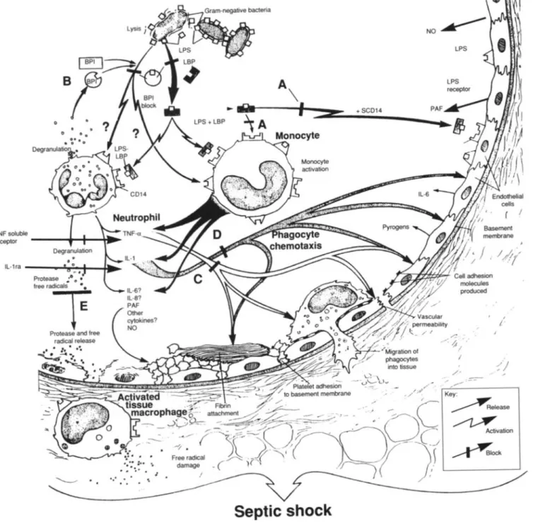
Figure 2. Experimental approaches to blocking septic shock.(A)Monoclonal antibodies to LPS prevent LPS from activating inflamma-tory reactions.(B)Bactericidal/permeability-increasing protein (BPI), a protein from neutrophil granules, binds LPS. Neither BPI nor antibodies to LPS function when gram-positive organisms invade the bloodstream. (C)TNF-ais inhibited by antibodies or solubleTNF-a
receptors.(D)IL-I is blocked by soluble IL-I receptors or IL-I receptor antagonist (IL-I ra), a naturally occurring human protein.(E)Tissue damage later in the septic shock cascade is minimized by protease inhibitors and free-radical scavengersor agents blocking other cytokines, including IL-6 and IL-8. LBP
=
LPS-binding protein; PAF=
platelet-activating factor; NO=
nitric oxide; and SCDl4=
soluble CDI4.organisms or substances) results in the conversion of prekal-likrein to kalprekal-likrein. In turn, kalprekal-likrein cleaves high-molecu-lar-weight kininogen to release bradykinin, a potent hypo-tensive agent [57]. The hypotension and DIC of bacteremia may be mediated-at least in part-by the activation of the contact system via the release of bradykinin and the
activa-tion of Factor XI. Recently, a monoclonal antibody to Fac-tor XII was used to block contact activation in baboons; the results showed that the contact system contributes to hypo-tension but not to DIC in lethal bacteremia [58]. The fact that tissue factor produced upon stimulation of macrophages and endothelial cells by LPS plays a major role in inducing
CID 1994: 18 (Suppl 2) Pathogenesis and Treatment of Septic Shock S211
DIC is indicated by the prevention of LPS-induced DIC by antibody to tissue factor in animals [59, 60]. Evidence also exists that protein C can be activated during gram-negative bacteremia [61] and that activated protein C can prevent the coagulopathic and lethal effects ofE. coli infusion in ba-boons [62].
Finally, it is widely recognized that vascular endothelium plays an active role in the development of septic shock. The systemic effects of high doses of intravenously administered TNF or IL-l into animals include hypotension, decreased systemic vascular resistance, vascular leak, and infiltration of polymorphonuclear leukocytes (PMNs) into tissues [63]. En-hancement of the expression ofadhesion molecules on endo-thelial cells by TNF and IL-l facilitates PMN margination. TNF and IL-l increase the procoagulant activity of endothe-lium and depress the expression of fibrinolytic activity; these changes result in a trend towards intravascular coagulation [64]. Generally, in animals given antibody to TNF or IL-l receptor antagonist (IL-l ra), an attenuation and/or blockade of many pathophysiological changes is associated with acti-vation of the coagulation cascade mediated by endothelial cells and cytokines [65, 66].
The Cytokine Network
Monocytic cells appear to have a pivotal role in mediation of the biological effects of LPS (figure 2). First, LPS can be removed from the blood and detoxified by monocytes-events of benefit to the host [67]. Second, LPS-stimulated monocytes produce cytokines such as TNF and IL-l. Several binding sites for LPS on the macrophage surface have been described [68]. LPS can also interact with monocytes after binding to plasma molecules. An acute-phase protein called LPS-binding protein (LBP) has been shown to bind to the lipid A moiety ofLPS [69]. LPS-LBP is a ligand for the CD 14 receptors on monocytes and macrophages [70]. Further-more, a soluble form of the CD 14 receptor in serum has been shown to promote the binding of LPS to endothelial cells and to stimulate these cells to produce cytokines and adhe-sion molecules [71, 72]. The role played by soluble CD 14 in vivo is still unknown. When complexed with LBP and at-tached to monocytes, LPS can stimulate the production of TNF by macrophages at concentrations far below those re-quired for stimulation by LPS alone [69, 70, 73]. Recently, another active component of human plasma, septin, has been described; septin may share with LBP the capacity to enhance the presentation of low concentrations of LPS to monocytes [74]. This information suggests that recognition of the presence ofLPS in plasma is important for an effective response to infection with gram-negative bacteria. Therefore, a principal function of LBP/septin may be to enhance the ability of the host to detect LPS early in infection [69, 70, 74]. No information is available on the role ofLBP/septin or CD 14 in vivo, but studies of the blocking ofLBP or CD 14 in
experimental models will help to define the early steps of interaction of LPS and monocytes in the development of shock.
Bactericidal/permeability-increasing protein (BPI), a pro-tein that has significant amino acid sequence homology with LBP (figure 2), appears to have therapeutic potential; it is found in PMN azurophilic granules that bind to lipid A and LPS in a way similar to that documented for LBP. BPI is an antagonist of LBP because it inhibits rather than amplifies the activity of LPS. In addition, BPI has been shown to in-hibit the limulus lysate assay mediated by LPS or the LPS-induced production of cytokines in blood [75, 76]. Experiments in animals challenged with endotoxin and gram-negative bacteria are now planned with a cloned recom-binant protein [77], and preliminary data on BPI-mediated protection of mice challenged with LPS have been pre-sented [78].
The intravascular activation of inflammatory systems in-volved in septic shock is mainly the consequence of an over-production of various cytokines. Several cytokines are pro-duced not only by macrophages but also by lymphocytes, endothelial cells, and other cells stimulated by microbial products. The systemic release oflarge amounts of cytokines is associated with death from septic shock in humans [79-81 ]. TNF is regarded as a central mediator of the pathophysio-logical changes associated with the release of LPS and possi-bly with shock caused by microorganisms that do not contain LPS. In animal models, antibodies to TNF-given either prophylactically (before intravenous bolus injections of LPS or gram-negative bacteria) or therapeutically (after chal-lenge )-have effectively increased the rate of survival. A po-tentially important advantage of making TNF (rather than endotoxin) a target in intervention strategies in patients is its possible role in the pathogenesis of shock caused by gram-positive bacteria. For example, septicemia associated with group A Streptococcus is clinically indistinguishable from "classic" gram-negative septic shock [82]. Cell-free superna-tants from cultures of gram-positive bacteria have been shown to induce the release ofTNF from human peripheral-blood monocytes in vitro [83], and concentrations ofTNF in the serum of patients with gram-positive sepsis are as high as those in the serum of patients with gram-negative sepsis [84]. However, antibodies to TNF have not been universally effec-tive in models of gram-posieffec-tive sepsis [85-87].
Cytokines other than TNF are involved in the induction of a shock-like state in animals. Considerable interest has also been focused on IL-I as a mediator of shock and of the asso-ciated "acute-phase" responses [65]. Circulating levels ofIL-I are elevated in shock; together with elevated levels ofTNF, these increased levels of IL-I correlate with the severity of disease. Direct proof of the central role of IL-l in septic shock comes from experiments with animals in which spe-cific blocking of the binding of IL-l to its cell receptor by
S212 Glauser et at. CID 1994; 18 (Suppl 2)
IL-I ra prevented the detrimental effects of inoculation of LPS orE.coli [88-90].
The overlap of proinflammatory functions and the synergy ofTNF and IL-l are probably important clues to the patho-genesis of septic shock. The administration of low doses of IL-l does not mediate severe shock and tissue injury; when IL-l is combined with TNF, however, the former increases the toxicity of the latter [91]. More important, LPS itself potentiates the lethal effects of TNF [92]. Interferon'Y has also been implicated in the synergy of TNF and IL-I [93]. Since several cytokines are probably involved in the patho-genesis of septic shock, blocking of TNF alone may not be sufficient to reverse the relevant conditions; therefore, phar-macotherapeutic "cocktails" may prove necessary.
The elucidation of these pathophysiological events has prompted the development of strategies to counteract exces-sive production or release of TNF and IL-I and hence to prevent or treat septic shock. Many studies have indicated that such therapy is beneficial in animals (including mice, rats, pigs, rabbits, and baboons). However, other important observations regarding potential therapeutic strategies must be considered. First, TNF, IL-l, and other cytokines are re-leased into the bloodstream during the first hour after bolus injection of LPS or live bacteria; TNF disappears rapidly from the circulation thereafter-several hours before the ani-mal's death [94]. An identical pattern has been found in children with fulminant meningococcemia [80]. Should it be documented in most cases of septic shock in humans, this pattern would suggest that levels of TNF may be elevated before shock develops. If so, then perhaps antibodies to TNF could not be administered soon enough to effectively treat patients with full-blown shock.
Moreover, intravenous bolus challenge in animal models and the fulminant course of meningococcal septicemia in children may not reflect most of the clinical situations in which septic shock develops. In most typical cases, a focus of infection may be present for hours or days and may cause the release of LPS or bacteria to be more sustained and subacute than during fulminant shock. Indeed, when serum concen-trations ofTNF and other cytokines were measured prospec-tively in 70 patients with septic shock, levels of TNF and IL-l were rather low and were detectable for up to 10 days after the onset of shock in patients who ultimately died [81]. These results indicate that concentrations ofTNF and other cytokines are sustained in patients presenting with gram-ne-gative shock-a picture unlike that seen after LPS or bacte-rial challenge in animal models, in which these concentra-tions are elevated and transient. Moreover, experimental models of severe, subacute, focal gram-negative bacterial in-fection have exhibited a pattern of TNF release different from that observed after bolus injection, and antibodies to TNF have failed to prevent death in these models [95-97]. Thus the release ofTNF in most clinical cases ofseptic shock is probably different from that in fulminant gram-negative
infections, and anticytokine therapies should be devised ac-cordingly.
In such anticytokine therapies, the role of cytokines as a defense against infection must be taken into account. TNF, IL-I, and other cytokines participate in the defense of the host; they are mediators that increase natural resistance by, for example, upregulating the cytolytic activity of lympho-cytes and the expression of complement receptors, enhanc-ing the oxidative burst of neutrophils, activatenhanc-ing macro-phages, and stimulating the proliferation of B cells, T cells, and progenitor cells. Indeed, while mice rendered deficient in the 55-kD receptor for TNF became resistant to challenge with endotoxin, they meanwhile became extremely sensitive to infection with Listeria monocytogenes [98]. Thus the blocking of cytokines in patients with septic shock for the purpose of counteracting "harmful" concentrations may in-terfere with the control of infection by physiological concen-trations. As a result, the infections causing septic shock may worsen, or the patient may become more susceptible to sec-ondary infections.
Two major clinical studies of the blocking of cytokines are in progress, the first examining a murine antibody to TNF and the second evaluating IL-I ra. Preliminary results indi-cate that anticytokine reagents may have detrimental effects in SUbgroups of patients; however, it is not known whether the failure observed in patients not presenting with shock is due specifically to a deleterious effect of these reagents on the host defenses against intracellular pathogens. When fur-ther results become available, the cause of the deaths of pa-tients in the various SUbgroups will be analyzed.
The large-scale clinical trial of murine antibody to TNF was initiated in patients presenting with sepsis syndrome and septic shock. While interim analyses revealed a beneficial (although statistically insignificant) effect of treatment on mortality among patients with septic shock, a deleterious ef-fect was found in some patients with sepsis syndrome [99]-a less cl[99]-assic form of the c[99]-asc[99]-ade of events th[99]-at le[99]-ads to de[99]-ath from infection. As a result, this study was discontinued, and a new trial of antibody to TNF including only patients with septic shock is being planned.
The results of the phase 3 trial of IL-I ra have recently been presented [100]. This randomized, double-blind, pla-cebo-controlled, multicenter clinical trial included 901 pa-tients with sepsis syndrome and septic shock. At enrollment, patients were randomized to receive either an intravenous loading dose of IL-l ra (100 mg) or placebo, which was fol-lowed by a continuous 72-hour intravenous infusion of IL-Ira (1 mg/[kg· h] or 2 mg/[kg· h]) or placebo. Patients were evaluated at 28 days for all-cause mortality. Treatment with IL-l ra did not significantly improve the rate of survival, which was the primary end point of the study. However, an individual patient/risk assessment approach, which took into account the APACHE III classification system and specific risk factors for sepsis, was used to analyze outcome as a
sec-CID 1994; 18 (Suppl2) Pathogenesis and Treatment of Septic Shock S213
ondary end point [101]. With this new method of risk predic-tion, treatment with IL-l ra appeared to be beneficial for pa-tients with a mortality risk of >0.24 but not for those with a mortality risk of <0.24.
Both of these studies aimed at inhibiting cytokines. The results suggest that, while this approach may be useful for the most severely affected patients, it is potentially harmful to patients who are less severely affected. Obviously, these find-ings need to be confirmed, and the subgroup of patients who might benefit from these therapies needs to be precisely de-lineated. The results of the IL-l ra study suggest that defining the risk of death at the time of intervention-rather than at the time of clinical presentation-may be a simple tool for identifying such patients.
Soluble forms of cytokine receptors offer an alternative approach to the blocking of cytokines. The potential of this approach was shown in experiments and clinical studies with IL-l ra: soluble TNF receptors may also be of value (figure 2). These receptors inhibit the bioactivity of TNF in vivo [102, 103]. Furthermore, it has recently been shown that mice deficient in the 55-kD TNF receptor are resistant to endotoxic shock [98]. Clinical trials with soluble TNF recep-tors are therefore being planned.
Currently, two other cytokines, IL-8 and IL-I 0, are being evaluated as possibly important mediators in shock. IL-8 has been characterized primarily as a PMN chemoattractant and a proinflammatory mediator; it has been detected in healthy volunteers after intravenous injection of endotoxin [104] and in patients with gram-negative shock [105]. Its precise role in vivo has not yet been fully elucidated. Recently, the anti-inflammatory IL-IO has been suggested as a candidate for treatment of bacterial sepsis. IL-l 0 was found to decrease the production of IL-l, IL-6, and TNF in vitro and to sup-press cytokines and provide protection in mice challenged with lethal doses of endotoxin [106, 107]. It is interesting that protection was documented when the administration of IL-IO was delayed after LPS challenge-an effect that is hardly evident with antibodies to TNF. To investigate the potential of IL-l 0 as a candidate for the treatment of bacte-rial sepsis, these preliminary results in endotoxemia should now be extended to bacterial infections.
Finally, as has been mentioned, the synergy that exists among cytokines (especially TNF and IL-I) and between cytokines and cell wall fragments (mainly TNF and LPS) suggests that a combined approach aimed at blocking various triggers and mediators may have the greatest potential for improving the outcome of septic shock.
Conclusion
Several approaches to the treatment and prevention of septic shock that are now being considered aim to suppress and/or inhibit one or another of a range of cytokines and other inflammatory mediators. However, since the syndrome
most likely results from complex interactions involving all these pathways and cytokines, the roles that each mechanism plays in the pathogenesis of septic shock must be delineated. This information will help to identify the subsets of patients who might benefit from the administration of antibodies to endotoxin and cytokines and to ascertain the need for other cytokine inhibitors or anti-inflammatory agents.
References
I. Bone RC. Sepsis. the sepsis syndrome. multi-organ failure: a plea for comparable definitions. Ann Intern Med 1991;1 14:332-3. 2. Bone RC, Fisher CJ. Clemmer TP. Siotman GJ. Metz GA. Balk RA. A
controlled clinical trial of high-dose methylprednisolone in the treatment of severe sepsis and septic shock. N Engl J Med 1987;317:653-8.
3. Bone RC, Sprung CL. Sibbald W J. Definitions for sepsis and organ failure. Crit Care Med 1992;20:724-6.
4. Ispahani P. Pearson NJ. Greenwood D. An analysis ofcommunity and hospital-acquired bacteremia in a large teaching hospital in the United Kingdom.QJ Med 1987;241 :427-40.
5. Calandra T. Glauser MP. Schellekens J, VerhoefJ. the Swiss-Dutch J 5 Immunoglobulin Study Group. Treatment of gram-negative septic shock with human IgG antibody to Escherichia coli J5: a prospec-tive. double-blind. randomized trial. J Infect Dis 1988; 158:312-9. 6. BrandtzaegP,Kierulf P. Gaustad P. et al. Plasma endotoxin as a pre-dictor of multiple organ failure and death in systemic meningococ-cal disease. J Infect Dis 1989; 159: 195-204.
7. Galanos C, Freudenberg MA. Matsuura M. Mechanisms of the lethal action of endotoxin and endotoxin hypersensitivity. In: Friedman H. Klein TW. Nokano M. Nowotny A. eds. Endotoxin: advances in experimental medicine and biology. New York: Plenum Press. 1990;603-19.
8. Stutzp.Liehl E. Lipid A analogs aimed at preventing the detrimental effects of endotoxin. Infect Dis Clin North Am 1991;5:847-74. 9. Lynn W A. Golenbock DT. Lipopolysaccharide antagonists. Immunol
Today 1992; 13:271-6.
10. Ziegler EJ. McCutchan JA. Fierer J. et al. Treatment ofgram-negative bacteremia and shock with human antiserum to a mutant
Esche-richia coli. N Engl J Med 1982;307: 1225-30.
II. Baumgartner JD. Glauser MP. McCutchan JA. et al. Prevention of gram-negative shock and death in surgical patients by prophylactic antibody to endotoxin core glycolipid. Lancet 1985;2: 59-63. 12. J5 Study Group. Treatment of severe infectious purpura in children
with human plasma from donors immunized with Escherichia coli J5: a prospective double-blind study. J Infect Dis 1992;165:695-701.
13. McCutchan JA. Wolf JL Ziegler EJ, Braude AI. Ineffectiveness of single-dose human antiserum to core glycolipid(E.coli J 5) for
pro-phylaxis ofbacterernic,gram-negative infections in patients with prolonged neutropenia. Schweiz Med Wochenschr 1983; I 13 (Suppl 14):40-5.
14. Greenman RL Schein RM. Martin MA. et al. A controlled clinical trial of E5 murine monoclonal IgM antibody to endotoxin in the treatment of gram-negative sepsis. The XOMA Sepsis Study Group.
lAMA 1991;266:1097-102.
15. Ziegler EJ, Fisher CJ. Sprung CL. et al. Treatment of gram-negative bacteremia and septic shock with HA-I A human monoclonal anti-body against endotoxin. A randomized. double-blind. placebo-con-trolled trial. N Engl J Med 1991;324:429-36.
16. Wenzel R. Bone R. Fein A. et al. Results of a second double-blind. randomized. controlled trial of antiendotoxin antibody E5 in
gram-S214 Glauser et al. CID 1994; 18 (Suppl 2)
negative sepsis [abstract no 1170]. In: Program and abstracts of the 31st Interscience Conference on Antimicrobial Agents and Chemo-therapy (Chicago). Washington, DC: American Society for Microbi-ology, 1991.
17. Warren HS, Danner RL, Munford RS. Anti-endotoxin monoclonal antibodies. N Engl J Med 1992;326:1153-7.
18. Kolata G. At market's edge: how a $1 billion drug fell flat. The New York Times, 15 March 1993:A20.
19. Heumann D, Baumgartner JD, Jacot-Guillarmod H, Glauser MP. An-tibodies to core lipopolysaccharide determinants: absence of cross-reactivity with heterologous lipopolysaccharides. J Infect Dis 1991; 163:762-8.
20. Baumgartner JD, Heumann D, Calandra T, Glauser MP. Antibodies to lipopolysaccharides after immunization of humans with the rough mutantEscherichia coliJ5. J Infect Dis 1991;163:769-72. 21. Fujihara Y, Lei MG, Morrison De. Characterization of specific
bind-ing of a human immunoglobulin M monoclonal antibody to lipo-polysaccharide and its lipid A domain. Infect Immun 1993;61: 910-8.
22. Mascelli MA, Frederick B, Ely T, et al. Reactivity of the human an-tiendotoxin immunoglobulin M monoclonal antibody HA-IA with lipopolysaccharides from rough and smooth gram-negative organ-isms. Infect Immun 1993;61: 1756-63.
23. Baumgartner JD. Immunotherapy with antibodies to core lipopolysac-charide: a critical appraisal. Infect Dis Clin North Am 1991;5: 915-27.
24. KriegerJI,Fletcher RC, Siegel SA, et al. Human endotoxin anti-body HA-I A mediates complement-dependent binding of
Esche-richia coliJ 5 lipopolysaccharide to complement receptor type I of
human erythrocytes and neutrophils. J Infect Dis 1993; 167:865-75.
25. Tonoli M, Davies KA, Norsworthy PJ, Cohen J, Walport MJ. The anti-lipid A antibody HA-I A binds to rough gram-negative bacte-ria, fixes complement and facilitates binding to erythrocyte CR I (CD35). Clin Exp ImmunoI1993;92:232-8.
26. Wood DM, Parent J8, Gazzano-Santaro H, et al. Reactivity of mono-clonal antibody E5 with endotoxin. I. Binding to lipid A and rough lipopolysaccharides. Circ Shock 1992;38:55-62.
27. Parent JB, Gazzano-Santoro H, Wood OM, et al. Reactivity of mono-clonal antibody E5 with endotoxin. II. Binding to short- and long-chain smooth lipopolysaccharides. Circ Shock 1992;38:63-73. 28. Warren HS, Amato SF, Fitting e. et al. Assessment of ability of
mu-rine and human anti-lipid A monoclonal antibodies to bind and neutralize lipopolysaccharide. J Exp Med 1993; 177:89-97. 29. Teng NNH, Kaplan HS, Hebert JM, et al. Protection against
gram-negative bacteremia and endotoxemia with human IgM antibodies. Proc Nat! Acad Sci USA 1985;82: 1790-4.
30. Baumgartner JD, Heumann D, Gerain J, Weinbreck P, Grau GE, Glauser MP. Association between protective efficacy of anti-lipo-polysaccharide (LPS) antibodies and suppression of LPS-induced tumor necrosis factor a and interleukin 6. Comparison of
°
side chain-specific antibodies with core LPS antibodies. J Exp Med 1990; 171:889-96.31. Cornelissen JJ, Makel I, Algra A, et al. Protection against lethal endo-toxemia by anti-lipid A murine monoclonal antibodies: compari-son of efficacy with that of human lipid A monoclonal anti-body HA-IA. J Infect Dis 1993;167:876-81.
32. Quezado ZMN, Natanson C, Alling DW, et al. A controlled trial of HA-I A in a canine model of gram-negative septic shock. JAMA 1993;269:2221-7.
33. Young LS, Gascon R, Alam S, Bermudez LEM. Monoclonal antibod-ies for treatment of gram-negative infections. Rev Infect Dis 1989; I I(suppl 7):S 1564-71.
34. Romulo RLC, Palardy JE, Opal SM. Efficacy ofanti-endotoxin mono-clonal antibody E5 alone or in combination with ciprofloxacin in neutropenic rats withPseudomonassepsis. J Infect Dis 1993; 167: 126-30.
35. Rothstein JL, Schreiber H. Tissue damage caused by TNF and com-plement. Immunol Ser 1992;56:453-81.
36. Jacobs HS. The role of activated complement and granulocytes in shock states and myocardial infarction. J Lab Clin Med 1981;98:645-54.
37. Hack CE, Nuijens JH, Felt-Bersma RJF, et al. Elevated plasma levels of the anaphylatoxins C3a and C4a are associated with a fatal out-come in sepsis. Am J Med 1989;86:20-6.
38. Reines HD, Halushka PV, Cook JA, Wise We. Rambo W. Plasma thromboxane concentrations are raised in patients dying with septic shock. Lancet 1982;2: 174-5.
39. Welbourn CRB, Young Y.Endotoxin, septic shock, and acute lung injury: neutrophils, macrophages, and inflammatory mediators. Br J Surg 1992;79:998-1003.
40. Springer TA. Adhesion receptors of the immune system. Nature 1990;346:425-34.
41. Saukkonen K, Sande S, Cioffe C, et al. The role of cytokines in the generation of inflammation and tissue damage in experimental gram-positive meningitis. J Exp Med 1990; 171:439-48. 42. Chang SW, FeddersonCO, Henson PM, et al. Platelet-activating
fac-tor mediates hemodynamic changes and lung injury in endotoxin-treated rats. J Exp Med 1990; 171:439-48.
43. Ooebber TW, Wu MS, Robbins JC, et al. Platelet activating factor (PAF) involvement in endotoxin-induced hypotension in rats. Stud-ies with PAF-receptor antagonist kadsurenone. Biochem Biophys Res Commun 1985;127:799-808.
44. Anderson BO, Bensard DD, Harken AH. The role of platelet activat-ing factor and its antagonists in shock, sepsis and multiple organ failure. SurgGynecol Obstet 1991;172:415-24.
45. Dhainaut JF, Tenaillon A, Letzulo Y, et al. Efficacy ofP.A.F. antago-nist (BN 52021) in reducing mortality of patients with severe gram-negative sepsis. Circ Shock 1993; Suppl 1:42.
46. Nathan e. Nitric oxide as a secretory product of mammalian cells. FASEB J 1992;6:3051-64.
47. Vane J R, Angaard EE, Botting RM. Regulatory functions ofthe vascu-lar endothelium. N Engl J Med 1990;323:27-36.
48. Hotchkiss RS, Parker JL, Adams HR. Inhibition of NO synthesis in septic shock. Lancet 1992;339:434-5.
49. Evans T, Carpenter A, Silva A, Cohen J. Differential effects of mono-clonal antibodies to tumor necrosis factor alpha and gamma inter-feron on induction of hepatic nitric oxide synthase in experimental gram-negative sepsis. Infect Immun 1992;60:4133-9.
50. Evans TE, Carpenter A, Kinderman H, et al. Evidence of increased nitric oxide production in patients with the sepsis syndrome. Circ Shock 1993 (in press).
51. Shultz PJ, Raij L.Endogenously synthesized nitric oxide prevents endotoxin-induced glomerular thrombosis. J Clin Invest 1992;90:
1718-25.
52. Teale DM, Atkinson AM. Inhibition of nitric oxide synthesis im-proves survival in a murine peritonitis model of sepsis that is not cured by antibiotics alone. J Antimicrob Chemother 1992;30: 839-42.
53. Cobb JP, Natanson e. Hoffman WD, et aI. N omega amino-t-argin-ine, an inhibitor of nitric oxide synthase, raises vascular resistance but increases mortality in awake canines challenged with endo-toxin. J Exp Med 1992; 176: 1175-82.
54. Petros A, Bennett D, Vallance P. Effect of nitric oxide synthetase inhibitors on hypotension in patients with septic shock. Lancet 1991;338: 1557-8.
CID 1994; 18 (Suppl 2) Pathogenesis and Treatment of Septic Shock S215
55. Kalter ES, van Dijk
we
Timmerman A, Verhoef J, Bouma BN. Acti-vation of purified human plasma prekallikrein triggered by cell wall fractions of Escherichia coli and Staphylococcus aureus. J Infect Dis1983; 148:682-91.
56. Spika JS, Peterson PK. Wilkinson BJ, et al. Role of peptidoglycan from Staphylococcus aureus in leukopenia, thrombocytopenia, and complement activation associated with bacteremia. J Infect Dis 1982; 146:227-34.
57. Colman R W. The role of plasma proteases in septic shock. N Engl J Med 1989;320: 1207-9.
58. Pixley RA De La Cadena R, Page JD, et al. The contact system contributes to hypotension but not disseminated intravascularcoag-ulation in lethal bacteremia. In vivo use of a monoclonal anti-fac-tor XIIantibody to block contact activation in baboons. J Clin Invest 1993;91:61-8.
59. Warr TA Rao LV, Rapaport SI. Disseminated intravascular coagula-tion in rabbits induced by administracoagula-tion of endotoxin or tissue factor: effect of anti-tissue factor antibodies and measurement of plasma extrinsic pathway inhibitor activity. Blood 1990;75:
1481-9.
60. Taylor FBJr, Chang A Ruf W, et al. LethalE.coli septic shock is
prevented by blocking tissue factor with monoclonal antibody. Circ Shock 1991;33: 127-34.
61. Esmon CT. The regulation of natural anticoagulant pathways. Science 1987;235: 1348-52.
62. Taylor FB, Chang A Esmon T, et al. Protein C prevents the coagulo-pathic and lethal effects of Escherichia coli infusion in the baboon. J C1in Invest 1987;79:918-25.
63. Dinarello CA, The proinflammatory cytokines interleukin-I and tu-mor necrosis factor and treatment of the septic shock syndrome. J Infect Dis 1991;163:1177-84.
64. Cotran RS, Pober JS. Effects of cytokines on vascular endothelium: their role in vascular and immune injury. Kidney Int 1989;35: 969-75.
65. Dinarello CA, Interleukin-I and interleukin-I antagonism. Blood 1991;77: 1627-52.
66. Tracey KJ, Lowry SF. The role of cytokine mediators in septic shock. Adv Surg 1990;23:21-56.
67. Munford RS, Hall CL. Uptake and deacylation of bacterial Iipo poly-saccharides by macro phages from normal and endotoxin-hypore-sponsive mice. Infect Immun 1985;48:464-73.
68. Lei MG, Chen TY, Morrison DC. Lipopolysaccharide/lipid A recep-tors on lymphocytes and macrophages. Int Rev Immunol 1990;6:223-35.
69. Schumann RR, Leong SR, Flaggs GW, et al. Structure and function of lipopolysaccharide binding protein. Science 1990;249: 1429-31. 70. Wright SO, Ramos RA, Tobias PS, Ulevitch RJ, Mathison Jc. CDI4,
a receptor for complexes oflipopolysaccharide (LPS) and LPS bind-ing protein. Science 1990;249: 1431-3.
71. Frey EA, Miller OS, Jahr TG, et al. Soluble CD 14 participates in the response of cells to lipopolysaccharide. J Exp Med 1992;176: 1665-71.
72. Pugin J, Schiirer-MalyCC,Leturcq0, et al. Lipopolysaccharide acti-vation of human endothelial and epithelial cells is mediated by lipopolysaccharide-binding protein and soluble CD 14. Proc Natl Acad Sci USA 1993;90:2744-8.
73. Heumann D, Gallay P, Barras C, et al. Control of lipopolysaccharide (LPS) binding and LPS-induced tumor necrosis factor secretion in human peripheral blood monocytes. J Immunol 1992; 148:3505-12.
74. Wright SD, Ramos RA Patel M, et al. Septin: a factor in plasma that opsonizes lipopolysaccharide-bearing particles for recognition by CDl4 on phagocytes. J Exp Med 1992;176:719-27.
75. Ooi CE, Weiss J, Doerfler ME, Elsbach P. Endotoxin-neutralizing properties of the 25 kD N-terminal fragment and a newly isolated 30 kD C-terminal fragment of the 55-60 kD bactericidal/ permeability-increasing protein of human neutrophils. J Exp Med 1991; 174:649-55.
76. Marra MN, WildeCG,Grifith JE, Snable JL, Scott RW. Bactericidal/ permeability-increasing protein has endotoxin-neutralizing activity. J Immunol 1990; 144:662-6.
77. Weiss J, Elsbach P, Shu C, et al. Human bactericidal/permeability-increasing protein and a recombinant NH2-terminal fragment cause killing of serum-resistant gram-negative bacteria in whole blood and inhibit tumor necrosis factor release induced by the bacte-ria. J C1in Invest 1992;90: 1122-30.
78. Opal SM. Bactericidal/permeability-increasing protein as an endoge-nous anti-endotoxin molecule. Circ Shock 1993; Suppl 1:47. 79. Girardin E, Grau GE, Dayer JM, Roux-Lombard P, Lambert PH.
Tumor necrosis factor andinterleukin-I in the serum of children with severe infectious purpura. N Engl J Med 1988;319:397-400. 80. Waage A, Halstensen A, Espevik T. Association between tumor ne-crosis factor in serum and fatal outcome in patients with meningo-coccal disease. Lancet 1987; I :355-7.
81. Calandra T, Baumgartner JD. Grau GE, et al. Prognostic values of tumor necrosis factor/cachectin, interleukin-l , interferon-alpha and interferon-gamma in the serum of patients with septic shock. Swiss-Dutch J 5 Immunoglobulin Study Group. J Infect Dis 1990;161:982-7.
82. Stevens DL, Tanner MH. Winship J, et al. Severe group A streptococ-cal infections associated with a toxic shock-like syndrome and scar-let fever toxin A, N Engl J Med 1989;321: 1- 7.
83. Bayson KF, Tomlinson M, Cohen J. In vitro stimulation of TNF-a from human whole blood by cell-free supernatants of gram positive bacteria. Cytokine 1992;4:397-402.
84. Marks JD, Marks CB, Luce JM, et al. Plasma tumor necrosis factor in patients with septic shock. Mortality rate, incidence of adult respira-tory distress syndrome, and effects of methylprednisolone adminis-tration. Am Rev Respir Dis 1990;141:94-7.
85. Wayte J, Silva AT, Krausz T, et al. Observations on the role ofTNF in a murine model of shock due to Streptococcus pyogenes. Crit Care Med 1993 (in press).
86. Martin RA Silva AT, Cohen J. Effect of anti-TNF alpha treatment in an antibiotic treatment model of shock due to Streptococcus
pyo-genes. FEMS Microbial Lett 1993 (in press).
87. Hinshaw LB, Emerson TE Jr. Taylor FB Jr, et al. Lethal
Staphylococ-cus aureus-induced shock in primates: prevention of death with
anti-TNF antibody. J Trauma 1992;33: 568-73.
88. McIntyre KW. Stepan GJ. Kolinsky KD, et al. Inhibition ofinterleu-kin I (I L-I)binding and bioactivity in vitro and modulation of acute inflammation in vivo by IL-I receptor antagonist and
anti-IL-I receptor monoclonal antibody. J Exp Med 1991; 173:931-9. 89. Wakabayashi G, Gelfand JA Burke JF. Thompson R'C,Dinarello
CA. A specific receptor antagonist for interleukinIprevents
Esche-richia coli-induced shock in rabbits. FASEB J 1991;5:338-43.
90. Alexander HR. Doherty GM. Buresh CM. et al. A recombinant hu-man receptor antagonist to interleukin I improves survival after lethal endotoxemia in mice. J Exp Med 1991;173:1029-32. 91. Waage A Espevik T. Interleukin I potentiates the lethal effect of
tumor necrosis factor-alpha/cachectin in mice. J Exp Med 1988; 167: 1987-92.
92. Rothstein JL, Schreiber H. Synergy between tumor necrosis factor and bacterial products causes hemorrhagic necrosis and lethal shock in normal mice. Proc Natl Acad Sci USA 1988;85:607-1I. 93. Billiau A. Gamma-interferon: the match that lights the fire? Immunol
S216 Glauser et aJ. CID 1994; 18 (Suppl2)
94. Michie HR, Manogue KR, Spriggs DR, et al. Detection of circulating tumor necrosis factor after endotoxin administration. N Engl 1 Med 1988;318: 1481-6.
95. Bagby G1, PlessalaKJ,Wilson LA, Thompson 11, Nelson S. Diver-gent efficacy of antibody to tumor necrosis factor-a in intravascular and peritonitis models of sepsis. 1 Infect Dis 1991;163:83-8. 96. Echtenacher B, Falk W, Mannel ON, Krammer PH. Requirement of
endogenous tumor necrosis factor/cachectin for recovery from ex-perimental peritonitis. 1 ImmunoI1990;145:3762-6.
97. Zanetti G, Heumann 0, Gerain 1, et aJ. Cytokine production after intravenous or peritoneal gram negative bacterial challenge in mice. Comparative protective efficacy of antibodies to tumor necrosis fac-tor-alpha and to lipopolysaccharide. 1 Immunol 1992; 148:1890-7. 98. Pfeffer K, Matsuyama T, Kiindig TM, et al. Mice deficient for the 55 kD tumor necrosis factor receptor are resistant to endotoxic shock, yet succumb toL.lIlonocytogenesinfection. Cell 1993;73:457-67. 99. Cross AS. Biotech strategies for treatment of sepsis. Endotoxin
News-letter 1993;3:2-3.
100. Fisher C1, Siotman G1, Opal SM, et al. Human recombinant interleu-kin-I receptor antagonist (IL-I ra) in the treatment of patients with sepsis syndrome. Circ Shock 1993; Suppl 1:42.
101. Knaus W, Harell F, Fisher C1, et al. An individual patient risk assess-ment approach to design and analysis of clinical trials for patients with sepsis and organ failure. Circ Shock 1993; Suppl I: 14. 102. Ashkenazi A, Marsters SA, Capon 01, et al. Protection against
endo-toxin shock by a tumor necrosis factor receptor immunoadhesion. Proc Natl Acad Sci USA 1991;88:10535-9.
103. Lesslauer W, Tabuchi H, Gentz R, et al. Recombinant soluble tumor necrosis factor receptor proteins protect mice from lipopolysaccha-ride-induced lethality. Eur 1 ImmunoI1991;21:2883-6. 104. Martich GO, Danner RL, Ceska M, Suffredini AF. Detection of
inter-leukin 8 and tumor necrosis factor in normal humans after intrave-nous endotoxin: the effect of antiinflammatory agents. 1 Exp Med 1991;173: 1021-4.
105. Halstensen A, Ceska M, Brandtzaeg P, Redl H, Naess A, Waage A. Interleukin-8 in serum and cerebrospinal fluid from patients with meningococcal disease. 1 Infect Dis 1993;167:471-5.
106. Howard M, Muchamuel T, Andrade S, Menon S. Interleukin 10 pro-tects mice from lethal endotoxernia. 1 Exp Med 1993; 177: 1205-8. 107. GerardC,BruynsC,Marchant A, et al. Interleukin 10 reduces the release of tumor necrosis factor and prevents lethality in experimen-tal endotoxernia. 1 Exp Med 1993; 177:547-50.