FEATURE ARTICLE
Inter- and Intrahemispheric Dissociations in Ideomotor Apraxia: A Large-Scale
Lesion
–Symptom Mapping Study in Subacute Brain-Damaged Patients
Aurelie L. Manuel1, Narges Radman2, Delphine Mesot1, Leila Chouiter2, Stephanie Clarke1, Jean-Marie Annoni2 and Lucas Spierer1,2
1Department of Clinical Neurosciences, Neuropsychology and Neurorehabilitation Service, Vaudois University Hospital Center and University of Lausanne, 1011 Lausanne, Switzerland and2Department of Medicine, Neurology Unit, University of Fribourg, 1700 Fribourg, Switzerland
Address correspondence to Aurelie Manuel, Faculty of Biology and Medicine–UNIL, Neuropsychology and Neurorehabilitation Service–CHUV, av. Pierre-Decker 5 1011, Lausanne, Switzerland. Email: [email protected]
Pantomimes of object use require accurate representations of move-ments and a selection of the most task-relevant gestures. Prominent models of praxis, corroborated by functional neuroimaging studies, predict a critical role for left parietal cortices in pantomime and advance that these areas store representations of tool use. In con-trast, lesion data points to the involvement of left inferior frontal areas, suggesting that defective selection of movement features is the cause of pantomime errors. We conducted a large-scale voxel-based lesion–symptom mapping analyses with configural/spatial (CS) and body-part-as-object (BPO) pantomime errors of 150 left and right brain-damaged patients. Our results confirm the left hemi-sphere dominance in pantomime. Both types of error were associ-ated with damage to left inferior frontal regions in tumor and stroke patients. While CS pantomime errors were associated with left tem-poroparietal lesions in both stroke and tumor patients, these errors appeared less associated with parietal areas in stroke than in tumor patients and less associated with temporal in tumor than stroke patients. BPO errors were associated with left inferior frontal lesions in both tumor and stroke patients. Collectively, our results reveal a left intrahemispheric dissociation for various aspects of pantomime, but with an unspecific role for inferior frontal regions. Keywords: frontal, ideomotor apraxia, lesion, pantomime, parietal, voxel-based lesion–symptom mapping
Introduction
Pantomime of object or tool use is the act of pretending to use an object by adopting the same limb configurations and producing the same sequences of movements as if the object were actually held and used. Pantomime of object or tool use is the act of pretending to use an object by adopting the same limb configurations and producing the same sequences of movements as if the object were actually held and used. Within the model of praxis byRothi et al. (1991,1997), panto-mime to verbal command is distinguished from other types of motor productions based on the fact that it neither requires a visual analysis of the gesture to be produced nor a compari-son between the visual input with a lexicon of action (as would be the case for, e.g., imitation of new or familiar ges-tures). Rather, the analysis of the auditory/verbal command is directly followed by the selection of the spatiotemporal attri-butes of the gesture to be performed from an action output lexicon and the programming and implementation of the motor action (see also Peigneux and Van der Linden 2000). Because the production of pantomimes involve semantic, ex-ecutive, and spatial/configural level of motor processing
(to respectively understand the gestures, select the relevant movements representing the action, and represent accurately the relationships between the body parts involved in the movement and of how they interact with the object (Golden-berg 2009)), pantomime constitutes a sensitive task to detect ideomotor apraxia following a brain lesion (Heilman and Rothi 1993). In the current article, we refer to “ideomotor apraxia” using the definition proposed byRothi et al. (1991, 1997):“an impairment in the timing, sequencing, and spatial organization of gestural movements” (Rothi et al. 1991).
Starting from the seminal hypotheses of Liepmann (1908) in stroke patients, most prominent models of praxia advance that left parietal areas store the motor representations of tool use guiding action and therefore predict that these structures play a central role in pantomime (Moll et al. 2000;Peigneux et al. 2004). Functional neuroimaging studies corroborate these models by consistently observing correlations between left parietal areas activity and pantomiming (Vingerhoets, Acke et al. 2012, Vingerhoets, Vandekerckhove et al. 2011; seeLewis (2006)for a meta-analysis of activation studies).
In contrast, lesion studies report that accurate pantomime depends on the integrity of left inferior frontal areas (Golden-berg et al. 2007) and less consistently of parietal areas (Kertesz and Ferro 1984; Goldenberg and Hagmann 1997; Peigneux et al. 2000). Although not directly for pantomimes, parietal areas have been involved in ideomotor apraxia (Basso et al. 1985;Haaland et al. 2000;Buxbaum et al. 2007) or coordination of arm movements in ideomotor apraxia (Mutha et al. 2010). Therefore, lesion studies conclude that pantomime critically depends on the selection of a limited, task-relevant set of features among the many features in-volved in the actual tool use to be mimed (Goldenberg et al. 2007;Goldenberg 2009;Bohlhalter et al. 2011). The disparity between thefindings of neuroimaging and lesion approaches about the involvement of parietal regions has been hypoth-esized to follow from the pantomimes being realized under different conditions in each type of study. Because of the con-straints induced by the scanner on participant’s movements, the pantomimes require additional spatial transformations of movements to unusual reference frames, which in turn in-crease the involvement of parietal structures (Andersen et al., 1997; Goldenberg et al., 2007). Rumiati et al. (2004), however, reported an involvement of left parietal areas for the pantomiming of visually presented objects in patients with deficit in the organization of sequences relative to tool use (ideational apraxia), suggesting that these structures might trigger tool use-related motor programs. Pantomime are © The Author 2012. Published by Oxford University Press. All rights reserved.
Cerebral Cortex December 2013;23:2781–2789 doi:10.1093/cercor/bhs280
globally sensitive to left hemisphere lesions (Bickerton et al. 2012), but have been found to be sensitive to lenticular stroke and associated to impaired working memory, suggesting that correct pantomime execution necessitates an efficient lexical route but also a dedicated workplace subserved by subcortical structures (Bartolo et al. 2003). Of note, electroencephalogra-phy studies manipulating the production of pantomimes in naturalistic conditions showed evidence for parietal activation in preparing tool-use movements, suggesting that this region is not only involved in spatial transformation, but also in plan-ning tool-related motor actions (Wheaton, Shibasaki et al. 2005;Wheaton, Yakota et al. 2005). During the neuropsycho-logical assessments, the patients produce the movements within a natural body-centered reference frame and with visual feedback (Goldenberg et al. 2007;Goldenberg 2009), which involves only routine support from parietal areas.
However, several other hypotheses could account for the discrepancy between the results of functional and lesion studies. The contribution of parietal structures to pantomime might have been underestimated in previous lesion studies due to the assessment having been conducted in chronic patients, that is, more than 1 month after lesion onset (Gold-enberg 2003a,2003b;Dovern et al. 2011). Specificity of net-works may indeed be revealed in the postacute phase only, after the resorption of the ischemic penumbra (Witte et al. 2000) and before the occurrence of major plastic anatomo-functional reorganizations (Adriani et al. 2003; Rey et al. 2007). The type of evaluation (conceptual vs. production components) may be more sensitive to parietal or to frontal lesions (Halsband et al. 2001). Furthermore, previous neurop-sychological studies included only a limited number of patients with lesions covering only limited portion of the brain (Goldenberg 2003a,2003b), patients selected based on a priori hypotheses on the region of interest (Dovern et al. 2011; Hanna-Pladdy et al. 2001) or patients with aphasia (Goldenberg et al. 2007). Finally, previous studies dichoto-mized behavioral data on apraxia (with vs. without apraxia) instead of considering the scores as continuous data, leading to a loss of power and reduced effect sizes (Cohen 1983). Col-lectively, these potential caveats could have lead to false-negative results in current lesion data on pantomime, poten-tially concerning the involvement of parietal areas.
In addition, attempts tofind common substrates for differ-ent types of pantomime errors and the rarity of some kinds of errors motivated researchers to collapse together various types of error in neuropsychological scoring of pantomime. As lesions to distinct areas may induce distinct types of error (Rumiati and Humphreys 1998;Halsband et al. 2001; Hanna-Pladdy et al. 2001;Rumiati et al. 2001), the use of compound scores might have in turn contributed to obscure putative in-trahemispheric dissociations for different types of pantomime errors. For instance, body-part-as-object (BPO) pantomime errors, consisting in representing objects with a part of the body rather than pretending to use an “invisible” object as specified in the test instructions (Goodglass and Kaplan 1963) have been suggested to depend on frontal but not parietal components (Peigneux and Van der Linden 1999;Arzy et al. 2006). The study of BPO errors could thus help to further reveal intrahemispheric dissociations between frontal and par-ietal contributions to pantomime.
To test these hypotheses, we conducted large-scale retro-spective voxel-based lesion–symptom-mapping analyses
(VLSM;Bates et al. 2003) on a group of subacute, unselected, hemispheric brain-damaged patients and pantomime scores differentiating the typical spatial/configural (CS) and BPO pantomime error types. We used highly selective inferential statistical analyses of lesion–symptom mapping based on continuous scores rather than descriptive comparisons between lesion patterns of patients’ groups defined by behav-ioral cutoffs (i.e., with or without apraxia). Because the inclusion of tumor and stroke patients in VLSM analyses might yield different results, we analyzed separately these 2 types of brain-damaged patients (e.g., Karnath and Steinbach 2011).
Methods Patients
One hundred andfifty right-handed patients with a first right or left unilateral hemispheric lesion (demographic data in table1) were se-lected retrospectively from consecutive in-patients admitted to the Neuropsychology and Neurorehabilitation Service of the Centre Hos-pitalier Universitaire Vaudois or the Hôpitaux Fribourgeois between 2007 and 2011. Patients with bilateral lesions were excluded to facilitate the interpretability of our results in terms of hemispheric specialization of pantomime. On average, the pantomime assessment was conducted 2.3 ± 6.9 weeks (mean ± SD) after the lesion onset or tumor diagnosis or removal and was part of the formal neuropsy-chological assessment carried out by experienced psychologists specialized in neuropsychology. All patients met the following criteria: 1) first unilateral hemispheric lesion without damage to the brain stem or cerebellum documented by CT-scan and/or MRI; 2) no prior neurological illness; 3) no psychiatric illness; 4) good cooperation and absence of major behavioral or attentional pro-blems; 5) sufficient understanding of the instructions; and 6) assess-ment of at least 4 pantomimes. Inclusion in the study was neither determined by the lesion characteristics nor by the pattern of behav-ioral deficit. The study was carried out in agreement with the rec-ommendations of the Ethics Committee of the Faculty of Biology and Medicine, Lausanne. Data were handled according to Swiss-Federal law on data protection.
Neuropsychological Assessment of Pantomime
The production of pantomime on verbal command was assessed by asking the patient to mime the use of an imaginary tool. In the case of hemiparesis, the patient was asked to use only the nonparetic ipsi-lesional hand. The evaluation of paresis was based on the Medical Research Council Scale for Muscle Strength (e.g.,Pizzi et al. 2009). In the present study, if the patient’s strength was below 3, the nondomi-nant (left) hand was used. Pantomimes were tested with items of the
Table 1
Demographic data of the 150 patients included in the study N = 150 Patients
Age, mean (±SD) 60.5 (±15.3), 16–89 years
Gender
Female 68
Male 82
Damaged hemisphere
LBD 84 (38 with aphasia: 32 patients withfluent aphasia and 6 with
nonfluent aphasia)
RBD 66
Etiology
Stroke (LBD, RBD) 81 (42, 39)
Tumor (LBD, RBD) 69 (42, 27) (44 before and 25 after surgery) Postlesion delay
(weeks ± SD)
2.3 (±6.9)
screening batteries by Peigneux and Van der Linden (2000) or
Mahieux-Laurent et al. (2009). The former included 4 items: brushing the hair with a comb; brushing the teeth with a toothbrush; planting a nail with a hammer; and sawing a branch of wood (Peigneux and Van der Linden 2000). The latter included 5 items: planting a nail with a hammer, tearing a piece of paper in two, lighting a match, brushing the hair with a comb, and drinking a glass (Mahieux-Laurent et al. 2009). On average 4.3 ± 0.5 (mean ± SD; range 4–5) items were probed for the assessment of pantomimes.
Two different types of errors were documented and analyzed in the present study: First, CS errors refer to inaccurate limb con figur-ations during pantomiming at the level of the sequencing, timing, and/or amplitude of the gestures, and of the relationships between the different body parts engaged in the movements. CS errors result in imprecise or unrecognizable gestures. For example, if the patient mimed brushing his hair with an imaginary comb with one hand but placed the imagined comb far away from his head, the pantomime was considered as incorrect. The second type of errors consisted in the use by the patients of their body parts as the object (BPO errors). BPO were considered as errors only when the patients did not correct it after reinstruction from the examiner. The patient was reinstructed after every BPO error. This reinstruction condition was implemented because BPO are common among healthy controls, but neurologically healthy population correct BPO after being reinstructed (Heilman and Rothi 1993;Raymer et al. 1997;Peigneux and Van der Linden 1999). For example, for combing the hair, if the patient used hisfist as the comb and brushed his hair with it, the examiner reinstructed him to pretend he was holding an imaginary comb in his hand rather than using his forelimb as the comb. If the patient did not correct the error and continued to use his limb as the object, the BPO was considered as pathological and counted as a BPO error (Heilman and Rothi 1993;
Raymer et al. 1997). We would note that because the present study was based on a retrospective analysis of data collected during routine neuropsychological assessments, the scoring of pantomime was not as precise and controlled as what could have been obtained with a specifically designed prospective study. For this reason, not all poss-ible error types [including, e.g., semantic content errors (Rumiati and Humphreys 1998); sequence or conceptual errors (Rumiati et al. 2001); or parapraxic errors (Halsband et al. 2001)] were analyzed sep-arately. However, the pantomime errors were scored by trained and experienced specialists in neuropsychology, according to strict pub-lished procedures (Peigneux and Van der Linden 2000; Mahieux-Laurent et al. 2009). Because each patient did not have to produce the same number of pantomimes (4 items for Peigneux’s battery and 5 items for Mahieux-Laurent’s battery), the scores used in the VLSM were the standardized numbers of BPO and CS pantomime error types (%). Pantomime scores obtained with Peigneux’s battery or Mahieux-Laurent’s battery did not statistically differ, neither for BPO errors (t(148) = −0.43; P = 0.66) nor for CS errors (t(148) = 0.56; P = 0.57).
Voxel-Wise Statistical Analysis of Lesion–Symptom Mapping Brain lesions were manually reported on axial slices of the standard Montreal Neurological Institute’s (MNI) brain template using the MRIcro software (Rorden and Brett 2000), according to previously de-scribed methods (Karnath et al. 2004; Spierer et al. 2009). Lesions were reported on the template brain by trained assistants naive to the clinical profiles of the patients (Fiez et al. 2000). These normalized lesions were then submitted to statistical mapping analyses using VLSM algorithms implemented in the MRICroN and NPM softwares (Rorden et al. 2007) to determine brain areas where damage yielded each type of pantomime errors. Because each patient did not have to produce the same number of pantomime, the scores used in the VLSM were the standardized numbers of BPO and CS pantomime error types (%). The t-tests on the continuous CS and BPO scores were performed on a voxel-by-voxel basis to compare performance in patients with versus without a lesion in each voxel, only testing voxels damaged in at least 4 patients. The results of the t-tests were then color-coded and mapped on the MNI template brain using the software package (Rorden and Brett 2000). Only voxels surviving a
conservative false discovery rate (FDR) corrected significance threshold of P < 0.05 were considered in the results (though a threshold of 0.01 was applied for the analyses of the tumor and stroke patients collapsed together presented in the Fig.1because we reached a much larger sample size, see the Results and discussion section).
The overall distribution of lesion among our patient sample is de-picted in Supplementary Figure S1a.
Results
We conducted the VLSM analyses on the groups of stroke and tumor patients separately (see Table1for demographic infor-mation of each subgroup).
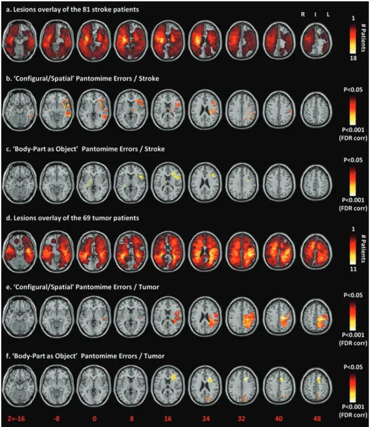
In stroke patients, CS pantomime error types were associ-ated with lesions to a network centered on inferior frontal and temporal areas, with sparse evidence for a role of parietal areas (Fig.1b). In tumor patients, CS error types were associ-ated with lesions to a more posterior network extending from inferior frontal to parietal areas, mostly including parietal white matter (Fig.1e).
In stroke patients, BPO pantomime errors were associated with lesions to the left middle and inferior frontal gyri, the rolandic and inferior frontal opercula, and the underlying white matter, mainly including the superior longitudinal fasci-culus (Fig.1c). In tumor patients, BPO errors were associated with the same network, but extending higher to the sup-plementary motor area (Fig.1f; see Supplementary Fig. S2 for the double dissociation between CS and BPO errors).
We also conducted the same VLSM analyses as above with stroke and tumor patients collapsed together. CS pantomime error types were associated with lesions to the left inferior parietal and angular gyri, postcentral and supramarginal gyri, and portions of the underlying white matter (Supplementary Fig. S1c). BPO pantomime errors were associated with lesions to the left middle and inferior frontal gyri, the rolandic and inferior frontal opercula, and the underlying white matter, mainly including the superior longitudinal fasciculus (Sup-plementary Fig. S1d) (see Sup(Sup-plementary Fig. S2 for the double dissociation between CS and BPO errors). Comparison between the results of the analyses of the lesions associated with CS and BPO errors revealed that the left inferior frontal regions predicted the occurrence of both types of error (Sup-plementary Fig. S1cd). We further tested putative effects of lesion size on the occurrence of CS and BPO errors. Lesion size differed between patients with versus without CS errors (P < 0.05, uncorrected) but not for patients with versus without BPO errors (P = 0.29, uncorrected). However, there was no evidence for correlations between the CS or BPO errors and the size of lesions (r(26)=0.16; P = 0.40; r(11) = −0.10; P = 0.74, respectively).
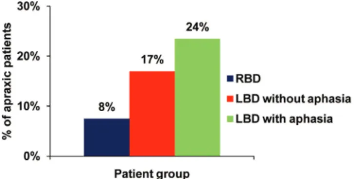
The incidence of the different patterns of error (CS, BPO, or CS + BPO) across patients is displayed in Table 2. The relationship between impaired pantomiming and aphasia is depicted in Figure2. There were more apraxic patients in the group “left hemispheric with aphasia” than in the group “left hemispheric without aphasia” and in the group “right-hemispheric.” The analysis of the incidence of at least 1 pan-tomime error in these 3 groups of patients further reveal that very few patients show both CS and BPO errors (Supplemen-tary Fig. S3).
Figure 1. Voxel-based lesion–symptom mapping on the stroke patients and tumor patients separately shows the relationship between performance in pantomime and brain lesions. (a) Overlap lesion plot of the 81 stroke patients. The number of overlapping lesions is coded with colors ranging from dark red (n = 1) to light yellow (n = 18 patients). (b) Only voxels significant at P<.05 FDR corrected are color-coded ranging from red to white. Configural/ spatial errors were associated with lesions to a network centered on left inferior frontal and temporal areas, with sparse evidence for a role of left parietal areas. (c) Body-part-as-object errors were associated to lesions of the left middle and inferior frontal gyri and the rolandic inferior frontal opercula, and the underlying white matter mainly including the superior longitudinal fasciculus. (d) Overlap lesion plot of the 69 tumor patients. The number of overlapping lesions is coded with colors ranging from dark red (n = 1) to light yellow (n = 11 patients). (e) Only voxels significant at P<.05 FDR corrected are color-coded ranging from red to white. Configural/ spatial errors were associated with lesions to the left inferior frontal and inferior and superior parietal gyri, angular gyrus, postcentral and supra marginal gyri, largely including the underlying white matter. (f ) Body-part-as-object errors were associated to lesions of the left middle and inferior frontal gyri and the underlying white matter mainly including the superior longitudinal fasciculus. Brain slices are displayed from z-coordinates−16 to 48 of the MNI space, with the left hemisphere on the right side.
Discussion
We conducted VLSM analyses based on a large cohort of 150 unselected patients with unilateral left or right hemispheric brain damage and their continuous scores in BPO and CS errors types in a classical neuropsychological assessment of pantomime. Our results reveal that distinct lesion sites within the left hemisphere predicted the occurrence of CS and of BPO pantomime error types. Both types of error were associ-ated with damage to left inferior frontal regions in tumor and stroke patients. CS errors were associated with lesions of left inferior parietal areas, whereas BPO errors were associated with lesions extending from left superior to inferior frontal gyri and a large portion of the underlying white matter in both tumor and stroke patients. Of note, we put forward a differential pattern of deficits according to etiology for CS errors: CS errors were less associated with parietal areas in stroke than in tumor patients, and temporal areas were less associated with CS errors in tumor than stroke patients.
Most of the previous investigations of pantomime in brain-damaged patients included only left brain-brain-damaged patients and/or samples selected based on the presence of aphasia (Schnider et al. 1997;Hanna-Pladdy et al. 2001;Goldenberg 2003a,2003b;Goldenberg et al. 2007;Dovern et al. 2011). In contrast, our study includes unselected consecutive patients sustaining both left and right unilateral brain damage. Our finding for a left hemispheric dominance in pantomime thus provides robust lesion evidence for the prominent involve-ment of left but not right hemispheric structures in panto-mime and corroborates functional imaging studies documenting a left hemispheric specialization for pantomime (Hermsdorfer et al. 2007;Vingerhoets, Acke et al. 2012, Vin-gerhoets, Vandekerckhove et al. 2011). A limitation of our
results in this regard is that because we included only right-handed patients, the present study cannot disentangle poten-tial interactions between handedness, the hand used for the pantomime and the side of the lesion. However, our results are consistent with previous evidence for a left lateralization in pantomimes in both left- and right-handed individuals (Vingerhoets, Acke et al. 2012) and for a similar left lateraliza-tion in studies comparing left- and right-hand pantomimes in right-handed participants (Moll et al. 2000;Choi et al. 2001). Of note, the VLSM analysis revealed the brain regions indu-cing the“more severe” pantomime impairments (Fig.1, Sup-plementary Fig. S1), which does not rule out that other regions play a role in pantomime. Although the left parieto-frontal network revealed in our results is the region which, when damaged, induces the most robust increase in the number of pantomime errors, right-hemispheric brain regions (or left nonfrontal, nonparietal regions) might be involved in praxis as well. Indeed, impaired pantomiming manifest in 8% of the right brain damaged patients (Fig. 2) and the lesion overlap of patients showing a deficit in pantomime reveals associations between right hemispheric lesions and apraxia (see Supplementary Fig. S2b).
A separated VLSM analysis of the lesion sites associated with the commission of distinct types of pantomime errors re-vealed a left intrahemispheric dissociation between the contri-bution of parietal and frontal areas to CS and BPO error types, respectively.
In tumor patients, our finding for a role of left parietal areas in CS pantomime errors is in line with findings from functional imaging approaches (Moll et al. 2000;Hermsdorfer et al. 2007;Vingerhoets, Acke et al. 2012,Vingerhoets, Vande-kerckhove et al. 2011). These studies interpreted the left par-ietal activity during pantomime as supporting the storage of knowledge about manipulation of familiar objects and learned gestures, both mechanisms being specific to praxia and necessary for accurate pantomime. Damage to this region is thus conceivably at the origin of the CS error types we ob-served (Buxbaum et al. 2007;Vingerhoets, Acke et al. 2012, Vingerhoets, Vandekerckhove et al. 2011). However, our ana-lyses revealed a critical role for the parietal white matter in pantomime in tumor patients, suggesting that a disruption of the functional interactions between the subparts of the fronto-parietal network involved in pantomime would yield even more CS errors than focal damage to their constitutive regions (Peigneux et al. 2001; Wheaton, Shibasaki et al. 2005; Wheaton, Yakota et al. 2005). In line with thisfinding, fronto-parietal and basal ganglia damage induced by corticobasal degeneration have been shown to induce severe apraxic symptoms (Leiguarda et al. 2000).
In contrast to these activation studies and to ourfinding in tumor patients, lesion data so far mostly report that the integ-rity of left frontal but less consistently parietal areas are necessary for pantomime (Goldenberg et al. 2007; see also Bohlhalter et al. 2011for supporting Transcranial Magnetic Stimulation data). Moreover, sparse evidence from single case reports describe patients with left parietal damage but pre-served pantomime (Goldenberg and Hagmann 1997; Peigneux et al. 2000). These lesion data have been interpreted in terms of the involvement of inferior frontal regions in the selection of task relevant gestures among all gestures possibly related to a given tool or object use (Goldenberg et al. 2007). Goldenberg et al. (Goldenberg et al. 2007;Goldenberg 2009)
Table 2
Incidence and range of each error type
Error (≥1) Total (n = 150), % Mean % of error (± SD) + range
No error 76 –
CS only 15 36.6 ± 18.0% (10–75%)
BPO only 6 45.4 ± 16.6% (20–75%)
CS + BPO 3 CS: 43.8 ± 24% (25–75%)
BPO: 31.3 ± 12.5% (25–50%) CS, configural/spatial error; BPO, body-part-as-object error.
Figure 2. Distribution of apraxic patients as a function of the damaged hemisphere and aphasia. Right brain-damaged (RBD) patients are reported in blue, left brain-damaged patients (LBD) without aphasia are reported in red, and LBD with aphasia are reported in green.
advanced that the disparities between the findings of func-tional and lesions studies on the role of parietal regions in pantomime could follow from the gestures being realized under different conditions in the scanners when compared with during neuropsychological assessments. In the scanner, more spatial transformation would be required because the movements have to be performed with constrained limb pos-itions, within unusual portions of space and without visual feedback. These additional demands would have artificially increased the involvement of the parietal structure supporting spatial processing and transformations into coordinates (Sack 2009; though seeRumiati et al. 2004for evidence of parietal activity even when participant were not instructed to perform the gestures in the scanner). However, our result for a parietal involvement in the absence of any extra demand on spatial transformation calls for additional accounts for the lack of associations between parietal damage and CS pantomime errors observed in previous lesion studies. The following hypotheses could be put forward in this regard. First, pre-vious studies included only stroke patients; our results for a much stronger association between CS errors and parietal areas in the tumor than in the stroke group suggest that the etiology of the lesion might play a role in their functional con-sequences on apraxia. While some evidence suggest that stroke and tumor results in the same deficits (Haaland and Delaney 1981), other pointed out that these 2 etiologies could yield distinct patterns of deficits, even if lesion size and location is controlled (Anderson et al. 1990). Our results suggest that lesion location associated with pantomime errors might differ depending on whether the VLSM analyses are based on stroke versus tumor lesions. Lesion–symptom mapping based on tumoral lesions has been argued to induce different patterns as when stroke lesions are analyzed. A poss-ible reason for these discrepancies could be that in tumor patients, infiltrations could yield functional loss while being invisible to the MRI or CT scans used to delineate the lesion loci during the lesion reconstruction, in turn confounding the mapping between lesion and symptoms (for discussion, see Anderson et al. 1990; Karnath and Steinbach 2011; Shallice and Skrap 2011). Interestingly, in line with the previous lesion data reviewed above (e.g.,Goldenberg et al. 2007), the result for a parietal involvement in CS error almost vanished when analyses were conducted in stroke patients only. However, we would note that specifically designed studies should be conducted to elucidate the differential role of lesion versus tumor patients (Duffau 2011). The results of VLSM analyses are indeed highly dependent on the spatial distribution of the lesion because it not only determines where in the brain the VLSM tests are actually conducted, but also the distribution of the statistical power of the statistical tests conducted at each voxel between the behavioral scores of lesioned versus intact patients (Kimberg et al. 2007; see Method section). This factor possibly account for our differen-tial pattern of results in the 2 groups of patients as evident from the difference in the lesion overlap in Figure 1a,d showing that lesion location are not strictly identical in stroke and tumor patients.
In this regard, the fact our study included a large cohort considerably increased the statistical power of our analyses and the portion of the brain covered by lesions. This factor could also explain why, in contrast to previous lesion studies, we reveal a parietal involvement in pantomime (though
mostly in tumor patients). The inclusion of both left and right brain-damaged patients in the VLSM further strengthened the sensitivity of our statistical tests by increasing the number of data-points (i.e., the behavioral scores) in the groups of the intact and lesioned patients compared at each voxel. We also analyzed continuous data instead of dichotomizing the scores into normal versus impaired based on behavioral cutoffs, thereby taking into account the severity of pantomime impair-ment in the VLSM and maximizing the statistical power of the analyses (Cohen 1983).
Finally, although the following reasoning only applies to stroke patients where the association between parietal areas and CS error was weak, our results might have revealed a par-ietal involvement because pantomime was assessed during the subacute phase and not during the chronic phase as in most previous studies. In the literature so far, pantomime scores were collected at postlesion delays of about 28 weeks (Goldenberg 2003a, 2003b); 20 weeks (Goldenberg et al. 2007); or 4 weeks (Schnider et al. 1997). In contrast, panto-mime scores in our study were collected on average 2 weeks after lesions onset, a period corresponding to the subacute phase. The specificity of parietal networks for pantomime was possibly revealed in the present study because panto-mime was assessed after—or at least during—the release of areas surrounding damaged regions from ischemic penumbra (Witte et al. 2000), but before major plastic anatomo-functional reorganization took place (Adriani et al. 2003). Although highly speculative, parsimonious explanations for the fact that the functions subserved by parietal but not frontal areas recovered in chronic patients would be that 1) parietal mechanisms could be hierarchically subordinated to frontal selection processes and more specific to the panto-mime task; and/or 2) the largely acquired and mnesic nature of parietal movement representations could be more prone to be recovered and taken over by other areas than frontal ex-ecutive selection mechanisms. We would note, however, that our interpretation of the results on the influence of the postle-sion delay is not made by directly comparing subacute versus chronic patients but in the light of previous literature. Conse-quently, the present study does not allow drawing definitive conclusions on the influence of postlesion delay functional re-covery in pantomime, but rather calls for further investi-gations specifically designed to disentangle the precise influence of this factor.
The results of the VLSM revealed a role of temporal regions in CS error, mostly in stroke patients. This finding is in line with models positing that the knowledge on tool use required to perform accurate pantomime on verbal command as in the current study depends on the semantic memory, notably in-stantiated within temporal areas (Kellenbach et al. 2003; Lewis 2006;Frey 2007;Canessa et al. 2008;Goldenberg and Spatt 2009). Our analyses further reveal that left frontal but not parietal lesions correlated with BPO error type. This finding is consistent with previous lesion studies reporting higher rates of BPO in left than right brain damaged patients (Mozaz et al. 1993). We also observed that lesions predicting BPO extended largely to the white matter underlying inferior and middle frontal cortices, including the superior longitudi-nal fasciculus (SLF). This finding substantiates the obser-vations by Hanna-Pladdy et al. (2001)of more BPO after left subcortical than cortical damage and evidence that lesions to the SLF induce severe apraxia (Mori et al. 2002;Schmahmann
and Pandya 2011). Lesions to frontal white matter tracks have been interpreted as inducing apraxia by disconnecting parie-tal and fronparie-tal motor areas (Pramsparie-taller and Marsden 1996).
Several candidate mechanisms have been advanced to explain BPO errors. First, because BPO are considered as errors only when they persist after reinstruction, they could be interpreted as perseveration and be accounted for by mere executive dysfunctions. A neighbor hypothesis by Peigneux and Van der Linden (1999) assumes that BPO could follow from difficulties in inhibiting automatic acti-vations of often used emblematic gestures (e.g., using his hand to represent the handset to signify a phone call). Sup-porting the hypothesis that BPO are due to a lack of inhibi-tory control, healthy elderly individuals with weaker inhibitory control also show more BPO errors than young and healthy adults (Peigneux and Van der Linden 1999). However, if BPO errors resulted from perseveration only, they should manifest in both left and right frontal damaged patients and not selectively in left hemispheric patients as in our results (Freedman et al. 1998).
Alternatively, BPO could be committed due to a pathologi-cal embodiment of the tool in the patient’s limbs, echoing phenomenon occurring during the rubber hand illusion (Bot-vinick and Cohen 1998).Kondo et al. (2009)further advanced that the inability to precisely formfinger postures to perform the gesture follows from the contamination of the motor command by the information concerning the shape of the objects. Such effect could possibly follow from damage to frontal regions (Arzy et al. 2006;Kondo et al. 2009).
A third candidate mechanism for BPO is advanced by Raymer et al. (1997), who suggests that BPO errors could be linked to difficulties of representing and/or selecting the ap-propriate object features necessary to produce the correct hand postures used to hold the object. In turn, such deficits would make the patients portraying the object itself instead of imagining it and adapting their gestures accordingly. In this regard, BPO would be in an attempt to circumvent the task difficulty by using limbs as a concrete rather than as an ab-stract representation of the object (see also Bartolo et al. 2003).
Of note, the overlap between patients’ lesions show that 5 of the 9 patients with only BPO errors are right-brain damaged, suggesting that right hemispheric structures might also play a role in BPO errors (Supplementary Fig. S2b). Because our study includes only unilateral patients, this result might explain the very limited number of patients showing both BPO and CS errors. However, the role of right-hemispheric structures in BPO error does not appear in the VLSM where the severity of the deficits (i.e., the number of BPO) and the patients with both CS and BPO errors are taken into account. Further studies including bilateral patients are required to investigate this question.
Another limitation of the present study is that because it was based on a retrospective approach, only information on CS and BPO error types were available. Previous neuropsy-chological investigations of pantomime deficits identified several other types of error, which revealed other types of mechanisms involved in pantomime. For instance, investi-gation of the relationships between pantomime and actual tool use or tool recognition showed that these 2 processes correlated to a certain extent (Bartolo et al. 2003; Rumiati et al. 2004for discussion), suggesting that pantomime deficits
may not solely follow from semantic processing impairments but also from deficits of the output lexicon (Cubelli et al. 2000).
Taken together, ourfindings reveal that pantomime is sub-served by a distributed, left-lateralized, frontoparietal network and that lesions to subparts of this network induce distinct error types. Furthermore, the results point out that the postle-sion delay and the etiology of the brain damage might be important to consider in the study of apraxia in brain-damaged patients.
Supplementary Material
Supplementary material can be found at: http://www.cercor.oxford journals.org/.
Funding
This work was supported by a grant from the Pierre Mercier Foundation for Science to L.S. and by grants from the Swiss National Science Foundation (Grant #320030-124897 to S.C.L. and #325100–118362 to J.M.A.).
Notes
The authors thank Francisco Bravo and Mélanie Manchon for their help in collecting the data, Francoise Colombo for providing parts of the neuropsychological data and David Magezi for the proofreading of the manuscript. Conflict of Interest: None declared.
References
Adriani M, Maeder P, Meuli R, Thiran AB, Frischknecht R, Villemure JG, Mayer J, Annoni JM, Bogousslavsky J, Fornari E et al. 2003. Sound recognition and localization in man: specialized cortical networks and effects of acute circumscribed lesions. Exp Brain Res. 153:591–604.
Andersen RA, Snyder LH, Bradley DC, Xing J. 1997. Multimodal rep-resentation of space in the posterior parietal cortex and its use in planning movements. Annu Rev Neurosci. 20:303–330.
Anderson SW, Damasio H, Tranel D. 1990. Neuropsychological im-pairments associated with lesions caused by tumor or stroke. Arch Neurol. 47:397–405.
Arzy S, Thut G, Mohr C, Michel CM, Blanke O. 2006. Neural basis of embodiment: distinct contributions of temporoparietal junction and extrastriate body area. J Neurosci. 26:8074–8081.
Bartolo A, Cubelli R, Della SS, Drei S. 2003. Pantomimes are special gestures which rely on working memory. Brain Cogn. 53:483–494. Basso A, Faglioni P, Luzzatti C. 1985. Methods in neuroanatomical
research and an experimental study of limb apraxia. In: Roy EA, editor. Neuropsychological studies of apraxia and related dis-orders. North Holland, Amsterdam/New York/Oxford. P 179–202. Bates E, Wilson SM, Saygin AP, Dick F, Sereno MI, Knight RT,
Dron-kers NF. 2003. Voxel-based lesion-symptom mapping. Nat Neuro-sci. 6:448–450.
Bickerton WL, Riddoch MJ, Samson D, Balani AB, Mistry B, Hum-phreys GW. 2012. Systematic assessment of apraxia and functional predictions from the Birmingham Cognitive Screen. J Neurol Neu-rosurg Psychiatry. 83:513–521.
Bohlhalter S, Vanbellingen T, Bertschi M, Wurtz P, Cazzoli D, Nyffeler T, Hess CW, Muri R. 2011. Interference with gesture production by theta burst stimulation over left inferior frontal cortex. Clin Neuro-physiol. 122:1197–1202.
Botvinick M, Cohen J. 1998. Rubber hands‘feel’ touch that eyes see. Nature. 391:756.
Buxbaum LJ, Kyle K, Grossman M, Coslett HB. 2007. Left inferior par-ietal representations for skilled hand-object interactions: evidence from stroke and corticobasal degeneration. Cortex. 43:411–423. Canessa N, Borgo F, Cappa SF, Perani D, Falini A, Buccino G,
Tetta-manti M, Shallice T. 2008. The different neural correlates of action and functional knowledge in semantic memory: an fMRI study. Cereb Cortex. 18:740–751.
Choi SH, Na DL, Kang E, Lee KM, Lee SW, Na DG. 2001. Functional magnetic resonance imaging during pantomiming tool-use ges-tures. Exp Brain Res. 139:311–317.
Cohen J. 1983. The cost of dichotomnization. Appl Psychol Measure. 7:249–253.
Cubelli R, Marchetti C, Boscolo G, Della SS. 2000. Cognition in action: testing a model of limb apraxia. Brain Cogn. 44:144–165.
Dovern A, Fink GR, Saliger J, Karbe H, Koch I, Weiss PH. 2011. Apraxia impairs intentional retrieval of incidentally acquired motor knowledge. J Neurosci. 31:8102–8108.
Duffau H. 2011. Do brain tumours allow valid conclusions on the localization of the human brain functions? Cortex. 47:1016–1017. Fiez JA, Damasio H, Grabowski TJ. 2000. Lesion segmentation and
manual warping to a reference brain: intra- and interobserver reliability. Hum Brain Mapp. 9:192–211.
Freedman M, Black S, Ebert P, Binns M. 1998. Orbitofrontal function, object alternation and perseveration. Cereb Cortex. 8:18–27. Frey SH. 2007. What puts the how in where? Tool use and the divided
visual streams hypothesis. Cortex. 43:368–375.
Goldenberg G. 2003a. Apraxia and beyond: life and work of Hugo Liepmann. Cortex. 39:509–524.
Goldenberg G. 2009. Apraxia and the parietal lobes. Neuropsycholo-gia. 47:1449–1459.
Goldenberg G. 2003b. Pantomime of object use: a challenge to cer-ebral localization of cognitive function. Neuroimage. 20(Suppl 1): S101–S106.
Goldenberg G, Hagmann S. 1997. The meaning of meaningless ges-tures: a study of visuo-imitative apraxia. Neuropsychologia. 35:333–341.
Goldenberg G, Hermsdorfer J, Glindemann R, Rorden C, Karnath HO. 2007. Pantomime of tool use depends on integrity of left inferior frontal cortex. Cereb Cortex. 17:2769–2776.
Goldenberg G, Spatt J. 2009. The neural basis of tool use. Brain. 132:1645–1655.
Goodglass H, Kaplan E. 1963. Disturbance of gesture and pantomime in aphasia. Brain. 86:703–720.
Haaland KY, Delaney HD. 1981. Motor deficits after left or right hemisphere damage due to stroke or tumor. Neuropsychologia. 19:17–27.
Haaland KY, Harrington DL, Knight RT. 2000. Neural representations of skilled movement. Brain. 123:2306–2313.
Halsband U, Schmitt J, Weyers M, Binkofski F, Grützner G, Freund HJ. 2001. Recognition and imitation of pantomimed motor acts after unilateral parietal and premotor lesions: a perspective on apraxia. Neuropsychologia. 39:200–216.
Hanna-Pladdy B, Heilman KM, Foundas AL. 2001. Cortical and sub-cortical contributions to ideomotor apraxia: analysis of task demands and error types. Brain. 124:2513–2527.
Heilman KM, Rothi LJG. 1993. Apraxia. In: Heilman KM, Valenstein E, editors. Clinical neuropsychology. New York: Oxford University Press. P. 141–163.
Hermsdorfer J, Terlinden G, Muhlau M, Goldenberg G, Wohlschlager AM. 2007. Neural representations of pantomimed and actual tool use: evidence from an event-related fMRI study. Neuroimage. 36 (Suppl 2):T109–T118.
Karnath HO, Fruhmann BM, Kuker W, Rorden C. 2004. The anatomy of spatial neglect based on voxelwise statistical analysis: a study of 140 patients. Cereb Cortex. 14:1164–1172.
Karnath HO, Steinbach JP. 2011. Do brain tumours allow valid con-clusions on the localisation of human brain functions? —Objec-tions. Cortex. 47:1004–1006.
Kellenbach ML, Brett M, Patterson K. 2003. Actions speak louder than functions: the importance of manipulability and action in tool rep-resentation. J Cogn Neurosci. 15:30–46.
Kertesz A, Ferro JM. 1984. Lesion size and location in ideomotor apraxia. Brain. 107(Pt 3):921–933.
Kimberg DY, Branch Cosletti H, Schwartz MF. 2007. Power in Voxel-based Lesion-Symptom Mapping. J Cogn Neurosci. 19:1067–1080. Kondo M, Mochizuki S, Kobayakawa M, Tsuruya N, Kawamura M.
2009. [Possible mechanism of body part as object and hand closing-in in apraxia]. Brain Nerve. 61:196–202.
Leiguarda R, Merello M, Balej J. 2000. Apraxia in corticobasal degeneration. Adv Neurol. 82:103–121.
Lewis JW. 2006. Cortical networks related to human use of tools. Neu-roscientist. 12:211–231.
Liepmann H. 1908. Drei Aufsätze aus dem Apraxiagebiet. Berlin: Kräger. Mahieux-Laurent F, Fabre C, Galbrun E, Dubrulle A, Moroni C. 2009. [Validation of a brief screening scale evaluating praxic abilities for use in memory clinics. Evaluation in 419 controls, 127 mild cogni-tive impairment and 320 demented patients]. Rev Neurol. (Paris). 165:560–567.
Moll J, de Oliveira-Souza R, Passman LJ, Cunha FC, Souza-Lima F, Andreiuolo PA. 2000. Functional MRI correlates of real and ima-gined tool-use pantomimes. Neurology. 54:1331–1336.
Mori S, Kaufmann WE, Davatzikos C, Stieltjes B, Amodei L, Frederick-sen K, Pearlson GD, Melhem ER, Solaiyappan M, Raymond GV et al. 2002. Imaging cortical association tracts in the human brain using diffusion-tensor-based axonal tracking. Magn Reson Med. 47:215–223.
Mozaz MJ, Pena J, Barraquer LL, Marti J. 1993. Use of body part as object in brain-damaged subjects. Clin Neuropsychol. 7:39–47. Mutha PK, Sainburg RL, Haaland KY. 2010. Coordination deficits in
ideomotor apraxia during visually targeted reaching reflect im-paired visuomotor transformations. Neuropsychologia. 48:3855–3867.
Peigneux P, Salmon E, Garraux G, Laureys S, Willems S, Dujardin K, Degueldre C, Lemaire C, Luxen A, Moonen G et al. 2001. Neural and cognitive bases of upper limb apraxia in corticobasal degener-ation. Neurology. 57:1259–1268.
Peigneux P, Van der Linden M. 1999. Influence of ageing and edu-cational level on the prevalence of body-part-as-objects in normal subjects. J Clin Exp Neuropsychol. 21:547–552.
Peigneux P, Van der Linden M. 2000. Présentation d’une batterie neu-ropsychologique et cognitive pour l’évaluation de l’apraxie ges-tuelle. Revue Neuropsychol. 10:311–362.
Peigneux P, Van der Linden M, Andres-Benito P, Sadzot B, Franck G, Salmon E. 2000. [A neuropsychological and functional brain imaging study of visuo-imitative apraxia]. Rev Neurol (Paris). 156:459–472.
Peigneux P, Van der Linden M, Garraux G, Laureys S, Degueldre C, Aerts J, Del FG, Moonen G, Luxen A, Salmon E. 2004. Imaging a cognitive model of apraxia: the neural substrate of gesture-specific cognitive processes. Hum Brain Mapp. 21:119–142.
Pizzi A, Carrai R, Falsini C, Martini M, Verdesca S, Grippo A. 2009. Pronostic value of motor evoked potentials in motor function re-covery of upper limb after stroke. J Rehab Med. 41:654–660. Pramstaller PP, Marsden CD. 1996. The basal ganglia and apraxia.
Brain. 119(Pt 1):319–340.
Raymer AM, Maher LM, Foundas AL, Heilman KM, Rothi LJ. 1997. The significance of body part as tool errors in limb apraxia. Brain Cogn. 34:287–292.
Rey B, Frischknecht R, Maeder P, Clarke S. 2007. Patterns of recovery following focal hemispheric lesions: relationship between lasting deficit and damage to specialized networks. Restor Neurol Neuro-sci. 25:285–294.
Rorden C, Brett M. 2000. Stereotaxic display of brain lesions. Behav Neurol. 12:191–200.
Rorden C, Karnath HO, Bonilha L. 2007. Improving lesion-symptom mapping. J Cogn Neurosci. 19:1081–1088.
Rothi LJ, Ochipa C, Heilman KM. 1991. Cognitive neuropsychological model of limb apraxia. Cogn Neuropsychol. 8:443–458.
Rothi LJ, Ochipa C, Heilman KM. 1997. A cognitive neuropsychologi-cal model of limb praxis and apraxia. In: Rothi LJG, Heilman KM, editors. Apraxia; the neuropsychology of action. Hove, UK: Psy-chology Press. p 29–50.
Rumiati RI, Humphreys GW. 1998. Recognition by action: dissociating visual and semantic routes to action in normal observers. J Exp Psychol Hum Percept Perform. 24:631–647.
Rumiati RI, Weiss PH, Shallice T, Ottoboni G, Noth J, Zilles K, Fink GR. 2004. Neural basis of pantomiming the use of visually pre-sented objects. Neuroimage. 21:1224–1231.
Rumiati RI, Zanini S, Vorano L, Shallice T. 2001. A form of ideational apraxia as a delective deficit of contention scheduling. Cogn Neu-ropsychol. 18:617–642.
Sack AT. 2009. Parietal cortex and spatial cognition. Behav Brain Res. 202:153–161.
Schmahmann JD, Pandya DN. 2011. Fiber pathways of the brain. New York: Oxford University Press.
Schnider A, Hanlon RE, Alexander DN, Benson DF. 1997. Ideomotor apraxia: behavioral dimensions and neuroanatomical basis. Brain Lang. 58:125–136.
Shallice T, Skrap M. 2011. Localisation through operation for brain tumour: a reply to Karnath and Steinbach. Cortex. 47:1007–1009.
Spierer L, Bellmann-Thiran A, Maeder P, Murray MM, Clarke S. 2009. Hemispheric competence for auditory spatial representation. Brain. 132:1953–1966.
Vingerhoets G, Acke F, Alderweireldt AS, Nys J, Vandemaele P, Achten E. 2012. Cerebral lateralization of praxis in right- and left-handedness: same pattern, different strength. Hum Brain Mapp. 33:763–777.
Vingerhoets G, Vandekerckhove E, Honoré P, Vandemaele P, Achten E. 2011. Neural correlates of pantomiming familiar and unfamiliar tools: action semantics versus mechanical problem solving? Hum Brain Mapp. 32:905–918.
Wheaton LA, Shibasaki H, Hallett M. 2005. Temporal activation of par-ietal and premotor areas related to praxis movements. Clin Neuro-physiol. 116:1201–1212.
Wheaton LA, Yakota S, Hallett M. 2005. Posterior parietal negativity pre-ceding self-paced praxis movements. Exp Brain Res. 163:535–539. Witte OW, Bidmon HJ, Schiene K, Redecker C, Hagemann G. 2000.
Functional differentiation of multiple perilesional zones after focal cerebral ischemia. J Cereb Blood Flow Metab. 20:1149–1165.