The clinical relevance of antibody-mediated rejection: a new era of
heart transplantation
Paul Mohacsi
a,*, Michele Martinelli
a, Yara Banz
band Claudia Boesch
aa Department of Cardiology, Swiss Cardiovascular Center, University Hospital Bern (Inselspital), Bern, Switzerland b Institute of Pathology, University of Bern, Bern, Switzerland
* Corresponding author. Department of Cardiology, Swiss Cardiovascular Center, University Hospital Bern (Inselspital), Freiburgstrasse, CH 3010 Bern, Switzerland. Tel: +41-31-6324464; e-mail: paul.mohacsi@insel.ch (P. Mohacsi).
Keywords:Heart transplantation• Antibody-mediated rejection • Antiendothelial-cell antibodies • Non-HLA antibodies
This issue reports a case where the authors can be congratulated on having saved the life of a patient who originally suffered from giant cell myocarditis and had been treated with cyclosporine (CyA), azathioprine (AZA) and corticosteroids (PDN) for 4 years before clinically deteriorating and been subjected to heart
trans-plantation (HTx) [1]. The pretransplant work-up included human
leukocyte antigen (HLA)-typing of both patient and donor, screening for anti-HLA antibodies and cross-match with regard to T- and B-cells being negative. Postoperatively, conventional immunosuppression (CyA, AZA and PDN) was introduced. There were three HLA Class I and one HLA Class II allele mismatches between donor and recipient.
The first endomyocardial biopsy (EMB) showed mild rejection
(ISHLT 1R) without C4d deposition in immunohistochemistry. The second EMB (3 weeks postoperatively) revealed severe macrophage
(CD68+), and eosinophilic and lymphocytic infiltration, again
without C4d deposition. The patient developed fever without clin-ical signs of infection and was treated under the presumption of acute cellular rejection (ACR). After initial stabilization, the patient developed cardiogenic shock 2 days later, which required urgent treatment with extracorporeal membrane oxygenation, followed by the implantation of a biventricular assist device the next day. At that time, EMB showed severe ACR (ISHLT 3R) and
antibody-mediated rejection (AMR), strongly positive for C4d [2,3]. The
cyto-toxic T- and B-cell cross-match tests remained negative.
The endothelial precursor cells cross-match test (EPC-XM;
XM-ONE®, AbSorber AB, Sweden) was synchronously performed
with EMBs, measuring serum levels of the amount of antiendothelial-cell antibodies (AECAs), either against
donor-specific EPC’s or against autoreactive EPC’s (the patient’s own
EPCs). After initially being negative, donor-specific AECA became
positive at the first EMB (ISHLT 1R), but autoreactive AECAs
were still negative. At the time of acute graft failure, the test was
surprisingly positive for both donor-specific and autoreactive
IgM antiendothelial AECA, but not for donor IgG AECA. The authors conclude that severe AMR was caused by non-HLA, AECAs. Epicrisis was successfully treated by plasmapheresis, in-travenous immunoglobulin (IVIgG), rituximab, muronomab-CD3, bortezomib and re-HTx. One month after re-HTx, the patient had ACR (ISHLT 2R), but remained free of AMR.
During the last 15 years, AMR became a topic of major clinical relevance. Potential explanations are improved diagnostic tests and the rising population of patients having undergone VAD implant-ation before HTx. Its pathophysiology remains unknown to date.
AMR is often associated with haemodynamic compromise, increased mortality and development of graft coronary artery disease (CAD). Younger age, congenital heart disease, positive
donor-specific cross-match, positive panel reactive antibody
(PRA) titres, sensitization to OKT3, CMV seropositivity, previous
blood transfusions and female gender have been identified as
risk factors for AMR [4].
Ongoing discussions finally led to the consensus conference
on April 20, 2010 in Chicago [5]. They also address the question
of non-HLA antibodies, such as vimentin, endothelial cells, MICA/MICB, with a very low incidence (2/128) by using the current XM-One technique to separate EPCs from donor blood
[6]. It was mentioned that‘more work is required to understand
whether these assays can be used to diagnose AMR’.
The association of AMR with cardiovascular mortality, such as death resulting from acute rejection, myocardial infarction, con-gestive heart failure, graft failure, arrhythmia or CAD, was
recent-ly shown in paediatric HTx [7]. Such an observation supports
protocol screening for AMR. However, the need for the treat-ment of measurable but clinically silent AMR and the algorithm to do so, still remains to be determined. Most of the transplant centres continue to perform event-driven and individualized strategies, as shown in the current case report.
Due to improved diagnostic testing and recently developed therapeutic strategies, we do have the chance to avoid or to
treat AMR individualized and sufficiently, either in the case of
preformed (such as in ABO incompatibility) or acquired HLA antibodies (such as via pregnancy, prior transfusion or trans-plant). Some may argue that most of the measures against AMR
need intravenous administration, which is difficult to maintain
over the years. However, we can assume that ‘accommodation’
will occur [8]. To provide an example of how to manage such
cases successfully, we would like to share the case of accidental B to O incompatible HTx having been rescued successfully by anti-B antibody absorption, plasma exchange with AB fresh frozen plasma, IVIgG, by using an anti-B antibody
© The Author 2012. Published by Oxford University Press on behalf of the European Association for Cardio-Thoracic Surgery. All rights reserved.
CA SE R E PO R T S
European Journal of Cardio-Thoracic Surgery 42 (2012) 1047–1049
EDITORIAL COMMENT
immunoadsorption column and the C1 esterase inhibitor
(C1inh, Berinert®) [9]. The HTx patient survived 5 years and
developed a histo-blood group type change of the graft from B
to O [10].
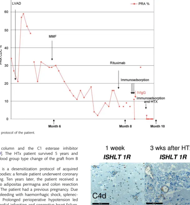
Another example is a desensitization protocol of acquired anti-HLA Class I antibodies: a female patient underwent coronary artery bypass grafting. Ten years later, the patient received a gastric bypass due to adipositas permagna and colon resection due to diverticulitis. The patient had a previous pregnancy. Due to intra-abdominal bleeding with haemorrhagic shock, splenec-tomy was required. Prolonged perioperative hypotension led to an anterior myocardial infarction and congestive heart failure. As a consequence, left ventricular assist device (LVAD) implant-ation was required. Preoperative serum PRA levels were 43%,
reaching postoperative serum PRA levels of 59%. Inflow
cytome-try 10 days post-LVAD implantation, 21 different HLA Class I
antibodies were detected. The virtual cross-match (etrl.
eurotransplant.org/cms/index.php?page=services) yielded a very low likelihood for receiving a matching cardiac allograft. As de-sensitization protocol, we administered mycophenolic mofetil, rituximab and repetitive Protein-A immunoadsorption. After single rituximab administration, the absolute B-cell count
dropped to <25/µl. Figure1depicts the desensitization protocol
with a consecutive decrease of the PRA levels. The patientfinally
underwent HTx with two unfavourable matches (HLA-B8 and B27); we continued during the postoperative phase with Protein-A immunoadsorption procedures. The EMB 1 week after HTx showed slightly swollen endothelial cells, which led to the
administration of IvIgG and C1inh. Figure2shows
immunohisto-chemical staining of the heart biopsies. The EMB 2 weeks after HTx showed ACR (International Society of Heart and Lung Transplantation 2R), which was treated with methylprednisolone
Figure 2:Immunohistochemical staining of heart biopsies: positive staining is highlighted by a brown colour. Detection of the classical complement pathway component C4d and terminal membrane attack complex C5b-9 as well as tissue macrophages by CD68.
Figure 1:Desensitization protocol of the patient.
V. Sigurdardottiret al. / European Journal of Cardio-Thoracic Surgery 1048
and rATG-Fresenius. Six years after HTx, the patient continues to
befine.
The current case report by Sigurdardottiret al. and our
exam-ples show that AMR is a contemporary clinical challenge, which can be reasonable diagnosed, addressed and solved in an appro-priate fashion.
One week after HTx (left panel) only minimal complement de-position was observed in small capillaries and larger vessels. This
is an unspecific feature and may be observed in the immediate
peritransplant period, e.g. following graft ischaemia/reperfusion. CD68 marks some scattered macrophages. Endothelial cells are flat and general cellularity is low. A minimal perivascular
lymphocyte-predominant infiltrate suggests minor ACR.
Three weeks after transplantation (right panel), there is still only minimal C4d deposition, and slightly more C5b-9, however, mainly localized to larger blood vessels. Macrophage counts remain constant. The endothelial lining of only a few capillaries appears slightly swollen, accompanied by a perivascular and
interstitial lymphocyte-predominant infiltrate, suggesting ACR
(top right panel). Hallmark features of AMR, including a more generalized swelling of the endothelial lining, thrombi in small vessels, oedema and haemorrhage as well as intravascular accu-mulation of macrophages, were not noted.
ACKNOWLEDGEMENTS
This work was supported by the Katharina Huber-Steiner Foundation.
REFERENCES
[1] Sigurdardottir V, Kolsrud O, Hernandez N, Dellgren G. Endothelial cell antibody-mediated rejection and successful retransplantation in a heart transplanted patient. Eur J Cardiothorac Surg 2012;42:1044–6.
[2] Reed EF, Demetris AJ, Hammond E, Itescu S, Kobashigawa JA, Reinsmoen NLet al. Acute antibody-mediated rejection of cardiac trans-plants. J Heart Lung Transplant 2006;25:153–9.
[3] Berry GJ, Angelini A, Burke MM, Bruneval P, Fishbein MC, Hammond E et al. The ISHLT working formulation for pathologic diagnosis of antibody-mediated rejection in heart transplantation: evolution and current status (2005–2011). J Heart Lung Transplant 2011;30:601–11. [4] Uber WE, Self SE, Van Bakel AB, Pereira NL. Acute antibody mediated
re-jection following heart transplantation. Am J Transplant 2007;7:2064–74. [5] Kobashigawa J, Crespo-Leiro MG, Ensminger SM, Reichenspurner H,
Angelini A, Berry G et al. Report from a consensus conference on antibody-mediated rejection in heart transplantation. J Heart Lung Transplant 2011;30:252–69.
[6] Nowak G, Karrar A, Holmén C, Nava S, Uzunel M, Hultenby Ket al. Expression of vascular endothelial growth factor receptor-2 or Tie-2 on peripheral blood cells defines functionally competent cell populations capable of reendothelialization. Circulation 2004;110:3699–707. [7] Everitt MD, Hammond MEH, Snow GL, Stehlik J, Revelo MP, Miller DV
et al. Biopsy-diagnosed antibody-mediated rejection based on the pro-posed ISHLT working formulation is associated with adverse cardiovas-cular outcomes after pediatric heart transplant. J Heart Lung Transplant 2012;31:686–93.
[8] Soares MP, Lin Y, Sato K, Stuhlmeier KM, Bach FH. Accomodation. Immunol Today 1999;20:434–7.
[9] Mohacsi P, Rieben R, Sigurdsson G, Tschanz H, Schaffner T, Nydegger UE et al. Successful management of a B-type cardiac allograft into an 0-type man with 3 1/2 year clinical follow-up. Transplantation 2001;72:1328–30. [10] Koestner SC, Kappeler A, Schaffner T, Carrel TP, Nydegger UE, Mohacsi P. Histo-blood group type change of the graft from B to O after ABO mismatched heart transplantation. Lancet 2004;363:1523–5.
CA SE R E PO R T S