XIV. COGNITIVE INFORMATION PROCESSING
Academic and Research Staff
Prof.
Prof.
Prof.
Prof.
Prof.
Prof.
Prof.
T.
W.
T.
J.
B.
R.
S.
J.
A.
R.
W.
Prof. D. E. TroxelProf. I. T. Young
Dr. R. R. Archer
Dr. J. E. Green
Dr. K. R. Ingham
Dr. D. A. Sankowsky
Dr. O. J. Tretiak
Graduate Students
E. G. Guttmann
D. W. Hartman
L. Hatfield
L. P. A. Henckels
P. D. Henshaw
M. Hubelbank
J. I. Makhoul
G. P. Marston
G. G. Matison
W. McCandless
F. X. Carroll C. L. Fontaine E. R. Jensen A. J. Laurino D. M. Ozonoff E. Peper Sandra A. Sommers M. Eden J. Allen B. A. Blesser T. S. Huang F. F. Lee S. J. Mason W. F. Schreiber P. Barnwell III L. Bass R. Bourk E. Bowie E. Boyle L. Brenner A. Ellias R. Ellis, Jr. E. Filip E. Greenwood B. GrossmanA.
SINC
n- A FAMILY OF WINDOW FUNCTIONS
1. Introduction
Window functions have been useful in the estimation of power spectra from
finite-time records. The autocorrelation function is ordinarily multiplied by a window
func-tion in order to improve the spectral estimate. 1, 2 More recently, window funcfunc-tions
have been used to reduce "leakage"
3in the evaluation of short-time spectra as
com-puted by using the fast Fourier transform.
Finite window functions have the property that they assume nonzero values only
with-in a fwith-inite with-interval. Without loss of generality, the fwith-inite with-interval can be centered around
the origin (t=O), so that
h(t) = 0, ItI > T,
(1)
where h(t) is the window function, and 2T is the length of the interval where h(t) can assume nonzero values. Sometimes 2T is referred to as the "'window size."
Well-known window functions include the rectangular Barlett, Parzen, Hanning, and Hamming windows.1, 2 The Fourier transforms corresponding to the rectangular Barlett and Parzen windows have in common the fact that they are of the form sin xn, where
This work was supported principally by the National Institutes of Health (Grants 5 PO1 GM14940-03 and 5 PO1 GM15006-03), and in part by the Joint Services Elec-tronics Programs (U. S. Army, U. S. Navy, and U.S. Air Force) under Contract DA 28-043-AMC-02536(E) and the National Institutes of Health (Grant 5 TO1 GM01555-03).
F. Pfister S. Prerau Rashid M. Robbins L. Seitz D. Shapiro W. Stallings P. Wacks A. Walpert R. Willemain D. Willems 1
n
=
1 for the rectangular window, n
=
2 for the Barlett window, and n
=
4 for the Parzen
window.
This report derives a general closed-form expression in the time domain for
the family of window functions whose Fourier transforms are of the form
sin x n
sin
Trxwhere n is any positive integer.
Because the function
xis sometimes referred to
rxas sinc x, we shall call this family of window functions SINC
n2.
Derivation
A window function hn(t), corresponding to the n
t hmember of the family SINC
n, is
defined by its transform Hn (w) as
Hn()
sin a nn a '
)(
(2)
where n is a positive integer, and a is a constant. h (t) is the inverse Fourier trans-form of Hn(w):
h (t) = I
Hn(a) e Jt1+00
sin a n jt
The problem is to compute the integral in Eq. 3 for any n.
The integral can be simplified by the help of two expansions4
2m 1 sin x = 1 2
m-1
k=0 sin m+l x = 22m (m+k 1 cos (2m-2k)x + 2 k m) (-) +k sin (2m+1l-2k)x , (-1k m k! (m-k)!m
> 0.
It is clear that if Eqs. 4 and 5 are properly substituted in Eq. 3, the integral must be computed separately for odd and even n.
where
(4)
(XIV. COGNITIVE INFORTMATION PROCESSING)
(a) n even
Let m
=
-and x
=
aw in Eq. 4.
Then substitute Eq. 4 in Eq.
1 h
h (t)
2T -on
_
1
2 n-1 2 k=0n
-+k2
(-1)
(
) cos (an-2ak)w kn
2
3.
The integral reduces
e dt odo. (ao)n
Place the following substitution in Eq. 6.
1 j(an-2ak)w
-j(an-2ak)]
cos (an-2ak)w = -[e+(an e .j(an2ak)w
The integral can now be evaluated by making use of the Fourier transform pair
S - j n (t+b)n - u (t+b), (7)
W
(n-I)!
where u- 1(t) is the ordinary unit-step function, and b is a constant. The result of the integration can be shown to be
h (t)= 1 (2a) (n-i)!
k= lk
Lk=0
(t+an-2ak)n - 1 u (t+an-2ak) L -1 + (t-an+2ak)n 1 (t-an+ak)n
+ (-1)2 nn/2)
tn- 1 t u_1(t )Equation 8 can be simplified. Since H (w) is a real and even function of o, it follows that hn(t) is a real and even function of time. Therefore it suffices to compute hn(t) for t < 0 and then replace every t by (-t) in order to obtain h (t) for t > 0.
For t < 0 Eq. 8 reduces to
1
h (t) =
(
2a)n (n-1)!
n
--
1
2 k=0 k n-I1 (t+an-2ak) u1 (t+an-2ak). u_1(ta -a )(8)
(Note that 2k < n always applies.) h (t) for t > 0 can be obtained from Eq. 9 by replacing t by (-t). Therefore, it follows that for all t
h (t) =
(2a)
n(n-i)!
n 2 k=0u
1I 1(an-2ak- tI),
where It! - absolute value of t.
(b) n odd
Let = n- 1 and x = aw in Eq. 4. Then substitute Eq. 4 in Eq. 3. The integral reduces to
n-1
2 .+m + 00n-1
- o 2 k = 0 n-i (-1)n
sin (an-2ak)wk/
e do. (aw)n Substitutingsin (an;--2ak) I [ej(an-2ak)w 0-j(an-2ak)c]
in Eq. 11 and making use of the transform pair in Eq. 7, we obtain
1
h (t)=
(2a)n (n-l)! n-1 k=0 u_ (t+an-2ak) - (t-an+2ak)n - u(t-an+2ak).
By using the fact that hn(t) is an even function of t, Eq. 12 reduces to
1 h (t) -n (2a)n (n-1)! n-! 1 2 k=0
u_(an-2ak- t )
for all t.
(10)
1
h (t) =
n
2(11)
(12)(13)
k (an-2ak- t )n-1 k (-1k )'(t+an-2ak) n -kL
(-1)k ni (an-2ak- t ) n - 1\k /
(XIV. COGNITIVE INFORIATION PROCESSING)
(c) n even or odd
Equations 10 and 13 for even and odd n, respectively, are very similar. The only difference is the upper limit of the summation on k. By defining
n-V1
n-1
n-2 integer portion of n
1_ 1 j
2
both equations can be combined into one equation which is valid both for even and for odd n, and for all t:
h (t) =
1
(
2a)n (n-i)!
2i
k=0
)n-i ul(an-2ak
-t ).
(14)Equation 14 completely specifies the family of window functions SINCn in the time domain.
3. Discussion
From the properties of the unit-step function it is easy to show from Eq.
h (t) = 0, for it > an. n 14 that (15)Comparing Eq.
T = an.15 with Eq. 1, we conclude that
(16)
The family of functions h (t) is time-limited, and the window size is equal to 2an.n A property of hn(t) is that the area under the function is independent of a is equal to 1. This can be easily shown.
+an J-an +00o h (t)dt = h (t) dt = if (0)= 1. -and n -and
(17)
If the window is of constant size 2T, that is, a = T/n, then, as n increases, the win-dow shape gets "thinner," but it also gets more peaked in order to keep the area of the window equal to 1. For this case of constant window size, Hn(w) takes the form
Ksin
(T
1/n)1
n i L Tw// n
(18}
As n increases, the width of the main lobe increases proportionally with n, which
() n n(a - a k - tresults in an increase in the window spectral bandwidth.
At the same time, the sidelobe
amplitude levels decrease with increasing n, and the high-frequency spectral roll-off is
equal to -6n dB/octave.
Therefore, there is a trade-off between a finer spectral
reso-lution and a reduction in leakage.
For any particular value of n it would be possible to obtain hn(t) by an (n-l)-fold
convolution of the rectangular window.
The advantage of Eq. 14 is that it gives hn(t) for
any value of n by a simple summation.
The reader may be interested in checking the
validity of Eq. 14 for the well-known rectangular, Bartlett, and Parzen windows.
J. I. Makhoul
References
1. R. B. Blackman and J. W. Tukey, The Measurement of Power Spectra (Dover
Pub-lications, Inc.
,
New York, 1958).
2.
P. I. Richards, "Computing Reliable Power Spectra," IEEE Spectrum, pp. 83-90,
January 1967.
3.
G. D. Bergland, "A Guided Tour of the Fast Fourier Transform," IEEE Spectrum,
pp. 41-52, July 1969.
4.
I. S. Gradshteyn and I. M. Ryzhik, Tables of Integrals, Sums, Sequences and
Prod-ucts (FIZMATGIZ, Moscow, 1963).
B.
RELEVANCE OF PSYCHOLOGY TO CHARACTER
RECOGNITION
The two disciplines which may be considered to dominate the field of character
recognition are cognitive psychology and mathematical implementation of algorithms.
The ultimate goal of character recognition is to characterize the visual pattern and to
judge which of the possible legal set elements (in our case printed letters from an
alpha-bet) is being represented.
These algorithms are unfortunately very ad hoc and difficult
to relate to human perception.
Moreover, an algorithm is usually evaluated under
restrictive conditions such that a high statistical success is used as evidence of an
ade-quate solution. An algorithm yielding a 95% accuracy rate when letters are restricted
to 12 Point Bodoni type, but which confuses a Bodoni "a" with an "Old Style" "s" cannot
be considered as approaching a solution to the general character-recognition problem.
The argument that the algorithm should be psychologically related to human
percep-tion is often met with an apparently valid comment such as "Flying machines are not
built on the principle of a bird's flapping wing, rather they are based on engineering and
aerodynamic principles." The analogy is not good because there are no known physical
attributes that give a letter its unique value; equivalence of stimuli is purely perceptual.
Consequently, the test of whether two particular characters are the letter "z" must be
judged by a human observer.
Any ad hoc algorithm, of which there are many, that does
(XIV. COGNITIVE INFORMATION PROCESSING)
not use perceptually related features and attributes for making its decisions becomes burdened with many extra limitations, e. g. , the visual data must be very clear, the letters must be taken from one type font, and multiple definitions are needed to over-come the effects of stimulus and system noise. The best long-range solution to the problem of character recognition will be by developing an algorithm that, although math-ematical in formulation, incorporates essential perceptual cues.
Psychological experiments generally indicate that the information contained in a letter is not uniformly distributed and is very redundant; some parts of the letter give essential cues necessary for resolving possible confusions, while other parts of the letter can be easily ignored. For example, text can be read when the bottom half of a line is deleted, but not when the top half is removed. The top half clearly has more
relevant information than the bottom half. In the same way of thinking, the capital let-ter "A" can be described as having a cenlet-tered closure at the top with two legs extending downward. This gives the essence of the "A", but there is the possibility of ambiguity with the letter "R". The ambiguity is clearly resolved by the presence of any discon-tinuity in the position and derivative of the rightmost leg at its intersection with the lower right side of the closure. From this quick analysis we can specify the psychological
space and the boundary between these two letters so that this description will success-fully characterize any "A" or "R" that a human being would correctly recognize.
Our investigation is now being carried out in many directions.
1. Many letter features can be discovered by an introspective process that prompts one to ask, "How can I distort a letter so that it becomes another letter." Namely, find some style of letter in which a small change produces a new character. For example, a slight indentation in the capital "D" will make it be perceived as a "B".
2. In one experiment, subjects were asked to judge a series of defocused or obscured letters. The resulting confusion matrices portray a space with different groups of letters clustering together. For example, the letters "a", "c", "e", "x" , and "z" seem to cluster in one group, whereas, the letters "b", "d", "p", and "1" cluster in another group. This would lead to the tentative conclusion that under defocusing the attributes compactness vs extendedness are perceived by the subjects.
3. We are attempting to gain some insight into a person's ability to discard visual data that are not relevant to the percept, and his ability to fill in deletion in order to complete his percept. This appears to be most difficult to accomplish with computing systems.
4. An algorithm that represents letters as a sequence of topological features, such as end point, intersection, and closure, is being investigated. We hope that the resulting encoding will have a one-to-one correspondence with the letter set.
5. The ultimate limitations of single-letter recognition can be seen in an experiment in which subjects were unable to recognize defocused letters, but were able to read
defocused sentences containing the same letters with the same degree of defocusing. Similarly, when subjects were presented with a sequence of letters from a sentence in rapid succession they were not able to read it. The sentence could easily be read, how-ever, when all of the letters were presented simultaneously, even though the total expo-sure time was the same in both cases. These familiar examples of context show the inherent limitation of any mechanized letter-by-letter recognition algorithm.
B. A. Blesser, E. Peper
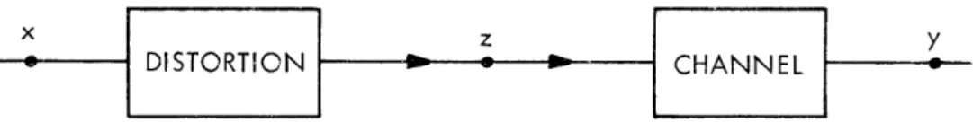
C. SOURCE RATE FOR A SIGNAL CORRUPTED BY NOISE
Consider a signal that is obtained know the rate that is required to code
x
DISTORTION
from an imperfect transducer. We would like to this signal within a certain fidelity. The situation
Y
CHANNEL
Fig. XIV-1. System under consideration.
is diagrammed in Fig. XIV-1. The true signal is x, we observe the corrupted signal,
z, and the signal at the output of the channel is y.
1.
Stereotype Formulation
We shall consider the problem of transmitting a single random variable.
Since the
channel and the transducer are disjoint, we assert that
F x,y/Z(
' b i = Px/z
( ') Py/zo(w,
).
(1)
From this we obtain the following relations:
Px, y('
W)
= d Px/z(, PVz (, y) . )By using the standard formulation, the distortion from the total system is
E = djdwP p(,co)
-(XIV. COGNITIVE INFORAL'TION PROCESSING)
where p(, o) is the distortion weighting function.
P z(u)
R = dtdc P (u,
~)
logt i tz Pr ( ) P ( )
ID z
The rate is to be minimized for a fixed value of E.
The rate is given by
(5)
The problem can be cast into a form for which the solution is available. By substituting (3) in (4), we obtain
E =e ddfdn P/ , , P , ).
If we define a new function
p'(c, ;) =
the new form of E becomes
C dcid P, z(W, ) p'(;s, ).
The solution for these equations is knoown,- and is
Pz, y ' ) = [(-) ef
(
')
(9)In this equation f is a parameter that determines the rate and the distortion and (_) is a function chosen so that
SPz,(u,
o) dy = 1.Z. Solution
Suppose that the source has a Gaussian power density function and the corruption is caused by adding an independent Gaussian random variable.
X/
p ()
---
exp-x P//'exp
Sn
L
i ___) °. 2 £. O a.(10)
(11)
d: Px/z@,0)
p , )We use squared difference as the error weighting function
p(x,y) = (x-y) . (12)
With these hypotheses, the function p' takes an attractive form
2 2 22
(T 0-
a-(w,
2)
2 2 2 n (13)x n x n
It follows from (9) that
22
P y( ,w) = c1 exp(, 2 2 - " (14)
z, + -cr
The various probability density functions that are required for finding the rate and the distortion are found by using moment-generating functions. We shall represent the
Fourier transform of g by the symbol g. With
Pzy (' 1(a-) (15a)
Pz( ) = f2() (15b)
the moment-generating function for P is
A f I()fl f2(k+av)
P (,'v) = (16)
y,z
a
vIn the case that is being considered
f1(6) = e (17a) f2(5) = e 2 (17b) 2 a 2 (17c) S+ 0-x n
(XIV. COGNITIVE INFORMATION PROCESSING)
2 2 x n
The joint distribution P z,y
P ,y((, z,y ) 1
(
Q
X)
aexp -
2
a2
X-
)
2 +W2
1 M t -jThe rate and the error may be evaluated directly. 2 2 0- G-x n 2 2 x+ x n 1
R =
2log
The integrals diverge if 4
0-2 2' x + x n
and for this critical value of X
E max max x
This result may be cast into a more suggestive form. We first observe that 2 2 o-x n 2 2 S+ -x n
is the error associated with a least mban square approximation of the corrupted signal. 2 The distortion is always greater than this amount, but is less than or equal to a x Distortion as a function of rate is given by
2 E axx (1-e-2R 2 2 n (22) (17d) (18) (19a) (19b) (20) (21)
For error twice the minimum value, the rate is
log
S .
3.dn3. Coding Sequences of Signals
Suppose we are to transmit a sequence of Gaussian random variables
{x
i} withvari-ances
(Ox.}
and corrupted by added Gaussian noise with
error
variance { Z}. We fix the total
2 2 x. n.1 + S 2 + 2 i e + cr i x. n. 1 1
and minimize the rate
4
x.
1 1R
log
2
i
2 2 x. n. 1 1This is very similar to the problem described by Kolmogorov.3 The
4 0 x. i 'nf O 2 2 x. n. 1 1
The sequence is divided into two subsequences {x ai} for which ai1l
kX.
= ,'s are chosen as
and {xpi) for which
4
o-x.
X. i cr 2 + 22 x. n. 1 1 The error isL
aij0 Z
{13
)
2 X. 1and the rate is
1
X.
(XIV. COGNITIVE INFORMATION PROCESSING)
4 -X.
2 log 2 '2 2 0
a
xi
i)
n
4. Rate for a Continuous Source
For a real Gaussian stationary source with spectrum S(f) and real noise N(f), the rate may be found through a procedure similar to that used by Kolmogorov.4 We define the associated function (f) given by the relation
X(f) = inf (fN S(f) + N(f)
and define the set of frequencies for which X(f) = 0 as R1. The mean squared error
S(f) N(f)
E
df (+ + df S(f),f E 1 S(f) + N(f)f R 1
and the rate per unit time is
1S2(f)
1
R
f
df logI Yf E R S(f) + N(f)
Another interesting case arises when the signal is passed through a filter whose fre-quency response is H(f) before N(f) is added. To reduce this to the problem solved above, we consider transmitting the spectrum S(f) with additive noise N(f) • H(f) -2. The result is X(f) = inf 0, 2H(f) 2 S2(f)
H(f),
S(f) + N(f)
R < H(f) 2 S2(f) 1 H(f) 2 S(f) + N(f) E = df H(f) 2 S(f) N(f) + df S(f) fE R1 I H(f) 2 S(f)+ N(f) f R11
g
S
2(f) H(f) 2
fER 1
S
2(f) IH(f) )2 + N(f)
This result may also be extended to the case in which the error is weighted with a
frequency weighting function.
0. J. Tretiak
References
1.
C. E. Shannon and W. Weaver, The Mathematical Theory of Communication
(Uni-versity of Illinois Press, Urbana, Ill.
,
1964), p. 111.
2.
Ibid. , p. 114.
3.
A. N. Kolmogorov, "On the Shannon Theory of Information Transmission in the Case
of Continuous Signals," IRE Trans.
,
Vol. IT-2, No. 4, December 1956, p. 106.
4.
Ibid.
,
p. 108.
D.
MODEL FOR THERAPEUTIC DIAGNOSIS OF DISEASE
1. Diagnostic Process
The steps taken by a physician to effect the cure of a patient who comes to him with
a complaint vary from physician to physician. Varying degrees of intuition or
experi-enced guessing are combined with principles of diagnosis that the physician learned in
medical school to arrive at a diagnosis and prescribe a course of treatment. The ability
of the physician to look at a patient and say he "looks sick" cannot be duplicated by a
machine.
A computer can, however, perform a statistical diagnosis if it is programmed
in a logical manner to do so, and thus act as a consultant to the physician.
Concentrating on the systematic approach to diagnosis, we find that information about
the state of a patient comes from four sources:
(i) his medical history, (ii) a physical
examination, (iii) laboratory tests, and (iv) careful observation of the course of his
illne s s.
There is a tendency on the part of the physician to collect more information than is
necessary to arrive at a probable diagnosis, sometimes with little efficiency in the
selection of laboratory tests. If a more systematic approach to test selection could be
made, the cost of making the diagnosis could be reduced. Also, the physician is
lim-ited in his ability to accurately consider more than a few symptoms and possible
dis-eases, and as a result may be led to an erroneous diagnosis if the disease is uncommon
and difficult to diagnose.
Moreover, the physician cannot easily assign approximate
probabilities or confidences to his final diagnosis.
Of itself, this is not of great
impor-tance; however, in the face of alternative treatments with risk to the patient, the
(XIV. COGNITIVE INFORMATION PROCESSING)
physician must once again make an experienced guess, and the chances of its being cor-rect or optimal might be improved by a quantitatively more systematic approach.
2. Modeling the Diagnostic Process
A consideration of the relative likelihood of diseases can be made through the use of Bayes' theorem. This probabilistic approach seems appropriate when we consider that medical knowledge itself is usually nondeterministic. We can determine the rela-tive probabilities of disease complexes Di, given the observed symptom complexes Sk' by Bayes' theorem.
P(Sk/Di) P(D i )
P(Di/Sk) S/ (1)
P(Sk/Di) P(Di
1
The second term in the numerator is the probability that the patient has disease com-plex D., regardless of any symptom that he might have (i. e. , the a priori probability of D.). This term varies from location to location, season to season, and is sensitive to the occurrence of epidemics. The denominator is just the scale factor of the uncon-ditional probability of symptom complex Sk
-Bayes' formula also provides a simple numerical means of performing the logical elimination of disease complexes. If disease Di and symptom Sk are logically incon-sistent, then P(Sk/Di) = 0, which from (1) yields P(Di/Sk) = 0.
With the assumption of the availability of the necessary probabilistic data, Bayes' formula is the key to most sequential differential diagnostic procedures. As knowledge
about the state of a patient increases through medical history taking, physical examin-ation, laboratory testing, observation of the course of the illness and the reaction to therapy, the probability of each disease complex can be adjusted accordingly. If no
dis-ease is indicated with sufficiently high probability, then, if possible, further informa-tion should be gathered. On the other hand, the high probability of one disease may signal the time to initiate treatment, and by using the probabilities the risk involved can be ascertained should the chosen therapy have contraindications for other diseases with lesser likelihoods. Should the risk be too great or the diagnosis still uncertain, the current disease probabilities help protide an indication of what information should be
sought (e. g. , what test to take) to move efficiently and with least expense toward a more conclusive diagnosis.
Previous studies have tended to consider diagnosis in a context somewhat divorced from the realities of the medical health care profession. Few have touched on the end purpose of diagnosis - to resolve uncertainty about the true state of the patient so that appropriate treatment can be chosen. The implication in considering the treatment -and not just a static diagnosis of the patient - is that the goal of the process has shifted
from that of ascertaining the probabilities of diseases to that of effecting the patient's
return to health.
Diagnosis-treatment is always subject to certain real-life constraints: testing costs,
treatment costs, risk to the patient, and so forth. Although the issue of cost in money
might be dismissed as insignificant because most of the population subscribes to some
form of medical insurance, the costs are nevertheless real if we consider not cost to
the patient but the cost of inefficient use of the increasingly scarce resources of the
phy-sician's time, facilities for treatment, and so on. It is this problem that I wish to
con-sider.
The rest of this report presents a model (admittedly only an approximation) of
the therapeutic diagnosis of a patient who has undergone a transition to a state of illness.
I shall be especially concerned with exploring and making explicit those difficulties or
alternatives that may exist in the implementation of the procedure.
3.
Model of Therapeutic Diagnosis
We can usually characterize a patient who visits his physician or is admitted to a
hospital for emergency treatment as someone who has undergone a transition from a
state of health (state I) to a state of illness (states 2, 3, .... , n), where the gravity of
the state increases with its number.
In reality these states form a continuum of patient
conditions, but in order to gain the simplifications of a discrete model I shall consider
5 states of health:
healthy, moderately ill, very ill, critically ill, and dead.
The
illus-tration below shows the possible transitions that a patient can have after an initial
tran-sition to the very ill state.
state 1 5
healthy
moderately
very
critically
dead
healthy
ill
ill
ill
The criteria for classifying a patient into one of these 5 states might be as follows.
State 1
Healthy or cured.
State 2
Probably would recover from his illness in time without
treat-ment.
State 3
Condition is as likely to improve as to worsen if not treated; or patient
will be permanently handicapped.
State 4
Probably would die in time without treatment.
State 5
Dead.
(XIV. COGNITIVE INFORMATION PROCESSING)
Should it be the case that these rough classifications presuppose a tentative diagnosis in order to make a classification, then we might use some other criterion such as the degree of patient discomfort.
Assume for the time being that the patient is in the ill state i (i ~ 2). What should the physician's course of action be? If P(ij/A) is the probability of the patient's going from state i to state j as a result of the physician's action A, then he might want to choose an A in order to
(i) Maximize P(il) (optimistic).
(ii) Minimize P(ij), where j > i; that is, minimize the possibility of the patient's condition worsening (pessimistic).
(iii) Maximize P(il) - f(cost of A) (more realistic). (iv) Optimize some more complex decision function.
The decision criterion used in this model will be developed for an area of medical spe-cialization with the following set of properties.
{Dm} m =1, .... , d the set of all disease classifications
{S } n =1, . .. s the set of all symptom complexes {Tq
}
q = , ... , t the set of all relevant diagnostic testsRx} x = 1 .. , r the set of all treatments useful in treating the above-mentioned set of diseases.
Let us assume that the patient has visited the doctor who has elicited, by history taking, from a computer questionnaire, or by physical examination, some initial infor-mation about the patient. This information can be put in the form of a symptom com-plex S n . From this information the doctor can formulate a probabilistic diagnosis, by using Bayes' theorem
P(S n/D ) P(Dm)
P(Dm/Snm n ) =
nm
(1)P(Sn/D ) P(Dm) m
where
P(Dm) = a priori probability of the occurrence of disease Dm P(Sn/Dm) = probability that a random patient with disease Dm
will exhibit symptom complex S . n
If the probability of one disease stands out from the rest (e. g. , Condition: sore elbow -Diagnosis: sprained elbow with probability .8) the physician may wish to proceed
directly to therapy (e. g.
,
immobilize the elbow).
Depending on the risks involved and
the available alternatives, however, he may wish to proceed further before treatment.
An x-ray photograph (which involves expense) may show a small fracture of the ulna.
This gives a new symptom complex and alters the probabilistic diagnosis (in this case
we might find P(fracture)
= .
99).
Although this is a very simple case history, the
ques-tion that is being raised is when should the gathering of informaques-tion stop and treatment
begin.
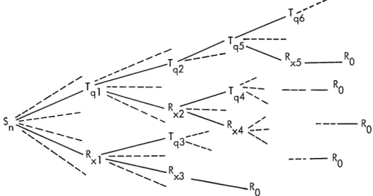
Schematically we may characterize the alternatives by a decision node.
Alternative
Consequences
ask more questions
more information + small cost
physical exam
more information + small cost
(partial or complete)
n
test T
more information + greater
cost + risk of testing
treatment Rx
probability of cure + risk +
cost + information from response
To simplify the notation, we assume that the set {T
}
includes the medical history and
physical examination, and that the set
{Rx}
includes the null treatment Ro (no further
treatment), as well as treatments in the general sense of the word (e. g.
,
wait N days).
More than one test may be in order before treatment, or an unsuccessful treatment may
be followed by a sequence of tests and a new treatment.
This gives rise to a decision
tree as a model of the diagnosis-treatment process (Fig. XIV-2). The branch that is
T
q6Tq
q
T0
qi
P
Tq5
9
xx2---RS
R
n x4 ---- RR
R
0
x3
(XIV. COGNITIVE INFORMATION PROCESSING)
followed until it eventually reaches a terminal node Ro is dependent upon the outcomes of the decisions made at all previous nodes, and probabilistically upon the true state of the patient. We would like to rank order all terminal points according to the decision criteria, and try to choose at each decision node that branch which leads to the best terminal node or nodes. Any such look-ahead procedure, however, creates the problem of computational complexity, because of a very large number of terminal points. Thus, for practical reasons, we are forced to limit look-ahead and may wish to limit consid-eration of only a few of the more promising branches at any given node.
A decision about how to limit the evaluation of the possibilities implied by the deci-sion tree might be made by considering the question, Should the physician continue testing and thereby obtain a more probable diagnosis, or should he forego further testing that is costly and likely to be unnecessary and initiate treatment? In terms of the model, how does the expected outcome, given that he administers the optimal treat-ment Rx without further testing, compare with the expected outcome of performing the most relevant test T and then administering the optimal treatment based on the probabilistic outcome of that test. Thus a decision based on this limited-depth look-ahead procedure is made by evaluating an inequality of the form
E(T followed by Rx) > E(Rx without testing). (2)
q x
The expected outcome will be a function of utility changes for the possible state transi-tions, monetary costs of treatment and tests, and risk involved.
Before stating the decision function we define the parameters: u . = utility of the transition from state i to state
j
CR = dollar cost of treatment R
x x
CT = dollar cost of performing test T , and the probabilities:
P(T , k) = probability that test Tq has outcome k = Z P(Tq, k D ) P(Dm) P(ij/Rx, Din) = probability that a patient with disease Dm will go from state i to
state j if treated with therapy Rx
P(ij/T q, D ) = probability that a patient with disease Dm will go from state i to state j if administered test T
k q
R q= given the observed state of the patient before administering test Tq, k
R q is the optimum treatment if the test has outcome k.
Eu(Rx) =
i
1
1 P(ij/R x) u..X
1] P(ij/R , D ) P(D m) u.. j Ilm(3)
The expected risk involved in administering test T can be written q E (Tq) =
i
i
I
j
P(ij/T q) u..ij q iiP(ij/T
,Dm) p(D m
uij
j mwhere P(Dm) is the probability of disease Dm computed from Bayes' formula. The inequality
k test
max E(Tq followed by R
q)
max E(R x x without further tests) treatcan be evaluated as follows.
E(R
xwithout further tests)
=
Eu(Rx) - CRx
P(Dm)]
= i
P(ij/Rx, Dm)
i
j m y kfollowed by R
k P(Tq,k) E(R - CRkq + Er(Tq) - CT q _uk
k
+
k
CR q] P(ij/Tq, Dm) P(Dm)q
m
m
u.. 3ij - CT qEquation 6 is evaluated for R x = 1, 2, ... , r and will have a maximum for some x = x. x
Thus, Rx,: is the optimal treatment if there is to be no further testing. Equation 7 is evaluated for T q = 1, 2, . . . , t. Each test T has a number of possible outcomes.
q q
E(T q
U.ij -1J CR
(XIV. COGNITIVE INFORMATION PROCESSING)
If Sn is the symptom complex before the results of Tq are known, then for every
out-come k of T we obtain a new symptom complex S n(k). For each outcome k we use Sn(k) and Bayes' formula (1) to compute a new a posteriori distribution for P(Dm). For each outcome k of T this new distribution is used to find that R which maximizes Eq. 6.
q
k xThat treatment is labeled R q, and the maximum value of (6) for outcome k of T is,
k k q
therefore, E (R q) - CR q This value is then used in evaluating Eq. 7 for each test T . For some q = q , Eq. 7 has a maximum, yielding T q : as the best test to
administer if we decide on further testing before treatment.
If Eq. 6 evaluated for Rx, is less than Eq. 7 evaluated for T ., then the model dic-tates further testing and suggests giving T as the next test. The results of T yields
q q
new information upon which the decision to treat or continue testing can again be made. If Eq. 6 evaluated for Rx, is greater than Eq. 7 evaluated for T ,, then the model dic-tates initiating treatment Rx*. If the patient is cured the process is terminated. If the patient is not cured, the additional information gained by his response to Rx* is lumped with the previous data Sn and used to decide whether to test or try a new treatment.
I have tried to describe briefly a model of the diagnosis-treatment procedure. There are many obstacles to implementing such a model, even on a small scale. Obviously, estimates must be obtained for several one- and two-dimensional parameters. It is my intention to pursue this model of the diagnosis-treatment process further, in the hope of simulating actual decisions so that its accuracy and implications can be properly deter-mined. In spite of the problem of data collection, parameter estimation, and practical application, the advantages of a well-defined decision procedure might be more efficient and accurate care in the face of an increasing scarcity of medical care resources, some implications for multiphasic screening, and, particularly, a visible and rational context in which to formulate decisions with regard to diagnosis and treatment of disease.
B. E. Boyle References
1. N. E. Betaque, Jr. , "Utility Theory Applied to Medical Diagnosis and Treatment," S. M. Thesis, Department of Electrical Engineering, M. I. T. , June 1969.
2. A. S. Ginsberg, et al. , "An Application of Decision Theory to a Medical Diagnosis-Treatment Problem," P3786, Rand Corporation, Santa Monica, California, Feb-ruary 1968.
3. G. A. Gorry, "A System for Computer-Aided Diagnosis," Project MAC Technical Report MIAC-44 (Thesis), Cambridge, Massachusetts, 1967.
4. R. S. Ledley, "Practical Problems in the Use of Computers in Medical Diagnosis," Proc. IEEE 57, 1900-1918 (1969).
5. D. R. Luce and H. Raiffa, Games and Decisions: Introduction and Critical Survey (John Wiley and Sons, Inc. , New York, 1968).
6. H. R. Warner, A. F. Toronto, and L. G. Veasy, "Experience with Bayes' Theorem for Computer Diagnosis of Congenital Heart Disease," Ann. N.Y. Acad. Sci. 115, 558-567 (1964).
E. LEUKOCYTE LOCATING PROCEDURE
Several members of our group are working on the development of a practical auto-mated leukocyte recognition system. The purpose of such a system is to perform a dif-ferential leukocyte (white blood cell) count on a smear prepared from human peripheral blood. An integral part of the system is the ability to rapidly determine whether leuko-cytes are present in a view and if so where. Working in Fortran IV on the PDP-9 com-puter, we have demonstrated the feasibility of a very simple locating procedure.
1. Background
When a prepared blood smear slide is examined under an oil-immersion microscope at 1000X magnification the appearance is similar to that of Fig. XIV-3, except for the loss of color. Many erythrocytes (red blood cells) can be seen surrounding and touching the single, roughly circular leukocyte distinguished by its optically dense nucleus. Leuko-cytes, 7-20 p. in diameter, are normally outnumbered by the erythrocytes 650:1. Several of the types of leukocytes have segmented or polymorphic nuclei as shown in Fig. XIV-3. A third type of cell found in mammalian blood is the blood platelet which is usually smaller than erythrocytes and of an optical density similar to leukocyte nuclei. For
1 2 3
further details see Davidsohn and Wells, Young, or Young.
172
-ROW
279'
279
DIAGONAL
1
1
108
COLUMN
DIAGONAL 2
Fig. XIV-3. Typical picture showing its four histograms and the
nucleus limits determined from the histograms.
(XIV.
COGNITIVE INFORMATION PROCESSING)
2. Pictorial DataThe pictorial data used in this study were prepared by Professor Young for his doc-toral research.2 4 A field of view approximately 70 p. X 50 . containing one leukocyte and the surrounding erythrocytes was photographed. The resulting transparencies were scanned by the on-line flying-spot color scanner SCAD and the output stored on magnetic tape in the form of quantized blue, green, and red optical transmittances at each of
18, 576 points (172 row X 108 column matrix) over the picture. For this study only the green record was used.
3. Image Features
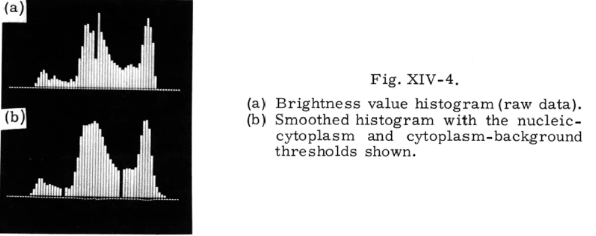
As can be seen from Fig. XIV-3, the nucleus of the leukocyte is the most guishing characteristic, and it alone is used to locate the leukocyte. In order to distin-guish between nucleic and non-nucleic material in the picture, a technique is used similar to that by which Young separated background from red and white cell points.
(a)
Fig. XIV-4.
(a) Brightness value histogram (raw data). (b (b) Smoothed histogram with the
nucleic-cytoplasm and nucleic-cytoplasm-background thresholds shown.
A histogram or probability density function of brightness values is computed from the entire picture (Fig. XIV-4). Three distinct peaks are seen: the first corresponds to a distribution of nucleus brightness values, the second to leukocyte and erythrocyte cyto-plasm, and the third to background points. Thus the minimum between the first and second peaks represents a threshold level by which to classify all points of the picture as either nucleic or non-nucleic.
The cytoplasm of the leukocyte provides a buffer zone of non-nucleic points around the nucleus, thereby eliminating the common problem of touching objects.
4. Location Algorithm
Determining bounds on the nucleus extent is accomplished by the use of spatial histograms (henceforth referred to as histograms, as distinct from the brightness value
histogram).
histograms
hr(r) =
The digital two-dimensional picture is transformed to four one-dimensional by a simple threshold-summing procedure defined by the following equations:
ROWS
r=l
T(bcr)cr
1 < r < ROWS
COLS
hc(c) = c=1 COLShm(m)=
c= 1COLS
h(p) :=c=l
T(bcr) T(bc, m+c-COLS) T(bc, p-c+1) 1 < c s COLS 1<
m < (COLS+ROWS-1) 1 p < (COLS+ROWS-1) h = row histogramr
h = column histogram c hm
= diagonal 1 histogramh
=diagonal 2 histogram
T(.) is a threshold function defined by
T(X) = 1 X
<
brightness value threshold T(X) = 0 otherwise{bcr) is the array of brightness values (i. e., the picture),
b is undefined for c, r not in the range (1, COLS), (1, ROWS).
cr
Although two histograms are sufficient for location, four are, at present, being used for reasons that will be mentioned. The spatial orientation of these histograms is rep-resented in Fig. XIV-3. The transformation above requires only one scan of the picture to examine the brightness value at each point and, when a bcr < threshold is encoun-tered, to increment the appropriate element in each histogram by one. Figure XIV-5 shows two typical histograms.
(a) 172
(b) 172
(b) 172
Fig. XIV-5. Typical spatial histograms: (a) from a picture containing a leukocyte with
a polymorphic nucleus; (b) from a picture containing a leukocyte with an
almost circular nucleus.
All of the 25 pictures tested gave exceptionally noise-free histograms with, in
gen-eral, no more than two or three extraneous peaks whose areas were much smaller than
that of the nucleus.
The nucleus limits are determined from the histograms by a two-step procedure.
First, the histograms are simplified by characterizing the information in the form of
disjoint peaks.
Each peak is represented in terms of four numbers: the two end points,
the area, and the maximum value.
Noise reduction takes the form of (i) eliminating any
peaks not wider than 2 units, and (ii) trimming the ends of the peaks inward to sharpen
boundaries and eliminate trailing edges.
The second step is to examine these
"simpli-fied histograms, " which are actually just lists of peaks, looking for possible groupings
of peaks that could constitute a nucleus.
The grouping operation is governed by the
fol-lowing criteria:
1.
The peak having the largest area is taken as the first candidate for inclusion in
the nucleus estimate.
2.
All remaining peaks with an area greater than 1/4 the area of the largest peak
are considered.
3.
To prevent grouping two nuclei together, a maximum width parameter and
maxi-mum area parameter are checked.
4.
If all peaks with area greater than 1/4 of the largest cannot be considered as one
nucleus, a second nucleus is postulated and the position of the largest peak that caused
the violation is stored.
The nucleus limits obtained from the histograms are checked for consistency with
two tests: (i) the area under each histogram between the proposed limits should be
approximately equal because each is an estimate of the number of nucleus points, and
(ii) the limits should define an octagon of area > average area from test (i).
All test
pictures considered thus far contained only one leukocyte.
Multiple cell location has
not yet been attempted.
In all of the 25 test cases, the nucleus was located. The octagonal enclosure was
never in error by more than 1 or 2 units inside or 3 units outside the nucleus boundary.
The usual diameter of a nucleus is 20-40 units.
5.
Summary
The important advantage of this method is that only the histograms need be in core
to access the presence and location of any leukocytes. Then when a leukocyte is located
the pertinent part of the picture can be brought into core.
With normal blood samples, a view usually contains at most one leukocyte.
Under
these conditions, only two histograms are necessary to do location.
Four histograms
are generated from each picture, however, to obtain a better estimate of the nucleus
(XIV. COGNITIVE INFORMATION PROCESSING)
area that is useful in leukocyte classification and to facilitate separation of leukocyte groupings.
As we have mentioned, several types of leukocytes have a polymorphic or segmented nucleus. This polymorphism can vary from a figure eight shape to four or more isolated segments. Much of the polymorphism of a particular nucleus is evidenced in the multi-lobed character of its histograms. A study is now under way to determine whether reli-able estimates of nucleus polymorphism can be made from the four histograms.
T. R. Bourk
References
. I. Davidsohn and B. Wells, Clinical Diagnosis by Laboratory Methods (W. B. Saunders Company, London, 13th edition, 1965).
2. I. T. Young, "Automated Leukocyte Recognition," Quarterly Progress Report No. 89, Research Laboratory of Electronics, M.I.T., April 15, 1968, pp. 231-242.
3. I. T. Young, "Automated Leukocyte Recognition," Ph. D. Thesis, M. I. T., June 1969. 4. I. T. Young, "Automated Leukocyte Recognition," Proceedings of the 8th
Interna-tional Conference on Medical and Biological Engineering and the 22nd Annual Con-ference on Engineering in Medicine and Biology, Chicago, Illinois, 1969.
F. DISCRIMINATION ALGORITHM FOR THE NORMAL AND SUSPICIOUS CELLS OF THE PAPANICOLAOU SMEAR
In our work of developing a discrimination algorithm, 'the data with which we are concerned are negative photomicrographs taken with a Leitz-Wetzlar Leica camera using Kodak Panatomic-X black and white film through a Leitz-Wetzlar ORTHOLUX
microscope with 1000X magnification.
The transparencies prepared from the photomicrographs have been scanned with the "new scanner." It is a black and white high-resolution flying-spot scanner. The scanned point density is approximately 4 points per micron. Several features have been investi-gated. Thus far, only three seemed to be of value: (i) integrated nuclear chromatin density, (ii) nuclear cytoplasmic ratio and (iii) nuclear size.
Integrated nuclear chromatin density seems to be the most promising of the three features. The first set of test data comprised 18 cells (see Fig. XIV-6), of which 14 were suspicious and 4 were normal cells. Discrimination based on the nuclear-size measure-ment alone placed 2 normal cells high up among the suspicious ones and 6 suspicious ones low down among the normal ones. The integrated nuclear chromatin density feature placed the two large normal nuclei much lower and raised the six smaller suspicious ones much higher among the suspicious cells. Actually, 100% separation was achieved. It is possible that small very darkly stained pyknotic normal nuclei will be difficult
I 0 1 * * ° ° * ° 0 ° ° 1000 2000 3000 4000 5000 100 200 300 400 500 600 INTEGRATED NUCLEAR CHROMATIN DENSITY NUCLEAR SIZE
Fig. XIV-6.
Test results.
X = normal cells. * = suspicious cells.
to separate from small suspicious cells just on the basis of their integrated nuclear chromatin density measurements. In this case the nuclear and cytoplasmic measure-ments will have to be taken into consideration, too.